Embed Size (px)
Citation preview
RESEARCH ARTICLE Open Access
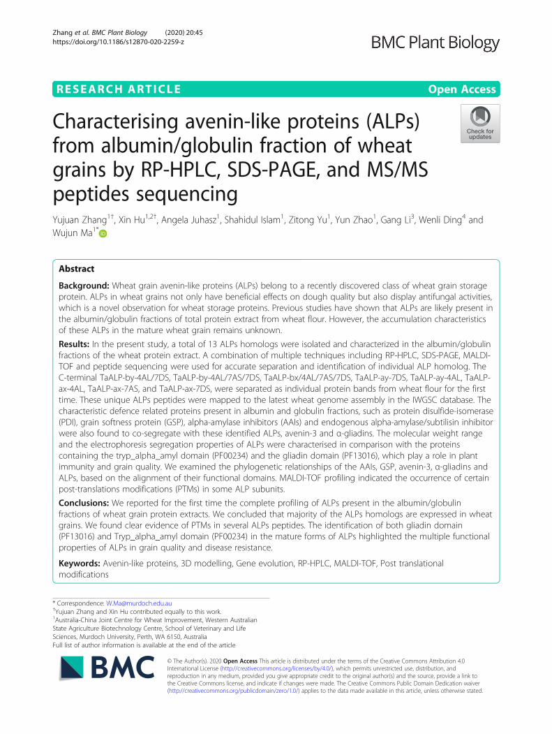
Characterising avenin-like proteins (ALPs)from albumin/globulin fraction of wheatgrains by RP-HPLC, SDS-PAGE, and MS/MSpeptides sequencingYujuan Zhang1†, Xin Hu1,2†, Angela Juhasz1, Shahidul Islam1, Zitong Yu1, Yun Zhao1, Gang Li3, Wenli Ding4 andWujun Ma1*
Abstract
Background: Wheat grain avenin-like proteins (ALPs) belong to a recently discovered class of wheat grain storageprotein. ALPs in wheat grains not only have beneficial effects on dough quality but also display antifungal activities,which is a novel observation for wheat storage proteins. Previous studies have shown that ALPs are likely present inthe albumin/globulin fractions of total protein extract from wheat flour. However, the accumulation characteristicsof these ALPs in the mature wheat grain remains unknown.
Results: In the present study, a total of 13 ALPs homologs were isolated and characterized in the albumin/globulinfractions of the wheat protein extract. A combination of multiple techniques including RP-HPLC, SDS-PAGE, MALDI-TOF and peptide sequencing were used for accurate separation and identification of individual ALP homolog. TheC-terminal TaALP-by-4AL/7DS, TaALP-by-4AL/7AS/7DS, TaALP-bx/4AL/7AS/7DS, TaALP-ay-7DS, TaALP-ay-4AL, TaALP-ax-4AL, TaALP-ax-7AS, and TaALP-ax-7DS, were separated as individual protein bands from wheat flour for the firsttime. These unique ALPs peptides were mapped to the latest wheat genome assembly in the IWGSC database. Thecharacteristic defence related proteins present in albumin and globulin fractions, such as protein disulfide-isomerase(PDI), grain softness protein (GSP), alpha-amylase inhibitors (AAIs) and endogenous alpha-amylase/subtilisin inhibitorwere also found to co-segregate with these identified ALPs, avenin-3 and α-gliadins. The molecular weight rangeand the electrophoresis segregation properties of ALPs were characterised in comparison with the proteinscontaining the tryp_alpha_amyl domain (PF00234) and the gliadin domain (PF13016), which play a role in plantimmunity and grain quality. We examined the phylogenetic relationships of the AAIs, GSP, avenin-3, α-gliadins andALPs, based on the alignment of their functional domains. MALDI-TOF profiling indicated the occurrence of certainpost-translations modifications (PTMs) in some ALP subunits.
Conclusions: We reported for the first time the complete profiling of ALPs present in the albumin/globulinfractions of wheat grain protein extracts. We concluded that majority of the ALPs homologs are expressed in wheatgrains. We found clear evidence of PTMs in several ALPs peptides. The identification of both gliadin domain(PF13016) and Tryp_alpha_amyl domain (PF00234) in the mature forms of ALPs highlighted the multiple functionalproperties of ALPs in grain quality and disease resistance.
Keywords: Avenin-like proteins, 3D modelling, Gene evolution, RP-HPLC, MALDI-TOF, Post translationalmodifications
© The Author(s). 2020 Open Access This article is distributed under the terms of the Creative Commons Attribution 4.0International License (http://creativecommons.org/licenses/by/4.0/), which permits unrestricted use, distribution, andreproduction in any medium, provided you give appropriate credit to the original author(s) and the source, provide a link tothe Creative Commons license, and indicate if changes were made. The Creative Commons Public Domain Dedication waiver(http://creativecommons.org/publicdomain/zero/1.0/) applies to the data made available in this article, unless otherwise stated.
* Correspondence: [email protected]†Yujuan Zhang and Xin Hu contributed equally to this work.1Australia-China Joint Centre for Wheat Improvement, Western AustralianState Agriculture Biotechnology Centre, School of Veterinary and LifeSciences, Murdoch University, Perth, WA 6150, AustraliaFull list of author information is available at the end of the article
Zhang et al. BMC Plant Biology (2020) 20:45 https://doi.org/10.1186/s12870-020-2259-z
BackgroundPolymorphic prolamins are composed of several groupsof structurally related proteins [1]. Most prolamins areknown to contain distinctive N- and C-terminals and re-petitive central domains [1]. The prolamin superfamilywas defined initially on the basis of a shared skeleton ofcysteine residues [1–7]. Recently, Juhász et al. [8] estab-lished a new reference map for immunostimulatorywheat grain prolamin and non-prolamin proteins basedon the new IWGSC bread wheat reference genome se-quence, RefSeq v1.0. Among these re-defined seed-borneallergens, the hydrophobic-seed domain-containing pro-teins show characteristics of antifungal properties, in-cluding cortical cell delineating protein [9], glycine-richprotein [10] and proline-rich protein [11]. Egg-cell se-creted protein [12] also has a prolamin-like domain. Thelipid transfer protein (LTP) and Non-specific LTP [13]have a LTP-2 domain. The 19 KDa Globulin [14, 15],small cysteine-rich proteins [16] belongs to the Domain-less Cys-rich proteins. By contrast, ω-gliadins andHMW-GSs are Domainless Cys-poor proteins. The α-amylase inhibitors (AAIs), α-trypsin inhibitors (ATIs)[17, 18], GSPs [19], Puroindolines [20, 21], α-gliadins[22] and avenin-like proteins (ALPs) all contain a Tryp-alpha-amyl domain (PF00234). Meanwhile, the Puroin-dolines, α-gliadins, LMW glutenins, γ-gliadins and ALPsalso have a Gliadin domain (PF13016). So far, the func-tional property of the gliadin domain was unknown, ex-cept for its nutrient’s reservoir activity during seedsgermination. Meanwhile, the extraction, quantificationand identification of the complete profile of these indi-vidual prolamin proteins in wheat posed a challenge,due to the complexity of the wheat flour proteins.Despite the fact that water and salt soluble proteins
from cereal grain were traditional extracted using dilutedsalt solutions [23–32], new methods were adopted toanalyse and identify components from these proteingroups. Water- and salt-soluble proteins from wheatflour have been characterized using a range of proteinanalytical methods, including SDS-PAGE, RP-HPLC,and differential precipitation by NH4Ac-MeOH followedby acetone enabled separation of the most abundant al-bumins from the gliadins [33–36]. Purothionins, GSP,and several AAIs proteins, as well as several CM3-typealpha-trypsin inhibitors (ATIs) and one protein relatedto the avenins from oats were identified in the albumin/globulin fraction [35]. Albumins are known to havemany different functions and share different types, e.g.glycoprotein, amylase inhibitors, serpins, purothionins,enzymes such as carbohydrases like α- and β-amylases,or proteolytic enzymes [37]. In the fraction of albumins,the representatives of individual protein components areshown to have functions in pathogen resistance. Albu-mins such as AAIs and ATIs [38, 39], serpins [40] and
purothionins [41] are considered to have a function ofnutrient storage and inhibitors of insect and pathogenattack on the germinating seed. Even though smalleramounts of ALPs were first found in the gluten extractsin other studies [42, 43], the close evolutionary relationsof ALPs with AAIs and ATIs and avenin-3, as well asthe lack of repetitive motifs compared with other glia-dins, suggests that they might also be enriched in thealbumin and globulin extracts. However, it is hard toisolate the ALPs from a mixture of wheat storage pro-teins in an efficient way, and the global ALPs proteinaccumulation profile in different wheat varieties stillremains unknown. Till now, the separation of varioussubunits of the homeologous ALPs in different wheatvarieties were rarely reported.Gluten are storage proteins found in the starchy endo-
sperm of wheat, barley and rye. In wheat, the ALPs canbe detected in the gluten-enriched fraction, includingamong others a range of gliadins, glutenins, proteaseinhibitors and LTPs [44]. The ALPs were named due tosequence homology with avenins of oats [45], mostclosely to avenin-3 [46], and α-, γ-gliadins [47]. Kasardaet al. [48] characterized a novel ALP called farinin, com-posed by two disulphide-linked small polypeptidessubsequent to a proteolytic cleavage of a precursor poly-peptide at an Asn-Glu (N-E) peptide bond. Researcheswere primarily on dough functional quality improvementvia incorporation of farinins (ALPs) into the gluteninmacropolymers (GMPs) [43, 48, 49]. Further, the func-tional allelic variations of TaALP-7A were found to beassociated with better processing quality [50]. In total,with the previous knowledge, modifying ALPs is apotential way to make better dough for grain industry.ALP proteins and its function in dough quality have
attracted an increasing amount of research attention. Guet al. [51] found that some storage proteins, such asHMW glutenin, globulins, and ALPs, show upregulatedexpression under water deficient environment, whichmight benefit bread making quality. A recent proteomicstudy indicated that drought stress affects the expressionof wheat storage proteins, such as gliadins, glutenins andALPs as early as 3 days after pollination (DAP), more-over, the misregulated expression is associated withcytoskeleton organization and grain quality proteins indeveloping seeds [52]. Using Mixolab-dough analysissystems, Wang et al. [53] reported that the starchsurface proteins (gliadins, b-type ALPs, LMW-GSs, andpartial globulins) in common wheat and waxy wheatdisplayed different performance to mixing and thermaltreatment. Recently, many storage proteins, includingHMW-GS, gliadins, globulins, ALPs, triticins, and ω-secalins have been identified in wheat endosperm andembryo, which displayed differential accumulation at theprotein level between two wheat species that are distinct
Zhang et al. BMC Plant Biology (2020) 20:45 Page 2 of 19
in grain weight and dough quality, suggesting that ALPsare responsible partly for the quality differences [54].Based on the study by Altenbach et al. [55], the farinins(ALPs) comprised from 2.6 to 3.1% of the protein in theSDS-extractable polymeric proteins (EPP) and 1.9–2.4%of the protein in the SDS-unextractable polymeric pro-teins (UPP), and they were influenced by post-anthesisfertilizer. Both type b subunits (bx and by) of ALPs werereported to have non-functional pseudogenes in Brachy-podium distachyon L accessions, Triticum dicoccoides,and T. aestivum [47, 56]. Most recently, more novelalleles of ALP were found in Aegilops tauschii Coss. ac-cessions [57]. Others have studied the multi-functionalproperties of ALPs despite their effects on dough quality.Gao et al. found [58] a potential protein-protein inter-action between a stress-responsive transcription factor,TaERFL1a, and a type a ALP by yeast two hybrid libraryscreening under water deficiency conditions. Meanwhile,Zhang et al. [47] have screened the WEW lines for poly-morphisms of ALPs and found the relationships betweenthe ALP gene evolution and environmental parameters.Further, a detailed phylogenetic analysis was performedon the genome-wide TaALPs genes and its close rela-tives to wheat and other monocots species [47], suggest-ing that ALPs might have the protease inhibition activitylike α-amylase inhibitors (AAIs), yet the substrates ofALPs can be further identified. Zhang et al. have studiedthe ALPs and its potential Fusarium head blight resistantfunctions [59], further illustrated their antifungal proper-ties. Other research suggest that ALP type b are minorstorage proteins which are important to protect endo-sperm starch reserves from degradation [60]. It isreported that, a putative ALP type b that comprises acereal-type AAIs, as well as serpin-Z1C like defenceproteins were increased by elevated CO2 [60, 61].Another novel study, indicated induction of one ALPand one chitinase in winter wheat (varieties. Bologna)grains, not only due to increased CO2, but might belinked to the microbial populations [62], as in the caseof accumulation of some multifunctional storage globu-lins, which exhibit antimicrobial activity [63].In this study, we identified the ALPs in two Australian
wheat varieties showing different grain quality, and char-acterized the ALPs and their electrophoretic mobilities,composition and extraction properties using the separ-ation techniques RP-HPLC and SDS-PAGE. We usedthe peptide and protein mass identification methodsMALDI-TOF and MS/MS to distinguish genome-widedifferent subunits of ALPs.
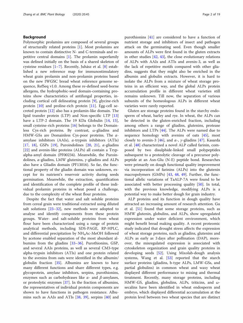
ResultsAllelic variations of ALPs in common wheat varietiesTo identify the allelic variation of TaALP genes amongdifferent wheat varieties, a total of 15 putative ALP
genes were cloned for Sanger sequencing. The allelicvariations of the deduced ALPs amino acid sequences intwo wheat varieties Spitfire and Mace were revealed bysequence alignment. Amino acid substitutions wereidentified only for 3 candidate genes: TaALP-bx-7AS,TaALP-by-7AS and TaALP-ax-4AL. As shown in Fig. 1a,Spitfire displayed a pre-mature codon for TaALP-by-7AS, while Mace contained a pre-mature stop codon forTaALP-bx-7AS. In addition, several non-synonymousmutations between Spitfire and Mace were also observedfor both TaALP-bx-7AS and TaALP-by-7AS. Based onthe sequence alignment (Fig. 1b), TaALP-ax-4AL allelescan be divided into three types (alleles -a, −b and -c).For this gene, Spitfire and Mace were identified asTaALP-ax-4AL-b and TaALP-ax-4AL-c, respectively. Inaddition to the selected three ALP genes, no variationwas found between Spitfire and Mace for all otherTaALP genes present in the wheat genome.The potential protein functional effect of the allelic vari-
ations of TaALP-ax-4AL in CS (−a), Spitfire (−b), andMace (−c) was investigated by sequence alignment andtertiary protein structural modelling analyses. As shown inFig. 2a, a total of 14 cysteine residues are strictly con-served in the three TaALPs. A total of 11 residue substitu-tions were identified between CS and Mace. A singleamino acid substitution (S169 N) is present in TaALP-ax-4AL proteins from CS and Spitfire. Tertiary structuralmodels were generated for TaALP-ax-4AL in CS andMace. The protein structure of TaALP-ax-4AL in Spitfireis represented by the CS model. Structural superimpos-ition (Fig. 2b) showed that the tertiary structures ofTaALP homologs are generally conserved. Both CS andMace protein models are consisted of 4 major alpha-helixes, plus 2 short helixes. The single amino acid substi-tution (S169 N) between CS and Spitfire was located atflexible loop region at the C-terminal, indicating littleeffect on the protein function. Of the 11 substitutionsbetween CS and Mace, 4 (Q79H, A92S, M136 T, andG137R) are located in the helix regions. Hydrophobicityprofile comparison (Fig. 2c) showed that all of these 4 sub-stitutions have caused hydrophobicity changes betweenthe 2 proteins, indicating a potential variance in theenzyme function. The other amino acid substitutions aremainly found in the flexible loop regions, with significanthydrophobicity changes identified in G125R (Fig. 2c). Thesingle amino acid substitution S169N between TaALP-ax-4AL proteins in CS and Spitfire displays no hydropho-bicity change, suggesting potentially identical enzymefunction for these 2 proteins.
Separation and identification of albumin and globulinsproteinsTo characterize the specific protein composition of cer-tain wheat grain storage protein groups, the albumin
Zhang et al. BMC Plant Biology (2020) 20:45 Page 3 of 19
and globulin fraction was extracted from wheat flour(variety Mace). A RP-HPLC method was developed forprotein separation. As shown in Fig. 3a, a total of 20HPLC peaks were chosen and collected for further ana-lysis. The collected peak samples were then loaded onSDS-PAGE gels for separation. The one-dimensionalSDS-PAGE patterns of each peak were shown in Fig. 3b(Peaks 1–11) and Fig. 3c (Peaks 12–20). Notably, mul-tiple bands were identified for each HPLC peak. Basedon the molecular weight (MW) prediction, putativeALPs proteins were identified as protein bands withMW around 17–19 kDa and 28–32 kDa, which corre-sponded to bands 1a, 1b, 6a, 11b and 13e (Fig. 3b).These proteins displayed a retention time (RT) of 17–26.5 min (Fig. 3b, Additional file 1). In addition, putativeAAIs such as CM2 and CM3 were suggested for proteinbands (2a, 6b, and 7a, Fig. 3b) of MW below 17 kDa.These proteins fell into peaks 2, 6 and 7 with RT at23.8–25 min (Fig. 3a, Additional file 1). Similarly, proteinbands 1b and 2a were identified as GSP (MW below 17kDa), which fell into peaks 1 and 2, with RT at 15.6–18.3 min (Fig. 3a, Additional file 1). α-gliadins have MWat around 31–40 kDa and were found in abundantamount in peaks 9–14, corresponding to RT at 22.4–27.5 min, whereas γ-gliadins (MW 31–40 kDa) were
found abundant in peaks 15–19, RT at 28.2–38 min(Additional file 1). Avenin-3 were found from the pro-tein bands 13 d and 16 d (Fig. 3c) in peaks 13 and 16,RT at 29.4–32.5 min (Additional file 1). Taken together,these results suggested that various types and subunitsof homologous ALPs were present and separated to-gether with a mixture of AAIs, GSP, α-gliadin, andavenin-3.
Classification of ALPs and other albumin and globulinproteinsALPs contain a signal peptide and two protein domains:Gliadin domain (PF13016) and Tryp_alpha_amyl domain(PF00234), which are also present in other albumin andglobulin proteins, such as the avenin-3, gliadins, GSPand AAIs. To investigate the evolutionary relationship ofthe different types of ALPs and their relationship withthe other co-segregated albumin and globulin proteins,two Maximum likelihood (ML) phylogenies were con-structed, based on the sequence alignments of the Glia-din domain (PF13016) (Fig. 4a) and Tryp_alpha_amyldomain (PF00234) (Fig. 4b), respectively. The domain se-quences could be found in Additional file 2. Noteworthy,the type b ALP sequences contain 2 cysteine-rich gliadindomains (R1 and R2) [45]. As shown in Fig. 4a and b,
Fig. 1 Diversity of TaALP-bx/by-7AS and ax-4AL genes in common wheat cultivars. a Amino acid sequences alignment of TaALP-bx/by 7AS ofwheat varieties Spitfire and Mace. b Amino acid sequences alignment of TaALP-ax-4AL genes in wheat varieties Living Stone, CS, Spitfire, Drysdale,RAC875, Lincoln, Kauz, Excalibur, Chara, Baxter, Mace, Bonnie Rock, Gladius, Greygory, Kukri, Westonia, Yitpi, Wyalketchem, Bethleyhem, Eagle Rock
Zhang et al. BMC Plant Biology (2020) 20:45 Page 4 of 19

the overall topology of both phylogenies were highlyconsistent and conserved, indicating these two domainshad evolved vertically and were present before the diver-gence among different protein subfamilies. The phylo-genetic branches representing GSP, AAIs, avenin-3/gliadin and ALPs could be clearly recognized. In bothphylogenies, GSP and AAIs diverged first, followed bythe avenin-3 and gliadin. The latter two protein groupsdisplayed a close relationship with each other. In both
phylogenetic trees, ALPs were found to be the latestevolved proteins, which further divided into six sub-branches: type c, ax, ay, bx-R1, bx-R2 and by. Of these,type c, ax and ay were grouped together, while the othertype b ALPs formed one branch. Interestingly, in bothphylogeny cases, the R2 domain of type by ALPs dis-played a closer relationship with the R2 domain of typebx ALPs, compared to the R1 domain of type by ALPs.This observation supported that type by ALPs may have
Fig. 2 Sequence alignment and protein modelling analyses. a Amino acid sequence alignment of TaALP-ax-4AL proteins in CS, Spitefire, andMace. Cysteine residues were highlighted in blue. Secondary structural elements based on protein modelling were displayed above the sequencealignment. Three predicted disulphate bonds were underlined in pink number 1, 2, and 3. b Superimposition of the tertiary structure models ofTaALP-ax-4AL in CS (green) and Mace (cyan). Disulphate bonds were displayed in sticks (yellow). The amino acid substitution sites were displayedin red. Only a single substitution (S169 N) exists between CS and Spitfire. c Displays the hydrophobicity profile. The substitution site residues withhydrophobicity change were shown in sticks, with red and white colours indicating the most hydrophobic and the most hydrophilic residues,respectively. Protein models were generated using the I-TASSER server [64, 65]. Structure visualization was implemented in PyMol (v1.7.4.5)
Zhang et al. BMC Plant Biology (2020) 20:45 Page 5 of 19

originated from the adjoining of type by-R1 and type bx-R2 domains.
ALP identification by RP-HPLC fractionation in wheatvarieties spitfire and MaceTo investigate the variations of ALPs composition in dif-ferent wheat varieties, total albumin and globulin pro-teins were extracted from two wheat varieties, Mace andSpitfire. Mace is a variety characterized as high andstable grain yield, whereas Spitfire is featured as slightlylower grain yield but higher grain protein content (≥13%) [66]. The two varieties possess different bread-making qualities [66]. For this study, the grain proteincontent for Mace and Spitfire are 12.14 and 14.22%, re-spectively. The moisture content for Mace and Spitfireare 12.67 and 12.31%, respectively. Identification of ALPswas carried out using RP-HPLC, SDS-PAGE, peptide-sequencing and MALDI-TOF techniques by followingthe method described above. The different chromato-graphic profiles for wheat cultivar Mace (Figs. 3 and 5)were resulted from the use of two RP-HPLC columns
(same model). We used the old column for the 20 peaks(from 15min to 38min) to target all the albumins andglobulins. Later we purchased a new column and ob-tained 36 peaks (0–38min) for the same sample. Asshown in Fig. 5b, there is no target protein for peaks 1–7 during 0–15min. So this time discrepancy does notaffect our results. Based on this observation, only 15–38 min were targeted in later runs. This second run wasused to identify the ALPs specifically.For Mace, a total of 36 elution peaks (Fig. 5a) were
identified by RP-HPLC separation. These peak fractionswere then loaded on SDS-PAGE gel for further separ-ation. As shown in Fig. 5b, most of the HPLC fractionscontained a mixture of proteins with different MWs.Those protein bands with MWs close to or lower thanthe predicted ALPs MWs (two domains ~ 33 kDa andone domain ~ 19 kDa) were selected as putative ALPs,and were extracted from the PAGE gel for furthercharacterization by peptide sequencing and MALDI-TOF analyses. Peptide sequencing revealed that 27 targetprotein bands were identified as genuine ALPs (Table 1,
Fig. 3 Separation of the wheat flour albumin and globulin extracts. a RP-HPLC analyses of albumin and globulin proteins in wheat variety Mace.b SDS-PAGE gel separation of albumin and globulin proteins from RP-HPLC peaks 1-11. c SDS-PAGE gel separtion of albumin and globulinproteins from RP-HPLC peaks 12-20. The numbers of the horizontal axis indicate the individual HPLC profile peaks; the band was named as peaknumber plus the characters labelled within each SDS-PAGE gel lanes and were sent for peptide sequencing. We loaded the eluates from RP-HPLCpeak 1 (retention time 18.2 min) in the first well of the SDS-PAGE below, while the eluates from peak 2 (retention time 15.8 min) were loaded inthe second well of the SDS-PAGE. The original SDS-PAGE gels can be viewed from supplementary data Additional file 5: Figs. S1-S2
Zhang et al. BMC Plant Biology (2020) 20:45 Page 6 of 19

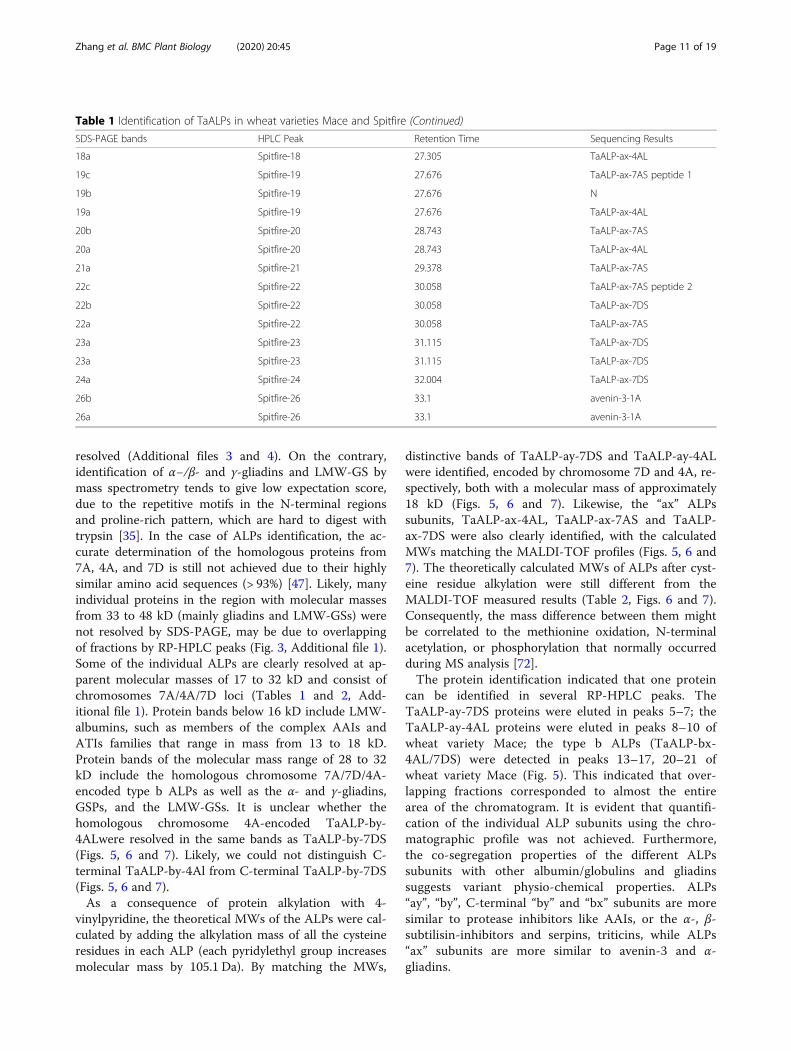
Fig. 5a-b, Additional file 1). These protein bands weredistributed in 21 HPLC peaks including peak number 8–20 and 24–30. Of these identified ALPs in Mace, bothtype a and b ALPs were present. Noteworthy, for wheatvariety Mace, type a ALPs displayed allelic variationscompared to the previously predicted ALP homologuesin wheat genome, with TaALP-ax-4AL-c allele, whiletype b ALPs had a pseudogene on chromosome 7A, theTaALP-bx-7AS silent allele. In particular, 5 type a ALPsparalogues (TaALP-ay-7DS/4AL, TaALP-ax-4AL/7AS/7DS) were found in 14 bands from HPLC peak number10–11, 17–20 & 24–30 (Table 1, Fig. 5a-b, Additionalfile 1). The MWs for these ALPs were verified byMALDI-TOF analyses (Fig. 6b-f), which were consistentwith the theoretically predicted MWs for TaALP-ay-7DS(18.42 kDa), TaALP-ay-4AL (18.47 kDa), TaALP-ax-4AL(19.47 kDa), TaALP-ax-7AS (18.75 kDa) and TaALP-ax-7DS (17.90 kDa) (Table 2). In addition, 15 protein bands(8a, 9a, 11a-18a, 20a) were identified as type b (by & bx)ALPs (Table 1). Interestingly, an additional 3 bands (8b,9b & 10 b) were identified as partial TaALP-by-4AL/7DS, displaying MW at ~ 18.34 kDa (Fig. 6a), whilst typ-ical full length type b ALPs have MW at 31.84 kDa and31.95 kDa for TaALP-by-4AL and TaALP-by-7DS,respectively (Table 2). This observation indicated theoccurrence of inter-domain cleavage specifically forTaALP-by-4AL/7DS. In addition, ALPs with two differ-ent MWs (~ 33.32 kDa & ~ 28.19 kDa) were found forprotein bands 8a, 9a, 11a and 12a. This observation
concerned TaALP-by-4AL/7AS/7DS only, and were veri-fied by MALDI-TOF analyses (Fig. 6a). Intriguingly, athird form of these 3 ALPs (TaALP-by-4AL/7AS/7DS)at MW of ~ 28.62 kDa were also detected during theMALDI-TOF analyses (Fig. 6c). These unusual forms oftype-by ALP may have resulted from a cleavage of thefull length type b ALP at the myristoylation sites,which were predicted to be present only in someALPs (Table 3). However, with myristoylation sitecleavaging, the theoretically calculated molecularweight for the “by” ALP subunits, TaALP-by-4AL,TaALP-by-7AS, TaALP-by-7DS would be 26.10 kDa,27.01 kDa and 27.41 kDa, which were smaller thantheir MALDI-TOF profiles (Table 2). In contrast tothe type-by ALPs, the identified type-bx ALPs(TaALP-bx-4AL/7DS), corresponding to bands 13a-18a, and 20a, were all found to be full length ALPsdisplaying MW at ~ 32.81 kDa (Fig. 6c), whilst fulllength “bx” ALPs have MW at 32.86 kDa and 32.46kDa for TaALP-bx-4AL and TaALP-bx-7DS, respect-ively (Table 2). This observation is consistent withthe fact that no myristoylation site has been predictedfor type-bx ALPs (Table 3). Even though the myris-toylation sites were identified for ALPs, the actualbiochemical reactions were hypothetical and need fur-ther investigation.In addition to Mace, similar analyses have been
performed on Spitfire. Only 30 HPLC peaks had beenidentified for the protein extraction in Spitfire (Fig. 5c),
Fig. 4 Phylogenetic analyses of the identified protein families from the wheat flour albumin and globulin extracts. a Maximum Likelihood (ML)phylogenetic relationship of the bread wheat (T. aestivum) PF13016 domain amino acid sequences from ALPs, CM3, GSP, alpha-gliadin andAvenin-3 sequences; b ML phylogenetic relationship of the bread wheat (T. aestivum) PF00234 domain amino acid sequences from ALPs, AAIs(CM2 and CM3), CM3, GSP, alpha-gliadin and Avenin-3 sequences
Zhang et al. BMC Plant Biology (2020) 20:45 Page 7 of 19
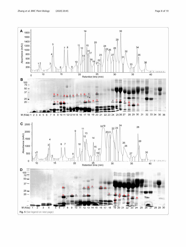
Fig. 5 (See legend on next page.)
Zhang et al. BMC Plant Biology (2020) 20:45 Page 8 of 19
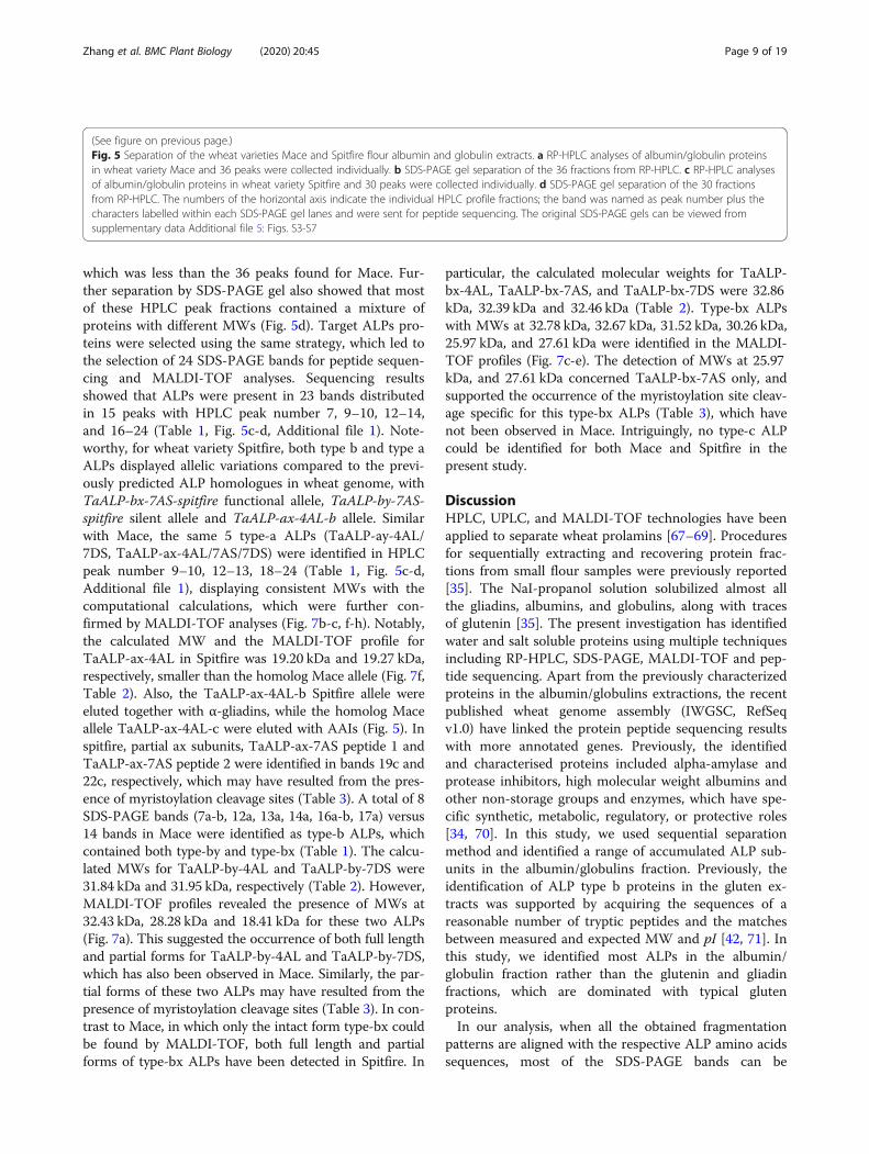
which was less than the 36 peaks found for Mace. Fur-ther separation by SDS-PAGE gel also showed that mostof these HPLC peak fractions contained a mixture ofproteins with different MWs (Fig. 5d). Target ALPs pro-teins were selected using the same strategy, which led tothe selection of 24 SDS-PAGE bands for peptide sequen-cing and MALDI-TOF analyses. Sequencing resultsshowed that ALPs were present in 23 bands distributedin 15 peaks with HPLC peak number 7, 9–10, 12–14,and 16–24 (Table 1, Fig. 5c-d, Additional file 1). Note-worthy, for wheat variety Spitfire, both type b and type aALPs displayed allelic variations compared to the previ-ously predicted ALP homologues in wheat genome, withTaALP-bx-7AS-spitfire functional allele, TaALP-by-7AS-spitfire silent allele and TaALP-ax-4AL-b allele. Similarwith Mace, the same 5 type-a ALPs (TaALP-ay-4AL/7DS, TaALP-ax-4AL/7AS/7DS) were identified in HPLCpeak number 9–10, 12–13, 18–24 (Table 1, Fig. 5c-d,Additional file 1), displaying consistent MWs with thecomputational calculations, which were further con-firmed by MALDI-TOF analyses (Fig. 7b-c, f-h). Notably,the calculated MW and the MALDI-TOF profile forTaALP-ax-4AL in Spitfire was 19.20 kDa and 19.27 kDa,respectively, smaller than the homolog Mace allele (Fig. 7f,Table 2). Also, the TaALP-ax-4AL-b Spitfire allele wereeluted together with α-gliadins, while the homolog Maceallele TaALP-ax-4AL-c were eluted with AAIs (Fig. 5). Inspitfire, partial ax subunits, TaALP-ax-7AS peptide 1 andTaALP-ax-7AS peptide 2 were identified in bands 19c and22c, respectively, which may have resulted from the pres-ence of myristoylation cleavage sites (Table 3). A total of 8SDS-PAGE bands (7a-b, 12a, 13a, 14a, 16a-b, 17a) versus14 bands in Mace were identified as type-b ALPs, whichcontained both type-by and type-bx (Table 1). The calcu-lated MWs for TaALP-by-4AL and TaALP-by-7DS were31.84 kDa and 31.95 kDa, respectively (Table 2). However,MALDI-TOF profiles revealed the presence of MWs at32.43 kDa, 28.28 kDa and 18.41 kDa for these two ALPs(Fig. 7a). This suggested the occurrence of both full lengthand partial forms for TaALP-by-4AL and TaALP-by-7DS,which has also been observed in Mace. Similarly, the par-tial forms of these two ALPs may have resulted from thepresence of myristoylation cleavage sites (Table 3). In con-trast to Mace, in which only the intact form type-bx couldbe found by MALDI-TOF, both full length and partialforms of type-bx ALPs have been detected in Spitfire. In
particular, the calculated molecular weights for TaALP-bx-4AL, TaALP-bx-7AS, and TaALP-bx-7DS were 32.86kDa, 32.39 kDa and 32.46 kDa (Table 2). Type-bx ALPswith MWs at 32.78 kDa, 32.67 kDa, 31.52 kDa, 30.26 kDa,25.97 kDa, and 27.61 kDa were identified in the MALDI-TOF profiles (Fig. 7c-e). The detection of MWs at 25.97kDa, and 27.61 kDa concerned TaALP-bx-7AS only, andsupported the occurrence of the myristoylation site cleav-age specific for this type-bx ALPs (Table 3), which havenot been observed in Mace. Intriguingly, no type-c ALPcould be identified for both Mace and Spitfire in thepresent study.
DiscussionHPLC, UPLC, and MALDI-TOF technologies have beenapplied to separate wheat prolamins [67–69]. Proceduresfor sequentially extracting and recovering protein frac-tions from small flour samples were previously reported[35]. The NaI-propanol solution solubilized almost allthe gliadins, albumins, and globulins, along with tracesof glutenin [35]. The present investigation has identifiedwater and salt soluble proteins using multiple techniquesincluding RP-HPLC, SDS-PAGE, MALDI-TOF and pep-tide sequencing. Apart from the previously characterizedproteins in the albumin/globulins extractions, the recentpublished wheat genome assembly (IWGSC, RefSeqv1.0) have linked the protein peptide sequencing resultswith more annotated genes. Previously, the identifiedand characterised proteins included alpha-amylase andprotease inhibitors, high molecular weight albumins andother non-storage groups and enzymes, which have spe-cific synthetic, metabolic, regulatory, or protective roles[34, 70]. In this study, we used sequential separationmethod and identified a range of accumulated ALP sub-units in the albumin/globulins fraction. Previously, theidentification of ALP type b proteins in the gluten ex-tracts was supported by acquiring the sequences of areasonable number of tryptic peptides and the matchesbetween measured and expected MW and pI [42, 71]. Inthis study, we identified most ALPs in the albumin/globulin fraction rather than the glutenin and gliadinfractions, which are dominated with typical glutenproteins.In our analysis, when all the obtained fragmentation
patterns are aligned with the respective ALP amino acidssequences, most of the SDS-PAGE bands can be
(See figure on previous page.)Fig. 5 Separation of the wheat varieties Mace and Spitfire flour albumin and globulin extracts. a RP-HPLC analyses of albumin/globulin proteinsin wheat variety Mace and 36 peaks were collected individually. b SDS-PAGE gel separation of the 36 fractions from RP-HPLC. c RP-HPLC analysesof albumin/globulin proteins in wheat variety Spitfire and 30 peaks were collected individually. d SDS-PAGE gel separation of the 30 fractionsfrom RP-HPLC. The numbers of the horizontal axis indicate the individual HPLC profile fractions; the band was named as peak number plus thecharacters labelled within each SDS-PAGE gel lanes and were sent for peptide sequencing. The original SDS-PAGE gels can be viewed fromsupplementary data Additional file 5: Figs. S3-S7
Zhang et al. BMC Plant Biology (2020) 20:45 Page 9 of 19
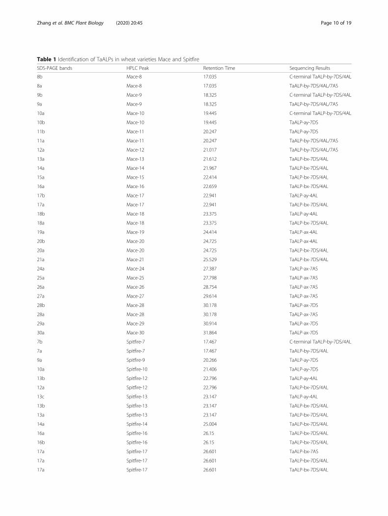
Table 1 Identification of TaALPs in wheat varieties Mace and Spitfire
SDS-PAGE bands HPLC Peak Retention Time Sequencing Results
8b Mace-8 17.035 C-terminal TaALP-by-7DS/4AL
8a Mace-8 17.035 TaALP-by-7DS/4AL/7AS
9b Mace-9 18.325 C-terminal TaALP-by-7DS/4AL
9a Mace-9 18.325 TaALP-by-7DS/4AL/7AS
10a Mace-10 19.445 C-terminal TaALP-by-7DS/4AL
10b Mace-10 19.445 TaALP-ay-7DS
11b Mace-11 20.247 TaALP-ay-7DS
11a Mace-11 20.247 TaALP-by-7DS/4AL/7AS
12a Mace-12 21.017 TaALP-by-7DS/4AL/7AS
13a Mace-13 21.612 TaALP-bx-7DS/4AL
14a Mace-14 21.967 TaALP-bx-7DS/4AL
15a Mace-15 22.414 TaALP-bx-7DS/4AL
16a Mace-16 22.659 TaALP-bx-7DS/4AL
17b Mace-17 22.941 TaALP-ay-4AL
17a Mace-17 22.941 TaALP-bx-7DS/4AL
18b Mace-18 23.375 TaALP-ay-4AL
18a Mace-18 23.375 TaALP-bx-7DS/4AL
19a Mace-19 24.414 TaALP-ax-4AL
20b Mace-20 24.725 TaALP-ax-4AL
20a Mace-20 24.725 TaALP-bx-7DS/4AL
21a Mace-21 25.529 TaALP-bx-7DS/4AL
24a Mace-24 27.387 TaALP-ax-7AS
25a Mace-25 27.798 TaALP-ax-7AS
26a Mace-26 28.754 TaALP-ax-7AS
27a Mace-27 29.614 TaALP-ax-7AS
28b Mace-28 30.178 TaALP-ax-7DS
28a Mace-28 30.178 TaALP-ax-7AS
29a Mace-29 30.914 TaALP-ax-7DS
30a Mace-30 31.864 TaALP-ax-7DS
7b Spitfire-7 17.467 C-terminal TaALP-by-7DS/4AL
7a Spitfire-7 17.467 TaALP-by-7DS/4AL
9a Spitfire-9 20.266 TaALP-ay-7DS
10a Spitfire-10 21.406 TaALP-ay-7DS
13b Spitfire-12 22.796 TaALP-ay-4AL
12a Spitfire-12 22.796 TaALP-bx-7DS/4AL
13c Spitfire-13 23.147 TaALP-ay-4AL
13b Spitfire-13 23.147 TaALP-bx-7DS/4AL
13a Spitfire-13 23.147 TaALP-bx-7DS/4AL
14a Spitfire-14 25.004 TaALP-bx-7DS/4AL
16a Spitfire-16 26.15 TaALP-bx-7DS/4AL
16b Spitfire-16 26.15 TaALP-bx-7DS/4AL
17a Spitfire-17 26.601 TaALP-bx-7AS
17a Spitfire-17 26.601 TaALP-bx-7DS/4AL
17a Spitfire-17 26.601 TaALP-bx-7DS/4AL
Zhang et al. BMC Plant Biology (2020) 20:45 Page 10 of 19
resolved (Additional files 3 and 4). On the contrary,identification of α−/β- and γ-gliadins and LMW-GS bymass spectrometry tends to give low expectation score,due to the repetitive motifs in the N-terminal regionsand proline-rich pattern, which are hard to digest withtrypsin [35]. In the case of ALPs identification, the ac-curate determination of the homologous proteins from7A, 4A, and 7D is still not achieved due to their highlysimilar amino acid sequences (> 93%) [47]. Likely, manyindividual proteins in the region with molecular massesfrom 33 to 48 kD (mainly gliadins and LMW-GSs) werenot resolved by SDS-PAGE, may be due to overlappingof fractions by RP-HPLC peaks (Fig. 3, Additional file 1).Some of the individual ALPs are clearly resolved at ap-parent molecular masses of 17 to 32 kD and consist ofchromosomes 7A/4A/7D loci (Tables 1 and 2, Add-itional file 1). Protein bands below 16 kD include LMW-albumins, such as members of the complex AAIs andATIs families that range in mass from 13 to 18 kD.Protein bands of the molecular mass range of 28 to 32kD include the homologous chromosome 7A/7D/4A-encoded type b ALPs as well as the α- and γ-gliadins,GSPs, and the LMW-GSs. It is unclear whether thehomologous chromosome 4A-encoded TaALP-by-4ALwere resolved in the same bands as TaALP-by-7DS(Figs. 5, 6 and 7). Likely, we could not distinguish C-terminal TaALP-by-4Al from C-terminal TaALP-by-7DS(Figs. 5, 6 and 7).As a consequence of protein alkylation with 4-
vinylpyridine, the theoretical MWs of the ALPs were cal-culated by adding the alkylation mass of all the cysteineresidues in each ALP (each pyridylethyl group increasesmolecular mass by 105.1 Da). By matching the MWs,
distinctive bands of TaALP-ay-7DS and TaALP-ay-4ALwere identified, encoded by chromosome 7D and 4A, re-spectively, both with a molecular mass of approximately18 kD (Figs. 5, 6 and 7). Likewise, the “ax” ALPssubunits, TaALP-ax-4AL, TaALP-ax-7AS and TaALP-ax-7DS were also clearly identified, with the calculatedMWs matching the MALDI-TOF profiles (Figs. 5, 6 and7). The theoretically calculated MWs of ALPs after cyst-eine residue alkylation were still different from theMALDI-TOF measured results (Table 2, Figs. 6 and 7).Consequently, the mass difference between them mightbe correlated to the methionine oxidation, N-terminalacetylation, or phosphorylation that normally occurredduring MS analysis [72].The protein identification indicated that one protein
can be identified in several RP-HPLC peaks. TheTaALP-ay-7DS proteins were eluted in peaks 5–7; theTaALP-ay-4AL proteins were eluted in peaks 8–10 ofwheat variety Mace; the type b ALPs (TaALP-bx-4AL/7DS) were detected in peaks 13–17, 20–21 ofwheat variety Mace (Fig. 5). This indicated that over-lapping fractions corresponded to almost the entirearea of the chromatogram. It is evident that quantifi-cation of the individual ALP subunits using the chro-matographic profile was not achieved. Furthermore,the co-segregation properties of the different ALPssubunits with other albumin/globulins and gliadinssuggests variant physio-chemical properties. ALPs“ay”, “by”, C-terminal “by” and “bx” subunits are moresimilar to protease inhibitors like AAIs, or the α-, β-subtilisin-inhibitors and serpins, triticins, while ALPs“ax” subunits are more similar to avenin-3 and α-gliadins.
Table 1 Identification of TaALPs in wheat varieties Mace and Spitfire (Continued)
SDS-PAGE bands HPLC Peak Retention Time Sequencing Results
18a Spitfire-18 27.305 TaALP-ax-4AL
19c Spitfire-19 27.676 TaALP-ax-7AS peptide 1
19b Spitfire-19 27.676 N
19a Spitfire-19 27.676 TaALP-ax-4AL
20b Spitfire-20 28.743 TaALP-ax-7AS
20a Spitfire-20 28.743 TaALP-ax-4AL
21a Spitfire-21 29.378 TaALP-ax-7AS
22c Spitfire-22 30.058 TaALP-ax-7AS peptide 2
22b Spitfire-22 30.058 TaALP-ax-7DS
22a Spitfire-22 30.058 TaALP-ax-7AS
23a Spitfire-23 31.115 TaALP-ax-7DS
23a Spitfire-23 31.115 TaALP-ax-7DS
24a Spitfire-24 32.004 TaALP-ax-7DS
26b Spitfire-26 33.1 avenin-3-1A
26a Spitfire-26 33.1 avenin-3-1A
Zhang et al. BMC Plant Biology (2020) 20:45 Page 11 of 19

The elution time differences between wheat cultivarMace and Spitfire might be due to the genotypic differ-ences. For the TaALP genes encoding loci, three genesdisplayed allelic variations and resulted in the differentdistribution of the corresponding proteins in the RP-HPLC profile and SDS-PAGE gels, as evidenced by thealleles TaALP-ax-4AL-b (Spitfire allele) and TaALP-ax-4AL-c (Mace allele), with retention times first identifiedat 24.41 min and 27.30 min, respectively (Table 1, Figs.5, 6 and 7). This is consistent with the 3D protein mod-elling results between the two alleles (Fig. 2). The differ-ent hydrophobicity profile explains their solubilityvariances in water and non-polar solvents. The othertwo alleles are silent alleles identified of TaALP-bx-7AS-Mace and TaALP-by-7AS-Spitfire encoding gene for
Mace and Spitfire, respectively, which resulted in the ab-sence of the actual protein product (Figs. 1, 5, 6 and 7a).Identification of the PTMs of ALPs was supported by
molecular mass based on MALDI-TOF analysis of RP-HPLC fractions. Specifically, the prediction of the myris-toylation sites of ALPs (Table 3) supported the posttranslational cleaving of ALPs at the myristoylation sites.Unfortunately, we have no direct experimental evidencefor the myristoylation of the ALPs. Whereas the inter-chain cleavage of “by” ALPs subunits were confirmed bythe C-terminal ALP peptides identified on the SDS-PAGE gels, which suggests that ALPs might function asprotease interacting substrates. As reported, the C-terminal by-7DS ALP are interacting with Fusarium gra-minearum beta-glucosidase and wheat metacaspase-4
Fig. 6 MALDI-TOF profiles of the peaks containing ALP proteins from wheat variety Mace. a The MALDI-TOF profile of C-terminal TaALP-by-4AL/7DS and TaALP-by-4AL/7AS/7DS in peak 8. b The MALDI-TOF profile of TaALP-ay-7AS in peak 11. c The MALDI-TOF profile of TaALP-ay-4AL,TaALP-by-4AL/7AS/7DS and TaALP-bx-4AL/7DS in peak 17. d The MALDI-TOF profile of TaALP-ax-4AL in peak 20. e The MALDI-TOF profile ofTaALP-ax-7AS in peak 26. f The MALDI-TOF profile of TaALP-ax-7DS in peak 29. Those peaks not identified as ALP and its derivatives in theMALDI-TOF profile were not labelled
Zhang et al. BMC Plant Biology (2020) 20:45 Page 12 of 19
based on a yeast two hybrid assay [59]. Further, the dif-ferences between the calculated MWs and the MALDI-TOF analysed results further indicated the occurrence ofmore than one PTMs, such as the acetylation, formyla-tion, methionine oxidation, phosphorylation, ubiquitina-tion and glycosylation, that are likely to happen to theALPs (Table 2, Figs. 6 and 7). Future research on thePTMs of ALPs can give more information to this area.The identities of individual proteins separated by RP-
HPLC here were also correlated with those of proteinsresolved by others work. Shewry et al. [73] characterizedcertain seed albumins from different wheat species byN-terminal sequencing and found that several belongedto the AAIs and ATIs family. By using wheat null gen-etic lines, Singh and Skerritt [33] established the locationof several of their genes on individual chromosomes foralbumin and globulin proteins. SDS-PAGE analysis ofwater-soluble proteins indicated the chromosomal loca-tion of polypeptides and proteins of different molecularweight were assigned on and 1D, 2A, 2B, 2D, 3AL, 3BS,
3DS, 4AL, 4BS, 4DS, 4DL, 5DL, 6DS, 7BS or 7DL [33].In our study, besides the identification of ALPs onchromosome arms 7DS, 4AL, and 7AS, it is alsodisplayed in our analysis that other water- and salt-soluble proteins were located to chromosomes 1A/1B/1D (Avenin-3, Gamma-gliadin B, γ-gliadins and LMW-GS), 2A/2B/2D (alpha-amylase/subtilisin inhibitor), 3A/3B/3D (Alpha-amylase inhibitor), 4B/4D (AAIs CM3),5A/5B/5D (GSP), 6A/6B (α-gliadins), 7A/7B/7D (60Sacidic ribosomal protein, AAIs CM2). Immunologicaland N-terminal sequencing characterisation identifiedmost of the water-soluble proteins belonged to a familyof AAIs, serine carboxypeptidase III homologous protein,while the salt-soluble proteins matched with barley em-bryo globulins, other proteins include, LTP, peroxidaseBP-1 precursor and histone H4 proteins [34]. Theprotein sequences identified could be used for molecularmarker development and selection in breeding pro-grammes. Information on the genetics and regulation ofthis fraction of proteins is necessary to understand their
Table 2 Summary of the identification of TaALPs in wheat varieties Spitfire and Mace
ALPs AAs Cysteine residues Main Peak of HPLC Retention time (Min) MW1 a (kDa) MW2 b (kDa) MW2 c (Da) MW2 d (Da)
C-terminal TaALP-by-7DS 152 11 Mace-8 17.03 17.39 18.54 – –
C-terminal TaALP-by-4AL 152 11 Mace-8 17.03 17.44 18.60 – –
TaALP-ay-7DS 154 14 Mace-11 20.24 16.96 18.42 9133.37 10,070.63
TaALP-ay-4AL 153 14 Mace-17 22.94 17.01 18.47 9198.44 10,135.7
TaALP-ax-4AL 162 14 Mace-20 24.72 18.01 19.47 10,198.54 11,135.8
TaALP-ax-7AS 156 14 Mace-26 28.75 17.29 18.75 9579.93 10,517.19
TaALP-ax-7DS 149 14 Mace-29 30.94 16.44 17.90 9914.25 10,851.51
TaALP-by-7DS 261 19 Mace-8-12 17.03–21.02 29.97 31.95 25,439.88 27,406.12
TaALP-by-4AL 261 19 Mace-8-12 17.03–21.03 29.87 31.84 24,533.1 26,095.2
TaALP-by-7AS 261 19 Mace-8-12 17.03–21.04 29.69 31.67 25,347.9 27,014.14
TaALP-bx-7DS 266 18 Mace-13-23 21.61–26.34 30.59 32.46 – –
TaALP-bx-4AL 267 19 Mace-13-24 21.61–26.35 30.88 32.86 – –
C-terminal TaALP-by-7DS 152 11 Spitfire-8 17.46 17.39 18.54 – –
C-terminal TaALP-by-4AL 152 11 Spitfire-8 17.46 17.44 18.60 – –
TaALP-ay-7DS 154 14 Spitfire-11 20.26 16.96 18.42 9133.37 10,070.63
TaALP-ay-4AL 153 14 Spitfire-15 23.14 17.01 18.47 9198.44 10,135.7
TaALP-ax-4AL 162 14 Spitfire-21 27.67 17.74 19.20 10,157.53 11,094.79
TaALP-ax-7AS 156 14 Spitfire-23 29.37 17.29 18.75 9579.93 10,517.19
TaALP-ax-7DS 149 14 Spitfire-25 31.11 16.44 17.90 9914.25 10,851.51
TaALP-by-7DS 261 19 Spitfire-8 17.46 29.97 31.95 25,439.88 27,406.12
TaALP-by-4AL 261 19 Spitfire-8 17.46 29.87 31.84 24,533.1 26,095.2
TaALP-bx-7DS 266 18 Spitfire-13-19 21.97–26.60 30.59 32.46 – –
TaALP-bx-4AL 267 19 Spitfire-13-19 21.97–26.60 30.88 32.86 – –
TaALP-bx-7AS 266 18 Spitfire-13-19 21.97–26.60 30.52 32.39 24,853.38 26,519.62aCalculated molecular weight of ALPs; b Calculated molecular weight of ALPs after molecule alkylation; c Calculated molecular weight of cleaved ALPs; dCalculatedmolecular weight of cleaved ALPs after molecule alkylation. Note: The theory was that each cysteine residue would combine with one 4-vp molecule and themolecular mass would increase 104.14 Da (the 4-vp molecular mass minus the mass of one hydrogen ion). The cleavage of ALP occurred at the predictedmyristoylation cleavage sites
Zhang et al. BMC Plant Biology (2020) 20:45 Page 13 of 19

role and function in the grain. It is likely that proteinswith similar physio-chemical properties are accumulatedin the same fraction. The ALPs identified together withother antifungal proteins in albumin and globulin frac-tion might indicate similar antifungal functions. Thisstudy provided separation solutions for future ALP func-tional study. The results can be utilized directly bybreeding programs aiming for wheat quality and diseaseresistance improvement.
ConclusionsWith the combination of multiple techniques, we re-ported for the first time the complete profiling of ALPspresent in the albumin and globulin fractions of wheatgrain protein extracts. We concluded that majority ofthe ALPs homologs are expressed in wheat grains. Wefound clear evidence of PTMs in several ALPs peptides.The identification of both gliadin domain (PF13016) andTryp_alpha_amyl domain (PF00234) in the mature formsof ALPs highlighted the multiple functional properties ofALPs in grain quality and disease resistance.
MethodPlant materials, reagents and chemicalsAll wheat materials were provided by Australian GrainResearch & Development Corporation. Australian PrimeHard (APH) variety Spitfire and Australian Hard (AH)
variety Mace from the 2014–2015 APH field trial wereharvested in Macalister of Queensland and Bellata ofNew South Wales, respectively. The unpolished maturitygrain samples were ground whole for protein extraction.All solvents and chemicals used for sample preparationwere either HPLC grade or analytical quality, unlessstated otherwise. Dithiothreitol (DTT), trifluoraceticacid, 4-vinylpyridine (4VP) and acetonitrile, Sinapinicacid (SA) were purchased from Sigma Chemical Co., St.Louis, MO, USA.
Gene cloning and sequencingThe primer pairs used in this study were the same as be-ing published by Zhang et al. [47] to amplify TaALPfragments from the genomic DNA of wheat varieties,Living Stone, Chinese Spring (CS), Spitfire, Drysdale,RAC875, Lincoln, Kauz, Excalibur, Chara, Baxter, Mace,Bonnie Rock, Gliadius, Greygory, Kukri, Westonia, Yitpi,Wyalketchem, Bethleyhem, and Eagle Rock. PCR ampli-fication cycles consisted of 1 cycle =3min 95 °C; 35 cy-cles = 30 s 95 °C, 30 s 60–62 °C, 1 min 72 °C; 1 cycle = 5min 72 °C. The target PCR products were separated by1.5% (w/v) agarose gel electrophoresis, and the expectedfragments were purified from the gel using a Gel Extrac-tion Kit (Promega, Madison, WI, USA). Subsequently,the purified PCR products were amplified using Big-Dye@version 3.1 terminator mix (Applied Biosystems)and submitted for Sanger sequencing. Alignment ofALPs was carried out using the MUSCLE add-on tool inGeneious Pro software (v10.2.2).
Sequence alignment and protein modellingAmino acid sequence alignment was carried out usingthe Multiple Sequence Alignment tool [74] at http://multalin.toulouse.inra.fr/ and was further annotatedusing the ESPript 3.0 tool [75]. Tertiary structure model-ling was performed using the template threading methodwith default parameters implemented at the I-TASSERserver [64, 65] at (https://zhanglab.ccmb.med.umich.edu/I-TASSER/). The structure templates identified andused for modelling include PDB: 2LVF, 1W1Q, 1PSY,1SM7 and 5 U87. Five models were generated for eachsubmitted amino acid sequence, of which, the top-ranking model was used for structural analyses. The se-lected models for TaALP-ax-4AL in CS and Mace haveC-score at − 2.18 and − 2.26, respectively. Consideringthe flexible and un-modelled N- and C-terminal regions,the overall quality of the generated are of high-quality.Protein structure visualization was implemented usingPyMol V1.7.4.5 software [76].
PhylogenyThe PF00234 and PF13016 domains for AAIs (CM2 andCM3), Avenin-3, alpha-gliadin, GSP, and ALPs were
Table 3 N-myristoylation site prediction of TaALPs in wheatvarieties Mace and Spitfire
ALPs location N-myristoylation site
TaALP-ay-4AL 101–106 GQSFGQ
TaALP-ay-7AS 102–107 GQSFSQ
TaALP-ay-7DS 102–107 GQSFGQ
TaALP-ay-7DS 113–118 GQSFGQ
TaALP-ax-4AL 110–115 GQRFGQ
121–126 GQSFGQ
TaALP-ax-7AS 104–109 GQRFGQ
115–120 GQSFGQ
TaALP-ax-7DS 108–113 GQSFGQ
TaALP-by-4AL 62–67 GTPFSQ
239–244 GLRMSL
TaALP-by-7AS_Mace 239–244 GLRMSL
TaALP-by-7DS 239–244 GLRMSL
TaALP-bx-7AS_Spitfire 233–238 GMYQAQ
TaALP-c-4AL 28–33 GSEQCQ
115–120 GMSQSQ
TaALP-c-7AS 142–147 GIPMAR
150–155 GGWVCE
TaALP-c-7DS 28–33 GSEQCQ
Zhang et al. BMC Plant Biology (2020) 20:45 Page 14 of 19

identified by hmmscan search against Pfam database[77] and used for phylogeny development. Codon-basedCDS sequence alignments and amino acid sequencealignments were performed using MUSCLE software
with default settings. The phylogenetic analysis was doneusing Maximum likelihood (ML) [78] in MEGA7 [79].The JTT +G (5 categories) amino acid substitutionmodel was used with 500 times bootstrapping test.
Fig. 7 MALDI-TOF profiles of the peaks containing ALP proteins from wheat variety Spitfire. a The MALDI-TOF profile of C-terminal TaALP-by-4AL/7DS and TaALP-by-4AL/7DS in peak 7. b The MALDI-TOF profile of TaALP-ay-7AS in peak 9. c The MALDI-TOF profile of TaALP-ay-4AL, TaALP-by-4AL/7DS and TaALP-bx-4AL/7AS/7DS in peak 13. d The MALDI-TOF profile of TaALP-bx-7AS and TaALP-bx-4AL/7AS/7DS in peak 16. e The MALDI-TOF profile of TaALP-bx-7AS and TaALP-bx-4AL/7AS/7DS in peak 17. f The MALDI-TOF profile of TaALP-ax-4AL in peak 19. g The MALDI-TOFprofile of TaALP-ax-7AS in peak 21. h The MALDI-TOF profile of TaALP-ax-7DS in peak 23. Those peaks not identified as ALP and its derivatives inthe MALDI-TOF profile were not labelled
Zhang et al. BMC Plant Biology (2020) 20:45 Page 15 of 19
Near infrared transmission spectroscopy (NIRS) analysisWheat cultivar Mace and Spitfire grain samples wereused for NIRS analysis without grinding. Three repli-cates were recorded per sample. Grain protein content(%) and Moisture content (%) were determined by NIRSusing the CropScan 3000F Flour and Grain Analyser.
Protein extractionThe albumin/globulin proteins were extracted from 100mg of flour according to the procedure of Dupont et al.[35]. Briefly, 100 mg of flour was extracted with 1 mL of0.3M NaI, 7.5% 1-propanol (NaI-propanol), and centri-fuged at 4500 g for 10 min, after two extractions, thesupernatant fractions were pooled in 15 mL tubes, pre-cipitated with four volumes of ice-cold (− 20 °C) NH4Ac-MeOH (0.1M ammonium acetate in 100% methanol),stored at − 20 °C for at least 48 h, and centrifuged asabove. The supernatant fluids were transferred into 50mL tubes and precipitated with four volumes of ice-coldacetone and incubated at − 20 °C overnight. Followingincubation, the fluid was centrifuged as above to yield al-bumin/globulin fraction pellets. The yield estimation ofthe extract is 10%.
RP-HPLCFreeze-dried protein pellets were dissolved in 500 μL 6M guanidine HCl (with a concentration of 1 mgmL− 1)adjusted to pH 8.0 with TRIS, plus 50 mM DTT, andthen alkylated with 4VP, prior to HPLC analysis [35]. Al-bumin and globulin proteins extracted from Spitfire andMace seeds were analyzed by RP-HPLC. A 1200 SeriesQuaternary HPLC-System was used, together with a SB-C8 reversed-phase analytical column (5 μm, 4.6 × 250mm), and a diode array UV-Vis detector (Agilent Tech-nologies, Palo Alto, CA, USA). The column temperaturewas set at 40 °C. Two mobile solvents were used for lin-ear gradient separation, with solvent A and solvent Bconsisted of 0.1% TFA (v/v) in ultrapure water (18 MΩ)and 0.1% TFA (v/v) in ACN, respectively. The flow ratewas set at 0.6 mL/min. The protein absorbance was de-tected at 210 nm wavelength. The elution gradient con-ditions were set as follows: from 0 to 51 min, eluent Bwas increased from 20 to 60%; from 51 to 53 min, eluentB was increased from 60 to 80% and then maintained at80% for 5 min for washing the column, then decreasedto the starting B concentration in 1 min and maintainedfor 10 min for the next run. The injection volume was100 μL. The proteins eluted from individual peaks werecollected with reference to the chromatographic profilecaptured in real time and pooled from three runs. RP-HPLC chromatographic fingerprint profiles showed novariation between runs, thus the elution of each runcould be combined to increase the amount of protein inthe final sample for later analysis. Samples were
immediately frozen at − 80 °C for 24 h and lyophilized.Lyophilized samples were stored at room temperaturebefore MALDI-TOF and SDS-PAGE analyses.
MALDI-TOFMALDI-TOF-MS was used to obtain the mass spectraprofile of albumin/globulin fractions obtained fromindividual HPLC peaks (fractions) with and without4VP alkylation. The albumin/globulin fraction proteinextracts were prepared for MALDI-TOF-MS test,whereas the pelleted RP-HPLC eluted protein sampleswere diluted 20 times for MALDI-TOF-MS test. Eachindividual RP-HPLC eluates were lyophilized, thefreeze-dried eluates were dissolved with 10 μL ultra-pure water, 1 μL was used for MALDI-TOF-MS, andthe residues were saved for SDS-PAGE running.Sample preparation was carried out according to thedried droplet method [80], using sinapinic acid (SA)as matrix. The matrix solution was prepared bydissolving SA in ACN/H2O/MeOH (60:8:32 v/v) at aconcentration of 20 mg/mL. All samples, including theRP-HPLC eluates, the raw albumin/globulin extractsand the alkylated albumin/globulins extracts weremixed with SA at the ratio of 1:9 (v/v) individually,and firstly, 1 μL of this protein-SA mixture was de-posited onto a 100-sample MALDI probe tip. Afterdrying, another 1 μL of this protein-SA mixture wasadded, then dried at room temperature. The massspectra for each sample was recorded on a VoyagerDE-PRO TOF mass spectrometer (Applied Biosys-tems, Foster City, CA, USA) using a positive linearion mode at an accelerating voltage of 25 kV and adelay time of 700 ns by capturing 1000 spectra of asingle laser shot with a mass range of 15,000–45,000m/z.
SDS-pageTo identify the ALPs from RP-HPLC eluates, SDS-PAGE was used to separate the protein mixtures of eachRP-HPLC eluate, and SDS-PAGE bands of interest werecut for protein peptides sequencing. The 12% SDS-PAGE was prepared following Fling and Gregerson’smethod [81]. Pelleted samples of HPLC eluates de-scribed above were mixed with 10 μL 2 × laemmli samplebuffer SDS loading buffer (Bio Rad). Electrophoresis wascarried out in a modified Laemmli system [82]. Runswere performed with running buffer of 25 mM Tris-HCL, 192 mM glycine and 0.1% SDS at 120 V for 2 h.The gels were stained in Coomassie Brilliant Blue (CBB)solution (R-250). Protein standards (Bio-Rad) were usedto estimate the molecular size of the proteins. The gelswere scanned by a gel Proteomic Imaging System “Imagelab 5.0” (Bio-Rad).
Zhang et al. BMC Plant Biology (2020) 20:45 Page 16 of 19

Protein identification by MS/MSProtein bands of interest were manually excised fromthe SDS-PAGE gels and analysed by Proteomics Inter-national Ltd. Pty, Perth, Australia. Protein samples weretrypsin digested and the resulting peptides were ex-tracted as previously described [83]. For each proteinband sample, 125 nanograms of trypsin were added fordigestion. The protein spots identification by MS/MSwere as previously described [53].
Supplementary informationSupplementary information accompanies this paper at https://doi.org/10.1186/s12870-020-2259-z.
Additional file 1. Peptide sequencing results of albumin/globulinextracts proteins.
Additional file 2. PF00234 and PF13016 domain sequences of albumin/globulin extracts proteins.
Additional file 3. ALP proteins identification from wheat cv. Mace.
Additional file 4. ALP proteins identification from wheat cv Spitfire.
Additional file 5. Original SDS-PAGE gels for Figs. 3 and 5.
AbbreviationsACN: Acetonitrile; CS: Chinese Spring; DAP: Days after pollination;EPP: Extractable polymeric protein; KDa: Kilodalton; MALDI-TOF: MatrixAssisted Laser Desorption/Ionization - Time of Flight; MW: Molecular Weight;RP-HPLC: Reversed phase-High Performance Liquid Chromatography;TFA: Trifluoroacetic acid; UPP: Unextractable polymeric protein
AcknowledgementsNot Applicable.
DeclarationsNot applicable.
Authors’ contributionsWM conceived the study. YZ and WM developed the manuscript. YZ and XHperformed the gene sequencing. YZ, AJ and WD performed phylogeny,evolutionary and protein modelling analyses. YZ, XH, YunZ and ZYperformed the RP-HPLC experiments, XH ran MALDI-TOF analyses. SI and GLinterpreted the peptide sequencing results. All authors have read andapproved the manuscript.
FundingThe study is financially supported by Australian Grain Research &Development Corporation project UMU00043 and Murdoch UniversityStrategic PhD scholarship (For Yujuan Zhang). The funders were not involvedin the experimental design of the study, data collection, analysis andinterpretation, and in writing the manuscript.
Availability of data and materialsAll data generated or analysed during this study are included in thispublished article and its supplementary information files.
Ethics approval and consent to participateNot applicable.
Consent for publicationNot applicable.
Competing interestsThe authors declare that they have no competing interest.
Author details1Australia-China Joint Centre for Wheat Improvement, Western AustralianState Agriculture Biotechnology Centre, School of Veterinary and LifeSciences, Murdoch University, Perth, WA 6150, Australia. 2The Key Laboratoryfor Quality Improvement of Agricultural Products of Zhejiang Province,School of Agriculture and Food Science, Zhejiang A&F University, Linan,Zhejiang 311300, Hangzhou, China. 3School of Agriculture, Food and Wine,University of Adelaide, Adelaide 5005, Australia. 4Nutritional Crop Physiology,Institute of Crop Science, University of Hohenheim, 70599 Stuttgart,Germany.
Received: 28 July 2019 Accepted: 20 January 2020
References1. Kreis M, Forde BG, Rahman S, Miflin BJ, Shewry PR. Molecular evolution
of the seed storage proteins of barley, rye and wheat. J Mol Biol. 1985;183(3):499–502.
2. Kreis M, Shewry PR, Forde BG, Forde J, Miflin BJ. Structure and evolution ofseed storage proteins and their genes, with particular reference to those ofwheat, barley and rye, vol. 2. Oxford: Oxford University Press; 1985b.
3. Shewry PR, Tatham AS. The prolamin storage proteins of cereal seeds:structure and evolution. Biochem J. 1990;267(1):1–12.
4. Shewry PR, Tatham AS. The characteristics, structures and evolutionaryrelationships of prolamins. Seed Proteins.Springer. Dordrecht. 1999. p. 11–33.
5. Shewry PR, Jenkins J, Beaudoin F, ENC M. The classification, functions andevolutionary relationships of plant proteins in relation to food allergens.Oxford, UK: Blackwell Science; 2004.
6. Douliez JP, Michon T, Elmorjani K, Marion D. Mini review: structure,biological and technological functions of lipid transfer proteins andIndolines, the major lipid binding proteins from cereal kernels. J Cereal Sci.2000;32(1):1–20.
7. Josè-Estanyol M, Puigdomènech P. Plant cell wall glycoproteins and theirgenes. Plant Physiol Biochem. 2000;38(1–2):97–108.
8. Juhász A, Belova T, Florides CG, Maulis C, Fischer I, Gell G, Birinyi Z,Ong J, Keeble-Gagnère G, Maharajan A. Genome mapping of seed-borne allergens and immunoresponsive proteins in wheat. Scienceadvances. 2018;4(8):eaar8602.
9. Gijzen M, Miller SS, Kuflu K, Buzzell RI, Miki BL. Hydrophobic proteinsynthesized in the pod endocarp adheres to the seed surface. Plant Physiol.1999;120(4):951–60.
10. Gómez J, Sánchez-Martínez D, Stiefel V, Rigau J, Puigdomènech P, Pagès M.A gene induced by the plant hormone abscisic acid in response to waterstress encodes a glycine-rich protein. Nature. 1988;334(6179):262.
11. Bradley DJ, Kjellbom P, Lamb CJ. Elicitor-and wound-induced oxidativecross-linking of a proline-rich plant cell wall protein: a novel, rapid defenseresponse. Cell. 1992;70(1):21–30.
12. Sprunck S, Rademacher S, Vogler F, Gheyselinck J, Grossniklaus U,Dresselhaus T. Egg cell–secreted EC1 triggers sperm cell activation duringdouble fertilization. Science. 2012;338(6110):1093–7.
13. Wang SY, Wu JH, Ng T, Ye XY, Rao PF. A non-specific lipid transfer proteinwith antifungal and antibacterial activities from the mung bean. Peptides.2004;25(8):1235–42.
14. Shorrosh BS, Wen L, Zen K-C, Huang J-K, Pan JS, Hermodson MA, Tanaka K,Muthukrishnan S, Reeck GR. A novel cereal storage protein: moleculargenetics of the 19 kDa globulin of rice. Plant Mol Biol. 1992;18(1):151–4.
15. Miyahara K, Nishio T. Rice mutant lines lacking α-globulin. Japanese Journalof Breeding. 1998;48(1):45–9.
16. Terras FR, Eggermont K, Kovaleva V, Raikhel NV, Osborn RW, Kester A, ReesSB, Torrekens S, Van Leuven F, Vanderleyden J. Small cysteine-rich antifungalproteins from radish: their role in host defense. Plant Cell. 1995;7(5):573–88.
17. Warchalewski J. Purification and characteristics of an endogenous alpha-amylase and trypsin inhibitor from wheat seeds. Mol Nutr Food Res. 1987;31(10):1015–31.
18. Heidari R, Zareae S, Heidarizadeh M. Extraction, purification, and inhibitoryeffect of alpha-amylase inhibitor from wheat (Triticum aestivum Var. Zarrin).Pakistan J Nutr. 2005;4:101–5.
19. Jolly C, Rahman S, Kortt AA, Higgins T. Characterisation of the wheat Mr15000 grain-softness protein and analysis of the relationship between its
Zhang et al. BMC Plant Biology (2020) 20:45 Page 17 of 19
accumulation in the whole seed and grain softness. Theor Appl Genet.1993;86(5):589–97.
20. Giroux M, Morris C. A glycine to serine change in puroindoline b isassociated with wheat grain hardness and low levels of starch-surfacefriabilin. Theor Appl Genet. 1997;95(5–6):857–64.
21. Giroux MJ, Morris CF. Wheat grain hardness results from highly conservedmutations in the friabilin components puroindoline a and b. Proc Natl AcadSci. 1998;95(11):6262–6.
22. van Herpen TW, Goryunova SV, van der Schoot J, Mitreva M, Salentijn E,Vorst O, Schenk MF, van Veelen PA, Koning F, van Soest LJ. Alpha-gliadingenes from the a, B, and D genomes of wheat contain different sets ofceliac disease epitopes. BMC Genomics. 2006;7(1):1.
23. Perdon AA, Juliano BOJP. Properties of a major α-globulin of riceendosperm. Phytochemistry. 1988;17(3):351–3
24. Kumamaru T, Satoh H, Iwata N, Omura T, Ogawa M, Tanaka K. Mutants forrice storage proteins. Theor Appl Genet. 1988;76(1):11–6.
25. Chen S-CG, Chen J, Hwang L-Y, Cheng M-C. Biochemical and immunologicalproperties of indica Rice. Bot. Bull. Academia Sinica. 1987;28:237–46.
26. Limas GG, Salinas M, Moneo I, Fischer S, Wittmann-Liebold B, Méndez EJP.Purification and characterization of ten new rice NaCl-soluble proteins:identification of four protein-synthesis inhibitors and two immunoglobulin-binding proteins. Planta. 1990;181(1):1–9.
27. Matsuda T, Nomura R, Sugiyama M, Nakamura RJA. Immunochemicalstudies on rice allergenic proteins. Agric Biol Chem. 1991;55(2):509–13.
28. Steenson DF, Sathe SK. Characterization and digestibility of Basmati rice(Oryza sativa L. var. Dehraduni) storage proteins. Cereal Chem. 1995;72(3):275–80.
29. Usui Y, Nakase M, Hotta H, Urisu A, Aoki N, Kitajima K, Matsuda T. A 33-kDaallergen from rice (Oryza sativa L. japonica) cDNA cloning, expression, andidentification as a novel glyoxalase I. J Biol Chem. 2001;276(14):11376–81.
30. Lang GH, Kagiya Y, Ohnishi-Kamayama M, Kitta K. Evaluation of solutions forbiochemical analyses of the proteins in rice grains. Biosci BiotechnolBiochem. 2013;77(1):126–31.
31. Lookhart G, SJCC B. Separation and characterization of wheat proteinfractions by high-performance capillary electrophoresis. Cereal Chem. 1995;72(6):527–32.
32. Žilić S, Barać M, Pešić M, Dodig D, Ignjatović-Micić D. Characterization ofproteins from grain of different bread and durum wheat genotypes. Int JMol Sci. 2011;12(9):5878–94.
33. Singh J, Skerritt JH. Chromosomal control of albumins and globulins inwheat grain assessed using different fractionation procedures. J Cereal Sci.2001;33(2):163–81.
34. Singh J, Blundell M, Tanner G, Skerritt JH. Albumin and globulin proteins ofwheat flour: immunological and N-terminal sequence characterisation. JCereal Sci. 2001;34(1):85–103.
35. DuPont FM, Chan R, Lopez R, Vensel WH. Sequential extraction andquantitative recovery of gliadins, glutenins, and other proteins from smallsamples of wheat flour. J Agric Food Chem. 2005;53(5):1575–84.
36. Fu B, Kovacs M. Rapid single-step procedure for isolating total gluteninproteins of wheat flour. J Cereal Sci. 1999;29(2):113–6.
37. Mcwilliams CS. The chemistry and Technology of Cereals as food and feed -Matz. Sa J Home Econ. 1959;51(10):898.
38. Buonocore V, Debiasi MG, Giardina P, Poerio E, Silano V. Purification andproperties of an alpha-amylase tetrameric inhibitor from wheat kernel.Biochim Biophys Acta. 1985;831(1):40–8.
39. Shewry PR, Field JM, Faulks AJ, Parmar S, Miflin BJ, Dietler MD, Lew EJ,Kasarda DD: The purification and N-terminal amino acid sequence analysisof the high molecular weight gluten polypeptides of wheat. Biochimica etBiophysica Acta (BBA)-Protein Structure and Molecular Enzymology 1984,788(1):23–34.
40. Østergaard H, Rasmussen SK, Roberts TH, Hejgaard J. Inhibitory serpins fromwheat grain with reactive centers resembling glutamine-rich repeats ofprolamin storage proteins cloning and characterization of five majormolecular forms. J Biol Chem. 2000;275(43):33272–9.
41. García Olmedo F, Carmona Quiles MJ, Lopez Fando J, Fernandez JA,Castagnaro A, Molina Fernández A, Hernandez Lucas C, CarboneroZalduegui P. Characterization and analysis of thionin genes. Plant GeneResearch Genes Involved in Plant Defense. 1992:283–302.
42. De Caro S, Ferranti P, Addeo F, Mamone G. Isolation and characterization ofAvenin-like protein type-B from durum wheat. J Cereal Sci. 2010;52(3):426–31.
43. Vensel WH, Tanaka CK, Altenbach SB. Protein composition of wheat glutenpolymer fractions determined by quantitative two-dimensional gelelectrophoresis and tandem mass spectrometry. Proteome Sci. 2014;12(1):8.
44. Colgrave ML, Goswami H, Byrne K, Blundell M, Howitt CA, Tanner GJ. Proteomicprofiling of 16 cereal grains and the application of targeted proteomics to detectwheat contamination. J Proteome Res. 2015;14(6):2659–68.
45. Kan Y, Wan Y, Beaudoin F, Leader DJ, Edwards K, Poole R, Wang D, MitchellRAC, Shewry PR. Transcriptome analysis reveals differentially expressedstorage protein transcripts in seeds of Aegilops and wheat. J Cereal Sci.2006;44(1):75–85.
46. Egorov TA, Musolyamov AK, Andersen JS, Roepstorff P. The complete aminoacid sequence and Disulphide bond arrangement of oat alcohol-solubleAvenin-3. FEBS J. 1994;224(2):631–8.
47. Zhang Y, Hu X, Islam S, She M, Peng Y, Yu Z, Wylie S, Juhasz A, Dowla M,Yang R. New insights into the evolution of wheat avenin-like proteins inwild emmer wheat (Triticum dicoccoides). Proc Natl Acad Sci. 2018;115(52):13312–7.
48. Kasarda DD, Adalsteins E, Lew EJL, Lazo GR, Altenbach SB. Farinin:characterization of a novel wheat endosperm protein belonging to theprolamin superfamily. J Agric Food Chem. 2013;61(10):2407–17.
49. Ma F, Li M, Li T, Liu W, Liu Y, Li Y, Hu W, Zheng Q, Wang Y, Li K, et al.Overexpression of avenin-like b proteins in bread wheat Triticum aestivum L.improves dough mixing properties by their incorporation into gluteninpolymers. PLoS One. 2013;8(7):e66758.
50. Chen XY, Cao XY, Zhang YJ, Islam S, Zhang JJ, Yang RC, Liu JJ, Li GY, AppelsR, Keeble-Gagnere G, et al. Genetic characterization of cysteine-rich type-bavenin-like protein coding genes in common wheat. Sci Rep. 2016;6.
51. Gu A, Hao P, Lv D, Zhen S, Bian Y, Ma C, Xu Y, Zhang W, Yan Y. Integratedproteome analysis of the wheat embryo and endosperm reveals centralmetabolic changes involved in the water deficit response during graindevelopment. J Agric Food Chem. 2015;63(38):8478–87.
52. Yang M, Dong J, Zhao W, Gao X. Characterization of proteins involved inearly stage of wheat grain development by iTRAQ. J Proteome. 2016;136:157–66.
53. Wang X, Appels R, Zhang X, Diepeveen D, Torok K, Tomoskozi S, Bekes F,Ma W, Sharp P, Islam S. Protein interactions during flour mixing using wheatflour with altered starch. Food Chem. 2017;231:247–57.
54. Cao H, He M, Zhu C, Yuan L, Dong L, Bian Y, Zhang W, Yan Y. Distinctmetabolic changes between wheat embryo and endosperm during graindevelopment revealed by 2D-DIGE-based integrative proteome analysis.Proteomics. 2016;16(10):1515–36.
55. Altenbach SB, Tanaka CK, Whitehand LC, Vensel WH. Effects of post-anthesisfertilizer on the protein composition of the gluten polymer in a US breadwheat. J Cereal Sci. 2016;68:66–73.
56. Subburaj S, Luo N, Lu X, Li X, Cao H, Hu Y, Li J, Yan Y. Molecularcharacterization and evolutionary origins of farinin genes in Brachypodiumdistachyon L. J Appl Genet. 2016;57(3):287–303.
57. Cao D, Wang H, Zhang B, Liu B, Liu D, Chen W, Zhang H. Genetic diversity ofavenin-like b genes in Aegilops tauschii Coss. Genetica. 2018;146(1):45–51.
58. Gao T, Li G-Z, Wang C-R, Dong J, Yuan S-S, Wang Y-H, Kang G-Z. Functionof the ERFL1a transcription factor in wheat responses to water deficiency.Int J Mol Sci. 2018;19(5):1465.
59. Zhang Y, Cao X, Juhasz A, Islam S, Qi P, She M, Zhu Z, Hu X, Yu Z, Wylie S.Wheat avenin-like protein and its significant Fusarium head blight resistantfunctions. bioRxiv. 2018:406694.
60. Högy P, Zörb C, Langenkämper G, Betsche T, Fangmeier A. AtmosphericCO2 enrichment changes the wheat grain proteome. J Cereal Sci. 2009;50(2):248–54.
61. Arachchige PMS, Ang C-S, Nicolas ME, Panozzo J, Fitzgerald G, Hirotsu N,Seneweera S. Wheat (Triticum aestivum L.) grain proteome response toelevated [CO2] varies between genotypes. J Cereal Sci. 2017;75:151–7.
62. Verrillo F, Badeck F-W, Terzi V, Rizza F, Bernardo L, Di Maro A, Fares C, ZaldeiA, Miglietta F, Moschella A. Elevated field atmospheric CO2 concentrationsaffect the characteristics of winter wheat (cv. Bologna) grains. Crop andPasture Science. 2017;68(8):713–25.
63. Donaldson PA, Anderson T, Lane BG, Davidson AL, Simmonds DH. Soybeanplants expressing an active oligomeric oxalate oxidase from the wheat gf-2.8 (germin) gene are resistant to the oxalate-secreting pathogen Sclerotinasclerotiorum. Physiol Mol Plant Pathol. 2001;59(6):297–307.
64. Roy A, Kucukural A, Zhang Y. I-TASSER: a unified platform for automatedprotein structure and function prediction. Nature protocols. 2010;5(4):725.
Zhang et al. BMC Plant Biology (2020) 20:45 Page 18 of 19

65. Yang J, Yan R, Roy A, Xu D, Poisson J, Zhang Y. The I-TASSER Suite: proteinstructure and function prediction. Nature methods. 2015;12(1):7.
66. Yu Z. Sulphur and nitrogen fertilization strategy for wheat grain qualityimprovement and the underlying mechanism: Murdoch University; 2017.
67. Weegels P, Marseille J, Bosveld P, Hamer RJ. Large-scale separation ofgliadins and their bread-making quality. J Cereal Sci. 1994;20(3):253–64.
68. Chen J, Lan P, Tarr A, Yan Y, Francki M, Appels R, Ma WJ. MALDI-TOF basedwheat gliadin protein peaks are useful molecular markers for wheat geneticstudy. 2007;21:2913–7.
69. Han C, Lu X, Yu Z, Li X, Ma W, Yan Y. Rapid separation of seed gliadins byreversed-phase ultra performance liquid chromatography (RP-UPLC) and itsapplication in wheat cultivar and germplasm identification. BiosciBiotechnol Biochem. 2015;79(5):808–15.
70. Pence JW, Weinstein N, Mecham D. The albumin and globulin contents ofwheat flour and their relationship to protein quality. Cereal Chem. 1954;31:303–11.
71. Mamone G, Caro SD, Luccia AD, Addeo F, Ferranti P. Proteomic-basedanalytical approach for the characterization of glutenin subunits in durumwheat. J Mass Spectrom. 2009;44(12):1709–23.
72. Parker CE, Mocanu V, Mocanu M, Dicheva N, Warren MR. Mass spectrometryfor post-translational modifications. Neuroproteomics. 2010;2010:PMID:21882444.
73. Shewry PR, Lafiandra D, Salcedo G, Aragoncillo C, Garcia-Olmedo F, Lew EJ-L, Dietler MD, Kasarda DD. N-terminal amino acid sequences of chloroform/methanol-soluble proteins and albumins from endosperms of wheat, barleyand related species: homology with inhibitors of α-amylase and trypsin andwith 2 S storage globulins. FEBS Lett. 1984;175(2):359–63.
74. Corpet F. Multiple sequence alignment with hierarchical clustering. NucleicAcids Res. 1988;16(22):10881–90.
75. Robert X, Gouet P. Deciphering key features in protein structures with thenew ENDscript server. 2014;42(W1):W320–4.
76. Schrodinger LL. The PyMOL molecular graphics system. 2010;1(5):0.77. Finn RD, Coggill P, Eberhardt RY, Eddy SR, Mistry J, Mitchell AL, Potter SC,
Punta M, Qureshi M, Sangrador-Vegas A. The Pfam protein familiesdatabase: towards a more sustainable future. Nucleic Acids Res. 2015;44(D1):D279–85.
78. Jones DT, Taylor WR, Thornton JM. The rapid generation of mutation datamatrices from protein sequences. Comput Appl Biosci. 1992;8(3):275–82.
79. Kumar S, Stecher G, Tamura K. MEGA7: molecular evolutionary geneticsanalysis version 7.0 for bigger datasets. Mol Biol Evol. 2016;33(7):1870–4.
80. Kussmann M, Nordhoff E, Rahbek-Nielsen H, Haebel S, Rossel-Larsen M,Jakobsen L, Gobom J, Mirgorodskaya E, Kroll-Kristensen A, Palm L. Matrix-assisted laser desorption/ionization mass spectrometry sample preparationtechniques designed for various peptide and protein analytes. J MassSpectrom. 1997;32(6):593–601.
81. Fling SP, Gregerson DS. Peptide and protein molecular weightdetermination by electrophoresis using a high-molarity tris buffer systemwithout urea. Anal Biochem. 1986;155(1):83–8.
82. Schafer-Nielsen C, Rose C. Separation of nucleic acids and chromatinproteins by hydrophobic interaction chromatography. Biochimica etBiophysica Acta (BBA)-Gene Structure Expression 1982, 696(3):323–331.
83. Bringans S, Eriksen S, Kendrick T, Gopalakrishnakone P, Livk A, Lock R,Lipscombe R. Proteomic analysis of the venom of Heterometrus longimanus(Asian black scorpion). Proteomics. 2008;8(5):1081–96.
Publisher’s NoteSpringer Nature remains neutral with regard to jurisdictional claims inpublished maps and institutional affiliations.
Zhang et al. BMC Plant Biology (2020) 20:45 Page 19 of 19