Embed Size (px)
Citation preview

Chapter 32: The X STOP Interspinous Process Decompression System for theTreatment ofLumbar Neurogenic Claudication
Richard M. Thunder MD
Ken Y.Hsu MD
James F. Zucherman MD
St. Mary's Spine CenterOne Shrader, Suite 450San Francisco, CA 94117Ph: 415-750-5849
Fx: 415-750-8103
thet][email protected]'n

1
Neurogenic intermittent claudication is the most commonand characteristic syndrome of
lumbarspinal stenosis. Patientstypicallyobtainrelief fromsittingand positionsof flexion, and
exacerbate the pain while standingor walking. The X STOP(St. FrancisMedicalTechnologies
Inc., Alameda, California) is an interspinous spacer developed to treat patients with neurogenic
intermittentclaudication (Figure 1). The implantblocks terminalextensionof the stenotic levels
of the lumbar spine by means ofa spacer placed between the spinous processes. The procedure
typicallyrequiresno generalanesthesia and can be performedin under an hour. The X STOPis
an altemativetherapy to conservative treatmentand decompressive surgeryfor patientssuffering
from lumbar spinal stenosis.' The X STOP Interspinous Process Distraction (IPD) System (X
STOP) is indicated in patients whose symptoms are exacerbated in extension and relieved in
flexion. Implantedbetween the spinous processes,the X STOPreduces extensionat the
symptomatic level and allows motion in flexion, axial rotation and lateral bending.^
Historical Perspective
Although spinal stenosis had been observed inanimals^*^ and found in Egyptian
mummies,it was probably first described in 1803,by Portal of France,who observedthat
narrowed spinal canals were associated with leg pain and atrophy. Our understanding of this
condition was not gained until about fifty years ago when Verbiest described the anatomic
changes ofhypertrophic articular processes causing spinal canal stenosis.^ Subsequently,
Kirkaldy-Williswrote about the three-joint complex and the pathologic changes found in
degenerative spinal stenosis.' Degenerative processes may start inone, two, orthree joint
complexes, including the disc anteriorly and the two facet joints posteriorly. With time, all three
joints are involved.The degeneration of the joint also causes abnormal motion, which may

produce osteophyte fonnation. Ultimately, disc protrusionor osteophyteformation, hypertrophy
of facetjoints and ligamentum flavum result in spinalstenosis. Medical literatureregarding this
condition became more available after the mid 1970's.^ The significant increase inthe reported
cases of spinalstenosis is relatedto the introduction of axial imaging provided by CT andMRI
scans.
In the United States lumbar spinal stenosis is the leadingpreoperative diagnosis for adults
older than 65 years who undergo spine surgery.' In 1996 more almost 90,000 surgeries were
performed for lumbar spinal stenosis.' Symptoms present with upright posture activity and
includeunilateral or bilateralradicularpain, sensationdisturbance and loss ofstrengthin the
lower extremities.^ Symptoms aretypically relieved with flexion the lumbar spine.
The incidence ofdegenerative lumbar stenosis ranges from 1.7 %to 8%.'° There does not
appear tobe gender predominance; however, degenerative spondylolisthesis associated with
lumbar spinal stenosis is four times morecommon among women. Symptoms typically develop
in the fifth or sixth decade of life in association with osteoarthritic changes in the lumbar spine.
Noknown relationship exists between incidence of lumbar spinal stenosis andrace. Spinal
stenosis did not have the socioeconomic significance that we see today until the 1970's. The
aging of ourpopulation is resulting in an increased incidence ofdegenerative stenosis. In 1900
the lifeexpectancy was45 years. People olderthan65constituted less than4% of the
population.'' The estimated life expectancy in 2026 is86 years with 20% ofthe population
expected tobe over 65 years of age. TheU.S. Census Bureau projections estimate doubling of
thepopulation older thanage64 yearsto 64million by 2040.
Current Treatments

Symptoms of spinal stenosis may respond to non-operative management. Conservative
measures often begin with a period of rest for one or two days as well as non-steroidalanti-
inflammatoiymedication,physical therapy and sometimes,oral steroids. In physical therapy,
trunk stabilization and core muscle strengthening is typically the goal. Epidural steroid injection
is often used as an adjunctparticularly in those patientswith unremitting radiculopathy and
neurogenic claudication. There is no clear evidence of long-term efficacy of epidural steroids;
however, they can give significant short-term relief and allow participation in physical therapy.
Outcomeswith non-operative treatment reportedby Hurri et al. showed44% of patients
had atleast some improvement inneurologic symptoms.^^ Inother studies. Atlas etal. found that
45% percent of patients had improvement in leg pain with non-operative treatment while
Johnssonet al reported 32% of patients treated non-operatively consideredtheir condition
improved.
Operative treatment is the primary indication for patients with worsening pain that is
resistant to conservative treatment. Patients with moderate to severe stenosis that do not improve
with non-operative interventions are likely to improve with surgical decompression.
Historically, the literature supporting operative treatment has been shown to have
methodological flaws with respect to indications for surgery and surgical outcome.''* However, in
the last decade, prospective studies such as the Maine Lumbar Study have shown superior
outcomes for operative treatment of symptomatic lumbar stenosis compared to non-operative
treatment.'̂ Most series report a 64% to91% rate ofimprovement following surgery.
Operative treatment while offering great potential to improve quality of life for individuals
has the potential for significant complications. Post-operative complications may include
infection, epidural hematoma, instability, non-union, instrumentationfailure and the need for

future surgery due to the development of disease at adjacent levels. From a general medical
perspective there is also a cardiac and respiratory risk particularly in elderly patients with
extensive blood loss procedures. The risk for post-operative infection remains significant despite
the practice of antibiotic prophylaxis and strict sterile technique. In a study by Yuan et. al. cohort
undergoing lumbar decompression and arthrodesis, with or without internal fixation, revealed
and infection rate of 2-3%.'̂ In this same study, therisk ofnerve root injury jfrom placement of
pedicle screws was 0.4%. In addition to nerve root injury, dural tears are not uncommon during
decompressive procedures. A study by Wang et al. which evaluated641 patients undergoing
lumbar spine surgery, halfofwhich were revisions, revealed 13.7%incidence of dural tears.A
meta-analysis of the literature performed by Turner et al in 1992 showed the following
comphcation rates for neurogenic claudication surgery: perioperative mortality (0.32%), dural
tears (5.91%), deep infection (1.08%), superficial infection (2.3%), deep vein thrombosis
(2.78%), any complication (12.64%).
X STOP Design Rationale and Pre-CIinical Confirmation
Taking into account the relatively large void of treatment optionsbetween the safer, yet
less effective conservative care, and the more risky and more effective surgical care, the X STOP
was developed to fill this void by providing a safe and effective treatment option. Based on the
clinicalpresentation of LSS patients, the X STOP was designed specifically to limit the terminal
extension movement at only the individual level(s) that provokes symptoms,while allowing
unrestricted movement of the remaining motion axes of the treated level(s). In addition the
implant was designed to be placed using a minimally invasive surgical techniquewith the patient
under local anesthesia. Finally, it was designed to be placed without altering the functional
tissues and allowing very straightforward removal should revision surgery become necessary; in
other words X STOP placement does not preclude any further surgery.
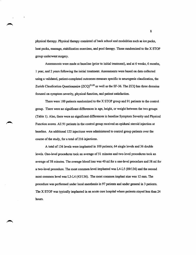
Several key design features allow for the implantation of the X STOP between the
spinous processes in a straightforward procedure that requires less than one hour ofoperative
time. The oval spacer separates the spinous processes and restricts terminal extension at the
implanted level (Figure 2). The two lateral wings prevent the implant from anterior or lateral
migration. The supraspinous ligament, which is retained during the procedure, prevents the
implant from posterior migration.
Biomechanical studies have shown that the implant significantly prevents narrowing of
the spinal canal and neural foramina, limits extension, and reduces intradiscal pressure and facet
loading.^*'In anMRI cadaver study, Richards etal. reported that XSTOP placement
increases theneural foramina areaby 26% andthespinal canal area by 18% during extension.'®
In addition, the foraminal width was increased by 41% and the subarticular canal diameter by
50% inextension.'® In a kinematics cadaver study, Lindsey etal reported that terminal extension
at the implant level was reduced by 62% following X STOP placement, while lateral bending
and axial rotation range ofmotion were unchanged. ^Ina cadaveric disc pressure study,
Swanson et al reported that the posterior annulus and nucleus pulposis pressures were reduced
by 63% and 41% respectively during extension, and by 38% and 20% respectively in the neutral,
standing position." Finally, Wiseman etal performed a cadaveric facet loading study and
reported that the mean facet force during extension decreased by68% during extension.^" In
each of these studies, the adjacent level measurements were not significantly changed from the
intact specimen state. These pre-clinical studies indicate that the X STOP increases spinal canal
and neural foramina space and also produces significant unloading of the disk and facets.

Operative Technique
Patientsare placed on a radiolucent table in a right lateral decubituspositionafter
administration of local anesthetic and intravenous sedation (Figure 3). The level to be treated is
identifiedby fluoroscopy. A mid-sagittalincision of approximately 4 cm is made over the
spinousprocessesof the stenotic level(s).This is carrieddown to the fasciawhich is incisedto
the right and to the left 2cm from the midline.The supraspinous ligamentis left intact.
Paraspinal musculature is then elevated off the spinous processesand medial laminabilaterally.
Occasionally, hypertrophied facets that blockentry into the anteriorinterspinous space are
partiallytrimmed to enableanteriorplacementof the implant. The small curveddilatoris
inserted across the interspinous space abutting the posterior facetjoints at the most anterior
marginof the interspinous space.After the correct level is verifiedby fluoroscopy, the small
dilator is removedand the larger curved dilator is insertedin the same interspinous hole created
by the small dilator. After the larger dilator is removed, the sizing instrument is insertedand
dilateduntil the supraspinous ligamentbecomesjust taught. The correct implantsize is indicated
on the sizing instrument. During the sizingand implantinsertion, thepatient is askedto
maximally flex to open the interspinous space.The appropriately sized X STOPis inserted
between the spinous processes to the pointwherethe wingis flushwith the rightside of the
spinous processes. Thescrew holefor theuniversal wing on the leftside is identified and the
universal wingscrewis engagedwith the main body hole.The two wingsare approximated
medially and the left sided universalwingscrew is securedwith a torque-limiting hex driver.
Anterior/posterior and lateral fluoroscopyviews are takento verify proper level and position.
The incision is thenclosed. The procedureis typically donewithina 24 hour hospitalization.

The X STOP multi-center randomized trial
A multi-centerprospective, randomized trial was performed comparing the outcomes of
mild to moderate neurogenic intermittent claudication patients treated with the X STOP
interspinous distraction system to those treated non-surgically.' There were 191 patients treated
in a prospective, controlled trial at 9 centers over a 15-monthperiod. Inclusion criteria included
age greater than 50 years; leg, buttock, or groin pain with or without back pain that could be
relieved during flexion; ability to sit for 50 minutes withoutpain; walk 50 feet or more;
completedat least six months of non-operative therapy; stenosis conflrmed by CT or MRI scan
at one or two levels; and ability to comply with scheduled clinical and radiographic follow-up
evaluations.
Exclusion criteria included fixed motor deficit; cauda-equina syndrome, significant
lumbarinstability; previouslumbarsurgery;significant peripheral neuropathy or acute
denervation secondaryto radiculopathy; scoliotic Cobbangle greaterthan 25 degrees;
spondylolisthesis greater than grade 1 (on a scale of 1 to4) at the affectedlevel(s);sustained
pathologic fi-actures or severe osteoporosis of the vertebrae and /or hips; obesity; active infection
or systemic disease; Paget's disease or metastasis to the vertebrae or steroid use for more than
one month within 12monthspreceding the study.Patientswere also excludedwho had anatomy
that would prevent implantationof the device, such as an ankylosedsegment, or spinal anatomy
that would cause the device to be unstable after implantation.
Eligiblepatientswere randomized to either the X STOP groupor the control group.
Those randomized to the control group received at least one epidural steroid injection and had
the option to receive non-steroidal anti-inflammatorymedications (NSAIDs), analgesics, and
8
physical therapy. Physical therapy consisted ofback school and modalities such as ice packs,
heat packs, massage, stabilizationexercises, and pool therapy.Those randomized to the X STOP
group underwent surgery.
Assessments were made at baseline (prior to initial treatment), and at 6 weeks, 6 months,
1 year, and 2 years following the initial treatment. Assessments were based on data collected
using a validated, patient-completed outcomes measure specific to neurogenic claudication, the
Zurich Claudication Questionnaire (ZCQ) '̂'̂ ^ as well as the SF-36. The ZCQ has three domains
focused on symptom severity, physical function, and patient satisfaction.
There were 100 patients randomized to the X STOP group and 91 patients to the control
group. There were no significant differences in age, height, or weight between the two groups
(Table 1). Also, there were no significant differences in baseline Symptom Severity and Physical
Function scores. All 91 patients in the control group received an epidural steroid injection at
baseline. An additional 125 injections were administered to control group patients over the
course of the study, for a total of216 injections.
A total of 136 levels were implanted in 100patients; 64 single levels and 36 double
levels. One-level procedures took an average of 51 minutes and two-level procedures took an
average of58 minutes. The average blood loss was 40 ml for a one-level procedure and 58 ml for
a two-levelprocedure. The most common level implanted was L4-L5 (89/136) and the second
most common level was L3-L4 (43/136). The most common implant size was 12 mm. The
procedure was performed under local anesthesia in 97 patients and under general in 3 patients.
The X STOP was typically implanted in an acute care hospital where patients stayed less than 24
hours.

At 2-yearsfollow-up, data from 93 of the 100X STOPpatients and 81 of the 91 control
patients were available for analysis. In the X STOPgroup, sevenpatients were lost to follow-up;
fourpatients died, twopatients failedto complete the ZCQ, andonepatient withdrew. In the
controlgroup, ten patientswere lost to follow-up; three patientsdied,one patientcould not
tolerate the initialepiduralsteroidinjectionwhich was aborted, andsix patientswithdrew.
Duringthe courseof the study,six patients in the X STOPgroupand24 patients in the control
groupunderwent decompressive surgery(laminectomy) for reliefof theirstenosis symptoms
duringthe 2-yearfollow-up period.Post-laminectomy outcomes wereavailablefor 28 of these
patients (6 X STOP and22 controls). Themeanfollow-up time for thelaminectomy group was
12.8 months (range 2.5 to 26.9 months).
Study Results
The X STOP grouphada significantly greaterpercentage of patients withan
improvement in Symptom Severity thandid the control group at each post-treatment visit. At the
24-monthevaluation, 56/93patients(60.2%) reporteda clinically significant reduction in the
severity ofsymptoms compared to 15/81 patients (18.5%) in the controlgroup (Figure 4). The X
STOP groupalsohad a significantly greaterpercentage of patients with an improvement in
Physical Function thandid thecontrol groupat eachpost-treatment visit. At the 24-month
evaluation, 53/93 patients (57.0%) reported a clinically significant improvement in theirphysical
function compared to 12/81 patients (14.8%) in the control group. TheX STOPgroup hada
significantly greaterpercentage of patientswho were at least"somewhat satisfied"in the Patient
Satisfaction domain than did the control group at each post-treatment visit. At the 24-month
evaluation, 68/93 patients(73.1%) were at least "somewhatsatisfied" comparedto only 28/78

10
patients (35.9%) in the control group. Sixteen of28 (57.1%) patients undergoinglaminectomy
had clinically significant improvement in Symptom Severity, 18/28 (64.3%) had clinically
significantly improvementin PhysicalFunction,and 15/28(53.6%)were satisfiedwith the
outcome of their treatment.
Results of the SF-36 scores show there were no significant differences in the pre-
treatment enrollment scores between the X STOP and NO- OP groups for any SF-36 domain
(Figure 5). At the 6-week, 6-month, and 1-yearpost-treatment follow-up time points, the X
STOP group scored significantlybetter than the NON OP group in every physical domain. In
addition, at each time point, the mean scoresin each category for the X STOPgroupwere
significantly better than the respective pretreatment scores,whereasin the NONOP group,none
of the mean scores was significantly better.
Three complications occurred intra-operatively or within 72 hours following surgeryin
theX STOPgroup.Therewas one episodeof respiratory distressand one ischemic coronary
episode whichresolved withoutclinicalsequelae. OneX STOPpatientwitha history of
cardiovascular diseasedeveloped pulmonary edema two days following deviceimplantation.
Therewere fourminor operative site-related complications in the immediate post-operative
period: onewounddehiscence, one swollen woundthatwas aspirated, one hematoma, andone
report of incisional pain.There werethreedevice-related complications in theX STOP group.
OneX STOPpatient suffereda fall which causedthe implantto dislodge. The dislodged implant
was removedwithout sequelae.An asymptomatic spinousprocess fi-acture was diagnosedin
anotherpatienton routine6-monthfollow-up radiographs, which required no further medical
treatmentor surgical intervention. One patient reportedworseningpain 382 days following

11
treatment, which was determined to be possibly related to the implant. Finally, one implant was
placed posterior enough to be considered malpositioned.
Radigraphic Study Images for single level with degenerative spondylolisthesis. Images
show improvement in disk-height, intervertebral angle, listhesis and foraminal dimension
Images from a two level procedure, pre-operative and one year post-operative.
MRI images ofdegenerative stenosis. Images at 6 weeks post-op show improved
foraminal space.
Discussion
A simple clinical observation that neurogenic intermittent claudication patients get symptom
relief from flexion and symptom exacerbation from extension led to the idea that restricting
extension at the symptomatic level(s) would likely relieve the patients' symptoms as well. The
concept developed into the X STOP interspinous process implant.
To date no randomized, prospective,multi-centerstudy has been performedfor either
conservative treatment or a decompressive laminectomy.The ZCQ outcomes measure used in
the above study provides a validated instrument to quantify a change in the symptoms, physical
function and patient satisfaction following an intervention for neurogenic intermittent
claudication.^Using the definition developed byStucki efal. ofa 0.5 change inboth the
Symptom Severity and Physical Function domains as representinga clinically significantchange,

12
the resultsof the presentstudy demonstrate that the X STOP improvessymptoms and function
significantly compared to epiduralsteroidinjections and conservative therapyin patientswith
mild to moderate symptoms of NIC after 6 weeks, 6 months, 12 monthsand 24 months post-
treatment.
Approximately 44% of control patients in the presentstudy experiencedsome
improvement in their pain symptoms,and 43% experienced some improvement in their physical
function. In addition, 24 of 91 (26%) patients in the control group elected to undergo a
laminectomy comparedto 6% in the X STOPgroup. This crossoverrate in the controlgroup is
consistent with those reported in the literature.
The outcomes assessed by the ZCQ scores in the present study for patients who
underwenta decompressive laminectomyare consistentwith the findings from the prospective
study reported by Katz etal. as well as data reported by others.^ '̂̂ ' The comparable outcomes for
the X STOPgroupand patientswho underwent a laminectomy in the currentstudyprovidea
basis againstwhich to compare the outcomesof the X STOPgroup in similarpatient
populations, usingthe same outcomes measureand success criteria.The resultsfor X STOP
patients and laminectomypatients reported by Katz et al. at two-year follow-up are very similar,
as are themean improvement scores for both Symptom Severityand PhysicalFunction (Figure
6). In the study by Katz et al., 63% of the patients were significantly improved in Symptom
Severity, 59% were improved inPhysical Function and 72% were satisfied.^®* '̂ Comparing these
results to resultsfor X STOPpatients, 59.8%were improvedin SymptomSeverity, 56.5%were
improved in Physical Function, and 72.8% were satisfied. Similar values are present for the 28
patients who went onto a decompressive laminectomyin the above study.

13
In light of similar outcomes between the X STOP and surgical decompression
procedures, there are important differences between the two surgical procedures. The procedural
aspects ofX STOP implantation compare favorably to those reported in the literature for
decompressive surgery. The mean operative time for the X STOP procedure was 51.2 minutes
for a single-level procedure and 58.1 minutes for a two-levelprocedure, which was considerably
less than the range of72 to278 minutes reported for laminectomy procedures.^®'̂ ^ Inaddition,
the mean blood loss of40.1 mL to 57.9 mL during the X STOP procedure was less than the
range of 115 to 1040 mL reported for decompressivesurgeryAdditionally, the ability to
perform a majority of the X STOP procedures under local anesthesia significantly reduces the
risks associated with the administration of general anesthesia.
Decompressive laminectomy is a relatively invasive surgical procedure and entails
significant risks for NIC patients with potential complications that include paralysis, myocardial
infarction, pulmonary embolism, pneumonia, hematoma,deep vein thrombosis, neurological
deficit, deep infection, superficial infection, dural tears, implant failure (when accompaniedby a
fusion) and pseudarthrosis.None of these complicationswere observed during or after the X
STOP procedure. Since the X STOP surgical technique is not performed adjacent to the nerve
roots or spinal cord, the risk ofneurologic deficit or paralysis may be considered minimal. No
incidence of either complication was reported in this study. Compared to the incidence and
severity ofcomplications cited in the laminectomy literature, the X STOP represents a much
safer procedure.
Because non-operative therapy served as a control in the above study, definitive
comparisons between the X STOP and decompressive laminectomy cannot be made. Tumer et
al conducted a meta-analysis of 74 stenosis studies, in which the authors note that no

14
randomized trials comparing surgery to conservative treatment hadbeen conducted.''' Few
studies were prospective, the follow-up data collection methods were unclear, rarely was the data
analyzed by someone other than the physician, and the outcomes were not assessed at consistent
time intervals. Subsequentclinical studies have somewhat rectified these shortcomings.
Medical therapy was selected as a control, both because it is a common treatment for patients
with mild to moderate NIC, and because implantation of the X STOP, like non-operative care,
does not require the patient to undergo a highly invasive procedure.
Summary
Implantation of the X STOP is an altemative to laminectomy, with clinical outcomes that
are comparable to and consistent with results reported for decompressive surgery. Results of a
randomized, prospective trial show that the X STOP improves symptoms and flmction
significantly compared to epidural steroid injections and conservativetherapy in patients with
mild to moderate symptoms. The absence of any major complications demonstrated that the X
STOP is relatively safe.
The X STOP provides an effective treatment option for patients suffering from mild to
moderate symptoms of lumbar spinal stenosis.
References
1. Zucherman J, Hsu K, Hartjen C, Mehalic T, Implicito D, Martin M, Johnson D, Skidmore G,
Vessa P, Dwyer J, Puccio S, Cauthen J, Ozuna R. A multicenter, prospective, randomized

15
trial evaluating the X STOP interspinous process decompression system for the treatment of
neurogenic intermittent claudication: two-year follow-up results. Spine 2005;In Press
2. Lindsey DP, Swanson ICE, Fuchs P, Hsu KY, Zucherman JF, Yerby SA. The effects of an
interspinous implant on the kinematics of the instrumented and adjacent levels in the lumbar
spine. Spine 2003;28:2192-7
3. Breit S, Kunzel W. Breed specific osteological features of the canine lumbosacral junction.
Ann Anat 2001;183:151-7
4. Tarvin G, Prata RG. Lumbosacral stenosis in dogs. J Am Vet Med Assoc 1980;177:154-9
5. Watt P. Degenerative lumbosacral stenosis in 18 dogs. J Small Anim Pract 1991;32:125-34
6. Verbiest H. A radicular syndrome from developmental narrowing of the lumbar vertebral
canal. J Bone Joint Surg 1954;36B:230-7
7. Kirkaldy-Willis WH, Wedge JH, Yong-Hing K, Reilly J. Pathology and pathogenesis of
lumbar spondylosis and stenosis. Spine 1978;3:319-28
8. Verbiest H. Chapter 16. Neurogenic intermittent claudication in cases with absolute and
relative stenosis of the lumbar vertebral canal (ASLC and RSLC), in cases with narrow
lumbar intervertebral foramina, and in cases with both entities. Clin Neurosurg 1973;20:204-
14
9. DartmouthMedical School. Center for the EvaluativeClinical Sciences. The quality of
medical care in the United States: a report on the medicare program; the Dartmouth atlas of
health care 1999ed. [Chicago, 111.]: AHA Press; 1999
10. Hilibrand AS, Rand N. Degenerative lumbar stenosis: diagnosis and management. J Am
Acad Orthop Surg 1999;7:239-49.
11. Bakshi S, Miller DK. Assessment of the aging man. Med Clin North Am 1999;83:1131-49

16
12. Simotas AC. Nonoperative treatment for lumbar spinal stenosis. Clin Orthop 2001;384:153-
61.
13. Simotas AC, Dorey FJ, Hansraj KK, Cammisa F, Jr. Nonoperative treatment for lumbar
spinal stenosis. Clinical and outcome results and a 3-year survivorship analysis. Spine
2000;25:197-203; discussions -4.
14. Turner JA, Ersek M, Herron L, Deyo R. Surgery for lumbar spinal stenosis. Attempted meta-
analysis of the literature. Spine 1992;17:1-8.
15. Atlas S, Deyo R, Keller RB, Chapin AM, Patrick DL, Long JM, Singer D. The Maine
Lumbar Spine Study, Part III. 1-Year Outcomes of Surgical and Nonsurgical Management of
Lumbar Spinal Stenosis. Spine 1996;21:1787-95
16. Yuan HA, Garfin SR, Dickman CA, Mardjetko SM. A Historical Cohort Study ofPedicle
Screw Fixation in Thoracic, Lumbar, and Sacral Spinal Fusions. Spine 1994;19:2279S-96S
17. Wang JC, Bohlman HH, Riew KD. Dural tears secondary to operationson the lumbar spine.
Management and results after a two-year-minimum follow-up of eighty-eight patients. J
Bone Joint Surg Am 1998;80:1728-32
18. Richards JC, Majiundar S, Lindsey DP, Beaupre OS, Yerby SA. The treatment mechanism of
an interspinous process implant for lumbar neurogenic intermittent claudication. Spine
2005;30:744-9
19. Swanson KE, Lindsey DP, Hsu KY, Zucherman JF, Yerby SA. The effects of an interspinous
implant on intervertebral disc pressures. Spine 2003;28:26-32
20. Wiseman CM, Lindsey DP, Fredrick AD, Yerby SA. The effect of an interspinousprocess
implant on facet loading during extension. Spine 2005;30:903-7

17
21. Stucki G, Daltroy L, Liang MH, Lipson SJ, Fossel AH, Katz JN. Measurement properties of
a self-administered outcome measure in liunbar spinal stenosis. Spine 1996;21:796-803.
22. Stucki G, Liang MH, Fossel AH, Katz JN. Relative responsiveness of condition-specific and
generic health status measures in degenerative lumbar spinal stenosis. J Clin Epidemiol
1995;48:1369-78.
23. Atlas SJ, Keller RB, Robson D, Deyo RA, Singer DE. Surgical and nonsurgical management
of lumbar spinal stenosis: four- year outcomes from the maine lumbar spine study. Spine
2000;25:556-62.
24. Cuckler JM, Bemini PA, Wiesel SW, Booth RE, Jr., Rothman RH, Pickens GT. The use of
epidural steroids in the treatment of lumbar radicular pain. A prospective, randomized,
double-blind study. J Bone Joint Surg Am 1985;67:63-6
25. Gunzburg R, Keller TS, Szpalski M, Vandeputte K, Spratt KF. Clinical and psychofunctional
measures of conservative decompression surgeiy for lumbar spinal stenosis: a prospective
cohort study. Eur Spine J 2003;12:197-204
26. Katz JN. Spinal Stenosis Data. Boston: Harvard Medical School, 2003:1-33.
27. Katz JN, Stucki G, Lipson SJ, Fossel AH, Grobler LJ, Weinstein JN. Predictors ofsurgical
outcome in degenerative lumbar spinal stenosis. Spine 1999;24:2229-33.
28. Benz RJ, Ibrahim ZG, Afshar P, Garfin SR. Predicting complications in elderly patients
undergoing lumbar decompression. Clin Orthop 2001;384:116-21.
29. Iguchi T, Kurihara A, Nakayama J, Sato K, Kurosaka M, Yamasaki K. Minimum 10-year
outcome ofdecompressive laminectomy for degenerative lumbar spinal stenosis. Spine
2000;25:1754-9.

18
30. Khoo LT, FesslerRG. Microendoscopic decompressive laminotomy for the treatmentof
lumbar stenosis. Neurosurgery 2002;51:146-54
31. PostacchiniF, Cinotti G, Perugia D, Gumina S. The surgical treatmentof central lumbar
stenosis.Multiple laminotomy comparedwith total laminectomy. J Bone Joint SurgBr
1993;75:386-92.
32. Reindl R, Steffen T, Cohen L, Aebi M. Elective lumbar spinal decompression in the elderly:
is it a high-risk operation? Can J Surg 2003;46:43-6.
33. Hurri H, Slatis P, Soni J. et al. Lumbar spinal stenosis: assessment of long-term outcome 12
years after operative and conservative management.J Spinal Disord 1998:11:110-5.
34. Johnsson KB, Uden A, Rosen I. The effect of decompression on the natural course ofspinal
stenosis: A comparison of surgically treated and untreatedpatients. Spine 1991;16:615-9.
46. Wiltse LL. History of spinal disorders. In: FrymoyerJW, ed. Adult Spine. New York: Raven
Press, 1991:33-55.

19
Figure Legends
Figure 1. An image of the X STOP depicting the adjustable universal wing, tissue expander,
fixed wing, and spacer. The tapered tissue expander allows for easier insertion between the
spinous processes. The universal and fixed wings limit anterior and lateral migration. The
spacer limits extension of the treated spinous processes.
Figure 2. A) posterior, B) lateral, and C) axial views ofa lumbar motion segment with an
implanted X STOP. The implant is placed posterior to the lamina and away for the nerve roots
and spinal cord. The supreispinous ligament is retained to prevent posterior migration. The
implant is not fixed to any bony structures.
Figure 3. SurgicalTechnique. A) Patients are placed in a right lateral decubitus position and
mid-sagittal incision ofapproximately 4 cm is made over the spinous processes of the stenotic
level(s). B) The small curved dilator is inserted at the most anterior margin of the interspinous
space. C) The sizing instrument is inserted and dilated. D) The X STOP is inserted between the
spinous processes. E) The universal wing is attached to the tissue expander.
Figure 4. A bar chart depicting the percent of patients in the X STOP and Control groups who
had significant clinical improvement in the Symptom Severity and Physical Function domains
and those who were satisfied with the treatment. The X STOP outcomes are significantly greater
that those of the Control group for each domain.

20
Figure 5. There were no significantdifferences between the baseline pre-treatment scores of the
X STOP and Control groups. At 24 months, the X STOP group had significantly greater SF-36
scores in all domains except the GH, RE, and MCS domains. Physical Functioning (PF), Role
Physical (RP), Bodily Pain (BP), General Health (GH), Vitality (VT), Social Function SF), Role
Emotional (RE), Mental Health (MH), Physical Component Summary (PCS), Mental
Component Summary (MCS)
Figure 6. A comparison of the ZCQ domain outcomes for the 197 patients reported by Katz et
al, the X STOP patients, and 6 X STOP and 22 Control patients who underwent a laminectomy.
The outcomes are similar for each group in each domain.

Variable XSTOP Control p-value*
Age (years) 70.0 (9.8) 69.1 (9.9) 0.513
Height (cm) 170.9 (9.7) 168.4 (11.2) 0.117
Weight (kg) 80.4(15.8) 81.8 (18.9) 0.569
Baseline SS 3.14 (0.56) 3.10(0.51) 0.582
Baseline PF 2.48 (0.48) 2.48 (0.51) 0.938
Spondylolisthesis Present 35/100 24/90 0.272
mean (sd), *student's t-test
Symptom Severity (SS), Physical Function (PF)
21
AdjustableWing I
VTissue
Expander
Oval
Spacer
Fixed
Wing
J

•'i'r-iiRKIF?-