Embed Size (px)
Citation preview

(2) Vacuole① Definition: vacuole is a kind of complex aqueous solution
enveloped by tonoplast full of cellular fluid and containing a variety of organic and inorganic substances; it is one of the obvious differences between plant and animal cell structures.
② Function: Store cellular metabolites and excretions;The cellular fluid is hypertonic, so that cells are at full capacity of
carrying out normal physiological activities and resisting drought and cold.
Participate in the biochemical cycle of endomembrane system material, cell differentiation & aging and other important life processes.
It is conducive to gas and nutrients exchange between the protoplasts and the external environment.
Chapter II Plant Cells and Tissues - Morphology of Cells

TEM showing a cross-sectional view of spongy mesophyll cells in a bean leaf illustrating the large amount of cell volume occupied by the central vacuoles (V).
Chapter II Plant Cells and Tissues - Morphology of Cells

Parenchyma cell from a tobacco (Nicotiana tabacum) leaf, with its nucleus “suspended” in the middle of the vacuole by dense strands of cytoplasm. The dense granular substance in the nucleus is chromatin. (From Evert 2006)
Chapter II Plant Cells and Tissues - Morphology of Cells

Tannin-containing vacuole in leaf cell of the sensitive plant (Mimosa pudica). The electron-dense tannin literally fills the central vacuole of this cell. (From Evert 2006)
Chapter II Plant Cells and Tissues - Morphology of Cells

Changes of vacuole at various stages of cell growth
Chapter II Plant Cells and Tissues - Morphology of Cells

(3) Other organelles Mitochondrion (more than 100 types of enzyme, the power plant) Endoplasmic reticulum (smooth type, rough type; forming a tight
channel, protein synthesis, synthesis of cell wall substance.) Golgi apparatus (related to the secretory function of cells) Ribosome (free and attached, multiple series in protein
synthesis). Lysosome (containing a variety of hydrolases, storage material
utilization, cell differentiation and aging) Spheroplast (non-unit membrane, fat storage) Microsomes (peroxisome and glyoxysome) Microtubules and microfilaments (microtrabecular system,
related to cell shape maintenance, wall formation and growth, and cell and organelle movement)
Chapter II Plant Cells and Tissues - Morphology of Cells

(4) Relationship among various organelles
Organelles have functional division, but they are interconnected and interdependent.
They are also interconnected in structure and origin.
Evolutionary significance of endomembrane system membranes: compartmentalization; huge surface area, ensuring orderly and efficient biochemical reaction; substance and information transmission system.
Chapter II Plant Cells and Tissues - Morphology of Cells

Diagram illustrating the functional domains of the plant ER systems.
Chapter II Plant Cells and Tissues - Morphology of Cells

(II) Cell wall
1. Definition: cell wall is a tough shell enveloping the
cell protoplast and is composed of non-living
substances secreted by protoplasts. Participate in wall
growth, substance absorption, intercellular
recognition, wall decomposition at the time of cell
differentiation, and defend from pathogenic bacteria
invasion.
Chapter II Plant Cells and Tissues - Morphology of Cells

A developing cell can change its wall architectureto provide myriad forms.
Without its wall, a protoplast adopts a spherical form.
Chapter II Plant Cells and Tissues - Morphology of Cells
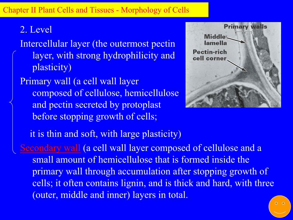
it is thin and soft, with large plasticity) Secondary wall (a cell wall layer composed of cellulose and a
small amount of hemicellulose that is formed inside the primary wall through accumulation after stopping growth of cells; it often contains lignin, and is thick and hard, with three (outer, middle and inner) layers in total.
2. LevelIntercellular layer (the outermost pectin
layer, with strong hydrophilicity and plasticity)
Primary wall (a cell wall layer composed of cellulose, hemicellulose and pectin secreted by protoplast before stopping growth of cells;
Chapter II Plant Cells and Tissues - Morphology of Cells

When they have achieved their final size and shape, some cells elaborate a multilayered secondary wall within the primary wall.
Chapter II Plant Cells and Tissues - Morphology of Cells

3. Pit and plasmodesmata
(1) Primary pit field: primary wall has some noticeably recessed areas.
(2)Plasmodesmata: plasmodesmata is the protoplasm fibril threading through pores on walls of adjacent cells; it is a direct connection bridge for substance and information among cell protoplasts, and an important guarantee for a multicellular plant to become a unitary organism in structure and function.
Chapter II Plant Cells and Tissues - Morphology of Cells

Electron micrograph of primary wall of a parenchyma cell from oat, Arena (Gramineae), coleoptile. The parallel-oriented microfibrils at top occurred in one of the angles of the cell. Other microfibrils show a random orientation. Plasmodesmatal pores are clustered in a primary pit field, 26,000. Reprinted with permission from Böhmer (1958), Planta 50, 461-497.
Chapter II Plant Cells and Tissues - Morphology of Cells

Electron micrographs of plasmodesmata in longitudinal and cross-sections (A), and a model of plasmodesmal substructure of tobacco leaves (B).
Chapter II Plant Cells and Tissues - Morphology of Cells

Light micrograph of plasmodesmata in the thick primary walls of persimmon (Diospyros) endosperm, the nutritive tissues within the seed. The plasmodesmata appear as fine lines extending from cell to cell across the walls. (620) (From Evert 2006)
Chapter II Plant Cells and Tissues - Morphology of Cells

(3) Pit: the area on primary wall that is not covered by the secondary wall at all at the time of secondary wall formation.
① Component:Pit cavity: the cavity formed by the secondary wall and opening
towards the cell compartment. Pit membrane: the primary wall and intercellular layer portion at
cavity bottom. Some membranes with thickened primary wall are specialized into pit plug, and the surrounding membranes are called plug edge.
② Category: Simple pit:Bordered pit: bordered pit is the pit with obviously decrescent orifice
due to the vaulted edge formed through cambered extension of secondary wall beyond the pit cavity.
③Pit pair: The pits of two adjacent cells often appear oppositely, and the two opposite pits are called pit pair.
Chapter II Plant Cells and Tissues - Morphology of Cells

Light microscope sectional view of vestured pits in
Terminalia chebula. ca.
3000. Abbreviations:
pm, pit membrane, sw,
secondary wall; v, vesture.
Chapter II Plant Cells and Tissues - Morphology of Cells

SEM of wood of eastern spruce (Picea) showing tracheids with circular bordered pit pairs on the lateral walls. Pit apertures (pa) and pit borders (pb) are visible, 1300
Chapter II Plant Cells and Tissues - Morphology of Cells

Photomicro-graph of chili fruit epidermis showing the pit pairs indicated by arrows.
Chapter II Plant Cells and Tissues - Morphology of Cells

4. Chemical components of cell wall:
Major components: cellulose, formed through glucosyl connection (-1, 4-glucosidic bond)
Minor components: pectin, hemicellulose, and other noncellulosic polysaccharides (all are hydrophilic substances)
Other substances: cutin, suberin, lignin, mineral;
Chapter II Plant Cells and Tissues - Morphology of Cells

5. Submicroscopic structure of cell wall
(1) Cellulose molecules Cellulose molecular beam (micelle)Microfibril (a structural unit composing cell wall)macrofibril
(2) Microfibril of different cell wall layer has different orientation.
Primary wall: reticular arrangement, and mostly in parallel with the long axis of cells.
Secondary wall: the same orientation in outer, medium and inner layers, and mostly inclined to the long axis at an angle.
Chapter II Plant Cells and Tissues - Morphology of Cells
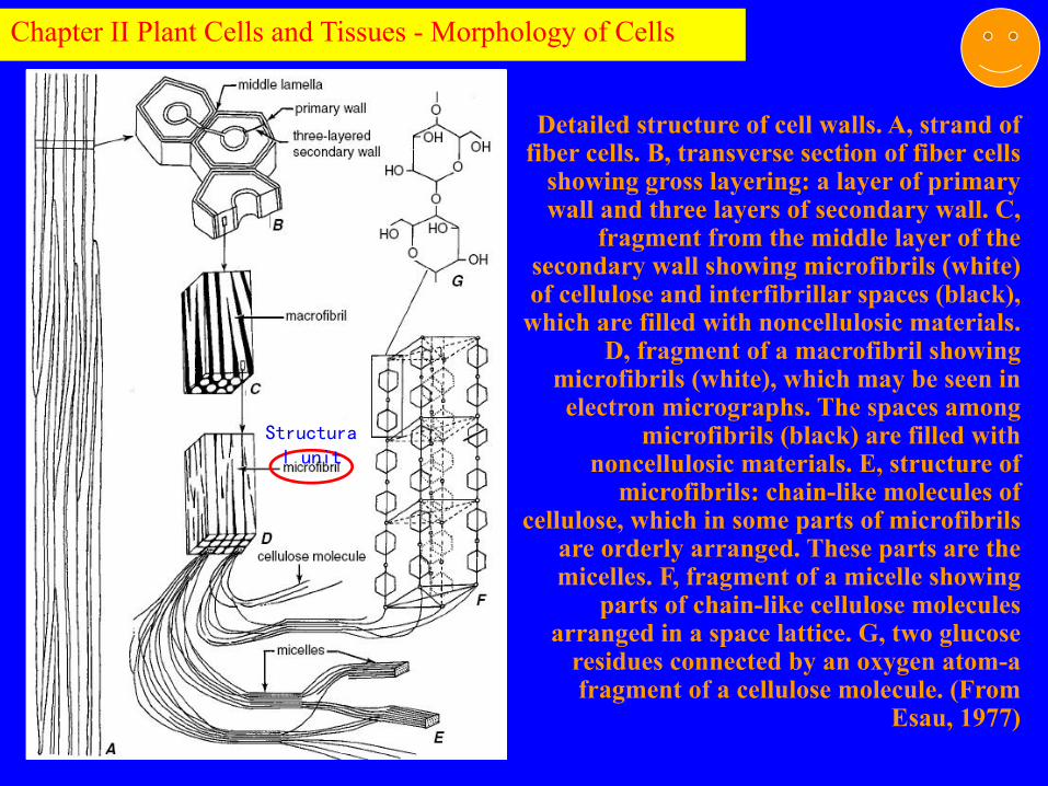
Detailed structure of cell walls. A, strand of fiber cells. B, transverse section of fiber cells
showing gross layering: a layer of primary wall and three layers of secondary wall. C,
fragment from the middle layer of the secondary wall showing microfibrils (white) of cellulose and interfibrillar spaces (black),
which are filled with noncellulosic materials. D, fragment of a macrofibril showing
microfibrils (white), which may be seen in electron micrographs. The spaces among
microfibrils (black) are filled with noncellulosic materials. E, structure of
microfibrils: chain-like molecules of cellulose, which in some parts of microfibrils
are orderly arranged. These parts are the micelles. F, fragment of a micelle showing
parts of chain-like cellulose molecules arranged in a space lattice. G, two glucose
residues connected by an oxygen atom-a fragment of a cellulose molecule. (From
Esau, 1977)
Chapter II Plant Cells and Tissues - Morphology of Cells
Structural unit

IV. Ergastic substances of plant cells (I) Definition: it is the product of protoplast metabolism of cells,
and can be produced and disappeared in different periods of cell activities; they can be stored products or waste.
(II) Starch: it is the most common storage form of carbohydrates, and is formed by polymerization of glucose molecules.
1. Starch grain: starch exists in cells in granular form, and is called starch grain. All parenchyma cells contain starch grains, which centrally exist in all kinds of storage organs. The morphological structure with annular striations
Chapter II Plant Cells and Tissues - Morphology of Cells

2. Type of starch grain
Simple starch grain: one hilum and multiple
annular striations
Compound starch grain: more than two hilums
and respective annular striations
Half compound starch grain: more than two
hilums and respective annular striations,
with common annular striations.
Chapter II Plant Cells and Tissues - Morphology of Cells

(III) Protein: storage protein in cells is in a solid state, and it can be crystal or amorphous.
Aleurone grain: amorphous protein is usually a spherical grain formed through being wrapped by a membrane, and is called aleurone grain.
Aleurone layer: aleurone grains are intensively distributed in one or several outmost cell layers of grain seeds, thus is called aleurone layer.
Chapter II Plant Cells and Tissues - Morphology of Cells

(IV) Fats and oils
Storage substance containing maximum energy and in
the smallest volume, which exists in cytoplasm, and
sometimes also in chloroplast.
Fats: solid state at room temperature.
Oils: liquid state at room temperature.
Chapter II Plant Cells and Tissues - Morphology of Cells

(V) CrystalsInorganic salts often form crystals. Calcium oxalate
crystal is one of the common crystals, while calcium carbonate crystal is one of the uncommon crystals. It is the accumulation of metabolic waste in vacuole, and can protect cells from being poisoned.
Types: Single crystal: prismatic or pyramidalAcicular crystal: acicular shape crystal with two sharp
tips, and is usually accumulated in beam. Cluster crystal: compound structure formed through
combination of many single crystals, and is spherical.
Chapter II Plant Cells and Tissues - Morphology of Cells

Calcium oxalate crystals seen in polarized light. A, prismatic crystals in phloem parenchyma of root of Abies. B, raphides in leaf of Vitis. C, druses in cortex of stem of Tilia. (A, 500; B, C, 750.) (From Evert 2006)
Chapter II Plant Cells and Tissues - Morphology of Cells
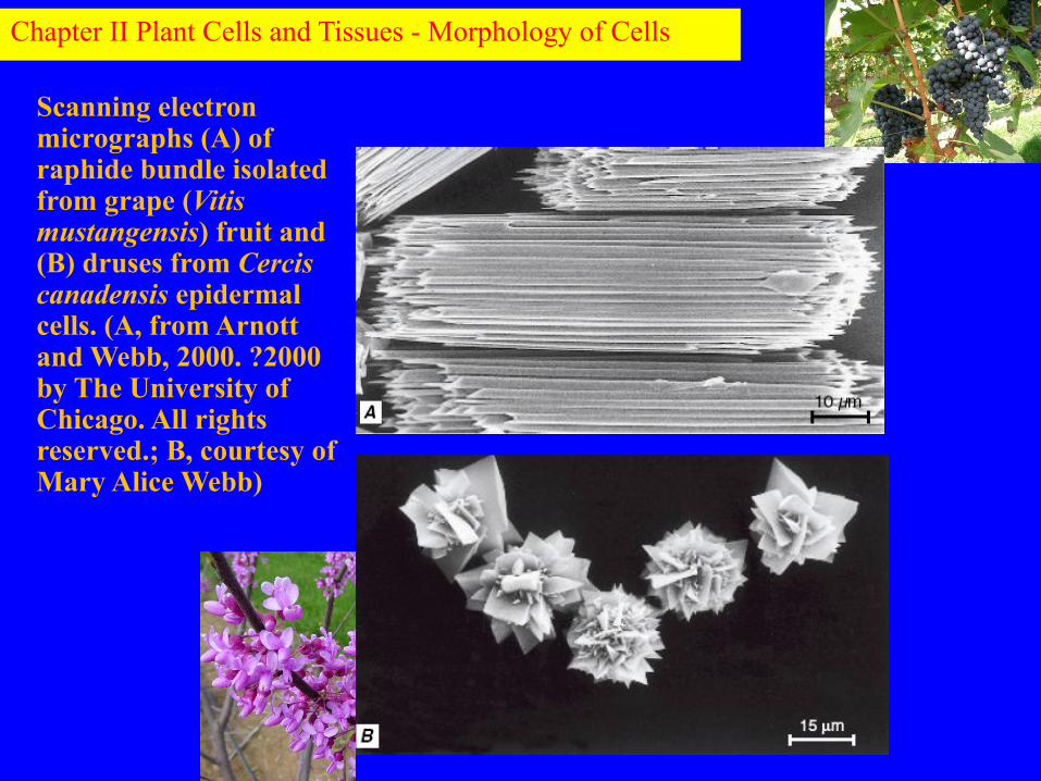
Scanning electron micrographs (A) of raphide bundle isolated from grape (Vitis mustangensis) fruit and (B) druses from Cercis canadensis epidermal cells. (A, from Arnott and Webb, 2000. ?2000 by The University of Chicago. All rights reserved.; B, courtesy of Mary Alice Webb)
Chapter II Plant Cells and Tissues - Morphology of Cells

Crocus cancellatus (Iridaceae), longitudinal section of leaf showing crystal idioblast containing styloid crystal (sc). Scale =50 m.
Chapter II Plant Cells and Tissues - Morphology of Cells

Calcium carbonate crystal. Transverse section of upper portion of rubber plant (Ficus elastica) leaf blade showing club-shaped cystolith in enlarged epidermal cell, the lithocyst. The cystolith consists mostly of calcium carbonatedeposited on a cellulose stalk. (155) (From Evert 2006)
Chapter II Plant Cells and Tissues - Morphology of Cells

I. Plant Cell Growth1. Definition: refers to cell volume growth, including
longitudinal and horizontal extension of cells. 2. In the growing process, cells will be obviously increased
in volume, accompanying with internal structure change: (1) With obvious increase in vacuolization and strong
metabolic capability, the product can be accumulated in the vacuole, resulting in absorption of large amount of water.
(2) Change of other organelles, .e.g thin polioplasm turning into thick polioplasm and proplastid turning into plastid, and change of the thickness and composition of cell wall.
3. Cell growth is limited, and is controlled by genes.
Chapter II: Plant Cells and Tissues--Cell Growth and Differentiation

II. Differentiation of plant cells1. Definition: the specialization of cell structure and
function is called cell differentiation.2. Individual development: it is the the result of the
continuous division, growth and differentiation of plant cells.
3. Phylogenetic development: with the evolution of plants, cell division becomes more detailed, cell differentiation becomes more violent, and the internal structure of frond becomes more complicated.
Chapter II: Plant Cells and Tissues--Cell Growth and Differentiation

Diagram illustrating some of the cell types that may originate from a meristematic cell
of the procambium or vascular cambium. The meristematic cell depicted here (at the
center), with a single large vacuole, is typical of the meristematic cells of the vascular
cambium. Procambial cells typically contain several small vacuoles. The meristematic
cells or precursors of all these cells had identical genomes. The different cell types become distinct from one another because they express sets of genes not expressed by
other cell types. Of the four cell types depicted here, the parenchyma cells are the
least specialized. Both the mature vessel element, which is specialized for the
conduction of water, and the mature fiber, which is specialized for support, lack a
protoplast. The mature sieve-tube element, which is specialized for the transport of
sugars and other assimilates, retains a living protoplast but lacks a nucleus and vacuole. It depends on its sister cell, the companion
cell, for life support. (From Raven et al., 2005)
Chapter II: Plant Cells and Tissues--Cell Growth and Differentiation

First 10 leaves from the main shoot of a potato plant (Solanum tuberosum). The leaves undergo a transition from simple to pinnately compound. (0.1. From McCauley and Evert, 1988)
Chapter II: Plant Cells and Tissues--Cell Growth and Differentiation

Increase in body complexity of charophyceans (A-F) and early divergent plants (G and H) is suggested by a phylogenetic model based on molecular data including tubulin (16) and rbcL sequences, a gene transfer event, and several intron insertion events (14). (A) Unicellular flagellate Mesostigma (whose divergence may, however, have preceded that of the charophycean lineage); (B) colonial Chlorokybus; (C) unbranched filament Klebsormidium; (D) unicellular desmid Netrium, belonging to a group (Zygnematales) that also includes unbranched filaments); (E) Chara, a branched filament with tissue at nodes (indicated by the presence of orange gametangia); (F) Coleochaete, a planar tissue-like species is shown; (G) Pallavicinia, representing liverworts, an early divergent plant group; (H) Lycopodium, an early divergent tracheophyte (vascular plant). (From Graham et al. 2000)
Chapter II: Plant Cells and Tissues--Cell Growth and Differentiation
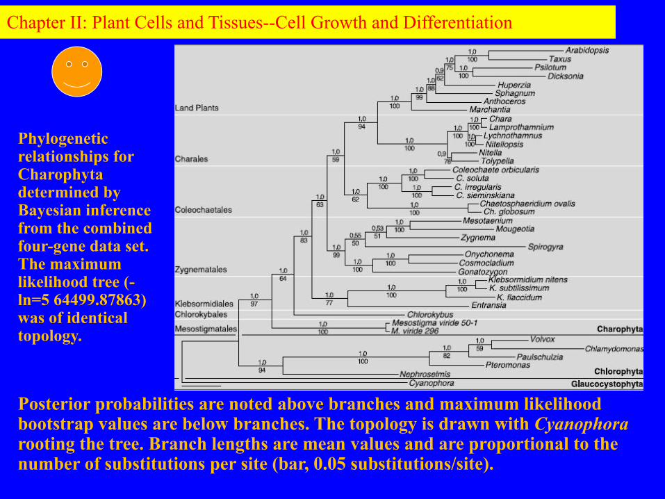
Posterior probabilities are noted above branches and maximum likelihood bootstrap values are below branches. The topology is drawn with Cyanophora rooting the tree. Branch lengths are mean values and are proportional to the number of substitutions per site (bar, 0.05 substitutions/site).
Phylogenetic relationships for Charophyta determined by Bayesian inference from the combined four-gene data set. The maximum likelihood tree (-ln=5 64499.87863) was of identical topology.
Chapter II: Plant Cells and Tissues--Cell Growth and Differentiation
•What are the similari t ies and differences between primary walls and secondary walls of plant cells?
•What are the same and different structures between plant and animal cells?
Chapter II: Plant Cells and Tissues--Cell Growth and Differentiation