Embed Size (px)
Citation preview

CHAPTER-I: INTRODUTION AND REVIEW OF LITERATURE 1
Chapter- I
INTRODUCTION AND REVIEW OF LITERATURE
1.1 INTRODUCTION
Advancement in technology has eased human life but these facilities have
increased competition and obscured life. The need to perform has resulted into increase in
stress as compared to early days.
Biologically, stress refers to consequences of failure of a human or animal body to
react appropriately to its emotional or physical threats. Stress in optimum quantum acts as
stimulator to achieve the best, but when it exceeds, it surely causes imbalance in the
biochemical parameters (Chandira et al, 2010). When stress is associated with oxygen it
is termed as oxidative stress. It plays a crucial role in the development of chronic and
degenerative diseases such as cancer, autoimmune disorders, rheumatoid arthritis,
cataract, aging, cardiovascular and neurodegenerative diseases (Willcox et al., 2004;
Pham-Huy et al., 2008). Chemically, oxidative stress is associated with increased
production of oxidizing species or a significant decrease in the effectiveness of
antioxidant defenses.
Oxygen is used by cells to generate energy, but paradoxically, as a by-product of
metabolism; it produces free radicals called reactive oxygen species (ROS) and reactive
nitrogen species (RNS) (Andersen, 2004). ROS and RNS exert beneficial effects at low
or moderate concentrations on cellular responses and immune function. But at high level,
these free radicals and oxidants generate oxidative stress, which is a deleterious process
that can damage cell structures, including lipids, proteins and DNA (Pham-Huy et al.,
2008).
To combat these free radicals, plants and animals maintain enzymatic antioxidant
system such as catalase, superoxide dismutase and various peroxidase and non enzymatic
complex systems such as glutathione, phenols, flavonoids and vitamins (Ganapaty et al.,
2007). Many synthetic antioxidants such as butylated hydroxyanisole (BHA) and
butylated hydroxytoluene (BHT) are also available in market. However, safety of these
synthetic antioxidants is now doubted (Moein et al., 2008). Thus, it is increasingly

CHAPTER-I: INTRODUTION AND REVIEW OF LITERATURE 2
emphasized to develop and utilize effective and non-toxic antioxidants of natural origin.
A great number of raw extracts or isolated pure compounds from medicinal plants
showed more effective antioxidant activity than BHA, BHT or vitamin E; hence they can
be potential sources of natural antioxidants (Pyo et al., 2004).
Arishtas and Asavas are complicated ayurvedic formulations, used as medicines
for over 3000 years to treat various disorders. They are taken as appetizers and
stimulants. They are alcoholic fermented preparations produced using microorganism in a
sugar/ jaggery/ honey or fruit base. It is a complicated biotechnological process
developed by ancient Indians. Charaka Samhita stated that, Arishta and Asava generally
promote courage, corpulence, satisfaction, imagination, strength of mind, body and
digestion. They alleviate sleeplessness, anxiety, and anorexia and are exhilarating.
Further, various Arishtas and Asavas available in market and claim their usefulness in
treatment of problems like backache, abdominal pain, irritation, improving strength and
stamina with rejuvenation as main claim (Govindarajan et al., 2008).
Though, traditional knowledge about preparation and applications of these
fermented polyherbal formulations exist in literature, standard parameters like alcohol
content, pH, acid value and other constituents are normally checked for validation.
However, bulk of knowledge about interesting antioxidant property of these fermented
medicines has remained unrecognized or not been studied in detail and validated.
1.2 AIMS AND OBJECTIVES
Considering above revealed views in mind, an investigation was carried out to study
Arishtas and Asavas with following aims and objectives:
1. To determine the antioxidant potential of Asavas and Arishtas.
2. To determine the biochemical parameters of Asavas and Arishtas.
1.3 REVIEW OF LITERATURE
1.3.1 Free Radicals
Excess stress leads to suppression in physical endurance as well as mental
capability for logical thinking and it also suppresses immunity leading to pathological
conditions (Kanoor et al., 2008). According to Hazra et al. (2008), oxidative stress is
most important reason behind today's modern diseases, which results from an imbalance

CHAPTER-I: INTRODUTION AND REVIEW OF LITERATURE 3
between formation and neutralization of pro-oxidants (Braca et al., 2002). This oxidative
stress leads to formation of free radicals, which seek stability through electron pairing
with biological macromolecules such as proteins, lipids and DNA. This ultimately cause
protein and DNA damage along with lipid peroxidation. These changes could lead to
cancer, atherosclerosis, cardiovascular diseases, ageing and inflammatory diseases
(Maxwell, 1995).
One major problem associated with inhaled oxygen is its pronounced tendency to
form free radicals. These free radicals are highly reactive molecules and hence
dangerous. Their role as culprits in causing disorders is being debated. These free radicals
are electrically charged molecules. So, to neutralize themselves, they attack our cells
tearing through cellular membranes to react and create havoc with the nucleic acid,
proteins, and enzymes present in our body. This attack by free radicals, collectively
known as oxidative stress, are capable of causing cells to lose their structure, function and
can eventually destroy them. They are continuously produced in our body, by continuous
use of oxygen such as in respiration and some cell-mediated immune functions. Free
radicals also generated due to environmental pollutions, cigarette smoke, automobile
exhaust, radiation, air pollution, pesticides etc. Stress is one of the oldest and chronic
disorders of mankind. Because of stress, patients often have lots of diseases that are
difficult to treat and manage or delay in treatment gives rise to other chronic conditions
and diseases (Chandira et al., 2010).
During oxidation, many free radicals are produced, which have an unpaired,
nascent electron. Free radicals are any atom (e.g. oxygen, nitrogen), molecule or
compound with at least one unpaired electron in their outermost orbit. Such a situation is
energetically unstable, making such species often highly reactive and short-lived. As a
result, they attempt to pair-up with other molecules, atoms or even individual electron to
create a stable compound. These free radicals achieve stability by removing or filching of
electron from surrounding molecule to produce an electron pair. They can also pinch
hydrogen atom from molecule which has bound to another molecule, or interact in
various ways with other free radicals to stabilize themselves (Wu and Cederbaum, 2003).
Though, by snatching an electron form surrounding atom, molecules or
compound these free radicals become stable, however, leftover of attacked atom,

CHAPTER-I: INTRODUTION AND REVIEW OF LITERATURE 4
molecules or compound then possess an unpaired electron and turned into a free radical.
These new born free radicals also want to stabilize themselves, so they fallow same path
of stabilization. In this way presence of a single radical may initiate a chain sequence of
electron transfer (redox) reactions. And until, subsequent free radicals are neutralized,
thousands of free radical reactions can occur within seconds of initial reaction. These
reactions leads to damages of important biomolecules such as proteins, lipids, nucleic
acids etc. This implicated in cause of several diseases like neurogenerative diseases,
cancer, arthritis, diabetes etc. Free radicals can have positive, negative or neutral charge.
They are generated as necessary intermediates in a variety of normal biochemical
reactions, but when generated in excess or uncontrolled, radicals can wreak havoc on a
broad range of macromolecules (Anonymous, 2012).
1.3.1.1 Reactive Oxygen Species
Various forms of activated oxygen are generally known as reactive oxygen
species (ROS). These lead to the oxidative cell damage and ends up in number of
pathological conditions like cancer, diabetes, arthrosclerosis and heart disease (Halliwell,
1998). ROS are classified as free radicals which includes superoxide anion (O2•),
hydroxyl radical (•OH), singlet oxygen (½O2), and non free radical i.e. Hydrogen
peroxide (H2O2) (Silva et al., 2010). These are produced from endogenous sources within
living organisms via various mechanisms like normal aerobic respiration, stimulated
poly-morpho-nuclear leukocytes, macrophages, and peroxisomes or from exogenous
sources like tobacco smoke, ionizing radiation, certain pollutants, organic solvents,
pesticides etc. (Zakaria, 2007). According to Ebrahimzadeh et al. (2010), these ROS are
responsible for cellular redox process.
The oxidation potential and reactivity of various ROS are given in the order
O2• < H2O2<
•OH (Fridovich, 1978).
Superoxide anions are formed when oxygen (O2) achieve an extra electron,
leaving the molecule with only one unpaired electron.
O2 + e- → O2
•
Though, within mitochondria, O2• is continuously being formed; however rate of
formation depends upon the amount of oxygen flowing through mitochondria at any
given time. Superoxide is capable of damaging cellular membranes (through

CHAPTER-I: INTRODUTION AND REVIEW OF LITERATURE 5
peroxidation), proteins (through varieties of mechanisms) and other macromolecules.
Superoxide radicals also play very important role in formation of hydroxyl radicals which
are most damaging radical within the body.
In-vivo hydrogen peroxide (H2O2) is produced by many reactions, either being
converted to highly damaging hydroxyl radical or catalyzed and excreted harmlessly as
water (Goldfarb, 1999). It may form by conversion of O2•.
O2• + 2H
+ + e
– → H2O2
The principal problem with H2O2 is that, it easily crosses cellular membrane
while receiving one more electron, normally originating from iron or copper, gives rise to
hydroxyl radical (•OH). It has ability to damage DNA and can lead to cancer-causing
illnesses, Down syndrome and other genetic disorders.
Though •OH is short-lived, they are most dangerous free radicals. Formed when
O2• and H2O2 react together, which causes an extreme reaction; they will attack any
molecule present around them. They can cause damage to cells where they react with
DNA, lipids, and proteins.
H2O → •OH
+
H
•
H2O2 → 2•OH
This type of free radicals can be formed from O2•, O
• and H2O2 via Harber-Weiss
and Fenton reaction respectively (Knight, 1999).
O2• + H2O2 →
•OH +
•OH + O2 Haber-Weiss reaction
Fe2+
+ H2O2 → Fe3+
+ •OH +
•OH Fenton reaction
1.3.1.2 Reactive Nitrogen Species
Nitric oxide (NO) is an important bio-regulatory molecule, which has a number of
physiological effects including control of blood pressure, neural signal transduction,
platelet function, antimicrobial and antitumor activity (Jagetia et al., 2004). In most
cases, low concentrations of NO are sufficient to affect these beneficial functions.
However, during infections and inflammations, formation of NO is elevated and may
bring about some undesired deleterious effects.
As a free radical and depending on the microenvironment, NO is oxidized,
reduced or form complex with other biomolecules. Though, NO does not directly interact
with the bio-organic macromolecules like DNA or proteins, but under aerobic condition,

CHAPTER-I: INTRODUTION AND REVIEW OF LITERATURE 6
NO molecule become very unstable and reacts with oxygen to produce intermediates
such as NO2, N2O4, N3O4 (Marcocci et al., 1994). When it comes in contact with O2•, it
forms highly reactive peroxynitrite anion (ONOO•) which leads to oxidative injury as
well as lung damage (Levi, 1987; Wink et al., 1991).
According to Jagita et al. (2004), there are increasing evidences which suggest
that NO and its derivatives produced by activated phagocytes may have a geno-toxic
effect and may contribute in multistage carcinogenesis process (Wink et al., 1991).
1.3.1.3 Transitional- metal ions
Ions are species which can lose or gain single electron as they change from one
valence state to another and because of their excited energy state; they have long been
recognized as powerful initiator of free radical. According to Fenton reaction, in vivo,
majority of •OH comes from metal catalytic breakdown of H2O2 (Valko et al., 2006).
Devasagayam et al. (2004) report some other sources of free radicals include
redox cycling of xenobiotics, exposure to physicochemical agents like ionizing radiations
and an endogenous compound or a drug that acts as photosensitizer. Most of the damage
induced by ionizing radiations in biological systems is indirect and mediated by products
of radiolysis of water including hydrogen radical (H•),
•OH, hydrated electron (eaq
–),
H2O2, peroxyl radical (ROO•), O2
•, ½O2 etc. (Von Sonntag, 1987; Devasagayam and
Kesavan, 1996).
Cigarette smoke contains a large amount of reactive species (Devasagayam and
Kamat, 2002). Cigarette tar contains quinone-hydroquinone-semiquinone system which
reduces O2 to O2•, H2O2 and
•OH. Cigarette smoke contains small oxygen and carbon
centered radicals as well as active oxidants such as NO• and nitrogen dioxide (NO2).
Wentworth et al. (2003) stated that antibodies convert ½O2 into H2O2 via a process that
they have postulated to involve dihydrogen trioxide (H2O3). During ischemia-reperfusion,
oxidants like O2•,
•OH and H2O2 are produced. This occurs during non-fatal myocardial
infarction, surgeries, stroke, transplantation, blockage of arteries under pathological
conditions. During ischemia in heart (in myocyte mitochondria) conversion of ATP to
adenosine causes generation of O2•, while in the blood vessels (endothelium) pathway
involved is transition from xanthine to uric acid (Yoshikawa et al 2000). Vogiatzi et al.
(2009) found some evidence which suggests that, common risk factors for atherosclerosis

CHAPTER-I: INTRODUTION AND REVIEW OF LITERATURE 7
increase production of ROS, not only from endothelial cells, but also from the smooth
muscle cells and the adventitial cells (Gozin et al., 1998). Thus, hypercholesterolemia,
diabetes mellitus (DM), arterial hypertension, smoking, age, and nitrate intolerance
increase the production of free ROS.
1.3.1.4 Free radicals in biological system
Free radicals, mostly in the form of ROS/RNS are known to play a dual role in
biological systems. They can either harmful or beneficial to living systems (Valko et al.,
2004). Beneficial effects of ROS/RNS comprise physiological role in cellular response to
anoxia. They play vital role against infectious agents and in function of a number of
cellular signaling systems. Furthermore, lower concentration of free radical is helpful in
induction of mitogenic response. In contrast, at high concentration, free radical can be
important mediators of damage to cell structures including lipids and membranes,
proteins and nucleic acids.
1.3.1.4.1 Free radicals in diseases
According to Hemnani and Parihar (1998) ROS and RNS have been implicated in
the patho-physiology of various clinical disorders including ischemia, reperfusion injury,
myocardial infarction, rheumatoid arthritis, neurodegenerative, atherosclerosis, acute
hypertension, hemorrhagic shock and diabetes mellitus. Some tumor cells also produce
ROS and RNS (Szatrowski and Nathan, 1991). Source of these products and their
contribution to transformed phenotypes are not known. Formation of oxidative stress may
result in damage of critical cellular macromolecules such as DNA, lipid and proteins and
causes many chronic diseases (Poli et al., 2004). Vogiatzi et al. (2009) stated that during
last decade, several studies have examined potential role of oxidative stress in
atherogenesis (Stephens et al., 1996; Ohara et al., 1993). Further, according to theory of
oxidative stress, oxidative modifications of low density lipoproteins (LDL) in the arterial
wall by ROS resulted in atherosclerosis.
Oxidative stress occurs in brain not only because of physical damage but also due
to free radicals produced by viruses, bacteria, fungi or parasitic diseases (Floyd, 1999).
According to Lushchak and Semchyshyn (2012), different types of physical or anoxic
trauma stimulate the generation of ROS/RNS that can recombine with metals and

CHAPTER-I: INTRODUTION AND REVIEW OF LITERATURE 8
produce powerful oxidants and also create other forms of ROS and RNS. These forms of
free radicals are well known to create major toxicities to brain and which cause
Parkinson’s disease, amyotrophic lateral sclerosis and Huntington’s disease.
Diabetes mellitus (DM) is caused due to excessive production of RNS; produced
due to oxidative metabolism. Such free radicals damage pancreatic β-cells which
produce, store and release insulin. So, once β-cells die, body becomes deficient in insulin.
These β-beta cells are then replace by α- cells (which produce glucagon) causes an
increase in blood glucose in the body. Once diabetic state is established, there is a
deficiency of NO in the vascular system; which is necessary to maintain proper blood
pressure. This condition leads to damage endothelial cells; which line the blood vessels.
Therefore, blood pressure increases and hypertension occurs (Van Dyke et al., 2010). The
role of transition metal ions is of particular interest because malignant diseases like
chronic inflammation produced changes in distribution in the body. (Halliwel and
gtteridge, 1989; Yagi, 1982). Some other examples originating from free radicals are
Alzheimer’s, Parkinson’s, Down’s syndrome, etc. (Das et al., 1997; Koleva et al., 2002).
1.3.1.4.2 Free radicals and Lipid
Lipids are major target for attack by free radical because oxygen is foremost
soluble in hydrophobic membrane. These free radicals easily break double bond present
in polyunsaturated fatty acid (PUFA). It leads to the peroxidation of PUFA in lipid
membrane; which severely damage cell membrane and thereby produces loss of fluidity
and breakdown of cell membrane. Such conditions make us to suggest possible role of
lipid peroxide in various pathological conditions and aging (Halliwell and Gtteridge,
1989). Significance of lipid peroxide on aging process is based on the fact that lipofuscin
age pigments accumulate almost linearly with advance age and these pigments
presumably results from polymerization of oxidized unsaturated lipids (Ohyashiki et al.,
1984).
It has been considered that, damage to lipid results in aging and pathological
disorders. Some phases of neuronal ceroid lipofuscinosis, intermittent claudication,
oxygen toxicity and liver injury caused by orotic acid, ethanol, phosphorus or chlorinated
hydrocarbon have been reported in relation to lipid peroxidation. Free radical mediated
lipid peroxidation has been proposed to be critically evolved in several diseases including

CHAPTER-I: INTRODUTION AND REVIEW OF LITERATURE 9
cancer, rheumatoid arthritis, drug associated toxicity and post-ischemic re-oxygenation
injury as well as in the degenerative diseases associated with aging (Halliwel and
gtteridge, 1989; Yagi, 1982).
1.3.1.4.3 Free radicals and Protein
Radical mediated damage to proteins may be initiated by electron leakage, metal-
ion dependant reactions and auto-oxidation of lipids and sugars. These reactive species
can interact with proteins directly, especially with their sulfhydryl groups (Herington,
1986). Among amino acids, histidine, tryptophan, methionine and tyrosine are more
reactive towards ROS resulting in sulphoxides and short-lived endo-peroxides that may
toxic to other cells. The consequent protein oxidation is O2 dependent and involves
several propagating radicals, especially alkoxyl radicals. This will results in modification
of enzyme activity (Bellomo et al., 1983). Damage to proteins lead to alterations in
intracellular calcium and potassium salts that will trigger a series of changes in cells
(Kerr et al., 1992). In selective cases, alteration of protein structure and the consequent
unfolding is associated with enhanced susceptibility to proteinases.
1.3.1.4.4 Free radicals and DNA
DNA damage is a result of extrinsic and intrinsic process including oxygen
derived free radicals which are normal consequences of cellular metabolism of oxygen.
Several types of damage including base lesions, sugar lesions, protein and DNA cross
link, breaking of single and double strand are produced by free radical induced reactions
(Kaneko et al., 1996). When these free radicals react with sugar moiety of DNA, some
sugar product and interacting bases are released; leads to DNA damage (Dizdaroglu et
al., 1975). Blake et al., (1987) stated that, when DNA is exposed to high fluxes of radical,
strand scission has been observed but changes in bases and deoxyribose sugars may be
more frequent. Hydroxyl radicals are thought to mediate these reactions. DNA damage
including fragmentation has been reported to occur during the respiratory burst and may
be a cause of neutrophil cell death and induction of 'autoimmune' processes (Birnboim
and Kanabus-Kaminska, 1985).

CHAPTER-I: INTRODUTION AND REVIEW OF LITERATURE 10
1.3.2 Defense against oxidative stress
To protect body cells and organs, from ill effect of free radicals, humans have
evolved a highly sophisticated and complex antioxidant defensive mechanism. This
defense system either naturally produced in-situ or externally supplied through foods
and/or medicines. These antioxidants act as free radical scavengers by preventing and
repairing damages caused by free radicals. Therefore they can enhance immune defense
and lower risk of cancer and degenerative diseases (Pham-Huy et al., 2008).
According to free radical theory, as a result of accumulation of oxidatively
damaged macromolecules and consequently cells or tissues due to aerobic metabolism to
which individuals are continuously exposed, ageing is initiated in human beings
(Harman, 1965). Thus, antioxidant defense may be one of major mechanism to combat
ageing and age-related problems (Mukherjee et al., 2011). They may be classified as-
Primary defense against free radicals
Secondary defense against free radicals
Primary defense is group of antioxidant enzymes, which are capable of
catalytically removing free radicals (Fridovich, 1978). On the other hand, the secondary
defense is group of macromolecules which act as free radical scavengers.
Now, antioxidants are substance, which when present in foods or body, markedly
delay or prevent oxidation of that substrate. Antioxidants may help body to protect itself
against various types of oxidative damage caused by ROS/RNS, which is linked to a
variety of diseases including cancer, diabetes, shock, arthritis, and acceleration of aging
process (Sanchez-Moreno et al., 1999). Thus, antioxidant defense systems along with
aerobic metabolism counteract oxidative damage rooted by free radicals (Gulcin et al.,
2002).
By definition antioxidants means “Against oxidation”; which has been broadly
explained by Halliwell (1998) as: “any substance when present at low concentrations
with those of an oxidisable substrate significantly delays or prevents oxidation of that
substrate”. Variety of biological activities are possessed by antioxidant compounds
including induction of drug metabolizing enzymes, inhibition of prostaglandin synthesis,
inhibition of carcinogen induced mutagenesis and scavenging of free radicals (Hirose et
al., 1994). In late 19th
and early 20th
century, extensive study was devoted to uses of

CHAPTER-I: INTRODUTION AND REVIEW OF LITERATURE 11
antioxidants in important industrial processes like prevention of metal corrosion,
vulcanization of rubber and polymerization of fuels (Matill, 1947).
Antioxidant compounds in food play vital role as a health-protecting factor.
Scientific evidence suggests that antioxidants reduce risk for chronic diseases including
cancer and heart disease. Whole grains, fruits and vegetables are primary basis of
naturally occurring antioxidants. Plant sourced food antioxidants like vitamin C and E,
carotenes, phenolic acids, phytate and phytoestrogens have been recognized as having
potential to diminish such diseases. Most of antioxidants, in a typical diet are derived
from plant sources and belong to various classes of compounds with a wide variety of
physical and chemical properties. Antioxidant compounds like polyphenols and
flavonoids scavenge free radicals such as peroxide, hydroperoxide or lipid peroxyl and
thus prevent body from oxidative damage (Miller et al., 2000). Numbers of clinical
studies conclude that antioxidants from fruits, vegetables, tea and red wine are major
source to overcome present problem of chronic diseases including cancer and heart
diseases.
Antioxidants are powerful electron donors; act as chain-breaker and also react
with free radicals before more important target molecules are damaged due to reaction of
free radicals. For example, antioxidant N-acetyl cysteine shows antimutagenic and
chemo-preventive activities in a variety of organs, such as lung, liver, skin and colon
(Flora et al., 1986; Izzotti et al., 1994). Antioxidant can alter gene expression induced by
ROS rather than their direct effect on transcription through Antioxidant Response
Element (ARE) present in promoters of many genes including phase II enzyme
glutathione stransferase (Primiano et al., 1997). Antioxidants show potent effect on
ability of several transcription factors to bind DNA (Winyard and Blake, 1997). They
have been shown to trigger apoptosis in smooth muscle cells independent of oxidative
reactions (Liu et al., 1998 and Tsai et al., 1996). Antioxidants can inhibit tumor initiation,
tumor promotion and cell transformation (Steele et al., 1990). Changes in antioxidant
defense enzymes such as SOD, GPx, CAT and Glutathione S-Tansferase (GST) have
been widely described in cancerous cells (Cerutti et al., 1994). Evidence suggests that
low molecular weight antioxidant enzymes and anti-inflammatory agents that inhibit
arachidonic acid metabolism are anti-carcinogenic (Kensler et al., 1983).

CHAPTER-I: INTRODUTION AND REVIEW OF LITERATURE 12
Specific type of free radical can be reduced by specific antioxidant compound or
enzyme. For example Super oxide dismutase (SOD) reduces O2• to H2O2, which can
further reduced by Glutathione Peroxidase (GPx) and Catalase (CAT). Vitamin E
prevents the propagation of free radical damage in biological membrane through its
ability to peroxyl radical (Babber and Harris, 1994). Table 1.1 indicates the mechanism
of antioxidant activity with respect to antioxidant compounds.
Table 1.1: Mechanisms of antioxidant activity.
Antioxidant class Mechanism of antioxidant
activity Example of antioxidant
Proper antioxidants Inactivating lipid free radicals Phenolic compounds
Hydroperoxide stabilizers Preventing decomposition of
hydroperoxides into free radicals Phenolic compounds
Synergists Promoting activity of proper
antioxidants Citric acid, ascorbic acid
Metal chelators Binding heavy metals into
inactive compounds
Phosphoric acid, Millard
compounds, citric acid
Singlet oxygen quenchers Transforming singlet oxygen into
triple oxygen Carotenes
Substances reducing Hydro-
peroxides
Reducing hydroperoxides in a
non-radical way Proteins, amino acids
Source: Pokorny et al. (2001)
All antioxidants are either endogenous, produced in the body e.g. enzymes like
super oxide dismutase (SOD), catalase (CAT), polyphenol oxidase (PPO) etc. or
exogenous antioxidants obtain from food like vitamins, phenols, flavonoids etc.
1.3.2.1 Endogenous antioxidants
Human body relies on several endogenous defense mechanisms to help protect
against free radical-induced cell damage. Body mainly have five types of endogenous
antioxidants like superoxide dismutase (SOD), α-lipoic acid (ALA), catalase (CAT) and
glutathione peroxidase (GPX). Out of which SOD, CAT and GPX are three most
important of these because body can produce more of them when certain free radicals are
present. Antioxidant enzymes catalyze breakdown of radical species usually in

CHAPTER-I: INTRODUTION AND REVIEW OF LITERATURE 13
intracellular environment (Fig. 1.1). Preventive antioxidants bind transition metal ions
such as iron and copper; avoid their interaction with H2O2 and O2• to produce highly
reactive •OH.
Fig. 1.1: Free Radicals and Antioxidant Enzymes
1.3.2.1.1 Superoxide Dismutase
In 1969, the work of Mc Cord and Fridovich in USA showed that erythrocyte
protein was able to remove O2• catalytically and thus identified as a superoxide dismutase
(SOD) enzyme. They include Mn++
enzyme in mitochondria and Cu++
/Zn++
enzyme
present in cytosol. Copper-zinc containing Superoxide Dismutase (SOD) having two
protein subunits, each of which bears one copper ion and one zinc ion for stability of
apoenzyme.
Superoxide Dismutase is a metallo-enzyme acting as primary preventive inhibitor
by catalyzing the conversion of O2• to H2O2 (Symons and Gutteridge 1998). Yagi (1982)
proposed two possible mechanisms for action of SOD that inhibit transformation-
1. It is likely to act at level of cell membrane and remove or prevent radical formation
that would cause membrane lipid peroxidation and leads to chain of extra-nuclear and
nuclear events, ultimately expressed as transformation.
2. It has been suggested that, oxygen radicals could be formed in growth medium. These
radicals may act as promoters in transformation process that would be removed by
SOD.
1.3.2.1.2 Catalase
Most aerobic cells contain Catalase activity (CAT) and are largely located in
peroxisomes. Catalase activity of tissue varies greatly; highest in liver and kidney and

CHAPTER-I: INTRODUTION AND REVIEW OF LITERATURE 14
lowest in connective tissue. Catalase activity is a metal-containing enzyme and has most
efficient enzyme that promotes redox reaction:
2H2O2 → 2H2O + O2
It also acts as hydrogen donors, for example methanol, ethanol, formic acid,
phenol with consumption of peroxide.
ROOH + AH2 → H2O + ROH + A
Hydrogen peroxide itself is not particularly reactive with most biologically
important molecules; however, it may acts as a precursor for more reactive oxidants such
as •OH. When concentration of H2O2 is high, it is removed by CAT (Bragadottir, 2001).
1.3.2.1.3 Glutathione system
Glutathione, a major non-protein thiol in living organism, plays role in
coordinating body’s antioxidant defense system. Glutathione peroxidase (GPX) is a
selenium-containing enzyme (Symons and Gutteridge, 1998) that catalyses the reduction
of hydrogen of lipid peroxides (LOOH) with reduced glutathione (GSH).
2GSH + H2O2 → 2H2O + GSSG
GSH-PX is capable of reacting with both lipid and hydrogen peroxides (Halliwell and
Gtterridge, 1989). Selenium, which is required for GPX activity, is obtained from the diet
and glutathione peroxidase activity can be enhanced in muscle by increasing the level of
selenium in the diet (Devore et al. 1983). GPX is only antioxidant enzyme, which has
been reported to be influenced by animal diet (Decker and Xu, 1998). Antioxidative
enzymes have little significance in food application and they are currently not of
commercial significance in the food industry (Meyer and Isaksen, 1995).
1.3.2.1.4 Lipoic acid
Lipoic acid, yet another important endogenous antioxidant, categorized as a
“thiol” or “biothiol”. It is a sulfur-containing molecule that known for its involvement in
reaction that catalyzes oxidative decarboxylation of α-keto acids and α-keto-glutarate in
Krebs Cycle. Lipoic acid and its reduced form i.e. dihydrolipoic acid (DHLA), are
capable of quenching free radicals in both lipid and aqueous domains and thus called as
“universal antioxidant”. It may also exert its antioxidant effect by chelating with pro-

CHAPTER-I: INTRODUTION AND REVIEW OF LITERATURE 15
oxidant metals. Research further suggests that lipoic acid has a sparing effect on other
antioxidants (Percival, 1998; Packer, 1995; Kagen, 1992).
1.3.2.2 Exogenous antioxidants
1.3.2.2.1 Antioxidant vitamins
Antioxidant vitamins show number of biological activities such as immune
stimulation, inhibition of nitrosamine and an alteration of metabolic activations of
carcinogens (Poppel and Berg, 1997). Experimental evidence suggests that, carotenoids
and other retinoids act in earlier and later stages respectively; that inhibit mutagenesis
and gene expression (Sies, 1993).
Vitamin ‘E’: Vitamin ‘E’ (α-tocopherol) is an important chain-breaking antioxidant
which can directly scavenge ROS. It is a major lipid-soluble antioxidant present in all
cellular membranes, which protects against lipid peroxidation. It also protects membrane
of Poly-Unsaturated Fatty Acid (PUFA) and Low Density Lipoprotein (LDL) (McCay,
1985). Vitamin ‘E’ can directly react with variety of oxygen radicals like peroxyl radical
ROO•, CCl3
•,
•OH, O2
• and singlet oxygen (Fukuzawa and Gebicki, 1983). Vitamin ‘E’
donates hydrogen from chromane ring to free radical. The toco-peroxyl radical formed
can be reduced to tocopherol by interaction with reductants serving as hydrogen donors.
ROO• + Vit. E-OH ROOH + Vit. E-O
•
Vit. E-O• + AH Vit. E-OH + A
•
Vitamin ‘E’ induces growth inhibition of cancer cell by scavenging free radicals,
thereby stabilizing cell membrane (Blot et al., 1993). Because of the antioxidant
properties, vitamin ‘E’ neutralizes ROS and RNS and reduces oxidative DNA damage
and mutation (Frei, 1994).
Vitamin ‘C’: Vitamin ‘C’ (ascorbic acid) is an important water-soluble antioxidant in
biological fluids and an essential micronutrient required for normal metabolic functioning
of body. Vitamin ‘C’ readily scavenges ROS, Ozone, ONOO•, NO2, NO
• and
hypochlorous acid (Noroozi et al., 1998) and reduces oxidative damage and mutations
(Frei, 1994). The most striking chemical activity of ascorbate is its ability to act as a
reducing agent (electron donor). Donation of one electron by ascorbate gives the semi-
dehydro-ascorbate radical, which can be further oxidized to give dehydroascorbate.
Epidemiological studies have indicated an inverse association between intake of vitamin

CHAPTER-I: INTRODUTION AND REVIEW OF LITERATURE 16
‘C’ and risk of cancers. Vitamin ‘C’ can act as a co-antioxidant by regenerating
tocopheroxyl radical produced during scavenging of ROS. Reports suggest that, Vitamin
‘C’ may prevent formation of ONOO• by reaction with O2
• and may help to release NO
•
from endothelial cell. Some studies have shown that, Vitamin ‘C’ restores all parameters
to normal by reducing oxidative stress and may help to reduce the risk of developing
diabetic complications. Ascorbic acid supplementation has also been shown to improve
glycemic control and decrease fasting blood glucose, triglycerides and cholesterol levels
in non-insulin dependent diabetes (Eriksson, 1995).
Antioxidants like ascorbic acid and various forms of vitamin E (tocopherols and
tocotrienols) inhibit the auto-oxidation of glucose and glycation of blood proteins e.g.
Hemoglobin. Therefore, antioxidant therapy can ease toxic state which occurs in diabetes
Type I or II (Van Dyke et al., 2010).
1.3.2.2.2 Phenols and Flavonoids
In recent years, there is an increasing interest in finding antioxidant
phytochemicals, because they can inhibit propagation of free radical reactions, protect
human body from diseases. The most effective components seem to be flavonoids and
phenolics found in almost all plants. Their metal-chelating capabilities and radical-
scavenging properties have enabled phenolic compounds to be thought of as effective
free radical scavengers and inhibitors of lipid peroxidation (Terao and Piskula, 1997). It
has been found that phytochemicals like polyphenols and flavonoids are good
antioxidants and exert protective effects against development of cancer and
cardiovascular diseases (Cao et al., 1996).
Cheniany et al., (2010) noted that, phenolic compounds occur in abundance, as
secondary metabolites, in all plant (Kefeli et al., 2003). These compounds belong to a
large and heterogeneous group of biologically active and non-nutrient compound (Jay-
Allemand et al., 2001). The polyphenolic antioxidants either trap initiating radical,
propagating lipid peroxyl radicals, recycling α-tocopherol or deactivating excited
photosentizers, etc. (Tiwari, 2001). The electron and H+ donating capacity of flavonoids
seem to contribute to termination of lipid peroxidation chain reaction based on their
reducing power (Tiwari, 2001). It is well known that all aerobic organisms possess such

CHAPTER-I: INTRODUTION AND REVIEW OF LITERATURE 17
antioxidant mechanism to protect against oxidative damages and various types of
enzymes responsible for removal or repair of damaged molecules (Auroma, 1998).
1.3.2.2.3 Synthetic antioxidants
Many synthetic antioxidants have been used for stabilization of foods. Synthetic
antioxidants includes N-acetyl (NAC), butylated hydroxyanisole (BHA) and butylated
hydroxytoluene (BHT), which are added to fatty and oil foods to prevent oxidative
deterioration. NAC has anti-mutagenic and chemo-protective activity in the verity of
organs such as lung, liver skin etc. NAC regulate gene expression and modify cellular
signaling induced by ROS (Lin et al., 2012). BHA inhibits carcinogenicity and
mutagenicity of many chemicals in various experimental animals. BHT has been shown
to inhibit carcinogenic effect of DMBA, 4-DMBA and azoxymethane. (Loliger, 1991).
However, use of these chemical compounds has begun to be restricted because of their
induction of DNA damage and their toxicity (Ito et al., 1986; Sasaki et al., 1986).
Moreover, Zamani et al. (2009) noted that making process of such synthetic
antioxidants is very expensive and some of them like BHA and BHT have confirmed
undesirable effects. These synthetic antioxidants though acts as free radical scavengers by
breaking chain reaction, still some scientist reported that safety of such synthetic
antioxidants are now in doubt (Moein, 2008). Watts (1975) affirmed that, at high dose,
synthetic antioxidants such as BHA and BHT suffer from some disadvantage due to their
toxicity. According to Wettasinghe and Shahidi (1999) potential health hazards caused by
use of synthetic antioxidants in food products has led to scrutiny of natural antioxidants.
Human metabolism counts on antioxidant defensive system involving enzymes
and proteins to prevent adverse effects of oxidative stress. However, defenses can be
overwhelmed in certain circumstances, so that harmful effects occur. It is accepted that
intake of antioxidant substances reinforces defenses against free radicals. Use of synthetic
antioxidants has been limited because of their toxicity (Valentao et al., 2002). Botterweck
et al. (2000) reported that, synthetic antioxidants appear to be involved in tumor
promotion and hence use of a natural antioxidant source become crucial.
So, it is of great significant and necessity that research focuses on discovering
potential natural, effective antioxidants to replace synthetic ones. Therefore, researchers
turn their point of view to find naturally occurring antioxidants for use in foods to swap

CHAPTER-I: INTRODUTION AND REVIEW OF LITERATURE 18
such synthetic antioxidants, which are being restricted due to their carcinogenicity
(Velioglu et al., 1998; Singh et al., 2002; Gulcin et al., 2004; Rezaeizadeh et al., 2011).
Thus finding of natural anti-oxidant from new sources is very important.
All human cells protect themselves against free radical damage by antioxidant
enzymes such as superoxide dismutase (SOD) and catalase, or compounds such as
ascorbic acid, tocopherol and glutathione (Niki et al., 1994). Sometimes these protective
mechanisms are disrupted by various pathological processes. When ROS are produced in
huge quantities, aforesaid natural mechanism can be ineffective. In such cases dietary
intake of antioxidants becomes essential. So, recently much attention has been directed
towards the development of ethno medicines with strong antioxidant properties but low
cytotoxicities (Hazra et al., 2008).
1.3.3 Ayurveda
Ayurveda is a traditional Indian medicinal system being practiced for thousands of
years. Along with traditional Chinese System of Medicine, Ayurveda is regarded as one
of two living great traditions of the world (Sekar, 2007). Atharveda (around 1200 BC),
Charak Samhita and Sushrut Samhita (100 - 500 BC) (Dash and Sharma, 2001) are main
classics that given detailed descriptions of over 700 herbs (Shrikumar and Ravi, 2007).
Ayurvedic Pharmacopoeia comprise with more than 1,200 species of plants,
nearly 100 minerals and over 100 animal products. Numerous drugs have entered into the
international pharmacopoeial through considerable research on Ayurveda in respect with
pharmacognosy, chemistry, pharmacology and clinical therapeutics. World Health
Organization (WHO) specifies that, primary health needs of countries in Africa, Asia and
Latin America are met by traditional medicines. Such traditional medicines are adapted to
industrialized countries as Complementary or Alternative Medicines (CAM). Ayurvedic
system of treatment has been estimated to meet 70-80% of the healthcare needs of India
(Viswanathan et al., 2003).
Ayurvedic system of treatment is designed to attain svasthya (to establish one’s
own natural state on perfect health) and advocates samadosa (structural and physiological
equilibrium), samagni (equilibrium of metabolic processes), samadhatu (equilibrium of
body tissues), samamalakriya (equilibrium of eliminative systems), prasannendriya
(equilibrium of senses), prasannamana (equilibrium of mind) and prasannatma (state of
pure awareness or contended self). Ayurveda contains 8 branches of sciences and 10

CHAPTER-I: INTRODUTION AND REVIEW OF LITERATURE 19
different diagnostic tools based on tridosha theory (three humours of body). Ayurveda
also advocates a system of prevention of diseases by stipulating a set of practices as daily
routine (Dinacharya) and seasonal routine (Ritucharya). Ayurvedic treatment system also
takes into account individual variations. Herbal drugs have been used by mankind since
time immemorial to treat various disorders and offer an alternative to synthetic
compounds, as they have been considered either non-toxic or less toxic (Tiwari and
Upadhyay, 2009).
Ayurvedic medicines are of various types, so as to meet diverse requirements in
treatment of human illness. They are Swarasa (expressed juice), Kalka (paste), Hima
(cold infusion), Phanta (hot infusion), Kwatha (decoction), Churna (powder), etc. along
with Arishtas (fermented decoctions) and Asavas (fermented infusions) (Dhiman, 2004).
1.3.3.1 Research on Ayurvedic Formulations
Ayurveda is becoming one of the best alternatives for modern medicines. Many
medical practitioners started integrating modern and ayurvedic systems for the benefit of
both systems. In short, Ayurveda not only deals with diseases and health problems but it
also deals with an integrated concept of health which include psychology, sociology,
anthropology, spirituality, tradition, custom, ritual, profession, food, family, social
relation and so on. According to Parekh et al. (2005) mainly in developing countries,
herbal medicine is still mainstay of about 75-80% of whole population for primary health
care because of better cultural acceptability, better compatibility with human body and
fewer side effects. Herbal medicines are claimed to be an oldest system of medicines in
the world and used for curing various diseases. Search for new chemical entities obtained
by screening natural sources such as plant extracts and microbial fermentation had led to
discovery of many clinically useful drugs that play major role in the treatment of human
diseases (Anonymous, 1994; Chaudhari, 1996).
Ayurvedic literatures are very rich in therapeutic formulations for all sorts of
disorders. Likewise references are also available on standardization of formulations and
their bioactivity. Naik et al. (2005) made phytochemical analysis and antioxidant
potential of ayurvedic formulation Triphala. Further, they examined Triphala extract for
its radio protective activity and confirmed that free radical reactions, antioxidant and
radio protecting activity of Triphala arise from polyphenols, which reduce oxidative
stress by converting reactive oxygen species and free radicals to non reactive products.

CHAPTER-I: INTRODUTION AND REVIEW OF LITERATURE 20
Recently, Patel et al. (2010) made detailed study and developed HPLC method for the
identification of such phenolic compounds e.g. Ellagic acid and Gallic acid in Triphala.
More recently, Samarakoon et al. (2011) evaluated antioxidant potential of
Amakajayas by using different antioxidant modules and some preliminary phytochemical
like phenols. Amalakayas Rasayana is a polyherbal composition in which Phyllanthus
emblica is the principal ingredient. Borde et al. (2011) analyzed some ayurvedic
formulations for their gallic acid contain.
Mukherjee et al. (2011) reported that, Rasayana therapy is one of the major
methods for preservation of health and delaying the process of ageing as described in
Ayurvedic system of medicine. Mukherjee et al. (2011) check the free radical quenching
property of Vayasthapana Rasayana and concluded that it possess good antioxidant
activity to fight age-related problems. In last decade Jagetia et al. (2004) evaluate NO
scavenging activity of certain herbal formulations like abana, chyavanaprasha, triphala,
geriforte, septilin and mentat. Velioglu et al. (1998) examined 28 plant products and
found a significant relationship between antioxidant activity and phenolics.
Various scientists have actively participated to validate Ayurvedic formulations
(Yadav and Dixit, 2008; Shenoy and Yoganarasimhan, 2008; Dubey et al., 2008). Kumar
et al. (2011) intended a method for evaluation of Nisamalaki Churna tablet which is a
Ayurvedic formulation used for anti-diabetic and consists of Curcuma longa and Emblica
officinalis (Anonymous, 2000). Waghmare et al. (2011) standardized parameters such as
physicochemical, chemo profiles as preliminary analysis, TLC fingerprint profiles and
safety evaluation as microbial contamination. Heavy metal determination was also
evaluated with marketed formulations of Varunakwatha Churna. Parameswaran and
Mandar (2010) standardized Ayurvedic formulation Sanjivani Vati through RP-HPLC.
Jaiprakash et al. (2012) worked on polyherbal formulation Vyaghriharitaki Avaleha with
a classical point of view. Somanathan et al. (1989) reported a chemical method for
standardization of Dasamulam Kasayam, which can be of general applicability to other
formulations.
Origin of Ayurveda indeed difficult to pinpoint, it have been placed by scholars of
Ayurveda and ancient Indian literature at around 6000 B.C. (Sayyad et al., 2012; Thatte
and Dahanukar, 1986). Ayurvedic remedy considered as cost effective, inherently safe
and with lesser side effects (Bouldin et al., 1999). This traditional system comprises of
CHAPTER-I: INTRODUTION AND REVIEW OF LITERATURE 21
various types of medicines including fermented forms, namely, Arishtas and Asavas.
(Vyas et al., 2010).
1.3.3.2 Arishta and Asava
Arishta and Asava are liquid preparations containing self generated alcohol, thus
contain water soluble as well as alcohol soluble substances of drugs. They are alcoholic
medicaments prepared by allowing herbal juices or their decoctions to undergo
fermentation with the addition of sugars, jaggery or honey (Sekar and Mariappan, 2008).
Due to their medicinal value, sweet taste, and easy availability, people are prone to
consume higher doses of these drugs for longer periods (Narayana and Subhose, 2005).
Arishtas and Asava differ from each other owing to their difference in method of
preparation. Arishtas are preparations which are subjected to fermentation for a specific
time after adding main decoction of herbs and adding other ingredients. While, in Asavas
infusion of main herbs are subjected to fermentation with other ingredients for allotted
period (Shastri, 1968; Nadkarni, 1976; Srikantha, 1989; Dash and Hashyap, 2002;
Viswanathan et al., 2003; Patwardhan et al., 2004; Dhiman, 2004). Fermentation of both
preparations is brought about by addition of sugar or jagerry with Dhataki (Woodfordia
fruticosa) flowers and honey. Many contain additional spices for improving their
assimilation. They are moderately alcoholic (up to 12% by volume) and mostly sweetish
with slight acidity and agreeable aroma. These medicinal wines have several advantages,
like better keeping quality, enhanced therapeutic properties, improvement in efficiency of
extraction of drug molecules from herbs and improvement in drug delivery into human
body sites (Sekar and Mariappan, 2008).
Literature survey on Arishtas and Asavas
Weerasooriya et al. (2006) studied quantitative parameters of Arishta and Asava
to guarantee quality and safety of product to consumer and established quality and
standard parameters like alcohol level, pH, acid value and other constituents of these
preparations. Weerasooriya et al. (2006) determined level of alcohol, acidity and pH in
commercially available Ashwagandharishta and Aravindasava to establish a routine
procedure for standardisation of these Ayurvedic preparations. They further stated that
these medicinal wines have several advantages like better keeping quality, enhanced
therapeutic properties, improvement in efficiency of extraction of drug molecules from
herbs and improvement in drug delivery into the human body sites. Govindarajan et al.
(2008) studied chemical constituents of polyherbal formulation (Ashokarishta) and

CHAPTER-I: INTRODUTION AND REVIEW OF LITERATURE 22
validated this by RP–LC method. Tatiya et al. (2008) prepared Arishta and Asava by
fermentation method and standardized these formulations based upon physicochemical
and phytochemical parameters. Kushwaha and Karanjekar (2011) prepared
Ashwagandharishta by traditional method and standardized by TLC method. They also
studied physicochemical and phytochemical parameters to confirm chemical constituents
from Ashwagandharishta. Sayyad et al. (2012) formulated Arjunarishta by traditional
method and evaluated its quality assessment. Mishra et al. (2012) assessed different
marketed brands of Ashokarishta and analyzed gallic acid concentration in different
brands by using HPTLC. Kumar et al. (2009) studied effect of Ashokarishta on
endocrine, bone, blood and biochemical profile of postmenopausal women.
Gharate et al. (2011) believed that establishing quality and standard parameters
like alcohol level, pH, acid value, total viable count and boiling point of such fermented
medicaments are highly significant. So they determined level of alcohol, acidity and pH
in commercially available Kanakasava and established routine procedure for
standardization of this Ayurvedic preparation.
Jain et al. (2009) standardized Dashamularishta, while, Singh et al. (2010a)
standardized Arjunarishta by using TLC method. Kalaiselvan et al. (2010) assessed
different marketed brands of Dashamularishta. They further declared that herbal
formulations, available in the market are usually not properly standardized and are not
assessed for their quality. As use of herbal formulations by patients is increasing, there is
an urgent need for pharmacists and physicians to have knowledge about safety and
efficacy of these preparations. Rajalakshmy and Sindhu (2011) screened preliminary
phytochemical and evaluated antioxidant property of Balarishtam. Kulkarni et al., (2012)
evaluated physicochemical characters and compared efficacy of Saratswatarishta by
process variations. Asava and Arishta were screened for antimicrobial activity by Farooq
and Pathak (1998). Hepatoprotective effect of Kumariasava on carbon tetrachloride
induced hepatic damage in rats was studied by Kataria and Singh (1997).
Though traditional knowledge about preparation and applications of such
medicaments exists in literature, as well as standard parameters like alcohol level, pH,
acid value and other constituents are normally practiced for validation. However, bulk of
knowledge about interesting antioxidant properties on these fermented medicines has
remained unrecognized or not been studied in detail and validated.
CHAPTER-I: INTRODUTION AND REVIEW OF LITERATURE 23
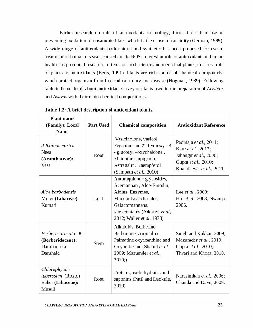
Earlier research on role of antioxidants in biology, focused on their use in
preventing oxidation of unsaturated fats, which is the cause of rancidity (German, 1999).
A wide range of antioxidants both natural and synthetic has been proposed for use in
treatment of human diseases caused due to ROS. Interest in role of antioxidants in human
health has prompted research in fields of food science and medicinal plants, to assess role
of plants as antioxidants (Beris, 1991). Plants are rich source of chemical compounds,
which protect organism from free radical injury and disease (Hogman, 1989). Following
table indicate detail about antioxidant survey of plants used in the preparation of Arishtas
and Asavas with their main chemical compositions.
Table 1.2: A brief description of antioxidant plants.
Plant name
(Family): Local
Name
Part Used Chemical composition Antioxidant Reference
Adhatoda vasica
Nees
(Acanthaceae):
Vasa
Root
Vasicinolone, vasicol,
Peganine and 2' -hydroxy - 4
- glucosyl –oxychalcone ,
Maiontone, apigenin,
Astragalin, Kaempferol
(Sampath et al., 2010)
Padmaja et al., 2011;
Kaur et al., 2012;
Jahangir et al., 2006;
Gupta et al., 2010;
Khandelwal et al., 2011.
Aloe barbadensis
Miller (Liliaceae):
Kumari
Leaf
Anthraquinone glycosides,
Acemannan , Aloe-Emodin,
Aloins, Enzymes,
Mucopolysaccharides,
Galactomannans,
latexcontains (Adesuyi et al,
2012; Waller et al, 1978)
Lee et al., 2000;
Hu et al., 2003; Nwanjo,
2006.
Berberis aristata DC
(Berberidaceae):
Daruhadrika,
Daruhald
Stem
Alkaloids, Berberine,
Berbamine, Aromoline,
Palmatine oxyacanthine and
Oxyberberine (Shahid et al.,
2009; Mazumder et al.,
2010;)
Singh and Kakkar, 2009;
Mazumder et al., 2010;
Gupta et al., 2010;
Tiwari and Khosa, 2010.
Chlorophytum
tuberosum (Roxb.)
Baker (Liliaceae):
Musali
Root
Proteins, carbohydrates and
saponins (Patil and Deokule,
2010)
Narasimhan et al., 2006;
Chanda and Dave, 2009.

CHAPTER-I: INTRODUTION AND REVIEW OF LITERATURE 24
Plant name
(Family): Local
Name
Part Used Chemical composition Antioxidant Reference
Cinnamomum
tamala Nees &
Eberm. (Liliaceae):
Tejpatra
Leaf
Carbohydrates, Glycoside,
Alkaloid, Amino Acids,
Flavanoids, Fixed Oil,
Tannins, Mucilage,
Saponins, Terpenoids,
Phytosterols (Palanisamy et
al., 2011;Jain et al., 2011a;
Mishra et al., 2010)
Chakraborty and Das,
2010; Eswaran et al.,
2010; Mathew and
Abraham, 2006; Chanda
and Dave, 2009; Smerq
and Sharma, 2011;
Khandelwal et al.,2011.
Cinnamomum
zeylanicum Blume
(Liliaceae): Tvak
Stem Bark
Ascorbic Acid, α-
Tocopherol, Total
Carotenoids, Lycopene,
Reduced Glutathione, Total
Phenols and Flavonoids
(Varalakshmi et al., 2012)
Varalakshmi et al., 2012;
Jayaprakasha et al.,
2007; Pandey et al.,
2010;
Cuminum cyminum
L. (Apiaceae):
Ajaji, Sweta Jiraka,
Jire
Fruit
Terpenoids (Iacobellis et al.,
2005; Aina et al. 2012),
Carbohydrates, Thiamin
(Vit. B1), Riboflavin (Vit.
B2), Niacin (Vit. B3),
Vitamin B6, Vit. C, Vi. E, α-
Pinene, Pmentha- 1, 3-dien-
7-ol, D-terpinene, Cuminic
aldehyde, Cuminyl Alcohol
(Muhammad and Riaz,2012)
Koppula and Choi,
2011; Jagtap and Patil,
2010; Khandelwal et al.,
2011; Muhammad and
Riaz, 2012.
Curcuma longa L.
(Zingiberaceae):
Haridra, Haladi.
Rhizomes.
Curcumin (Jain et al., 2011);
Curcuminoids (Curcumin,
Bisdemethoxycurcumin,
Demethoxycurcumin),
Phenolic volatile oils (Cai et
al., 2003)
Cho et al., 2011; Kim et
al., 2005; Selvam et al.,
1995; Nishinaka et al.,
2007; Yu et al., 2002;
Khandelwal et al., 2011;
Sanhita et al., 2012.

CHAPTER-I: INTRODUTION AND REVIEW OF LITERATURE 25
Plant name
(Family): Local
Name
Part Used Chemical composition Antioxidant Reference
Cyperus rotundus
Linn. (Cyperaceae):
Mustaka, Musta
Rhizome
Carbohydrate, Tannins,
Saponins, Flavonoids,
Alkaloids, β-Cyanins,
Quinones, Terpenoids,
Phenols, Coumarins,
Proteins, Steroids (Lydia
and Sundarsanam, 2012;
Sharma and Singh, 2011)
Lydia and Sundarsanam,
2012;
Nagulendran et al.,,
2007.
Elettaria
cardamomum (L.)
Maton
(Zingiberaceae):
Ela
Seeds Total phenols (Abbas, 2011)
Jamal et al., 2006; Verma
et al., 2009; Jain et al.,
2011b; Khalaf et al.,
2008.
Glycyrrhiza glabra
L. (Leguminaceae):
Madhuka
Root
Triterpene, Saponins,
Flavonoids,
Polysaccharides, Pectins,
simple sugars, Amino acids,
Mineral salts (Meena et al.,
2010; Obolentseva et al.
(1999); Liquiritin,
Isoliquiritin (Yamamura et
al., 1992); Glabridin,
Glabrene (Tamir et al.,
2001); Glycyrrhetic acid
(Ploeger et al., 2001)
Naik and Satav, 2003;
Rafi, 2004; Wittschier et
al., 2009; Siracusa et al.,
2011; Khandelwal et al.,
2011; Vaya et al., 1997.
Jasminum officinale
L. (Oleaceae): Jati Flower
Secoiridoid glucoside,
Oleuropein (Teerarak et al.,
2010)
Tsai et al., 2006
Mangifera indica L.
(Anacardiaceae):
Amra, Amba
Ripe and
unripe
fruits
Alkaloids, Steroids,
Carbohydrates, Flavonoids,
Saponins, Amino acids,
Proteins, Phenols and tanins
(Latha, 2011)
Ribeiro et al., 2008;
Ghosal, 1996;
Maisuthisakul and
Gordon, 2009; Chanda
and Dave, 2009; Sanhita
et al., 2012.

CHAPTER-I: INTRODUTION AND REVIEW OF LITERATURE 26
Plant name
(Family): Local
Name
Part Used Chemical composition Antioxidant Reference
Nymphaea stellata
Burm. f.
(Nymphaeaceae)
Utpala
Flower
Nymphayol (Subhash et al.,
2009); Gallic acid (Rakesh
et al., 2009);
Shajeela et al., 2012;
Rajagopal and Sasikala,
2008; Raja et al.,
2011;Rakesh et al., 2010;
Alam et al., 2012.
Ocimum sanctum L.
(Lamiaceae) Tulsi Leaves
Rosmarinic acid,
Lithospermic acid, Eugenol,
Methyleugenol, Urosolic
acid, Phenolics and
Flavonoids such as Orientin,
Vicenin ( Kelm et al., 2000).
Kelm et al., 2000;
Yanpallewar et al., 2004;
Adhvaryu et al., 2007.
Phyllanthus emblica
L.
(Euphorbiaceae):
Amla, Amaliki.
Fruit
Vitamin C, Flavanoids,
Kaempferol, Ellagic Acid
and Gallic Acid (Singh,
2008)
Ghosal et al., 1996; Liu
et al., 2008; Chanda and
Dave, 2009; Scartezzini
et al., 2006; Yokozawa et
al., 2007; Mahesh et al.,
2009; Srikumar et al.,
2005; Rasool and Sabina,
2007; Chanda and Dave,
2009; Sanhita et al.,
2012.
Piper nigrum L
(Piperaceae):
Marika
Seeds,
Fruits
Alkaloid, Glycosides,
Terpenoid, Steroid,
Flavonoid, Tannins,
Reducing Sugar and
Anthraquinones (Nahak and
Sahu, 2011).
Gulcin, 2005; Singh et
al., 2008; Chanda and
Dave, 2009; Khalaf et
al., 2008; Nahak and
Sahu, 2011; Madhu et
al., 2012.
Pluchea lanceolata
(DC.) Oliv. & Hiern
(Asteraceae):
Rasana
Root, Leaf
Terpenoids, Anthroquinone
Glycoside, Coumarin,
(Arora et al., 2011)
Arora et al., 2011

CHAPTER-I: INTRODUTION AND REVIEW OF LITERATURE 27
Plant name
(Family): Local
Name
Part Used Chemical composition Antioxidant Reference
Pueraria tuberosa
DC. (Fabaceae):
Vidari
Root
Tuberosin (Pandey and
Tripathi, 2010); Isoflavone,
Coumestan (Ramakrishna et
al., 1998); Puetuberosanol,
epoxychalcanol (Pawan et
al., 1996); Pterocarponoids
anhydrotuberosin 3-O
methylanhydrotuberosin,tub
erostan (Prasad et al., 1985)
Santosh et al., 2010;
Pandey and Tripathi,
2010; Bharti et al.,
2012.
Ricinus communis L.
(Euphorbiaceae):
Eranda
Root
Alkaloids and flavonoids
(Kang et al., 1985); Phenols
(Khogali et al., 1992); (Jena
and Gupta, 2012).
Singh and Chauhan
200); Kadri et al., 201);
Gupta et al., 2006;
Ilavarasan et al., 2006;
Jena and Gupta, 2012.
Rubia cordifolia L.
(Rubiaceae):
Manjishtha
Root
Anthraquinones (Purpurin,
Alizarin, Munjistin, and
their Glycosides) (Cai et al.,
2003); Triterpenoids,
Anthraquinone (Prajapati
and Parmar, 2011) ;
Rubiadin (Mohana et al.,
2006; Deoda et al., 2011)
Cai et al., 2003; Prajapati
and Parmar, 2011; Son
et al., 2008.
Santalum album L.
(Santalaceae):
Candana
Heard
wood
Santene, Nortricyclo-
Ekasantalene, Alcohols-
Santenol, Teresantalol,
(Kirtikar and Basu, 1933)
(Shankaranaryana, 1980;
Brunke et al., 1995) and the
acids α- santalic acids, β-
Santalic acids and
Teresantalic acids
(Kaur, 2005)
Banerjee et al. 1993;
Sindhu et al., 2010;

CHAPTER-I: INTRODUTION AND REVIEW OF LITERATURE 28
Plant name,
(Family): Local
Name
Part
Used Chemical composition Antioxidant Reference
Saraca asoca (Roxb.)
Wilde (Fabaceae):
Ashoka
Stem
bark
Tannins, Proteins, Steroids,
Glycosides, Carbohydrates,
Saponins, Flavonoids (Saha
et al., 2012;
Pradhan et al., 2010)
Parihar et al., 2010;
Panchawat and Sisodia,
2010.
Syzygium aromaticum
(L.) Merrill & Perry
(Myrtaceae):
Lavanga
Flower
Bud
Total Phenols and
Flavonoids (Kim et al.,
2011).
Banerjee and Das, 2005;
Shyamala et al., 2003;
Khandelwal et al., 2011;
Wojdyło et al., 2007;
Kim et al.,2011.
Terminalia arjuna
(Roxb.) Wight & Arn.
(Combretaceae):
Arjuna
Stem
Bark
Sugars, Amino Acids,
Proteins, Phenols,
Terpenoids, Alkaloids,
Flavonoids, Quinones,
Steroids (Doorika and
Ananthi, 2012);Oxalic acid,
Inorganic Acid,
Carbohydrate (Nema et al.,
2012); Vitamin C, Vitamin
E, Carbohydrates, Tannins
Ellagic acid (Raj et al.
2012);
Doorika and Ananthi ,
2012; Singh et al., 2011;
Raj et al., 2012.
Terminalia belerica
(Gaertn.) Roxb.
(Combretaceae):
Bibhitaka
Plant
Gallic acid, Tannic acid and
Ascorbic acid (Singh et al.,
2008)
Yokozawa et al., 2007;
Mahesh et al., 2009;
Srikumar et al., 2005.
Terminalia chebula
Retz.
(Combretaceae):
Haritki
Plant,
Fruits
Chebulagic acid, Chebulinic
acid, Tannic acid, Ellagic
acid, Gallic acid, Ascorbic
acid (Singh, 2008); Tannins
(Ellagitannins), Phenolic
acids (Cai et al., 2003)
Yokozawa et al., 2007;
Mahesh et al., 2009;
Srikumar et al., 2005;
Yadav et al., 2011;
Chanda and Dave, 2009.

CHAPTER-I: INTRODUTION AND REVIEW OF LITERATURE 29
Plant name,
(Family): Local
Name
Part
Used Chemical composition Antioxidant Reference
Vitis vinifera L.
(Vitaceae): Draksha
Fruits,
Seeds
Lipids, Proteins, Pectin,
Carbohydrate, Vit B2, Vit C,
Phenols, Oxalic Acids,
Alkaloids (Khan et al.,
2008; Lutz et al., 2011).
Baydar et al., 2007;
Chanda and Dave, 2009;
Fauconneau et al., 1997;
Lutz et al., 2011,
Jayaprakasha et al.,
2001.
Withania somnifera
(L.) Dunal
(Solanaceae):
Ashvagandha
Root,
Leaf,
Seed
Alkaloids (Isopellertierine,
anferine), Steroidal Lactones
(Withanolides, Withaferins),
Saponins containing an
additional acyl group
(Sitoindoside VII and VIII),
Withaferin A (Singh et al.,
2010b)
Ichikawa et al., 2006;
Maitra et al., 2009;
Udayakumar et al., 2010;
Bhattacharya et al.,
2000; Khandelwal et al.,
2011; Sanhita et al.,
2012.
Woodfordia fruticosa
(L.) Kurz
(Lythraceae): Dhatki
Flowers
Tannins, Cardiac
Glycosides, Steroids,
Saponins, (Vaghasiya et al.
2011; Finose and K.Devaki,
2011), Anthraquinonoes,
Flavons, Flavonols and
Chalcones, Terpenoids,
Phlobatanins and Cardiac
Glycosides (Khan et al.,
2011a).
Nitha et al., 2012;
Khan et al., 2011.
Zingiber officinale
(Zingiberaceae):
Sunthi
Rhizome
Phenolic, Volatile oils
(Gingerol Analogues:
Gingerols, Shogaol) (Cai et
al., 2003)
Kabuto et al., 2005;
Jiang et al., 2005; Rhode
et al., 2007; Shukla and
Singh, 2007; Ahmed et
al., 2008.
Many researchers, who have carried out work to prove antioxidant action of
drugs, have used either single in-vitro or in-vivo model. However, it is not advisable to
follow only one model and in many cases results are not reproducible. Various in-vitro
antioxidant models, which have proved efficient in confirming anti-stress properties of
extracts and formulations, would be of more helpful when used as a set of models at a
time for screening antioxidant potential. Such screening with various models would

CHAPTER-I: INTRODUTION AND REVIEW OF LITERATURE 30
enlighten anti-stress prospective of any drug or extract under various stress conditions.
Therefore, it is very important to use as much possible antioxidant models at a time to
generate more reliable results, to prove claims of polyherbal drugs which are already in
market and to screen new formulations or products.
Arishtas and Asavas are considered as unique and valuable therapeutics in
Ayurveda. Though traditional knowledge in literature as well as in practice exists about
Arishtas and Asavas, there was little effort to document, preserve and improve this
knowledge for betterment of mankind. A Literature survey revealed that although,
Arishta and Asava have been studied for ability to cure number of diseases, information
on quantitative parameters of Arishta and Asava to guarantee quality of the product to
consumer is less. Carrying out in detail studies on antioxidant potential and antioxidant
related biochemical parameters of Arishta and Asava may find applications of these
fermented polyherbal drugs in the management of diseases caused due to oxidative stress.
Therefore, it was intended to investigate the antioxidant potential of Arishtas and
Asavas by way of different in-vitro models to trough light on their possible exploitation
in stress management, cancer and heart diseases as well as general health problems.
1.4. SCOPE OF THE PRESENT STUDY
Antioxidants are micro-constituents of diet that are involved in structural
maintenance of DNA and cell and their repair. They protect DNA and cell membranes
against oxidative damage, including that induced by carcinogenic agents. It is therefore,
biologically believable that diets rich in antioxidants protect against cancer and other
chronic diseases. Individual and population requirements for antioxidants are determined
by the level of exposure to oxidative stress. Not only diets rich in vegetables and fruits
but regular consumption of fermented polyherbal medicine also give protection against
oxidative damage. Present study is the major attempt on antioxidant activity of Arishta
and Asava related with their biochemicals.