Embed Size (px)
Citation preview

1
CHAPTER I
Introduction
Abstract
This chapter covers historical background, fundamentals, classifications and
advantages of drug delivery systems. It introduces hydrogels and various stimuli
responsive hydrogels as controlled release (CR) systems. The most common methods
of preparations of micro/nanoparticles of polymeric blends, graft copolymers and
interpenetrating polymer networks are discussed along with drug loading, drug release
kinetics and release mechanisms.

2
I.1. Historical Background of Drug Delivery System
Perspective drug delivery systems can be defined as mechanisms to introduce
therapeutic agents into the body. Chewing leaves and roots of medical plants and
inhalation of soot from the burning of medical substances are examples of drug
delivery from the earliest times. However, these primitive approaches of delivering
drugs lacked a very basic need in drug delivery; that is, consistency and uniformity (a
required drug dose). This has led to the development of different drug delivery
systems during the later part of the eighteenth and early nineteenth century. Those
methods include pills, syrups, capsules, tablets, solutions, extracts, emulsions and
suspensions [1,2].
The classical era of medicine development started with the discovery of
vaccines in 1885 and techniques for purification of drugs from plant sources in the
late nineteenth century, followed by macromolecular drug delivery. Along with drug
development, the mode of systemic administration and drug delivery systems were
also developed to improve therapeutic efficacy of drugs to reduce toxic effects by
augmenting the amount and persistence of drugs in the vicinity of the target cells,
while reducing drug exposure to non-target cells. This basic rationale gave birth to
controlled drug delivery science. The controlled drug delivery system requires
simultaneous consideration of factors such as drug property, route of administration,
nature of delivery vehicle, mechanism of drug release, ability of targeting and
biocompatibility [3,4].
I.2. Fundamentals of Drug Delivery System
The main purpose of using a drug delivery system (DDS) is not only to deliver
a biologically active compound in a controlled manner (time period and releasing
rate) without altering their original properties, but also to maintain the drug

3
concentration in the body within therapeutic window (Figure I.1.). To obtain a given
therapeutic response, a suitable amount of the active drug must be absorbed and
transported to the site of action at the right time and the rate of input can then be
adjusted to produce the concentrations required to maintain the level of the effect for
as long as necessary. Besides, one can tune the release kinetics and direct drug
targeting towards a specific organ or diseased tissue (targeted drug delivery). The first
two features were addressed by using drug carriers, usually biopolymers or synthetic
polymers, whose properties can be manipulated to improve the DDS efficiency.
Owing to rapid advances in recent years, the application of polymers to drug delivery
has grown rapidly [5,6].
Figure I.1. Scheme of the effect of drug concentration in the body when using
different administration methods
I.3. Classification of Drug delivery Systems
Drug delivery systems can be broadly classified into temporal and targeted
drug delivery systems. Temporal drug delivery systems are designed to release
therapeutic levels of drugs from a matrix for the desired period of time. There are two
Pretended therapeutic time
4
strategies to achieve controlled release using temporal DDS. First approach is to
prepare the matrix that release drugs over extended duration. Numerous works have
been done based on biodegradable polymers in which the rate of drug release matches
the rate of drug elimination. Therefore, drug concentration is within the therapeutic
range for a longer time. This release pattern is highly beneficial for drugs that are
rapidly metabolized and eliminated from the body after administration [7-10]. The
second approach is to prepare a feedback controlled devices that release the
appropriate amount of drug in response to a therapeutic marker.
In recent years, several research groups have been developing responsive
systems [11-15]. These systems can be classified as external regulated and self-
regulated systems. The external controlled devices apply external triggers for pulsed
delivery such as: magnetic, ultrasonic, thermal and electric triggers. In the self-
regulated system, the release rate is controlled by feedback information. The self-
regulated systems utilize several approaches such as pH-sensitive polymers, enzyme
substrate reactions and competitive binding as rate-control mechanisms. The
advantage of temporal delivery system is that therapeutic concentration of a drug can
be maintained in the body for longer times without repeated administration, thereby
eliminating the problems of drug under or over dosage. Furthermore, it is more
economical due to lower drug wastage, reproducible and it increases patient
compliance. Attempts are still being made to develop novel delivery systems that can
further control the drug release pattern by synthesizing novel polymers as matrix
systems as well as by developing smart systems that can deliver multiple drugs in a
controlled manner or under the effect of an external stimulus.
Targeted drug delivery systems are designed to deliver drugs at the proper
dosage for the required amount of time to a specific site of the body where it is

5
needed, thereby preventing any adverse effects of drugs on other organs or tissues.
There are two basic types of targeting systems: passive and active. Passive targeting
systems rely on non-specific interactions such as hydrophobic or electrostatic
interactions and the body physical characteristics. The size of drug carriers has been
extensively studied for passive targeting. It was found that particles larger than 5-7
μm in diameter usually become trapped in the lung [16] and particles smaller than 1
μm in diameter rapidly phagocytosed by the Kupffer cells of the liver [17]. When the
particle size is reduced below 100 nm, the particles can appear in the bone marrow
[18].
It was also demonstrated that drug carriers smaller than 200 nm can be
accumulated efficiently in tumor through enhanced permeability and retention (EPR)
effect due to the abnormality of tumor tissue, resulting in the enhanced vascular
permeability compared to healthy tissues [19-21]. On the other hand, active targeting
systems utilize specific interactions, such as antigen-antibody and ligand receptor
binding, to achieve specific targeting goals. In this approach, the therapeutic index of
drugs could be enhanced by keeping drugs away from healthy cells. The types of
receptors that have been utilized for this purpose include transferrin receptors (tumor
cells) [22], folate receptors (tumor cells) [23], albumin receptors (cardiac and lung)
[24] and growth factors receptors [25]. Targeted delivery assumes great importance
particularly in the case of highly toxic drugs such as chemotherapeutic drugs and
highly active and fragile biotechnological molecules such as peptides and proteins.
Furthermore, targeted drug delivery systems can be employed to deliver drugs to sites
that are inaccessible under normal conditions such as the brain.
6
I.4. Advantages of Controlled Drug Delivery
In the past 30 years, controlled drug delivery technology has represented one
of the most rapidly advancing research areas. The field is driven by the belief that
controlled drug delivery will contribute significantly to human health [26-30]. These
drug delivery systems offer numerous advantages compared to conventional dosage
forms:
♦ increasing the efficacy of currently used drugs
♦ providing opportunities for the use of new agents currently precluded from
clinical use due to challenges including low drug solubility and systemic toxicity
♦ reducing harmful side effects
♦ precise control of dose
♦ decreasing number of dosages
♦ improving patient compliance and convenience
I.5. Hydrogels as Drug Delivery Systems
Hydrogels are hydrophilic polymer networks which absorb water or biological
fluids from 10-20 % (an arbitrary lower limit) up to thousands of times their dry
weight. Hydrogels may be chemically stable or they may degrade and eventually
disintegrate and dissolve. They are called reversible or physical gels when the
networks are held together by molecular entanglements, and/or secondary forces
including ionic, H-bonding or hydrophobic forces. Physical hydrogels are not
homogeneous, since clusters of molecular entanglements, or hydrophobically- or
ionically-associated domains, can create inhomogeneities. Free chain ends or chain
loops also represent transient network defects in physical gels. Hydrogels are called
permanent or chemical gels when they are covalently-crosslinked networks [31-35].

7
I.5.1 Classification of Hydrogels
Based on their nature hydrogels are classified as:
I.5.1.1. pH Sensitive Hydrogels as CR Systems
The pH sensitive hydrogels can be neutral or ionic in nature. In neutral
hydrogels, the driving force for swelling arises from the water-polymer
thermodynamic mixing contributions and elastic polymer contributions. In ionic
hydrogels, swelling is due to the previous two contributions as well as ionic
interactions between charged polymer and free ions [35]. The presence of ionizable
functional groups like carboxylic acid, sulfonic acid or amine groups renders the
polymer more hydrophilic and results in high water uptake. In the case of anionic
polymeric network containing carboxylic or sulfonic acid groups, ionization takes
place, as the pH of the external swelling medium rises above the pKa of that ionizable
moiety [36-38].
The dynamic swelling change of the anionic hydrogels can be used in the
design of intelligent controlled release (CR) devices for site-specific drug delivery of
therapeutic proteins to large intestine, where the biological activity of the proteins is
prolonged. The change in the pH of the external environment will act as a stimulus
and the response to the stimulus leads to the change in swelling properties of the
hydrogels, causing the release of the encapsulated bioactive species. The cationic
hydrogels show swelling at pH values below pKa of the cationic group. The amine
groups are protonated at pH lower than pKa and become hydrophilic and absorb
water. At pH greater than pKa, the polymer is hydrophobic and excludes water [39-
40].
Numerous researchers have studied dynamic swelling of pH-Sensitive
networks. Katchalsky and Michaeli [41] established that the collapse and expansion of
poly(methylacrylic acid) (PMA) hydrogels occurred reversibly by adjusting the pH of
8
the fluid. Ohmine and Tanaka [42] observed the sudden collapse of ionic network in
response to sudden changes in the ionic strength of the swelling medium. Studies by
Khare and Peppas [43] examined pH and ionic strength dependent swelling kinetics in
case of hydrogels of PMA or poly(acrylic acid) (PAA) with poly(hydroxyethyl
methacrylate). Kim et al. [44] prepared the pH-sensitive anionic hydrogels based on
poly(methacrylic acid-co-methacryloxyethyl glucoside) and poly(methacrylic acid-g-
ethylene glycol). The hydrogels showed limited swelling in a pH 2.2 buffer but rapid
swelling was observed in the pH 7.0 buffer solution.
I.5.1.2. Temperature Sensitive Hydrogels as CR systems
Thermosensitive hydrogels are one of the widely studied responsive polymer
systems. Thermosensitive polymers are characterized by the presence of hydrophobic
groups, such as methyl, ethyl and propyl groups. The most widely studied temperature
sensitive polymer is poly(N-isopropylacrylamide) (PNIPAAm). PNIPAAm is a non-
biodegradable polymer with a LCST ~32° in water and cross-linked gels of this
material collapse around this temperature [45].
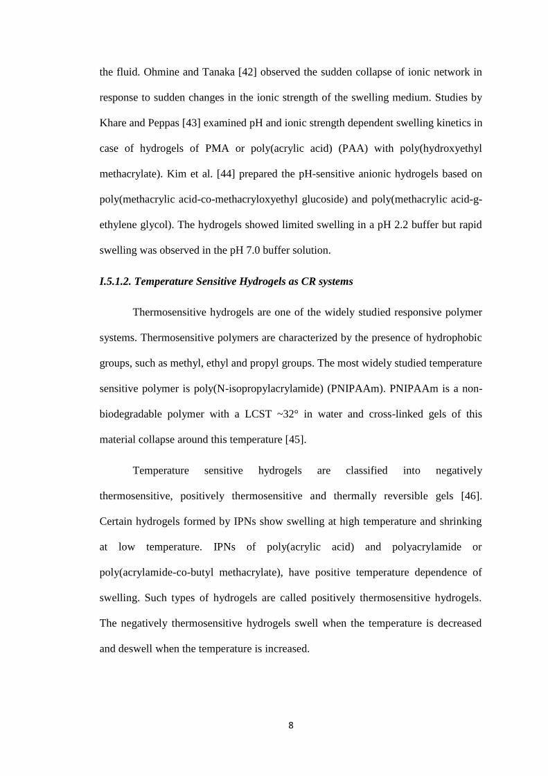
Temperature sensitive hydrogels are classified into negatively
thermosensitive, positively thermosensitive and thermally reversible gels [46].
Certain hydrogels formed by IPNs show swelling at high temperature and shrinking
at low temperature. IPNs of poly(acrylic acid) and polyacrylamide or
poly(acrylamide-co-butyl methacrylate), have positive temperature dependence of
swelling. Such types of hydrogels are called positively thermosensitive hydrogels.
The negatively thermosensitive hydrogels swell when the temperature is decreased
and deswell when the temperature is increased.
9
Figure I.2. Response of polymeric hydrogels to various environmental stimuli.
I.5.1.3. Enzyme Sensitive Hydrogels as CR Systems
Since many biodegradable polymers can be digested by specific enzymes,
enzyme sensitive hydrogels can be prepared from such biodegradable polymers. Some
enzymes are used as important signals for diagnosis to monitor several physiological
changes and specific enzymes in specific organs have become useful signals for site
specific drug delivery [47]. Therefore, the enzyme sensitive hydrogels are promising
candidates as enzyme sensitive drug delivery systems.
Hovgaard et al. [48] focused on the fact that microbial enzymes in the colon,
such as dextranases, can degrade the polysaccharide dextran. They prepared dextran
hydrogels cross linked with diisocynate for colon specific drug delivery. The dextran
hydrogels were degraded in vitro by a model dextranase, as well as in vivo in rats and
in human colonic fermentation model. Release of a drug from the dextran hydrogels
can be controlled by the presence of dextranase. Drug release from the dextran
hydrogels in the absence of dextranase was observed to be based on simple diffusion
process, however in the presence of dextranase it was mainly governed by the
degradation of the dextran. Thus, it follows that dextran hydrogels are dextranase
sensitive and may hold promise as intelligent systems for colon specific drug delivery.
change in
pH
Temperature
Electric Field Light
Ultrasound
Magnetic Field

10
I.5.1.4. Glucose Sensitive Hydrogels as CR Systems
Glucose sensitive hydrogels are very useful for the development of self
regulated insulin delivery systems and enable us to construct an artificial pancreas that
can administer the necessary amount of insulin in response to the blood glucose
concentration. Combining glucose oxidase with pH sensitive hydrogels to sense
glucose and regulate insulin release is the method that many researchers have used to
develop glucose sensitive insulin delivery systems. Within the pH sensitive hydrogels
containing glucose oxidase, glucose is converted to gluconic acid by glucose oxidase,
thus lowering the pH in the hydrogels. Insulin can be released by the pH sensitive
swelling of the hydrogels. Thus, the pH sensitive hydrogels containing glucose
oxidase can control insulin release in response to the glucose concentration [49].
I.5.1.5. Electrical Sensitive Hydrogels as CR Systems
Electric current can also be used as an environmental signal to induce
responses of hydrogels. Hydrogels, sensitive to electric current, are usually made of
polyelectrolytes. An electric field as an external stimulus has advantages, such as the
availability of equipment, which allows precise control with regards to the magnitude
of current, duration of electric pulses, intervals between pulses, etc.
The electrical behavior of hydrogels composed of sodium alginate (SA) and
oly(diallyldimethylammonium chloride) (PDADMAC) was studied by Kim et al.
[50]. The SA/ PDADMAC IPN hydrogel exhibited pH and electrolyte concentration
sensitive behavior. When an electric field is applied to a strip of the SA/PDADMAC
hydrogel in an aqueous HCl solution, the gel showed significant and quick bending
towards the cathode. It was concluded that the deformation of a polymer hydrogel
under an electric field was due to voltage-induced motion of ions and concomitant
expansion of one side of the polymer and the contraction of other side of the polymer.

11
I.5.1.6. Light Sensitive Hydrogels as CR Systems
Light (ultraviolet or visible) is a desirable external stimulus for drug delivery
systems because it is inexpensive and easily controlled. Light-sensitive drug carriers
are fabricated from polymers that contain photo-sensitizers such as azobenzene,
stilbene and triphenylmethane [51,52]. Suzuki and Tanaka [53] have investigated
visible light-responsive hydrogels using the trisodium salt of copper chlorophyllin in
PNIPAAm hydrogels. When light is applied to the hydrogels, the chromophore
absorbs the light, increasing the local temperature of the hydrogel. The resulting
temperature change alters the swelling behavior.
Vivero-Escoto et al. [54] prepared gold capped mesoporous silica nanospheres
for photo-induced intracellular release of drugs in human cells. The 100 nm silica
nanospheres were capped with 5 nm gold nanospheres and functionalized with a
cationic photo-reactive linker. Photoirradiation using ultraviolet light for 10 min at
0.49 mW/cm2 cleaved the photolabile linker, causing uncapping of the silica due to
charge repulsion between the gold and silica nanospheres, allowing drug to be
released [55]. Fomina et al. [56] developed a novel light-sensitive polymer containing
a quinone-methide moiety. Nile Red, a hydrophobic dye, was released from the
nanoparticles after only one minute of 350 nm light exposure. Light can be effective
in modulating drug release because it can be used to increase the local temperature
and to cleave bonds.
I.5.1.7. Ultrasound Sensitive Hydrogels as CR Systems
Ultrasound has been shown to trigger the drug release by raising the local
temperature or causing cavitation [57]. Both processes can increase the permeability
of cell membranes and accelerate polymer degradation [58]. Ultrasound sensitive
vehicles have the potential to treat tumorigenic cancers due to their invasive character,
ability to penetrate deeply into the human body and ease of control.
12
In 2002, Pruitt and Pitt [59] investigated ultrasound mediated doxorubicin
release using stabilized Pluronic P105 micelles. Doxorubicin was encapsulated within
polymeric micelles composed of 10% Pluronic P105 and N,N-diethylacrylamide and
delivered systemically to rats. Application of low-frequency ultrasound at the tumor
site resulted in doxorubicin release; this resulted in a significant reduction in tumor
volume. Lin et al. [60] have investigated the physical and chemical properties of lipid
membranes subjected to ultrasound treatment. They showed that high permeability
resulting from ultrasound treatment is correlated with lipid packing and can be useful
for efficient drug release and ultrasound-mediated DNA transfection. In 2007, Ferrara
et al. [61] reviewed that small gas bubbles, used to enhance ultrasound contrast, can
be used for drug delivery applications and monitoring. When driven by an ultrasonic
pulse, small gas bubbles oscillate with a wall velocity on the order of tens to hundreds
of meters per second and can be deflected to a vessel wall or fragmented into particles
on the order of nanometers. Also, a focused ultrasound beam can be used for
disruption of delivery vesicles and blood vessel walls, which offer the opportunity to
locally deliver a drug or gene. Ultrasound does not damage the surrounding tissue,
making it attractive for triggering the drug release.
I.5.1.8. Magnetically Sensitive Hydrogels as CR Systems
Magnetically modulated particulate systems have recently attracted much
attention for in vivo imaging and targeted drug delivery [62]. In this approach,
imaging agents or drugs can be localized to specific sites through the application of an
external magnetic field. Superparamagnetism in many biomedical applications such as
drug delivery is useful because the superparamagnetic iron oxide devices (SPIOD)
can be transported by electrical field effects to the desired site and once the external
magnetic field is removed, magnetization disappears and the SPIOD can remains at

13
the target site for a certain period [63]. Two types of iron oxide have mainly been
investigated for their use in magnetic formulation: maghemite (γ-Fe2O3) and
magnetite (Fe3O4) due to their high saturation magnetization and high magnetic
susceptibility; the magnetite (Fe3O4) particles are preferred because of their greater
saturation magnetization and biocompatibility that make them more promising
candidates for various biomedical applications [64].
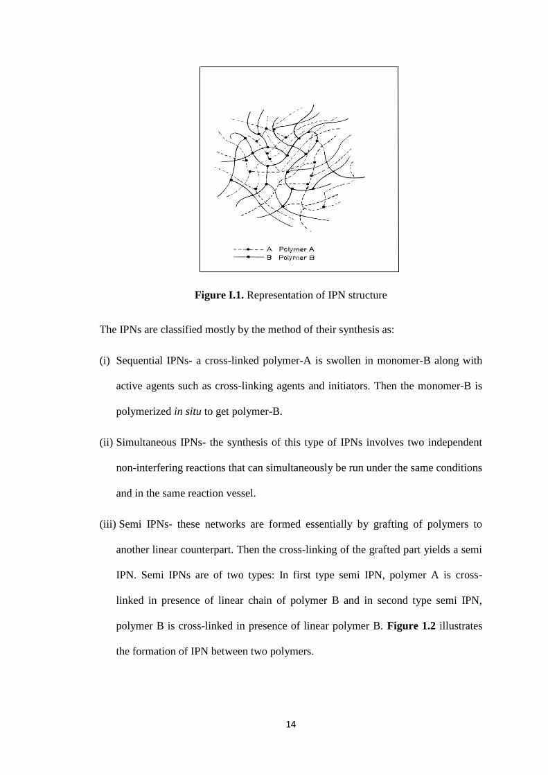
I.6. Interpenetrating Polymer Networks (IPNs)
The term “Interpenetrating Polymer Networks” (IPNs) was coined by Miller
[65] in 1960. IPNs have been extensively investigated over the past four decades due
to their ability to produce versatile materials with the required combination of
properties. Usually they are intimate mixture of two polymers which are in network
form; at least one is synthesized and /or cross-linked in the immediate presence of the
other. As the IPNs are cross-linked networks that are insoluble in water, but absorb
large quantity of water or biological fluid and they are soft and rubbery in nature
resembling those of living tissues in their physical properties and hence they are
highly biocompatible and non-toxic. In IPNs, two or more different polymers are
individually cross-linked in the immediate presence of other. So, the cross-linked
chains are intermingled resulting in considerable phase mixing through the restriction
of domain size. This result in an appreciable improvement in strength and dynamic
mechanical properties of IPNs compared to single polymeric hydrogels.
14
Figure I.1. Representation of IPN structure
The IPNs are classified mostly by the method of their synthesis as:
(i) Sequential IPNs- a cross-linked polymer-A is swollen in monomer-B along with
active agents such as cross-linking agents and initiators. Then the monomer-B is
polymerized in situ to get polymer-B.
(ii) Simultaneous IPNs- the synthesis of this type of IPNs involves two independent
non-interfering reactions that can simultaneously be run under the same conditions
and in the same reaction vessel.
(iii) Semi IPNs- these networks are formed essentially by grafting of polymers to
another linear counterpart. Then the cross-linking of the grafted part yields a semi
IPN. Semi IPNs are of two types: In first type semi IPN, polymer A is cross-
linked in presence of linear chain of polymer B and in second type semi IPN,
polymer B is cross-linked in presence of linear polymer B. Figure 1.2 illustrates
the formation of IPN between two polymers.
15
I.7. Polymeric Micro/Nanoparticles as Drug Delivery Systems
In recent years, polymeric micro/nanoparticles have attracted a considerable
attention as potential drug delivery devices in view of their applications in the CR of
drugs, drug targeting to particular organ/tissues, as carriers of DNA in gene therapy,
in the delivery of proteins and peptides through the peroral route of administration
[66-69].
I.7.1. Most Commonly Used Methods for Preparation of Polymeric
Micro/Nanoparticles
I.7.1.1. Emulsion Crosslinking Method
In this method, water-in-oil (w/o) emulsion is prepared by emulsifying the
polymer aqueous solution in the oil phase. Aqueous droplets are stabilized using a
suitable surfactant. The stable emulsion is crosslinked by using an appropriate
crosslinking agent such as gluteraldehyde to harden the droplets. Microspheres were
filtered and washed repeatedly with n-hexane followed by alcohol and then dried [70].
By this method, size of the particles can be controlled by controlling the size of
aqueous droplets. However, the particle size of the final product depends upon the
extent of crosslinking agent used, while hardening in addition to speed of stirring
during the formation of emulsion. In addition microparticles were prepared by
crosslinking the polymer to obtain a non-sticky glassy hydrogel followed by passing
through a sieve [71]. Schematic representation of the preparation of microspheres by
emulsion crosslinking method is in.
I.7.1.2. Solvent Evaporation Method
In this method, polymer is dissolved in an organic solvent like
dichloromethane, chloroform or ethyl acetate. Drug is dissolved or dispersed in the
performed polymer solution and the drug containing polymer solution is emulsified

16
into an aqueous solution to make an oil-in-water (o/w) emulsion by using a surfactant
or emulsifying agent like gelatin, poly(vinyl alcohol) (PVA), polysorbate-80 etc.
After the formation of stable emulsion, organic solvent is evaporated either by
increasing the temperature, under vacuum or by continuous stirring [72-74].
I.7.1.3. Spontaneous Emulsification/Solvent Diffusion Method
This is a modified version of solvent evaporation method [75], wherein water-
soluble solvents like acetone or methanol along with the water insoluble organic
solvents like dichloromethane or chloroform are used as an oil phase. Due to the
spontaneous diffusion of water-soluble solvent, an interfacial turbulence is created
between the two phases, leading to the formation of smaller particles. In this method,
the particle size can be varied by varying the concentration of water-soluble solvent.
I.7.1.4. Double Emulsion method
In this method, drug dissolved in an aqueous solvent is emulsified with the
non-miscible organic solution of polymer to form w/o emulsion. The organic solvent,
dichloromethane is mainly used and the homogenization step is carried out using
either high-speed homogenizer or sonicator. This primary emulsion is then rapidly
transferred to an excess of aqueous medium containing a stabilizer, usually PVA.
Again, homogenization or intensive stirring is necessary to initially form the double
emulsion of w/o/w. Subsequent removal of organic solvent by heat, vacuum or both
would result in the phase separation of polymer and the core to produce microspheres
[76].
I.7.1.5. Spray Drying Method
In this method, the polymer is dissolved in a volatile organic solvent such as
dichloromethane or acetone; the drug in solid form is then dispersed in polymer

17
solution by high speed homogenization or it can be dissolved in a solvent; then this
solution is atomized in a stream of heated air. From the droplets formed, the solvent
evaporates instantaneously yielding free flowing microparticles [77-79]. Size of
microparticles depends upon atomizing conditions, size of the nozzle, spray flow rate
and inlet air temperature. The microspheres are collected from air streams by cyclone
separator. Residual solvents are removed by vacuum drying.
I.7.1.6. Coacervation/Precipitation Method
This method utilizes the physiochemical properties of the polymers. For
instance, chitosan is insoluble in alkaline pH medium, but precipitates/coacervates
upon contact with the alkaline solution. Particles are produced by blowing chitosan
solution into an alkali solution like sodium hydroxide using a compressed air nozzle
to form coacervates droplets [80]. Separation and purification of particles was done
by filtration/centrifugation followed by successive washing with hot and cold water.
Varying compressed air pressure or spray-nozzle diameter controlled the size of the
particles and then by using the cross-linking agent to harden the particles could
control the drug release.
I.7.1.7. Emulsion-Droplet Coalescence Method
The novel emulsion-droplet coalescence method was developed by Tokumitsu
et al. [81], which utilizes the principles of both emulsion crosslinking and
precipitation. However, in this method, instead of cross-linking the stable droplets,
allowing the coalescence of chitosan droplets with NaOH droplets induces
precipitation. First, a stable emulsion containing aqueous solution of chitosan along
with drug is produced in liquid paraffin oil and then, another stable emulsion
containing chitosan aqueous solution of NaOH is produced in the same manner. When
18
both emulsions are mixed under high-speed stirring, droplets of each emulsion would
collide at random and coalesce, thereby precipitating the chitosan droplets to give
small size particles.
I.7.1.8. Ionic Gelation Method
The use of complexation between the oppositely charged polymers to prepare
microspheres has attracted much attention, because the process is very simple and
mild. In addition, reversible physical crosslinking by electrostatic interaction, instead
of chemical crosslinking, has been applied to avoid the possible toxicity of reagents
and other undesirable effects. Recently, many researchers [82,83] have explored the
ionic gelation technique for potential pharmaceutical usage. Cationic polymers such
as chitosan can undergo ionic gelation when reacted with polyanion such as
tripolyphosphate (TPP), whereas the anionic polymers such like sodium alginate and
gellan gum undergo ionic gelation with bivalent cations such as calcium, barium or
zinc [84].
I.7.1.9. Dispersion Polymerization Method
Couvreur et al. [85] reported the production of nanoparticles (≈200 nm) by
polymerizing mechanically the dispersed methyl or ethyl cyanoacrylate in an aqueous
acidic medium in the presence of polysorbate-20. The cyanoacrylic monomer is added
to an aqueous solution of a surface-active agent (polymerization medium) under
vigorous mechanical stirring to polymerize alkylcyanoacrylates at the ambient
temperature. Drug is dissolved in the polymerization medium either before the
addition of the monomer or at the end of the polymerization reaction. The suspension
is then purified by ultracentrifugation or by resuspending the particles in an isotonic
surfactant free medium.
19
I.7.2. Drug Loading in to Polymer Micro/Nanoparticles
Drug loading in micro/nanoparticulate systems can be done by two methods
i.e., during the preparation of particles (incorporation) and after the formation of
particles (incubation). In these systems, drug is physically embedded into the matrix
or adsorbed onto the surface. Various methods of loading have been developed to
improve the efficiency of loading, which largely depends upon the method of
preparation as well as physicochemical properties of the drug. Maximum drug loading
can be achieved by incorporating the drug during the formation of particles, but it
may get affected by the process parameters such as method of preparation, presence
of additives, etc. Both water-soluble and water-insoluble drugs can be loaded into
polymeric particulate systems. Water-soluble drugs are mixed with polymer solution
to form a homogeneous mixture, and then, particles can be produced by any of the
methods discussed earlier [86]. Water-insoluble drugs can be loaded by the soaking
method [87] or by using the multiple emulsion technique.
I.8. Graft Copolymers as Drug Delivery Systems
Recently, much attention has been paid to the graft copolymerization of
natural polysaccharides [88-90] in order to obtain novel tailored hybrid materials with
minimum loss of the initial properties of the substrate. Due to their structural diversity
and water solubility, natural polysaccharides could be interesting starting materials for
the synthesis of graft copolymers. A graft copolymer is a macromolecular chain with
one or more species of block connected to the main chain as side chain(s). Thus, it can
be described as having the general structure, where the main polymer backbone
(commonly referred to as the trunk polymer), has branches of another polymeric chain
emanating from different points along its length. This fascinating technique may be
considered as an approach to achieve novel polysaccharide-based materials with

20
improved properties including all the expected usefulness of these biomaterials. After
a thorough literature survey, it was found that polysaccharide-based graft copolymers
are mainly synthesized by free radical polymerization under the influence of different
chemical initiating systems [91,92]. These graft copolymers could be applied in the
design of various stimuli-responsive CR systems.
I.9. Drug Release and Release Kinetics
Drug release from micro/nanoparticles and subsequent biodegradation are
important in developing successful formulations. Drug release from the hydrogel-
based particulate systems depends upon the extent of crosslinking, morphology, size
and density of the particulate system, physiochemical properties of the drug as well as
the presence of adjuvants. In case of biodegradable polymer, the release depends on
matrix erosion and a combined erosion/diffusion process. In vitro release also depends
upon pH, polarity and the presence of enzymes in the dissolution media.
The release of drug from the particulate systems involves three different
mechanisms: (a) release of drug from the surface of particles, (b) diffusion through
the swollen rubbery matrix and (c) release due to polymer erosion. Drug release by
diffusion involves three steps. First, water penetrates into the particulate system,
which causes swelling of the matrix; secondly, the glassy polymer converts into
rubbery matrix, while the third step is the diffusion of drug from the swollen rubbery
matrix. Hence, the release is slow initially and later, it becomes fast.
To ascertain the kinetics of release parameters, following empirical equations
[94,95] were used to estimate the release kinetics parameters. According to zero order
release, we have:

21
where Q is the amount of drug at time, t; Q0 is the amount of drug at t = 0 and K0 is
zero order release constant. The first order equation is:
where K1 is the first order release constant. Higuchi square root equation is given as:
where Mt is the amount of drug released at time, t and KH is Higuchi rate constant.
Hixson-Crowell cube root equation is:
where Kc is cube root law release constant. Cumulative release data were also
analyzed using [96, 97]:
here, Mt/M represents the fractional drug release at time t, k is a kinetic parameter,
characterizing the drug-polymer interaction and n is an empirical parameter,
characterizing the release mechanism. For spheres, n values below 0.43 indicates that
the drug release is diffusion controlled, while the values of n between 0.43 and 0.85
are indicative of both diffusion controlled as well as swelling-controlled release
(anomalous), but the values >0.85 indicate swelling-controlled release that is related
to polymer relaxation phenomenon during swelling. If n value is >1, then drug release
follows Super Case II transport mechanism [98].
I.10. Present Thesis Research Problem
Careful literature analysis revealed that plethora of work is being done on
development of polymeric matrices as drug delivery systems in order to address
l n Q = ln Q 0 – K 1 t (7 ) (I.2)
M t = K H t
1/ 2 ( 8 ) (I.3)
Q
1 / 3 = Q 0
1 / 3 - K c t ( 9 ) (I.4)
M t / M = kt
n (10) (I.5)
Q = Q 0 - K 0 t ( 6 ) (I.1)
22
pharmaceutical hurdles to improve the human life. In the same view, the present
thesis covers newer approaches to develop CR drug delivery systems by
encapsulating different drugs. Different polymeric microspherical delivery systems
were developed and their release characteristics were tuned by modifying the
polymeric system by blending two different natured polymers. Such studies are
important in developing successful formulations for their large-scale
commercialization, once-a-day formulation, CR/sustained release tablets. Thus, theme
of the thesis is timely and presents the comprehensive approach to the above
mentioned problem. Details of each of these problems will be covered in subsequent
chapters.

23
I.11. Literature Cited
[1] N.A. Peppas, Adv. Drug Deliv. Rev. 65 (2013) 5.
[2] G. Camenisch, G. Folkers, H. van der Waterbeemd, Pharm. Acta. Helv. 71
(1996) 309.
[3] B. Jeong, Y.K. Choi, Y.H. Bae, G. Zentner, S.W. Kim, J. Control. Release. 62
(1999) 109.
[4] J. Tsung, D.J. Burgess, In: Fundamentals and Applications of Controlled
Release Drug Delivery, Editor: M.J. Rathbone, Springer. (2012) 107-123.
[5] K.W. Leong, R. Langer, Adv. Drug Del. Rev. 1 (1988) 199.
[6] I.I. Slowing, J.L. Vivero-Escoto, C.W. Wu, V.S.Y. Lin, Adv. Drug Del. Rev. 60
(2008) 1278.
[7] J. Panyam, V. Labhasetwar, Adv. Drug Deliv. Rev. 55 (2003) 329.
[8] R.H. Muller, M. Radtke, S.A. Wissing, Adv. Drug Deliv. Rev. 54 (2002) 131.
[9] C. Perez, A. Sanchez, D. Putnam, D. Ting, R. Langer, M.J. Alonso, J. Control.
Release. 75 (2001) 211.
[10] N. Kumar, M. Ravikumar, A.J. Domb, Adv. Drug Del. Rev. 53 (2001) 23.
[11] T. Mikashi, N. Asami, T. Uragami, Nature, 399 (1999) 766.
[12] K. Lee, M.C. Peters, D. Mooney, J. Adv. Mater.13 (2001) 837.
[13] D.S. Kohane, D.G. Anderson, C. Yu, R. Langer, Pharml. Res. 20 (2003) 1533.
[14] K. Kono, Adv. Drug Deliv. Rev. 53 (2001) 307.
[15] J. Kost, R. Langer, Adv. Drug Del. Rev. 46 (2001) 125.
[16] L. Illum, S.S. Davis, C.G. Wilson, M. Frier, J.G. Hardy, N.W. Thomas, Int. J.
Pharm. 12 (1982) 135.
[17] L. Illum, S.S. Davis, FEBS letter. 167 (1984) 79.
[18] A.G. Desai, M.L. Thakur, Semin. Nucl. Med. 3 (1985) 229.
[19] R. Diepold, J. Kreuter, P. Guggerenbuhl, J.R. Robinsin, Int. J. Pharm, 54 (1989)
149.

24
[20] T.A. Duncan, H. Connors, J. Drug Targeting, 3 (1996) 317.
[21] W.L. Monsky, D. Fukumura, T. Gohongi, Cancer Res. 59 (1999) 4129.
[22] H. Li, Z. Qian, Med. Res. Rev. 22 (2002) 225.
[23] J.A. Reddy, P.S. Low, Crit. Rev. Ther. Drug Carrier Syst. 15 (1998) 587.
[24] E. Tomlinson, J.J. Burger, Polymers in controlled Drug delivery, Wright,
Brostol, (1987) 25-48
[25] J. Chen, S. Gamou, A. Takayanagi, Y. Ohtake, M. Ohtsubo, N.H. Shimizu,
Gene Ther. 9 (1998) 2673.
[26] K.Y. Lee, M.C. Peters, D. Mooney, J. Adv. Mater. 13 (2001) 837.
[27] D.D. Lasic, Polymer news. 23 (1998) 367.
[28] L. Brannon-peppas, D.T. Birnbaum, J.D. Kosmala, Polymer news, 22 (1997)
316.
[29] K.E. Uhrich, S.M. Cannizaaro, R.S. Langer, Chem. Rev. 99 (1999) 3181.
[30] R. Langer, Nature. 392 (1998) 5.
[31] A.S. Hoffman. Adv. Drug Del. Rev. 43 (2002) 3.
[32] S.H Gehrke, P.I. Lee, Hydrogels for drug delivery systems. In: P. Tyle, Editor
Specialized Drug Delivery Systems, Marcel Dekker (1990) 333-392.
[33] P. Gupta, K. Vermani, S. Garg, Drug discov. Today. 7 (2002) 569.
[34] N.A. Peppas, Eur. J. Pharm. Biopharm. 50 (2000) 27.
[35] J. Kost, Inteligent drug delivery systems. In:E. Mathiowitz, Editor,
Encyclopaedia of Controlled Drug Delivery, John Wiley and Sons (1999) 445-
459.
[36] J. Ricka, T. Tanaka, Macromolecules, 17 (1984) 2916.
[37] E.S. Lee, Z. Gao, Y.H. Bae, J. Control. Release. 132 (2008) 164.
[38] A.R. Khare, N.A. Peppas, Biomaterials. 16 (1995) 559.
[39] B.A. Firestone, R.A. Siegel, Polym. Commun. 29 (1988) 204.
[40] R.A. Siegel, B.A. Firestone, Macromolecules. 21 (1988) 3254.
25
[41] I. Michaeli, A. Katchalsky, J. Polymer Sci. 23 (1957) 683.
[42] I. Ohmine, T. Tanaka, J. Chem. Phys. 77 (1992) 5725
[43] A.R. Khare, N.A. Peppas, Biomaterials. 16(1995) 559
[44] S.W. Kim, C.M. Pai, K. Makino, L.A. Seminoff, D.L. Holmberg, J.M. Gleeson,
D.E. Wilson, E.J. Mack, J. Control. Release. 11 (1990) 193.
[45] E.R. Gariepy, J.C. Leroux, Eur. J. Pharm. Biopharm. 58 (2004) 409.
[46] Y. Qui, K. Park, Adv. Drug Del. Rev. 53 (2001) 321.
[47] C.S. Leopold, Pharm. Sci. Techn. Today. 2(1999) 197.
[48] L. Hovgaar, H. Brondsted, J. Control. Release. 36 (1995) 159.
[49] L.A. Klumb, T.A. Horbett, J. Control. Release. 27 (1993) 95.
[50] S.J. Kim, S.G. Yoon, S.M. Lee, J.H. Lee, S.I. Kim, Sensors Actuators. B. 96
(2003) 1.
[51] A.S. Angelatos, B. Radt, F. Caruso, J. Phys.Chem. B. 109 (2005) 3071.
[52] C. Alvarez-Lorenzo, L. Bromberg, A. Concheiro, Photochem. Photobiol. 85
(2009) 848.
[53] A. Suzuki, T. Tanaka, Nature. 346 (1990) 345.
[54] J.L. Vivero-Escoto, I.I. Slowing, C.W. Wu, V.S. Lin, J. Am. Chem. Soc. 131
(2009) 3462.
[55] A.N.C. Klaikherd, S.Thayumanavan, J. Am.Chem.Soc. 131 (2009) 4830.
[56] N. Fomina, C. McFearin, M. Sermsakdi, O. Edigin, A. Almutairi, J.
Am.Chem.Soc.132 (2010) 9540.
[57] G.A. Husseini, M.A.D. de la Rosa, E.S. Richardson, D.A. Christensen, W.G.
Pitt, J. Control. Release. 107 (2005) 253.
[58] G.A. Husseini, W.G. Pitt, J. Pharm. Sci. 98 (2009) 795.
[59] J.D. Puitt, W.G. Pitt, Drug Deliv. 9 (2002) 253.
[60] H.Y. Lin, J.L. Thomas, Langmuir, 20 (2004) 6100.
[61] K. Ferrara, R. Pollard, M. Borden, Annu. Rev. Biomed. Eng. 9 (2007) 415.

26
[62] U.O. Hafeli, Int. J. Pharm. 277 (2004) 19.
[63] Y. Ge, Y. Zhang, J.G. Xia, M. Ma, S. He, F. Nie, N. Gu, Colloids Surf. B. 73
(2003) 294.
[64] D. Maity, D.C. Agarwal, J. Magn. Magn. Mater. 308 (2007) 46.
[65] J.R. Miller, J.Amer. Chem. Soc. 13 (1960) 11.
[66] A. Kumari, S.K. Yadav, S.C. Yadav, Coll. Surf. B: Biointerf. 75 (2010) 1.
[67] S.A. Agnihotri, N.N. Mallikarjuna, T.M. Aminabhavi, J. Control. Release. 100
(2004) 5.
[68] S. Nimesh, R. Manchanda, R. Kumar, A. Saxena, P. Chaudhary, V. Yadav, S.
Mozumdar, R. Chandra, Int. J. Pharm. 323 (2006) 146.
[69] I.M. El-Sherbiny, H.D.C. Smyth, Int. J. Pharm. 395 (2010) 132.
[70] S.A. Agnihotri, T.M. Aminabhavi, J. Control. Release. 96 (2004) 245.
[71] S. Gibaud, A. Bonneville, A. Astier, Inter. J. Pharm. 242 (2002)197.
[72] S. Nilkumhang, A.W. Basit, Int. J. Pharm. 377 (2009) 135.
[73] Z. Teixeira, N.D. Annatto, J. Chem. Educ. 85 (2008) 946.
[74] T. Phromsopha, Y. Baimark, Biotechnology. 9 (2010) 61.
[75] A. Lamprecht, N. Ubrich, M. Hombreiro Perez, C.M. Lehr, M. Hoffman, P.
Maincent, Int. J. Pharm. 196 (2000) 177.
[76] X. Li, Q. Guo, X. Zheng, X. King, S. Shi, L. Chen, X. Zhao, Y. Wei, Z. Qian,
Drug Deliv. 7 (2010) 705.
[77] L. Peltonen, H. Valo, R. Kolakovic, T. Laaksonen, J. Hirvonen, Expert Opin.
Drug Deliv. 7 (2010) 705.
[78] H. Mahajan, S. Guttani, S.Surana, Int. J. Pharm. Sci. Nanotechn.1 (2008) 267.
[79] K. Nishimura, S. Nishimura, H. Seo, N. Nishi, S. Tokura, I. Azuma, J. Biomed.
Mater.Res. 20 (1998) 1359.
[80] H. Tokumitsu, H. Ichikawa, Y. Fukumori, Pharm. Res. 16 (1999) 1830.
[81] K.G.H. Desai, H.J. Park, Drug Develop. Res. 64 (2005) 114.
[82] K.G.H. Desai, H.J. Park, J. Microencapsulation. 22 (2005) 377.
27
[83] A.A. Badwan, A. Abumalooh, E. Sallam, A. Abukalaf, O. Jawan, Drug
Develop. Ind. Pharm. 11 (1985) 239.
[84] M.O. Emeje, N.E. Eni-ike, S.A. Brown, S.I. Ofoefule, Asian J. Pharm. Sci. 3
(2009) 153.
[85] P. Couvreur, B. Kante, M. Roland, P. Goit, P. Bauduin, P. Speiser, J. Pharm.
Pharmacol. 31 (1979) 331.
[86] A.G. Sullad, L.S. Manjeshwar, T.M. Aminabhavi, Ind. Eng. Chem. Res.
50 (2011) 11778.
[87] S.G. Kumbar, A.R. Kulkarni, T.M. Aminabhavi, J. Microencapsu. 19 (2002)
173.
[88] F. Qian, F. Cui, J. Ding, C. Tang, C. Yin, Biomacromol. 7 (2006) 2722.
[89] S. Muschert, F. Siepmann, B. Leclercq, B. Carlin, J. Siepmann,
Eur.J.Pharm.Biopharm. 72 (2009) 130.
[90] K.S. Soppimath, T.M. Aminabhavi, Eur. J. Pharm. Biopharm. 53 (2002) 87.
[91] R.K. Mishra, P.B. Sutar, J.P. Singhal, A.K. Banthia, Polym. Plast. Techn. Eng.
46 (2007) 1079.
[92] R.C. Mundargi, S.A. Agnihotri, S.A. Patil, T.M. Aminabhavi, J. Appl. Polym.
Sci. 101 (2006) 618.
[93] M.H. Maswadesh, M.H. Semreen, A.A. Abdulhalim, Acta Pol. Pharm. Drug
Res. 63 (2006) 1149.
[94] K.B. Gudasi, R.S. Vadavi, N.B. Shelke, M. Sairam. T.M. Aminabhavi, React.
Funct. Polym. 66 (2006) 1149.
[95] S. Dash, P.N. Murthy, L. Nath, P. Chowdhury, Acta. Pol. Pharm. Drug Res. 67
(2010) 217.
[96] P.L. Ritger, N.A. Peppas, J. Control. Release. 5 (1987) 37.
[97] R.W. Korsmeyer, S.R. Lustig, N.A. Peppas, J. Polymer. Sci. B. Polymer. Phys,
24 (1986) 395.
[98] Q. Wanga, J. Zhang, A. Wang. Carbohydr. Polym. 78 (2009) 731.