Embed Size (px)
Citation preview

CChhaapptteerr 55
BBiiooaaccccuummuullaattiioonn ooff HHeeaavvyy MMeettaallss bbyy BBaacctteerriiaall IIssoollaatteess

Bioaccumulation ofBioaccumulation ofBioaccumulation ofBioaccumulation of heavy metals using bacterial isolatesheavy metals using bacterial isolatesheavy metals using bacterial isolatesheavy metals using bacterial isolates
205
Bioaccumulation of heavy metals by bacterial isolates
5.1 Introduction
5.1.1 Heavy metal pollution
Heavy metal pollution is a most important apprehension, as it leads to toxicity, risk
to human survival and disrupts ecological balance (WHO, 2007). The occurrence of toxic
heavy metals in the environment is geogenic or anthropogenic. The release of untreated
industrial wastewater into the environment has become a foremost concern in the
developing countries and is viewed as one of the most important environmental issues.
With rapid development of various industries such as mining and smelting of metals,
surface finishing industry, energy and fuel production, fertilizer and pesticide industry,
metallurgy, iron and steel, electroplating, electrolysis, electro-osmosis, leather industry,
photography, electric appliance manufacturing, metal surface treating, aerospace and
atomic energy installation, knowingly or unknowingly discharged the metal contaminated
pollutans to the surroundings causing serious threat to human beings and other life forms
(Volesky, 1990) and hence potent environmental stressors.
The metals are grouped into three kinds, including toxic metals (such as Hg, Cr,
Pb, Zn, Cu, Ni, Cd, As, Co, Sn, etc.), precious metals (such as Pd, Pt, Ag, Au, Ru etc.) and
radionuclides (such as U, Th, Ra, Am, etc.) (Wang and Chen, 2006). Toxic heavy metals
entering into the ecosystem leads to geo-accumulation, bio-accumulation and bio-
magnification processes which have intense ecological and public health implications
(Jarup, 2003). The heavy metal emission into the environment occurs via an array of
processes and pathways, to the air (during combustion, extraction and processing), to
surface waters (via runoff and releases from storage and transport) and to the soil (hence
into ground waters and crops) (Morgan and Stuum, 1991 and Ilyin and Travnikov, 2003).
Heavy metals are toxic to all groups of organisms from microbial to plant and animal. Due
to their smaller size and direct exposure to the environment, microorganisms are more
affected by toxic heavy metals (Patel et al., 2007 and Giller et al., 1999). Elevated levels
of heavy metals can result in decreased microbial community size and structure (Konopka
et al., 1999); decreased organic matter mineralization (Chander and Brookes, 1991) and
leaf litter decomposition (Strojan, 1978).

Chapter 5Chapter 5Chapter 5Chapter 5
206
5.1.2 Treatment strategies
In mitigating heavy metal polluted environments several treatment strategies and
methods are available such as chemical precipitation, filtration, ion exchange,
electrochemical treatment, membrane technologies, adsorption on activated carbon,
evaporation, phytoremediation etc. and these processes are neither effective nor
economical when the heavy metal ion concentration is very low (Wilde and Beneman,
1993). These methods have disadvantages like partial metal removal, high reagent and
energy requirements and production of toxic sludge (Ahalya et al., 2003). Sheng et al.
(2006) reported that in bioremediation by phytoremediation, the synthetic chelators used
to amplify the phytoavailability of metals are highly expensive. Compared with the above
methods the removal of metals by indigenous microorganisms have advantages such as
low cost and without secondary pollution (Jun-xia et al., 2007), and biosorption is an
ideal candidate for the treatment of high volume and low concentration complex
wastewaters (Wang and Chen, 2006).
Among the different techniques employed for metal removal from multi-elemental
system, biosorption has been found to be highly selective depending on the typical binding
capacity of the biosorbents (Knauer et al., 1997). A number of bacteria and fungi that can
accumulate an abundant range of metal species have been described (Fourest et al., 1994
and Volesky and Holan, 1995). It also plays a benevolent role in reclamation of heavy
metal contaminated soil and wastewater. Microorganisms uptake metals either actively
(bioaccumulation) and/or passively (biosorption) (Hussein et al., 2003). Magyarosy et al.
(2002) reported that the high surface to volume ratio of microorganisms and their ability to
detoxify metals make them potential alternatives for remediation. The most important
benefit of microbial biosorption over conventional treatment methods are low cost,
minimization of chemical and biological sludge, high efficiency, regeneration of
biosorbent and possibility of metal recovery (Kratchovil and Volesky, 1998).
5.1.3 Bioaccumulation of heavy metals using bacterial isolates
Among the various methods, bioaccumulation and biosorption are potential for the
removal of metals (Volesky and Holan, 1995). The active mode of metal accumulation by
living cells is usually referred as bioaccumulation which is dependent on intrinsic
biochemical and structural properties, physiological and genetic adaptation, environmental
modification of metal specification, availability and toxicity (Blackwell et al., 1995).

Bioaccumulation ofBioaccumulation ofBioaccumulation ofBioaccumulation of heavy metals using bacterial isolatesheavy metals using bacterial isolatesheavy metals using bacterial isolatesheavy metals using bacterial isolates
207
Bioaccumulation is defined as the uptake of toxicant by living cells and transport into the
cell (Malik, 2004) and is a growth dependent process mediated only by living biomass
(Karna et al., 1996). The process of biosorption is possible by both living and dead
biomasses (Brady et al., 1994). The ability of certain species of bacteria in accumulating
heavy metals was investigated thoroughly (Malekzadeh et al., 2002 and Chowdhury et al.,
2008). Hence the natural organisms, either indigenous or extraneous can be used for
bioremediation of heavy metals (Prescottet et al., 2002).
5.2 Review of literature
Even though certain heavy metals are required for various biochemical activities of
living organisms, their high concentration impose severe toxicity to living beings. The
microorganisms living in the heavy metal polluted environment are more resistant to
metals than the microbe from non-contaminated area (Frostegard et al., 1996; Kelly et al.,
1999; Macnaughton et al., 1999; Pennanen et al.,1996; Witter et al. 2000 and Bahig et al.,
2008). Das et al. (2006) reported that the microorganisms living in the marine extreme
environmental conditions have potential tolerance to heavy metals. Many researchers
reported the tolerance and environmental pollution of heavy metals (Hutchinson and
Symington, 1997 and Hernandez et al., 1998). The long term exposure of heavy metal
cause a selection pressure on bacterial population which enhances their tolerance level to
lead (Pb) (Lo et al., 1999 and Kasthuri and Chandran, 2009 ) and chromium (Cr) (De leo
and Ehrlich, 1994; McLean and Beveridge, 2001).
As heavy metal pollution is increasing day by day, the responses of microbial
communities are of serious concern (Rasmussen and Sorensen, 1998 and Nies, 1999).
Several researchers all over the world have noticed heavy metal resistance among many
bacterial species (Schmidt et al., 1991 and Liesegang et al., 1993). Goris et al. (2001)
isolated thirty one heavy metal resistant bacteria from industrial biotopes with
considerable variations in activity. Pseudomonas putida was able to remove 80% Cd+2,
Pb+2 , Zn+2, and Cu+2 from aqueous solution (Pardo, et al., 2003). Microbial populations in
metal polluted environments adapt to different concentrations of heavy metals and become
metal resistant (Prasenjit and Sumathi, 2005). Jankowska et al. (2006) isolated metal
resistant bacteria from coastal waters of Sopot beach, Gdańsk Bay (Poland). Congeevaram
et al. (2007) studied heavy metal resistant fungi and bacteria from the soil samples
adjoining electroplating industry and the bioaccumulations of Cr (VI) and Ni(II) by these

Chapter 5Chapter 5Chapter 5Chapter 5
208
isolates. The presence of heavy metal resistant bacterial strains in and around industrial
area in Kazanlı, Mersin, Turkey was investigated thoroughly (Sevgi et al., 2010). They
documented a total of 272 Pseudomonas sp. and 161 Bacillus sp. strains from soils.
Resistance to Cr was found in 73.9% of the bacterial strains, whereas 26% of the isolates
exhibited resistance to Ni, 18.4% to Zn, 11.5% to Cd, 9.2% to Co, and 7.3% to Cu.
Bacterial resistance to heavy metals might be chromosomal or plasmid mediated (Silver,
1996 and Gupta et al., 1999). Zolgharnein et al. (2003) reported that the occurrence of
plasmids in heavy metal resistant bacteria was more than that in common bacteria.
Several industries discharge enormous amounts of chromium and lead into the
environment. Unlike many other pollutants, heavy metals are difficult to be removed from
the environment (Ren et al., 2009). Hence bioaccumulation/ biosorption was an effective
tool to remove the metals from the contaminated site. Microbs uptake metals either actively
(bioaccumulation) and/or passively (biosorption) (Anders et al., 1992; Hussein et al., 2001
and Hussein et al., 2003). Several authors reported the bioaccumulation/biosorption
property of microorganisms against the heavy metals in laboratory condition, particularly
against chromium (Cr VI) and lead (Pb II). Alam and Ahmad (2011) studied the chromium
removal by bacteria through biosorption and bioaccumulation from tannery effluents
contaminated soil. The studied isolates are Stenotrophomonas maltophilia,
Exiguobacterium sp., Pantoea sp., and Aeromonas sp. and the results were substantiated
by transmission electron microscopy which confirmed the accumulation of chromium in
cytoplasm. The biosorption and bioaccumulation of Cr and Pb were elucidated by many
researchers worldwide (Gadd and White, 1993; Sharma and Forster, 1993 and Fude et al.,
1994). The lead removing potential of Brevibacterium sp. from aqueous solutions are
reported earlier (Andreoni et al., 1997). Uslu et al. (2003) reported that the culture was
able to remove 54.9% and 30.5% total Pb (II) ions at 50 and 75 mg/L-1initial metal ion
concentrations. Rhizopus arrhizus M1 during lead biosorption indicated that the cell
surface functional groups of the fungus might act as ligands for metal sequestration
resulting in removal of the metals from the aqueous culture media (Pal et al., 2010).
Srinath et al. (2002) reported the bioaccumulation capacity of Bacillus circulans and
Bacillus megaterium by about 34.5 and 32.0 mg Cr/g dry weight, respectively and brought
the residual concentration of Cr(VI) to the permissible limit in 24 hrs when the initial
concentration was 50 mg Cr (VI)/l. Above 80% of Cr (VI) reduction achieved in
bioreactor with an initial chromium concentration of 50 mg/L at hydraulic retention time

Bioaccumulation ofBioaccumulation ofBioaccumulation ofBioaccumulation of heavy metals using bacterial isolatesheavy metals using bacterial isolatesheavy metals using bacterial isolatesheavy metals using bacterial isolates
209
(HRT) of 8 hours (Rama Krishna and Philip, 2005). The bioaccumulation of heavy metals
by Gram negative bacteria are well established (Tohamy et al., 2006 and Noghabi et al.,
2007). El-Hendawy et al. (2009) studied the bioaccumulation of heavy metals by Vibrio
alginolyticus isolated from the wastes of Iron and Steel Factory, Helwan, Egypt. They
observed that the percentage reduction by V. alginolyticus was 20% for Cd, 31% for Cu+2,
40% for Pb+2 and 45% for Zn+2. A total of 90% chromium VI removal was obtained by
Micrococcus sp. at pH 7.0 (Shankar et al., 2007). Similrly S4 strain was able reduce
64.4% Cr (VI) at 160mg/L-1 within 72 hours (Farag and Zaki, 2010). It has been reported
that bioaccumulation for lead by B. licheniformis ranged from 0 to 1.1 mol metal/g
biomass (Issazadeh, 2011). Bioremediation of 74.5% Cr (VI) at 48 hours by B. cereus was
noted by Tripathi (2011).
5.3 Objectives of the study
1. To isolate and evaluate the metal resistance of bacterial strains from the sediment
samples of Vattakayal backwaters.
2. To evaluate the antibiotic sensitivity of the bacterial isolates.
3. To identify and characterise the metal resistant bacterial strains.
4. To study the bioaccumulation potentials of Bacillus cereus and Bacillus
lichiniformis against chromium and lead.
5.4 Materials and Methods
5.4.1 Sample collection
Surface sediment samples from Vattakayal backwaters (Station 6) were collected
using Peterson grab and transported on ice to the laboratory and processed within 5-7
hours of sample collection.
5.4.2 Isolation of heavy metal tolerant bacterial strains from the sediments
Isolation and enumeration of bacteria were carried out by standard serial dilution
plate technique. 10g of sediment was transferred to 90 ml normal saline and agitated
vigorously. Different aqueous dilutions, 10-1 to 10-7 of the suspensions were prepared.
Serially diluted samples were sown in Nutrient Agar (Hi-media India) ( beef extract 3gL-1,
Peptone 5gL-1, NaCl 5gL-1, Agar 20 gL-1), supplemented with 50µg/ml of heavy metals
individually and incubated at 37°C for 24- 48 hours (Ansari and Malik, 2007 and Narita et

Chapter 5Chapter 5Chapter 5Chapter 5
210
al., 2003). After incubation, morphologically distinct colonies were picked and purified.
Pure cultures of bacterial colonies were prepared and persevered at 4oC as slant cultures
for further analysis.
5.4.3 Preparation of metal stock solution
The stock solutions of chromium [Cr(VI)], copper [Cu(II)], Zinc [Zn(II)], cadmium
[(Cd(II)] and lead [Pb(II)] were prepared in deionized water and sterilized by filter
membrane (0.22 µm) and stored at 4°C. The salts used were potassium dichromate
(K2Cr2O7), copper (II) sulfate pentahydrate (CuSO45H2O), zinc sulphate hexahydrate
(ZnSO46H2O), Cadmium nitrate tetrahydrate (Cd(NO3)24H2O) and lead nitrate (PbNO3)2.
All working concentrations were obtained by diluting the stock solution with deionized
and steriled (Bahig et al., 2008) water and the pH were adjusted to the desired values
using 1M HCl and 1M NaOH solutions.
5.4.4 Determination of Minimal Inhibitory Concentration (MIC)
Resistance of the bacterial isolates to varying concentrations of heavy metals such
as lead, zinc, chromium, and cadmium were determined by agar dilution method (Luli et
al. 1983). Fresh overnight cultures of the isolates grown in peptone water were aseptically
inoculated into nutrient agar plates, which were supplemented with increasing
concentration of the aforesaid metals individually (5µg/ml to 500µg/ml). The plates were
incubated at room temperature and observed for bacterial growth. The lowest
concentration of heavy metals at which no growth occurred when compared with the
control plates was considered as the Minimal Inhibitory Concentration (MIC). All metal
salts were added to the medium after autoclaving and cooling to 45-50oC, from filter
sterilized stock solutions.
5.4.5 Sensitivity to antibiotics
Antibiotic sensitivity of heavy metal resistant isolates was determined by disc
diffusion method (Bauer et al., 1966). Antibiotic impregnated discs (6 mm diameter,
HiMedia) were placed on Muller - Hinton agar plates swabbed evenly with individual
isolates and incubated at 37oC for 24 hrs. The antibiotic diffuses out of the disk to form the
gradient; the test organism starts to divide, grow and progresses towards a critical mass of
cells. The so-called inhibition zone edge is formed at the critical time where a particular
concentration of the antibiotic is just able to inhibit the organism before it reaches an
overwhelming cell mass or critical mass (Acar and Goldstein, 1996). The diameter of the

Bioaccumulation ofBioaccumulation ofBioaccumulation ofBioaccumulation of heavy metals using bacterial isolatesheavy metals using bacterial isolatesheavy metals using bacterial isolatesheavy metals using bacterial isolates
211
inhibition zones around the discs was measured in mm. Antibiotic resistance was
determined by comparing the diameter of inhibition zone around each antibiotic disk with
zone size interpretive chart supplied by Hi-media laboratories, Bombay. The
concentrations of the antibiotic present in the disc were ampicillin (10 µg), tetracycline (25
µg), chloramphenicol (25 µg), penicillin (1 µg), Tetracyline (25 µg), streptomycin (10µg)
and Sulphatriad (300 µg) respectively (Raja et al., 2006).
5.4.6 Multiple Antibiotic Resistance (MAR) indexing of the isolates
The MAR index when applied to a single isolate is defined as a/b where ‘a’
represents the number of antibiotics to which the isolate was resistant and ‘b’ represents
the total number of antibiotics to which the isolate was exposed. Isolates with a MAR
index value higher than 0.2 is considered to have originated from high-risk source of
contamination such as human, commercial poultry farms, swine and dairy cattle where
antibiotics are often used.
5.5 Identification and characterization of heavy metal tolerant bacterial strains
5.5.1 Morphological
The morphological characteristics of the bacterial colonies including shape,
margin, elevation and pigment production were visually observed during isolation. Cell
shape of the bacterial strains were examined under microscope (1000x magnification) after
Gram staining. Motility of the isolates was tested by semisolid agar.
5.5.2 Biochemical characteristics
Bacterial isolates which are maintained as pure culture on Nutrient Agar were
characterized and identified by Biochemical tests of Oxidase, Catalase, Indole, Methyl
red, Voges Proakauer, Citrate, Urease, Nitrate reduction, D-Glucose gas production, D-
Glucose acid production, Lactose, Mannitol, Sucrose, Phenylalanine Deaminase, H2S from
LIA, Litmus milk reaction, Gelatine, Starch, Lipid etc. as per Bergey’s Manual of
Determinative Bacteriology: 9th edition (Holt et al., 1994) and 8th edition (Buchanan and
Gibbons, 1974).

Chapter 5Chapter 5Chapter 5Chapter 5
212
5.5.3 Molecular characterisation
5.5.3.1 Genomic DNA isolation from culture cells
Genomic DNA was isolated from the bacterial isolates. The pure bacterial colony
was inoculated in Luria Bertani broth. After incubation 1ml of the culture was transferred
to a sterile micro centrifuge tube and the cells were harvested by centrifugation (12000
rpm) for 10 minutes at room temperature. Supernatant was discarded and the pellet was
resuspended in 1 ml of 0.85% (w/v) NaCl solution and centrifuged as above. Discarded
the supernatant and added 600µl lysis buffer along with 7 µl of proteinase-K. Vortexed
the mixture and incubated at 65ºC for 1 hour. Added equal volume of chloroform: isoamyl
alcohol (24 : 1) with gentle mixing by inverting the tubes for 2-5 minutes. Centrifuged the
samples for 15 minutes (12000 rpm) at room temperature. The aqueous phase was
collected in another micro centrifuge tube without disturbing the interface and lower
phase. Repeated the steps of chloroform: isoamyl alcohol extraction. Again the aqueous
phase was collected and added 50µl volume of 3M Sodium Acetate (pH 5.2) followed by
equal quantities of ice cold isopropanol, so that the DNA gets precipitated and centrifuged
it again at room temperature for 5 minutes at 12000 rpm. The supernatant was discarded
and rinsed the pellet twice with 70% ethanol, followed by maintaining the tubes for 1hour
in vacuum desiccators. The desiccated DNA samples were completely resuspended in
50µl of DNA dissolving buffer (TE buffer) and stored at -20oC. The Ultraviolet (UV)
absorbance was checked at 260 and 280 nm for determination of DNA concentration and
purity. Concentration of DNA was estimated using the formula: Concentration of DNA
(mg/ml) = OD 260 ×50 × Dilution factor.
5.5.3.2 16S rDNA gene amplification
The bacterial 16S rDNA fragment was amplified from the extracted genomic DNA
by using 16S rDNA universal primer, 8F (5'-AGAGTTTGATCMTGG-3') and reverse
primer 1492r (5'-ACCTTGTTACGACTT-3'). PCR was performed in a final reaction
volume of 25 µl in 200 µl capacity thin wall PCR tube. PCR tubes containing the mixture
were tapped gently and spinned briefly at 10,000 rpm. The PCR tubes with all the
components were transferred to thermal cycler. The reaction mixture contain deionized
water - 16.5µl, Taq buffer without MgCl(10 X) - 2.5µl, MgCl22( 15 mM)- 1.0µl and
dNTPs mix (10 mM each) - 1.5µl. After the initial denaturation of 5 min at 95oC there
were 29 cycles consists of denaturation at 94oC for 30 seconds, annealing at 58oC for 30

Bioaccumulation ofBioaccumulation ofBioaccumulation ofBioaccumulation of heavy metals using bacterial isolatesheavy metals using bacterial isolatesheavy metals using bacterial isolatesheavy metals using bacterial isolates
213
seconds, extension at 72oC for 45 seconds and final extension at 72oC for 10 min. PCR
was carried out in ABI3730xl Genetic Analyzer (Applied Biosystems, USA) . The PCR
product were analysed by 1.5% (w/v) agarose gel electrophoresis in 1 X TBE (Tris-
Borate-EDTA; electrophoresis buffer) with Bromophenol blue loading dye. Viewed the
gels on UV transilluminator and photograph of the gel was taken. The PCR product was
sequenced by ABI3730xl Genetic Analyzer (Applied Biosystems, USA). Sequences were
matched with previously published bacterial 16S rDNA sequence in the NCBI database
using ADVACED BLAST (www.ncbi.nlm.nih.gov/BLAST) (Altschul et al., 1997).
5.6 Phylogenetic identification
A phylogenetic tree was constructed using the neighbour-joining distance method
with the MEGA4 software (Tamura et al., 2007) and the reliability of the bootstrap
consensus inferred from 1000 replicates. Some reference sequence from the Gene Bank of
most closely related to that of Bacillus were used in generating phylogenetic tree (Yang et
al., 2009).
5.7 Bioaccumulation of two heavy metals (Cr and Pb) by bacterial
isolates
Determination of metal accumulation by bacterial isolates was done based on the
procedure of Hernandez et al. (1998) with some modifications. The bacterial strains
growing in high concentration of Cr and Pb were selected for Cr and Pb removal studies.
Bioaccumulation method at different pH (6, 7 and 8) and different temperature (25oC,
30oC and 35oC) with living bacterial cells was used for the removal of Cr and Pb. To study
heavy metal removal with live cells, nutrient broth amended with initial concentration of
chromium and lead (100µg/ml) were inoculated from overnight grown cultures of selected
bacterial isolates. The inoculated flasks were incubated at room temperature for 96 hrs in a
shaking condition (150rpm). Control flasks without bacterial biomass were running
simultaneously with the experiment flasks. An aliquot of 5ml sample was taken daily (24
hrs interval) from each flask. Samples were centrifuged to remove suspended biomass and
concentration of heavy metals was determined in the supernatant using Atomic Absorption
Spectrophotometer (Perkin Elmer-A Analyst 300, Atomic Absorption Spectrophotometer).
Bacterial growth in the experimental flask was monitored as a function of biomass by
measuring the absorbance at 595 nm against the blank. The average value was obtained
and put into the following formula to get the removal rate ( η) ( Xiao-xi, 2009).

Chapter 5Chapter 5Chapter 5Chapter 5
214
( )100%
a b
aη
− = ×
Where a is the mass of metal in the solution without bacteria, and b is the mass of
metal in the solution with bacteria.
5.8 Results and Discussion
The present investigation was designed to determine the heavy metal tolerance of
the bacterial isolate from the heavy metal contaminated sediments of Vattakayal
backwaters near the industrial area of Chavara. It also aimed to determine the percentage
removal of heavy metal in the laboratory condition by selected bacterial strains. Therefore
in the present work heavy metal tolerant bacteria from the sediments were isolated,
identified and characterized. After isolation experiments were conducted to determine the
Minimum Inhibitory Concentration and the bioaccumulation property.
5.8.1 Isolation of heavy metal tolerant bacterial strains from the sediments
The bacterial strains were isolated from the heavy metal contaminated sediments
from station 6 (Plate II) where the wastewater from Kerala Minerals Metals Ltd.
(KMML) reaches the estuary. The microbes capable of tolerating individually 50µg/ml of
metals like chromium, copper, zinc, cadmium and lead were isolated. Totally 7 strains
(K1, K2, K3,K4, K5, K6 and K7) were isolated based on their difference in colony
morphology in the preliminary screening. Theses isolates were then subjected to further
determination of minimum inhibitory concentration.
5.8.2 Heavy metal resistance patterns of bacterial isolates
In the present work a total of seven metal resistant bacterial strains were isolated
from the lake sediments. Most of the isolates showed resistance to one or more heavy
metals selected, however, the patterns of tolerance among the seven cultures varied (Table
5.1). Metal resistance studies of the bacterial isolates showed that about 57% of the
isolates showed comparatively high tolerance (≥300µg/ml) to lead, zinc and copper.
Resistance to cadmium and chromium was in between 95µg/ml - 300µg/ml and 100µg/ml
-250µg/ml respectively. Sampling environments that contain elevated concentrations of
heavy metals are a potential source of heavy metal tolerant bacteria. Many researchers
reported that indigenous organisms isolated from heavy metal contaminated sites had

Bioaccumulation ofBioaccumulation ofBioaccumulation ofBioaccumulation of heavy metals using bacterial isolatesheavy metals using bacterial isolatesheavy metals using bacterial isolatesheavy metals using bacterial isolates
215
tolerance to heavy metals toxicity (Kamala-Kannan et al., 2006; El-Hendawy et al., 2009
and Andreazza et al., 2011).
Among the heavy metals Zn, Cu and Pb were found to be less toxic, whereas Cd and
Cr were highly toxic to all strains in the order of resistance Pb>Cu>Zn>Cd>Cr indicating
the adaptation of the organism to a stressed environment. Similar observation has earlier
been reported (Raja et al., 2006). The metal resistance of Ralstonia in lake sediments and
industrial biotopes has previously been studied (Diels and Mergeay, 1990 and Yong et al.,
2008). Ralstonia sp. CH34, formerly Alcaligenes eutrophus (Brim et al., 1999) is one
among the best known example of heavy metal resistant bacterial strain. Duan and Min
(2004) isolated a strain DKC1 of Ralstonia eutropha from a long-term cadmium treated
soil sample and found that it had high resistance to cadmium, could grow well on solid
medium supplemented with 3 mM cadmium. Strain DKC1 showed high resistance to Cd
but lower resistance to other heavy metals. Sun et al. (2009) isolated copper tolerant
strains Sphingomonas sp. (JM14, YM22 and YM12) from copper contaminated soils.
According to Gupta et al. (1999) the bacterial resistance to heavy metals were
chromosomal or plasmid mediated. The plasmids are frequently present in heavy metal
resistant bacteria than in common bacteria (Zolgharnein et al., 2007). In the present study
it was found that the isolates K1 and K4 showed high tolerance to heavy metals
(Table.5.1) and hence they were selected for bioremediation studies in the laboratory
condition. The metals Cr and Pb are selected for bioremediation as they are present in
higher concentration in the wastewater coming from the factory as well as in the station 6
where the isolates were collected from the sediments.
Table 5.1 Heavy metal resistance patterns of bacterial strains isolated from Vattakayal Lake Sediments.
Isolates Heavy metal concentration (µg/ml)
Cr Cd Zn Cu Pb Mean K1 200 300 350 360 450 332 K2 150 120 450 300 275 259 K3 100 100 220 290 185 179 K4 250 240 110 380 375 271 K5 175 105 230 280 250 208 K6 100 140 350 300 300 238 K7 110 95 340 270 400 243
Mean 155 157.1 292.9 311.49 319.3

Chapter 5Chapter 5Chapter 5Chapter 5
216
5.8.3 Resistance to antibiotics
A total of seven bacterial strains were isolated from the sediment samples and
tested for their sensitivity to 6 common antibiotics (ampicillin, penicillin, sulphatriad,
chlorampenicol, streptomycin and tetracycline). 57.14% of the isolates were resistant to at
least one antibiotic tested. Resistance to ampicillin (57.14%) and penicillin (57.14%) was
noted most frequently. Resistance to sulphatriad (14.28%) and chlorampenicol (14.28%)
were relatively low (Table 5.2, Fig.5.1 & Plate XVI). A total of 3 resistant patterns were
observed among the isolates and the most frequent (50%) resistant pattern was AMP
(ampicillin- penicillin) (Table 5.3). Out of the 7 bacterial isolates many isolates showed
multiple metal and antibiotic resistances. Numerous isolates had acquired multiple drug
resistance (MDR). The present study identified the varying multiple antibiotic resistances
(MAR) index and resistance patterns of the isolates (Table 5.3).
The result of the present study, in general, agrees with those of other works in
which varying frequencies of antibiotic resistance were detected with bacteria isolated
from natural environments (Schmitt et al., 2006 and Patterson, et al., 2007). Diverse
antibiotic resistance patterns of bacterial isolates may be due to the disparity in the
selection pressure in the formation of resistant mutants. There has been substantial anxiety
about environment containing antibiotics due to the possibility of antibiotic-resistant
strains becoming dominant in the bacterial communities in such ecosystem (Wittwer et al.,
2005 and Heuer, and Smalla, 2007). Even though resistant organisms can be found
naturally in the environment, most resistance is linked with man made impacts of some
type, either agricultural or direct human impact.
Pseudomonas aeruginosa isolated from various soils in Spain were resistant to
many antibiotics and had higher levels of resistance than isolates from nearby surface
waters and these isolates were resistant to ampicillin, chloramphenicol, kanamycin,
nalidixic acid, tetracycline Marques et al.,1979). In the present study it was found that
the various bacterial isolates exhibited varying degrees of resistance to the different
antibiotics (Table 5.2).
Bioaccumulation ofBioaccumulation ofBioaccumulation ofBioaccumulation of heavy metals using bacterial isolatesheavy metals using bacterial isolatesheavy metals using bacterial isolatesheavy metals using bacterial isolates
217
5.8.3.1 Association between metal tolerance and antibiotic resistance
Out of the 7 bacterial isolates many isolates showed multiple metal and antibiotic
resistance. The bacterial isolates K1 and K4 showed multiple antibiotic resistance against
3 antibiotics tested and they showed higher heavy metal resistance. The use of antibiotics
in medicine and agriculture clearly stimulates the proliferation of antibiotic resistance
(Neu, 1992). Heavy metals and other toxicants have also been suggested to play an
important role (Summers, 2002). Multiple genes encoding for metal and antibiotic
resistance are commonly found on the same plasmids and/or transposons, conferring
coresistance. In some cases, single enzymes function as efflux pumps for multiple metals
and antibiotics; this is defined as cross-resistance (Hayashi et al., 2000).
Table 5.2 Antibiotic sensitivity of bacterial strains isolated from Vattakayal lake
sediments.
Antibiotic disc Con. Isolates
(µg/disc) K1 K2 K3 K4 K5 K6 K7
Ampicillin 10 HR 20 (R) 36(S) 13 (R) 11(R) 21(I) 26(I)
Chloramphenicol 25 22(S) 17 (I) 29 (S) 11 (R) 14 (I) 16 (I) 20 S
Penicillin G 1 HR 19 (R) 30 (S) HR 11 (R) 22 (I) 26 (I)
Streptomycin 10 26(S) 26 (S) 22 (S) 17 (S) 19 (S) 22 (S) 21 (S)
Sulphatriad 300 HR 30 (S) 31 (S) 30 (S) 20 (S) 30 (S) 30 (S)
Tetracycline 25 21(S) 30 (S) HS 26 (S) 28 (S) 30 (S) 28 (S)
HR- Highly sensitive; I- Intermediate; S- sensitive; HS – Highly sensitive

Chapter 5Chapter 5Chapter 5Chapter 5
218
Plate XVI Antibiotic resistance pattern of the selected isolates
Table 5.3 MAR index and resistance pattern among the bacterial isolates from the
sediments of Vattakayal lake.
MAR Index Resistance Pattern No. of isolates showing the pattern
% of occurrence of pattern
0.5 AMPS3 1 25
0.5 AMCP 1 25
0.33 AMP 2 50 (AM- ampicillin, P- penicillin, S3 – sulphatriad, C - chlorampenicol,)

Bioaccumulation ofBioaccumulation ofBioaccumulation ofBioaccumulation of heavy metals using bacterial isolatesheavy metals using bacterial isolatesheavy metals using bacterial isolatesheavy metals using bacterial isolates
219
Fig. 5.1 Percentage of antibiotic resistance of bacterial isolates from Vattakayal lake
sediments.
0 10 20 30 40 50 60
AM
P
S3
C
S
T
% of resistance
An
tib
iotic
s
(AM- ampicillin, P- penicillin, S3 – sulphatriad, C - chlorampenicol, S - streptomycin and T - tetracycline).
Importantly, a substantial number of reports suggest that metal contamination in natural
environments could have an important role in the maintenance and proliferation of
antibiotic resistance (Alonso et al., 2001). This is of particular concern considering that
anthropogenic levels of heavy metals are currently several orders of magnitude greater
than levels of antibiotics (Stepanauskas et al., 2005). Berg et al. (2005) found that soil
microbes isolated from a copper-amended field were more resistant to copper and
antibiotics than strains isolated from control plots 21 months after copper amendment.
Additionally, copper resistant strains were significantly more resistant to ampicillin and
sulfonamide than copper sensitive isolates, which strengthened the argument that the traits
are co-selected. Elevated frequencies of microbial resistance to various antibiotics have
been observed in metal contaminated freshwater streams (McArthur and Tuckfield, 2000)
and metal contaminated ash settling basins (Stepanauskas et al., 2005).
Nakahara et al. (1977) have suggested that the combined expressions of antibiotic
resistance and metal tolerance may not be a fortuitous phenomenon but rather is caused by
selection resulting from metals present in an environment. Jon et al. (1984) reported that
there was a positive association of tolerance to Cu, Pb, and Zn with multiple antibiotic
resistances of bacterial isolates from drinking water sources, which is further supported by
the results of the present work.

Chapter 5Chapter 5Chapter 5Chapter 5
220
5.8.4 Identification and characterization of heavy metal tolerant bacterial
strains
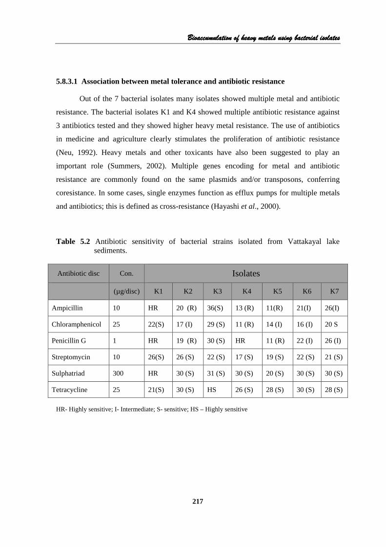
5.8.4.1 Morphological and Biochemical characteristics
The morphological and biochemical tests were carried out for the identification of
seven heavy metal tolerant bacterial isolates. These tests were performed following the
Bergey’s Manual of Determinative Bacteriology: 9th edition (Holt, et al., 1994) and 8th
edition (Buchanan and Gibbons, 1974. Based on the analysis the isolates were identified as
Bacillus cereus, Enterobacter aerogens, Pseudomonas putida, Bacillus licheniformis,
Klebsiella pneumonia , Alcaligens faecalis, and Citrobacter kosari (Table 5.4).
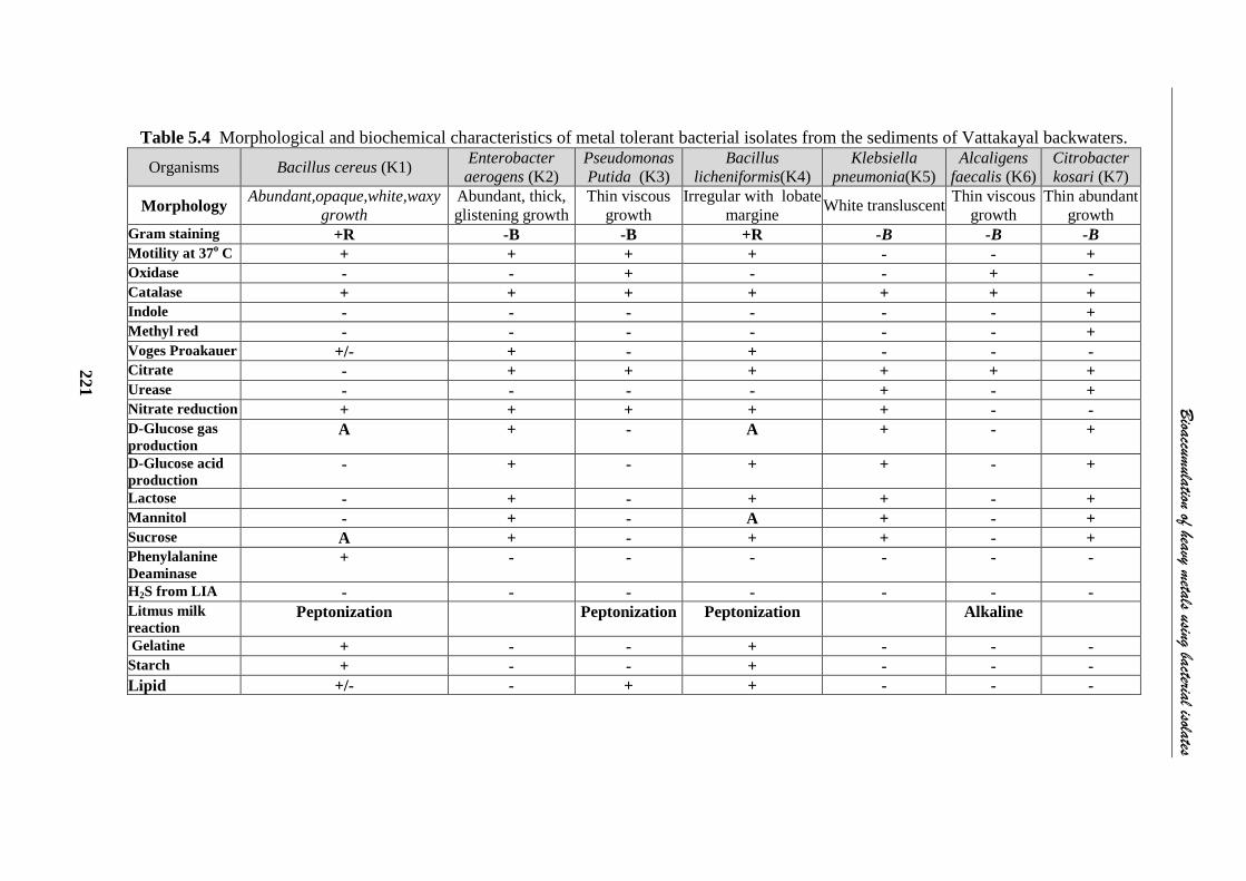
5.8.4.2 Molecular characterization of heavy metal tolerant bacterial strains
The Genomic DNA was isolated from the pure culture pellets of isolates K1 and
K4 and 16S rDNA fragment was amplified by PCR from the above isolated genomic DNA
using16S rDNA universal primers. The PCR amplicon was purified by column
purification in order to remove contaminants (Figs. 5.2a & b). The sequencing of purified
gene segment was done using ABI3730xl Genetic Analyzer (Applied Biosystems, USA)
(Figs. 5.3 a & b). The comparative analysis of the sequences of isolates with already
available database using BLAST (Basic Local Alignment Search Tool) showed that the
strains were close to the members of genus Bacillus. The highest sequence similarity of
the sediment bacteria are as follows: K1, Bacillus cereus (showed 99% similarity with
Bacillus cereus Accession No: 346665.1) and isolate K4 (96% similarity with Bacillus
licheniformis strain CICC 10180, accession No: AY859478.1).The 16S rDNA sequences
were submitted in the National Center for Biotechnology Information (NCBI) data bank
under the accession numbers JQ863364 (Bacillus cereus) and JQ863365 (Bacillus
licheniformis )
Bioaccumulation ofBioaccumulation ofBioaccumulation ofBioaccumulation of heavy metals using bacterial isolatesheavy metals using bacterial isolatesheavy metals using bacterial isolatesheavy metals using bacterial isolates
221
Table 5.4 Morphological and biochemical characteristics of metal tolerant bacterial isolates from the sediments of Vattakayal backwaters.
Organisms Bacillus cereus (K1) Enterobacter aerogens (K2)
Pseudomonas Putida (K3)
Bacillus licheniformis(K4)
Klebsiella pneumonia(K5)
Alcaligens faecalis (K6)
Citrobacter kosari (K7)
Morphology Abundant,opaque,white,waxy growth
Abundant, thick, glistening growth
Thin viscous growth
Irregular with lobate margine White transluscent
Thin viscous growth
Thin abundant growth
Gram staining +R -B -B +R -B -B -B Motility at 37 o C + + + + - - + Oxidase - - + - - + - Catalase + + + + + + + Indole - - - - - - + Methyl red - - - - - - + Voges Proakauer +/- + - + - - - Citrate - + + + + + + Urease - - - - + - + Nitrate reduction + + + + + - - D-Glucose gas production
A + - A + - +
D-Glucose acid production
- + - + + - +
Lactose - + - + + - + Mannitol - + - A + - + Sucrose A + - + + - + Phenylalanine Deaminase
+ - - - - - -
H2S from LIA - - - - - - - Litmus milk reaction
Peptonization Peptonization Peptonization Alkaline
Gelatine + - - + - - - Starch + - - + - - - Lipid +/- - + + - - -
Bioaccum
ulation of heavy metals using bacterial isolates
221

Chapter 5Chapter 5Chapter 5Chapter 5
222
Gel Image
Lane 1: 1 Kb •
Lane 2: 16S rDNA amplicon
Fig. 5.2a Gel image for K1 Fig. 5.2b Gel image for K1 Fig. 5.3a Consensus Sequence Data Culture K1: (877bp) TGCAGTCGAGCGAATGGATTAAGAGCTTGCTCTTATGAAGTTAGCGGCGGACGGGTGAGTAACACGTGGGTAACCTGCCCATAAGACTGGGATAACTCCGGGAAACCGGGGCTAATACCGGATAACATTTTGAACCGCATGGTTCGAAATTGAAAGGCGGCTTCGGCTGTCACTTATGGATGGACCCGCGTCGCATTAGCTAGTTGGTGAGGTAACGGCTCACCAAGGCAACGATGCGTAGCCGACCTGAGAGGGTGATCGGCCACACTGGGACTGAGACACGGCCCAGACTCCTACGGGAGGCAGCAGTAGGGAATCTTCCGCAATGGACGAAAGTCTGACGGAGCAACGCCGCGTGAGTGATGAAGGCTTTCGGGTCGTAAAACTCTGTTGTTAGGGAAGAACAAGTGCTAGTTGAATAAGCTGGCACCTTGACGGTACCTAACCAGAAAGCCACGGCTAACTACGTGCCAGCAGCCGCGGTAATACGTAGGTGGCAAGCGTTATCCGGAATTATTGGGCGTAAAGCGCGCGCAGGTGGTTTCTTAAGTCTGATGTGAAAGCCCACGGCTCAACCGTGGAGGGTCATTGGAAACTGGGAGACTTGAGTGCAGAAGAGGAAAGTGGAATTCCATGTGTAGCGGTGAAATGCGTAGAGATATGGAGGAACACCAGTGGCGAAGGCGACTTTCTGGTCTGTAACTGACACTGAGGCGCGAAAGCGTGGGGGAGCAAACAGGATTAGATACCCTGGTAGTCCACGCCGTAAACGATGAGTGCTAAGTGTTAGAGGGTTTCCGCCCTTTAGTGCTGAAGTTAACGCATTAAGCACTCCGGCCTGGGGAGTACGGGCCGCAAGGCTGAAACTCAAAGGAATTTGACGGGGG Fig. 5.3b Consensus Sequence Data Culture K4: (458 bp) TGCAGTCGAGCGGACAGATGGGAGCTTGCTCCCTGATGTCAGCGGCGGACGGGTGAGTAACACGTGGGTAACCTGCCTGTAAGACTGGGATAACTCCGGGAAACCGGGGCTAATACCGGATGCTTGATTGAACCGCATGGTTCAATTATAAAAGGTGGCTTTTACCTACCACTTACAAATGGACCCCCGGCGCATTACCTATTTGGGGAGGTAACGGCTCACCAAGGCAACAATGCTTACCCAACCTGAAAGGTGGATCGGCCACCCTGGAACTGAAACACGGCCCAAACTCCTACGGAAGGCACCATTAGGAAATCTTCCGCATGGAACAAAAGTCTGACGAACCACCCCCCCGGGATTGATGAAGGTTTTCGAATCGAAAAACTCTGTTGTTAGGAAAAAACAATTACCGTTCAATTAGGGGGGTACCTTGACGGTACTTAACCAAAAAGCAACGG
Bioaccumulation ofBioaccumulation ofBioaccumulation ofBioaccumulation of heavy metals using bacterial isolatesheavy metals using bacterial isolatesheavy metals using bacterial isolatesheavy metals using bacterial isolates
223
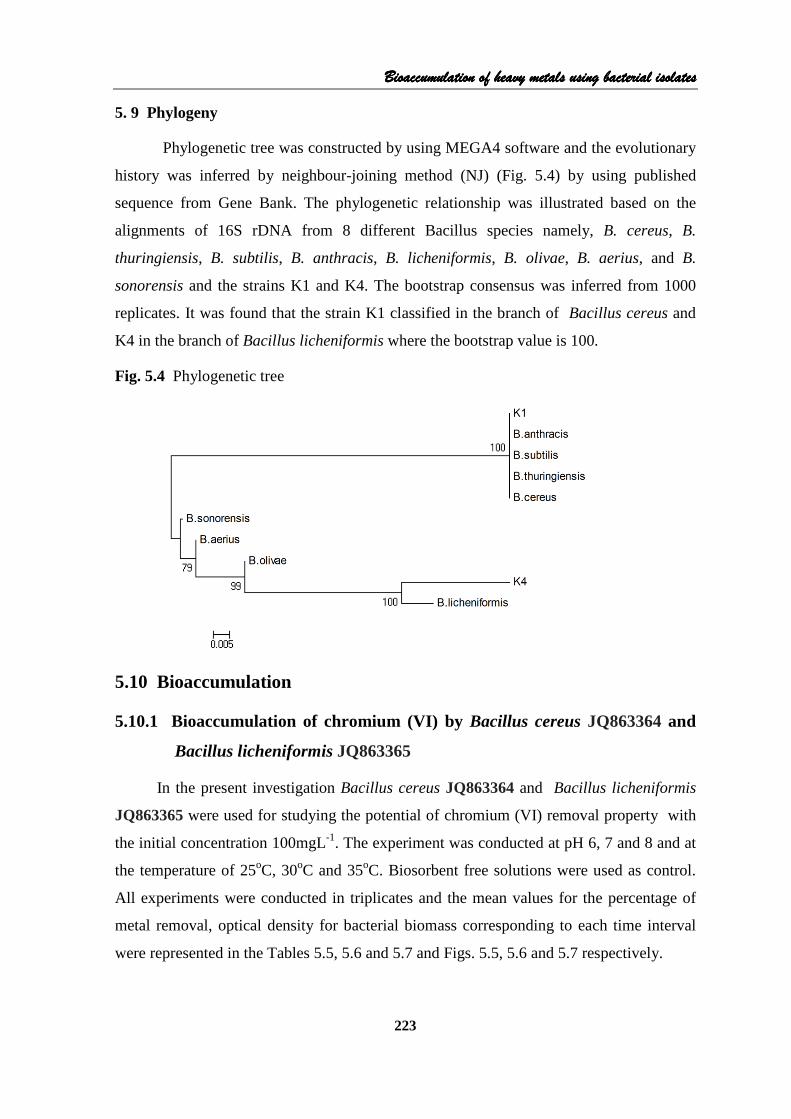
5. 9 Phylogeny
Phylogenetic tree was constructed by using MEGA4 software and the evolutionary
history was inferred by neighbour-joining method (NJ) (Fig. 5.4) by using published
sequence from Gene Bank. The phylogenetic relationship was illustrated based on the
alignments of 16S rDNA from 8 different Bacillus species namely, B. cereus, B.
thuringiensis, B. subtilis, B. anthracis, B. licheniformis, B. olivae, B. aerius, and B.
sonorensis and the strains K1 and K4. The bootstrap consensus was inferred from 1000
replicates. It was found that the strain K1 classified in the branch of Bacillus cereus and
K4 in the branch of Bacillus licheniformis where the bootstrap value is 100.
Fig. 5.4 Phylogenetic tree
5.10 Bioaccumulation
5.10.1 Bioaccumulation of chromium (VI) by Bacillus cereus JQ863364 and
Bacillus licheniformis JQ863365
In the present investigation Bacillus cereus JQ863364 and Bacillus licheniformis
JQ863365 were used for studying the potential of chromium (VI) removal property with
the initial concentration 100mgL-1. The experiment was conducted at pH 6, 7 and 8 and at
the temperature of 25oC, 30oC and 35oC. Biosorbent free solutions were used as control.
All experiments were conducted in triplicates and the mean values for the percentage of
metal removal, optical density for bacterial biomass corresponding to each time interval
were represented in the Tables 5.5, 5.6 and 5.7 and Figs. 5.5, 5.6 and 5.7 respectively.

Chapter 5Chapter 5Chapter 5Chapter 5
224
Bioaccumulation studies revealed that with increase in time, the biomass of the
selected bacterial isolates increased. Correspondingly, with increase in biomass, the heavy
metal bioaccumulation was also increased. The percentage removal of chromium (VI) by
Bacillus cereus at 25oC was minimum (3.1%) at pH 6 and maximum (13.5%) at pH 8
(Table 5.5). Table 5.6 represents the percentage removal of chromium (VI) by Bacillus
cereus at 30oC and pH 6, 7 and 8. The maximum chromium (VI) removal (57.33%) was
observed at pH 8 and minimum (48.93%) at pH 7. At pH 6 and 8 the chromium (VI)
removal was above 50% compared to pH 7. Table 5.7 represents the mean value of cell
growth and percentage removal of chromium (VI) by Bacillus cereus at pH 6, 7, and 8 and
temperature 35oC. The percentage removal of chromium (VI) was very low at 25oC and
35oC compared to 30oC (Table 5.6, Fig.5.6). The analysis of variance of bioaccumulation
of chromium between temperature and between time were significant (P<0.01). The
Duncan test (Table 5.8) showed significant variation in bioaccumulation except at 25oC
and 35oC.
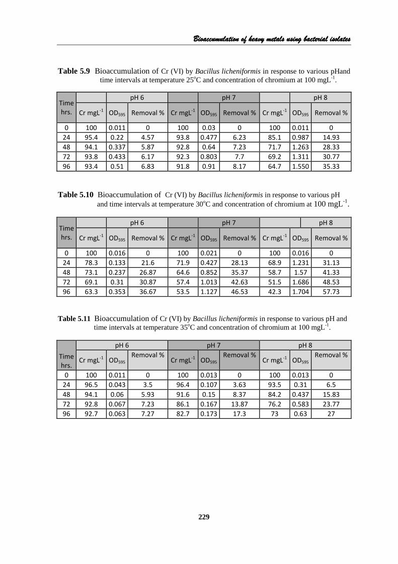
The data on percentage of chromium removed by Bacillus licheniformis at pH 6, 7,
and 8 at temperatures 25oC, 30oC and 35oC are represented in Tables 5.9, 5.10, 5.11 and
Figs. 5.8 and 5.9, 5.10. At temperature 25oC the percentage of removal of chromium (VI)
was maximum for pH 8 (35.33%) and minimum at pH 6 (6.83%). Similarly at temperature
30oC the percentage removal of chromium (VI) was high at pH 8 (57.73%) and
temperature 30oC and at pH 6 and 7 it was below 50% (Table 5.10). At 35oC chromium
removal was maximum (27%) at pH 8. Analysis of variance between temperature, pH and
time showed that there was significant difference at 1% level. Duncan test (Table 5.12) for
temperature showed no significant difference between temperatures 25oC and 35oC.
Duncan test (Table 5.12 & 5.13) for temperature and pH, indicates notable variation
between temperature and between pH. From the analysis it can be concluded that at pH 8
and temperature 30oC the percentage removal of chromium was maximum(Table 5.10).
Chromium exists in several oxidative states but the most stable forms are
chromium (III) and chromium (VI) and they have dissimilar chemical characteristics and
biological effect (Cervantes et al., 2001). The hexavalent chromium (Cr VI) is generally
recognized as mutagen and carcinogen, mainly respiratory tract and pancreatic cancer
(Ganguli and Tripathi, 2002). It is evident from the above observation that the microbe
Bacillus cereus has the ability to accumulate maximum chromium (VI) at 30oC when
compared with results at 25oC and 35oC. It was also found that the percentage of removal

Bioaccumulation ofBioaccumulation ofBioaccumulation ofBioaccumulation of heavy metals using bacterial isolatesheavy metals using bacterial isolatesheavy metals using bacterial isolatesheavy metals using bacterial isolates
225
was increased with increase in time, as observed by Kaushik et at. (2008). The maximum
uptake of chromium (57.33%) was observed at pH 8 and it is in agreement with the
observation made by Kocberber and Donmez (2007). Similarly Yang et al. (2009)
reported that Intrasporangium sp. Q5-1 grew in pH 6 to 10 and the maximum Cr (VI)
reduction observed at pH 8 and the removal was 98 % .
The heavy metal tolerance pattern of bacterial strain isolated from the metal
contaminated sediments of Vattakayal backwaters (station 6) showed a maximum
tolerance of 200 µg/ml for chromium. The resistance of the present strains may be due to
the presence of high concentration of heavy metals in the collection site, frequent presence
of plasmids or due to mutation (Ohta et al., 1971 and McLean and Beveridge., 2001).
Earlier studies revealed that Bacillus species can grow in medium containing high
concentration of heavy metals and have high ability to remove heavy metals (Kim, 2005).
Sultan and Hasnain (2007) isolated several groups of chromium resistant strains from
tannery effluent. Many researchers reported the metal resistance and metal removal
capabilities of Bacillus sp. (Mythili and Karthikeyan, 2011; Ashok Kumar, 2010 and
Rajkumar et al., 2012). The walls of Gram positive bacteria are efficient metal chelators
and have thicker peptidoglycan layer and Bacillus cereus has similar cell wall properties.
In B. subtilis, the carboxylic group of glutamic acid of peptidoglycan was the major site of
metal deposition (Gadd, 1990).
In the environment chromium (III) and chromium (VI) are the most prevalent
species and the environmental impact of chromium is at higher level, and its
environmental behavior pattern lies in its oxidation states (Losi et al., 1994). The present
study was conducted for analyzing the heavy metal resistance pattern of B. licheniformis
and its possible strategies for the bioaccumulation at varying pH and temperatures at
regular time intervals. Kavitha et al. (2011) reported that B. licheniformis exhibits growth
and tolerance (225mg/L) in the presence of higher concentration of chromium (VI). B.
licheniformis isolated from the sediments of Vattakayal backwaters contaminated with
heavy metals also exhibited high tolerance to chromium (VI) (250µg/ml) compared with
the Minimum Inhibitory Concentration (150mg/L) (Srinath et al., 2002). Similar trend was
shown by chromium resistant bacteria isolated from tannery effluent 250 µg/mL of Cr
(Basu et al., 1997).
The microbes survived in polluted soil are prone to show higher tolerance to heavy
metals as compared to the strains from non-contaminated sites (Zouboulis et al., 2004).

Chapter 5Chapter 5Chapter 5Chapter 5
226
The bacterial isolates with high chromium resistantce can be used for biosorption of
chromium VI contaminated environment (Polisak et al., 2009). At pH 8 and temperature
30oC the percentage removal of chromium by B. licheniformis isolated from the heavy
metal contaminated sediments of Vattakayal was 57.73%. The pH was a significant factor
to determine the adsorption of metal ion by bacterial strains. This result is in tune with the
adsorption of chromium by Staphylococcus sp. where the optimum pH was 8 (Mythili and
Karthikeyan, 2011). According to Udandi Boominadhan et al. (2009) the protease activity
of B. licheniformis was high at pH 8. B. licheniformis showed 31.31% chromium removal
within 24 hours and is comparable to Bacillus fumus RH109 (38.85%) (Rani et al., 2007).
The higher uptake of metals by B. licheniformis may be due to the presence of teichoic
and teichuronic acids, which are acting as important binding sites in B. licheniformis
(Gadd, 1990). The present study shows that B. licheniformis is an effective agent for the
removal of hexavalent chromium from aqueous solution and the results may be helpful for
the development and design of indigenous bioreactors for removal of chromium VI from
the industrial wastewater.

Bioaccumulation ofBioaccumulation ofBioaccumulation ofBioaccumulation of heavy metals using bacterial isolatesheavy metals using bacterial isolatesheavy metals using bacterial isolatesheavy metals using bacterial isolates
227
Table 5.5 Bioaccumulation of Cr (VI) by Bacillus cereus in response to various pH and
time intervals at temperature 25oC and concentration of chromium at 100 mgL-
1.
Time
hrs.
pH 6 pH 7 pH 8
Cr mgL-1 OD595 Removal % Cr mgL
-1 OD595 Removal % Cr mgL
-1 OD595 Removal %
0 100 0.019 0.0 100 0.02 0 100 0.023 0 24 98.7 0.95 1.3 98.8 1.063 1.17 93.4 1.103 6.57 48 97.8 1.124 2.23 96.6 1.193 3.43 90.2 1.437 9.77 72 97.3 1.131 2.7 95.2 1.26 4.83 87.4 1.49 12.6 96 96.9 1.142 3.1 94.8 1.287 5.2 86.5 1.467 13.5
Exp. Experimental
Table 5.6 Bioaccumulation of Cr (VI) by Bacillus cereus in response to various pHand
time intervals at temperature 30oC and concentration of chromium at 100 mgL-1.
Time
hrs.
pH 6 pH 7 pH 8
Cr mgL-1 OD595 Removal % Cr mgL
-1 OD595 Removal % Cr mgL-1 OD595 Removal %
0 100 0 0 100 0.001 0 100 0.037 0 24 69.8 0.83 30.2 72.3 0.85 27.67 80.8 0.907 19.23 48 61.5 1.051 38.47 62.6 1.06 37.37 65.1 1.373 34.93 72 51.6 1.09 48.37 56.5 1.097 42.47 50.8 1.54 49.17 96 43.4 1.12 56.63 51.1 1.177 48.93 42.7 1.617 57.33
Table 5.7 Bioaccumulation of Cr (VI) by Bacillus cereus in response to various pH and
time intervals at temperature 35oC and concentration of chromium at 100 mgL-
1.
Time
hrs.
pH 6 pH 7 pH 8
Cr mgL-1 OD595
Removal % Cr mgL
-1 OD595 Removal %
Cr mgL-1 OD595
Removal %
0 100 0.01 0 100 0.005 0 100 0 0 24 88.9 0.923 11.1 100 0.853 0 99.3 1.025 0.73 48 86.3 1.01 13.67 92.2 1.037 7.77 94.8 1.27 5.2 72 85.2 1.023 14.87 90.8 1.037 9.2 93 1.42 7 96 83 1.027 17.03 90.3 1.037 9.73 91.7 1.52 8.33

Chapter 5Chapter 5Chapter 5Chapter 5
228
Fig. 5.5 Cr (VI) removal by B. cereus at Fig. 5.6 Cr (VI) removal by B. cereus at
various pH and at temperature 25oC . various pH and at temperature 30oC.
0.0
5.0
10.0
15.0
0 24 48 72 96
% o
f R
em
ov
al
Time hrs
Temp 25 oC pH 6 Temp 25 oC pH 7
Temp 25 oC pH 8
0.0
20.0
40.0
60.0
80.0
0 24 48 72 96
% o
f R
em
ov
al
Time hrs
Temp 30 oC pH 6 Temp 30 oC pH 7
Temp 30 oC pH 8 Fig. 5.7 Cr (VI) removal by B. cereus in at various pH and at temperature 35oC . ANOVA
0.0
5.0
10.0
15.0
20.0
0 24 48 72 96
% o
f R
em
ov
al
Time hrs
Temp 35 oC pH 6 Temp 35 oC pH 7
Temp 35 oC pH 8
Table 5.8 Duncan test of Cr (VI) removal by Bacillus cereus at various temperatures
Subset
Temperature 1 2
25 OC 4.427
30 OC 32.718
35 OC 6.976
Temperature p< 0.01 Significant
pH P> 0.05 Not significant
Time P< 0.01 Significant
Bioaccumulation ofBioaccumulation ofBioaccumulation ofBioaccumulation of heavy metals using bacterial isolatesheavy metals using bacterial isolatesheavy metals using bacterial isolatesheavy metals using bacterial isolates
229
Table 5.9 Bioaccumulation of Cr (VI) by Bacillus licheniformis in response to various pHand time intervals at temperature 25oC and concentration of chromium at 100 mgL-1.
Time
hrs.
pH 6 pH 7 pH 8
Cr mgL-1 OD595 Removal % Cr mgL
-1 OD595 Removal % Cr mgL-1 OD595 Removal %
0 100 0.011 0 100 0.03 0 100 0.011 0 24 95.4 0.22 4.57 93.8 0.477 6.23 85.1 0.987 14.93 48 94.1 0.337 5.87 92.8 0.64 7.23 71.7 1.263 28.33 72 93.8 0.433 6.17 92.3 0.803 7.7 69.2 1.311 30.77 96 93.4 0.51 6.83 91.8 0.91 8.17 64.7 1.550 35.33
Table 5.10 Bioaccumulation of Cr (VI) by Bacillus licheniformis in response to various pH
and time intervals at temperature 30oC and concentration of chromium at 100 mgL-1.
Time
hrs.
pH 6 pH 7 pH 8
Cr mgL-1 OD595 Removal % Cr mgL
-1 OD595 Removal % Cr mgL-1 OD595 Removal %
0 100 0.016 0 100 0.021 0 100 0.016 0 24 78.3 0.133 21.6 71.9 0.427 28.13 68.9 1.231 31.13 48 73.1 0.237 26.87 64.6 0.852 35.37 58.7 1.57 41.33 72 69.1 0.31 30.87 57.4 1.013 42.63 51.5 1.686 48.53 96 63.3 0.353 36.67 53.5 1.127 46.53 42.3 1.704 57.73
Table 5.11 Bioaccumulation of Cr (VI) by Bacillus licheniformis in response to various pH and
time intervals at temperature 35oC and concentration of chromium at 100 mgL-1.
Time
hrs.
pH 6 pH 7 pH 8
Cr mgL-1 OD595
Removal % Cr mgL
-1 OD595 Removal %
Cr mgL-1 OD595
Removal %
0 100 0.011 0 100 0.013 0 100 0.013 0 24 96.5 0.043 3.5 96.4 0.107 3.63 93.5 0.31 6.5 48 94.1 0.06 5.93 91.6 0.15 8.37 84.2 0.437 15.83 72 92.8 0.067 7.23 86.1 0.167 13.87 76.2 0.583 23.77 96 92.7 0.063 7.27 82.7 0.173 17.3 73 0.63 27

Chapter 5Chapter 5Chapter 5Chapter 5
230
Fig. 5.8 Cr (VI) removal by B. licheniformis Fig. 5.9 Cr (VI) removal by B. licheniformis at various pH and at temperature 25oC . at various pH and at temperature 30oC .
0.0
5.0
10.0
15.0
20.0
25.0
30.0
35.0
40.0
0 24 48 72 96
% o
f R
em
ov
al
Time hrs
Temp 25 oC pH 6 Temp 25 oC pH 7
Temp 25 oC pH 8
0.0
10.0
20.0
30.0
40.0
50.0
60.0
70.0
0 24 48 72 96
% o
f R
em
ov
al
Time hrs
Temp 30 oC pH 6 Temp 30 oC pH 7
Temp 30 oC pH 8
Fig. 5.10 Cr (VI) removal by B. licheniformis at various pH and at temperature 35oC . ANOVA
0.0
5.0
10.0
15.0
20.0
25.0
30.0
0 24 48 72 96
% o
f R
em
ov
al
Time hrs
Temp 35 oC pH 6 Temp 35 oC pH 7
Temp 35 oC pH 8
Table 5.12 Duncan test of Cr (VI) removal by Bacillus licheniformis at various
temperatures
Subset
Temperature 1 2 25 OC 10.809 30 OC 29.827 35 OC 9.347
Table 5.13 Duncan test of Cr (VI) removal by Bacillus licheniformis at various pH
Subset
pH 1 2 3 6 10.891 7 15.011 8 24.08
Temperature p< 0.01 Significant
pH P< 0.01 Significant
Time P< 0.01 Significant

Bioaccumulation ofBioaccumulation ofBioaccumulation ofBioaccumulation of heavy metals using bacterial isolatesheavy metals using bacterial isolatesheavy metals using bacterial isolatesheavy metals using bacterial isolates
231
5.10.2 Bioaccumulation of lead (II) by Bacillus cereus JQ863364 and Bacillus
licheniformis JQ863365
Effect of temperature and pH on the removal of lead by Bacillus cereus are
presented in Table 5.14, 5.15, 5.16 and Figs. 5.11 and 5.12, 5.13. The percentage removal
of lead was maximum (88.6%) at pH 6 and temperature 25oC. At pH 7 and 8 also the
percentage of lead removal was found above 50%. At temperature 30oC and 35oC and pH
6 the maximum removal of lead was 68.27% and 76.83% respectively (Tables 5.15 and
5.16). ANOVA showed that the variation between temperature, pH and time intervals
were significant at 1% level. Duncan test for temperature and pH showed two subsets in
the study signifying notable variations. But no significant variation noticed between
temperature 25oC and 35oC and pH 7 and 8 (Tables 5.17 and 5.18). The study thus
revealed that the percentage of lead removal by Bacillus cereus was maximum (88.6 %) at
pH 6 and temperature 25oC (Table 5.14 and Fig. 5.11).
Bioaccumulation of lead by Bacillus licheniformis at pH 6,7, and 8 and
temperature 25oC, 30oC and 35oC in a medium amended with 100 mgL-1 lead at various
time intravels 0, 24, 48, 72 and 96 hours are presented in Tables 5.19, 5.20, 5.21 and Figs.
5.14, and 5.15, 5.16. At temperature 25oC the percentage of removal was maximum at pH
6 (71.17%) and minimum (27.13%) at pH 8 (Table 5.19). Table 5.20 represents the
percentage of lead removal at 30oC with a maximum of 65.53% at pH 7 and minimum of
35.87% at pH 8 for 96 hours. At temperature 35oC the concentration of lead reduced to
75.23% for 96 hours at pH 6 (Table 5.21). In all cases the percentage of reduction in the
concentration of lead in the experimental medium increases with the increase in time.
Barring the pH 8 at temperature 25oC and 30oC, B. licheniformis exhibited above 50%
removal of lead. The analysis of variance of bioaccumulation of lead between temperature,
pH and time intervals were found significant at 1% level (P<0.01). Duncan test also
showed significant variations (Table 5.22 & Table 5.23) but there was no significant
variation between pH 6 and 7. The result clearly indicated that in Bacillus licheniformis,
the percentage of accumulation was maximum at pH 6 and temperature 35oC.
The microorganisms have the ability to accumulate metallic elements and this has
been studied at first from toxicological point of view (Volesky, 1990a, b, c). In the present
study the Bacillus cereus isolated from the heavy metal contaminated sediments ( Station
6) of Vattakayal backwaters exhibited a high percentage of metal uptake, agreeing with

Chapter 5Chapter 5Chapter 5Chapter 5
232
the report that the strains isolated from polluted soil showed the capacity of biosorption
(Ozdemir, et al., 2004 and YU Xia, et al.,2003). The metal binding property may be due
to the phosphoryl groups and the carboxyl group of peptide chains which provide
negatively charged sites in the Gram positive bacterial cell wall (Urrutia et al., 1997 and
Moat et al., 2002). Ray et al. (2005) reported that the removal of Pb (II) ions with growing
cells of Bacillus Cereus M116 was maximum (85%) when initial lead concentration was 50
mg/L at pH 6 and temperature 30oC. Similarly in the case of Bacillus circulans only 78 %
lead uptake from 500mg/L-1 in 72 hours and also it increases with increase in pH upto 6
(Khanafari, et al., 2008). Parungao (2007) reported that Stenotrophomonas maltophilia
was able to remove only 42.75 % of lead from the primary solution and immobilized cells
of Bcillus sp. adsorbed 84.27% lead from the industrial wastewater (Rani, 2010). It may be
noted that in the present study the metal accumulation by Bacillus cereus was found
higher when compared to the results of other similar studies. Hence, it is found to be a
potential agent for the removal of lead from the wastewater in the industrial area.
Heavy metals are highly toxic to all biological systems from microbs to plants and
animals (Patel et al., 2007). They exerts a selective pressure on microbial population
leading to the emergence of resistant strains (McGrath et al., 2001). These microorganisms
can play a key role in the bioremediation of heavy metal contaminated soil and
wastewater. In the present study B. licheniformis accumulates a maximum of 75.23% lead
from 100mg/L-1 aqueous solution at pH 6 and temperature 35oC. Similar study had been
conducted by Issazadeh (2011) where there was 1.1 mol/g biomass for bioaccumulation of
lead by B. licheniformis. Gram-positive bacteria, such as B.licheniformis, B. subtilis,
Brevibacterium helovolum, and Rhodococcus elythropolis, exhibited a particularly high
capacity for accumulating samarium (Sm) and the accumulation increased with increase
in pH (Tsuruta, 2007). El-Hendawy, (2009) reported that the bioaccumulation of heavy
metals by Vibrio alginolyticus isolated from wastes of Iron and Steel factory could reduce
40% Pb2+. The high accumulation ability in B.licheniformis may be due to the presence of
teichoic and teichuronic acids which are the principal sites of metal binding in B.
licheniformis walls (Beveridg, 1982). From the above results it can be concluded that the
Gram positive bacteria B. licheniformis, is a potential agent for lead removal.

Bioaccumulation ofBioaccumulation ofBioaccumulation ofBioaccumulation of heavy metals using bacterial isolatesheavy metals using bacterial isolatesheavy metals using bacterial isolatesheavy metals using bacterial isolates
233
Table 5.14 Bioaccumulation of Pb(II) by Bacillus cereus in response to various pH and time intervals at temperature 25oC and concentration of chromium at 100 mgL-1
.
Time
hrs.
pH 6 pH 7 pH 8
Cr mgL-1 OD595 Removal % Cr mgL
-1 OD595 Removal % Cr mgL-1 OD595 Removal %
0 100 0.033 0 100 0.023 0 100 0.023 0 24 76.1 1.087 23.9 52.1 0.867 47.93 62.9 0.82 37.13 48 19.5 1.8 80.5 60.1 1.633 39.93 44.8 1.473 55.17 72 14.2 1.99 85.83 62.1 1.907 37.77 43.9 1.71 56.1 96 11.4 2.13 88.6 44.9 1.953 55.13 40.4 1.747 59.63
Table 5.15 Bioaccumulation of Pb(II) by Bacillus cereus in response to various pH and
time intervals at temperature 30oC and concentration of chromium at 100 mgL-1
.
Time
hrs.
pH 6 pH 7 pH 8
Cr mgL-1 OD595 Removal % Cr mgL
-1 OD595 Removal % Cr mgL-1 OD595 Removal %
0 100 0.04 0 100 0.021 0 100 0.017 0 24 91.9 1.193 7.93 91.4 1.197 8.63 81.6 1.227 18.43 48 62.5 1.707 37.63 77.2 1.443 22.83 66.9 1.49 33.13 72 35.6 1.743 64.43 63.8 1.477 36.2 55.2 1.507 44.83 96 31.7 1.78 68.27 55.5 1.493 44.53 50.1 1.533 49.87
Table 5.16 Bioaccumulation of Pb (II)by Bacillus cereus in response to various pH and time intervals at temperature 35oC and concentration of chromium at 100 mgL-
1.
Time
hrs.
pH 6 pH 7 pH 8
Cr mgL-1 OD595
Removal % Cr mgL
-1 OD595 Removal %
Cr mgL-1 OD595
Removal %
0 100 0.053 0 100 0.018 0 100 0.017 0 24 62.5 1.607 37.53 70.7 1.61 29.27 82.4 1.507 17.63 48 33.3 1.93 66.73 46.2 1.857 53.83 54.2 1.837 45.77 72 26.1 2.027 73.9 42. 1.963 57.23 48.1 1.927 51.87 96 23.2 2.093 76.83 40.6 2.033 59.37 44.8 1.937 55.17

Chapter 5Chapter 5Chapter 5Chapter 5
234
Fig. 5.11 Pb (II) removal by B. cereus at Fig.5.12 Pb (II) removal by B. cereus at various pH and at temperature 25oC. various pH and at temperature 30oC
0.0
20.0
40.0
60.0
80.0
100.0
0 24 48 72 96
% o
f R
em
ov
al
Time hrs
Temp 25 oC pH 6 Temp 25 oC pH 7
Temp 25 oC pH 8
Fig 5.13 Pb (II) removal by B. cereus at various pH and at temperature 30oC. ANOVA
Table 5.17 Duncan test of Pb removal by Bacillus cereus at various temperatures
Subset
Temperature 1 2
25 OC 44.509
30 OC 29.116
35 OC 41.676
Table 5.18 Duncan test of Pb removal by Bacillus cereus at various pH
Subset
pH 1 2
6 47.473
7 32.844
8 34.982
Temperature p< 0.01 Significant
pH P< 0.01 Significant
Time P< 0.01 Significant

Bioaccumulation ofBioaccumulation ofBioaccumulation ofBioaccumulation of heavy metals using bacterial isolatesheavy metals using bacterial isolatesheavy metals using bacterial isolatesheavy metals using bacterial isolates
235
Table 5.19 Bioaccumulation of Pb(II) by Bacillus licheniformis in response to various pH and
time intervals at temperature 25oC and concentration of chromium at 100 mgL-1.
Time
hrs.
pH 6 pH 7 pH 8
Cr mgL-1 OD595
Removal % Cr mgL
-1 OD595 Removal %
Cr mgL-1 OD595
Removal %
Exp. Exp. Exp.
0 100 0.027 0 100 0.02 0 100 0.055 0 24 71.7 1.147 28.33 81.2 0.943 18.77 98.9 0.937 1.13 48 50.8 1.723 49.23 60.9 1.31 39.07 95.4 1.223 4.57 72 36.7 1.78 63.33 47.8 1.453 52.23 84.4 1.273 15.57 96 28.8 1.803 71.17 44.9 1.547 55.13 72.9 1.297 27.13
Table 5.20 Bioaccumulation of Pb(II) by Bacillus licheniformis in response to various pH and
time intervals at temperature 30oC and concentration of chromium at 100 mgL-1.
Time
hrs.
pH 6 pH 7 pH 8
Cr mgL-1 OD595
Removal % Cr mgL
-1 OD595
Removal % Cr mgL
-1 OD595
Removal %
Exp. Exp. Exp.
0 100 0.097 0 100 0.127 0 100 0.147 0 24 63.7 0.913 37.27 76.5 1.01 23.53 84.1 1.353 15.87 48 53.8 1.21 46.23 48.5 1.233 51.53 73.7 1.673 26.33 72 49.7 1.427 50.33 43.2 1.407 56.83 69.2 1.76 30.77 96 49.1 1.457 50.87 34.5 1.447 65.53 64.1 1.763 35.87
Table 5.21 Bioaccumulation of Pb (II) by Bacillus licheniformis in response to various pH and
time intervals at temperature 35oC and concentration of chromium at 100 mgL-1.
Time
hrs.
pH 6 pH 7 pH 8
Cr mgL-1 OD595
Removal % Cr mgL
-1 OD595 Removal %
Cr mgL-1 OD595
Removal %
Exp. Exp. Exp.
0 100 0.027 0 100 0.04 0 100 0.026 0 24 75.1 1.323 24.87 71.5 1.033 28.47 65.3 1.317 34.73 48 51.1 1.71 48.93 46.2 1.363 53.83 47.6 1.75 52.37 72 34.1 1.927 65.93 42 1.413 57.97 43 2.033 57.03 96 24.8 1.973 75.23 41 1.47 58.97 41.5 2.073 58.4

Chapter 5Chapter 5Chapter 5Chapter 5
236
Fig. 5.14 Pb (II) removal by B. licheniformis Fig.5.15 Pb (II) removal by B. licheniformis at various pH and at temperature 25oC . at various pH and at temperature 30oC .
0.0
10.0
20.0
30.0
40.0
50.0
60.0
70.0
0 24 48 72 96
% o
f R
em
ov
al
Time hrs
Temp 25 oC pH 6 Temp 25 oC pH 7
Temp 25 oC pH 8
0.0
20.0
40.0
60.0
80.0
0 24 48 72 96
% o
f R
em
ov
al
Time hrs
Temp 30 oC pH 6 Temp 30 oC pH 7
Temp 30 oC pH 8
Fig.5.16 Pb (II) removal by B. licheniformis at various pH and at temperature 35oC . ANOVA
0.0
20.0
40.0
60.0
80.0
0 24 48 72 96
% o
f R
em
ov
al
Time hrs
Temp 35 oC pH 6 Temp 35 oC pH 7
Temp 35 oC pH 8
Table 5.22 Duncan test of Pb removal by Bacillus licheniformis at various temperatures
Subset
Temperature 1 2 3
25 OC 32.664
30 OC 28.378
35 OC 41.116
Table 5.23 Duncan test of Pb removal by Bacillus licheniformis at various pH
Subset
pH 1 2
6 40.716
7 37.458
8 23.984
Temperature p< 0.01 Significant
pH P< 0.01 Significant
Time P< 0.01 Significant

Bioaccumulation ofBioaccumulation ofBioaccumulation ofBioaccumulation of heavy metals using bacterial isolatesheavy metals using bacterial isolatesheavy metals using bacterial isolatesheavy metals using bacterial isolates
237
References
Acar, J. and Goldstein, F.W. 1996. Disk susceptibility test. In: Lorian, V. (Ed.) Antibiotics
in Laboratory Medicine. Williams and Wilkins, Baltimore, 1-51.
Ahalya, N., Ramachandran, T. V. and Kanamadi, R. D. 2003. Biosorption of heavy
metals. Res.J.Chem.Environ., 7 : 71-79.
Alam, M.Z, Ahmad, S. 2011. Chromium removal through biosorption and
bioaccumulation by bacteria from tannery effluents contaminated soil. Clean-Soil
Air Water, 39 : 226-237.
Alonso, A., Sanchez, P., Martinez, J. L. (2001). Environmental selection of antibiotic
resistance genes. Environ Microbiol., 3 : 1–9.
Altschul, S. F., Maddan, T. L., Schaffer, A. A., Zang, J., Zang, Z., Miller, W. & Lipman,
D. J. 1997. Gapped BLAST and PSIBLAST: a new generation of protein database
search programs. Nucleic Acids Research, 25 : 3389–3402.
Anders, M. Y. J. H. and Hubert, C. J. 1992. Bacterial biosorption and retention ofthorium
and uranyl cations by mycobacterium smegmatis. J. RadioAnal. Nucl. Lett., 166 :
431-440.
Andreazza, R., Pieniz, S., Okeke, B. C. and Camargo, F. A. O. 2011. Evaluation of
copper resistant bacteria from vineyard soils and mining waste for copper
biosorption . Brazilian Journal of Microbiology, 42 : 66-74.
Andreonian, V., Colombo, M., Di Simine, D., Finoli, C., Origgi, G., Vecchio, A. and
Carzaniga, R. 1997. Removal of lead from aqueous solutions by a Brevibacterium
strain. In: Rosen, D. (Ed.) Modern agriculture and the environment. Kluwer
Academic Publishers, Netherlands, 521-531.
Ansari, M. I. and Malik, A. 2007. Biosorption of nickel and cadmium by metal resistant
bacterial isolates from agricultural soil irrigated with industrial waste water.
Bioresource Technology, 98 (16) : 3149-3153.
Ashok Kumar., Balwant Singh Bisht and Vishnu Datt Joshi . 2010. Biosorption of Heavy
Metals by four acclimated microbial species, Bacillus spp., Pseudomonas sp.,
Staphylococcus spp. and Aspergillus niger. J. BIOL. ENVIRON. SCI., 4 (12) : 97-
108.

Chapter 5Chapter 5Chapter 5Chapter 5
238
Bahig, A. E., Aly, E. A., Khaled, A. A. and Amel, K. A. 2008. Isolation, characterization
and application of bacterial population from agricultural., soil at Sohag Province,
Egypt. Malaysian Journal of Microbiology, 4 (2) : 42- 50.
Basu, M., Bhattacharya, S. and Paul, A. K. 1997. Isolation and characterization of
chromium resistant bacteria from tannery effluents. Bulletin of Environmental
Contamination and Toxicology, 58 : 535-542.
Bauer, A.W., Kirby, W., Sherris, J. and Turck, M. 1966. Antibiotic susceptibility testing
by a standardized disc method. Am. J. Clin. Pathol., 45 : 493–496.
Berg, J., Tom-Petersen, A., Nybroe, O. 2005. Copper amendment of agricultural soil
selects for bacterial antibiotic resistance in the field. Lett Appl Microbiol., 40 :146–
151.
Beveridge, T. J., Forsberg, C. W. AND Doyle, R. J. 1982. Major Sites of Metal Binding
in Bacillus licheniformis Walls. Journal of bacteriology, 150 (3) : 1438-1448.
Blackwell, K.J., Singleton, I. and Tobin, J.M. 1995. Metal cation uptake by yeast: A
review. Appl. Microbial., Biotechnol., 43 : 571-584.
Brady, D., Stoll, A. and Duncan, J. R. 1994. Biosorption of heavy metal cations by non-
viable yeast biomass. Environ. Technol., 15 : 429-438.
Brim, H., Heyndrickx, M., De Vos, P., Wilmotte, A., Springael, D., Schlegel, H. G. and
Mergeay, M. 1999. Amplified rDNA restriction analysis and further genotypic
characterisation of metal-resistant soil bacteria and related facultative
hydrogenotrophs. Syst. Appl. Microbiol., 22 : 258-268.
Buchanan, R. E. and Gibbons, N. E. 1974. Bergey’s manual of determinative
bacteriology. (Eighth edition), The Williams and Wilkins Co., Baltimore.
Cervantes, C., Garcia, J. C., Devars, S. and Corona, F. G. 2001. Interactions of chromium
with microorganisms and plants. FEMS Microbiol. Ver., 25 : 335-347.
Chander, K. and Brookes, P. C. 1991. Effects of heavy metals from past applications of
sewage sludge on microbial biomass and organic matter accumulation in a sandy
loam soil and silty loam UK soil. Soil Biol. Biochem., 23 : 927-932.

Bioaccumulation ofBioaccumulation ofBioaccumulation ofBioaccumulation of heavy metals using bacterial isolatesheavy metals using bacterial isolatesheavy metals using bacterial isolatesheavy metals using bacterial isolates
239
Chowdhury, S., Mishra, M., Adarsh, V.K., Mukherjee, A., Thakur, A.R. and Chaudhuri,
S.R. 2008. Novel metal accumulator and protease secretor microbes from East
Calcutta Wetland. Am. J. Biochem. Biotechnol., 4: 255-264.
Congeevaram, S., Dhanarani, S., Park, J., Dexilin, M. and Thamaraiselvi, K. 2007.
Biosorption of chromium and nickel by heavy metal resistant fungal and bacterial
isolates. Journal of Hazardous Materials, 146 : 270-277.
Das, S., Lyla, P.S. and Khan, S. A. 2006. Marine microbial diversity and ecology:
importance and future perspectives. Current Science, 90 (10) : 1325-1335.
DeLeo, P. C. and Ehrilich, H. L. 1994. Reduction of hexavalent chromium by
Pseudomonas fluorescens LB300 in batch and continuous cultures. Appl.
Microbial Biotechnol., 40 : 756-759.
Diels, L. and Mergeay, M. 1990. DNA probe-mediated detection of resistant bacteria from
soils highly polluted by heavy metals. Appl. Environ. Microbiol., 56 : 1485-1491.
Duan, X. J. and Min, H. 2004. Isolation, identification and preliminary studies on the
resistance gene detection of a Cd 2+ resisting bacterium. Acta Sci. Circums., 1 :
154-158.
El-Hendawy, H. H., Ali, D.A., El-Shatoury, E. H. and Ghanem, S. M. 2009.
Bioaccumulation of heavy metals by Vibrio alginolyticus isolated from wastes of
Iron and Steal Factory, Helwan, Egypt. Egypt. Acad. J. Biolog. Sci., 1(1) : 23-28.
Farag, S. and Zaki, S. 2010. Identification of bacterial strains from tannery effluent and
reduction of hexavalent chromium. Journal of Environmental Biology, 31 (5) :
877-882.
Fourest, E., Canal, C. and Roux, J. C. 1994. Improvement of heavy metal biosorption by
mycelial dead biomasses (Rhizopus arrhizus, Mucor miehei and Penicillium
chrysogenum): pH control and cationic activation. FEMS Microbiol., Rev., 14 :
325–332.
Frostegard, A., Tunlid, A. and Baath, E. 1996. Changes in microbial community structure
during long-term incubation in two soils experimentally contaminated with metals.
Soil Biology and Biochemistry, 28 (1) : 55-63.
Chapter 5Chapter 5Chapter 5Chapter 5
240
Fude, L., Harris, B., Urrutia, M. M. and Beveridge, T. J. 1994. Reduction of Cr(VI) by a
consortia of sulfate-reducing bacteria (SRB-III). Applied and Environmental
Microbiology, 60 (5) :1525–1531.
Gadd, G, M. 1990. Heavy metal accumulation by bacteria and other microorganisms.
Experientia., 46 : 834–840.
Gadd, G. M. and White, C. 1993. Microbial treatment of metal pollution- a working
biotechnology? Trends Biotechnol., 11 : 353-359.
Ganguli, A. and Tripathi. A . 2002. Bioremediation of toxic chromium from
electroplating effluent by chromate-reducing Pseudomonas aeruginosa A2Chr in
two bioreactors. Applied Microbiology and Biotechnology, 58 : 416-420.
Giller, K. E., Wittwer, E. and McGrath, S. P. 1999. Assessing risks of heavy metal
toxicity in agricultural soils. Human Ecol Risk Assess., 5 : 683–689.
Goris, J., De Vos, P., Coenye, T., Hoste, B., Janssens, D., Brim, H., Diels, L., Mergeay,
M., Kersters, K. and Vandamme, P. 200. Classification of metal-resistant bacteria
from industrial biotopes as Ralstonia campinensis sp. nov., Ralstonia
metallidurans sp. nov. and Ralstonia basilensis Steinle et al. 1998 emend.
International Journal of Systematic and Evolutionary Microbiology, 51 : 1773-
1782.
Gupta, A., L. Phung., Chakravarty. L. and. Silver. S. 1999. Mercury resistance in Bacillus
cereus RC607: Transcriptional organization and two new open reading frames . J.
Bacteriol., 181 : 7080-7086.
Hayashi, S., Abe, M., Kimoto, M., Furukawa, S., Nakazawa, T. 2000. The DsbA-DsbB
disulfide bond formation system of Burkholderia cepacia is involved in the
production of protease and alkaline phosphatase, motility, metal resistance, and
multi-drug resistance. Microbiol. Immunol., 44 : 41-50.
Hernandez, A., Mellado, R. P. and AND Martinez, J. L. 1998. Metal Accumulation and
Vanadium-Induced Multidrug Resistance by Environmental Isolates of Escherichia
hermannii and Enterobacter cloacae. Applied and Environmental Microbiology, 64
(11) : 4317-4320.

Bioaccumulation ofBioaccumulation ofBioaccumulation ofBioaccumulation of heavy metals using bacterial isolatesheavy metals using bacterial isolatesheavy metals using bacterial isolatesheavy metals using bacterial isolates
241
Heuer, H., Smalla, K. 2007. Manure and sulfadiazine synergistically increased bacterial
antibiotic resistance in soil over at least two months. Environ Microbiol., 9 : 657–
666.
Holt, J. G., Krieg, N. R., Sneath, P. H. A., Staley, J. T. and Williams, S. T. 1994. Bergey’s
Manual of Determinative Bacteriology (Ninth edition). Williams and Wilkins.
London.
Hussein, H., Farag, S. and Moawad, H. 2003. Isolation and characterisation of
Pseudomonas resistant to heavy metals contaminants. Arab J. Biotechnol., 7 : 13-
22.
Hussein, H., Krull, R., Abou el-ela, S. I. and Hempel, D. C. .2001. Interaction of the
different heavy metal ions with immobilized bacterial culture degrading
xenobiotic wastewater compounds. In: Conference Proceedings: Int. Water
Assoc. World Water Conf., Berlin, Germany.
Hutchinson, T. C. and Symington, M. S. 1997. persistance of metal stress in a forested
ecosystem near Sudbury, 66 year after closure of the O’Donnell roast bed.
J.Geochem.Explore., 58 : 323-330.
Ilyin, I. and Travnikov, O. 2003. Heavy metals: Transboundary pollution of the
environment. Moscow, EMEP Meteorological Synthesizing Centre – East
(EMEP/MSC-E Technical Report 5/2003).
Issazadeh, K., Pahlaviani, M. R. M. K. and Massiha, A. 2011. Bioremediation of Toxic
Heavy Metals Pollutants By Bacillus spp.Isolated From Guilan Bay sediments,
North of Iran. International Conference on Biotechnology and Environment
Management., IPCBEE, 18, IACSIT Press, Singapore.
Jankowska, K., Olańczuk-Neyman, K. and Kulbat, E. 2006. The sensitivity of bacteria to
heavy metals in the presence of mineral ship motor oil in coastal marine sediments
and waters. Polish J. of Environ. Stud., 15 (6) : 935-941.
Jarup, L. 2003. Hazards of heavy metal contamination. British Medical Bulletin, 68 : 167-
182.
Jon, C., John, J., Armstrong, L., Ramon, N., Seidler, J. 1984. Association of Metal
Tolerance with Multiple Antibiotic Resistance of Bacteria Isolated from Drinking
Water. Appl. Environ. Microbiol., 47 : 1238-1242.

Chapter 5Chapter 5Chapter 5Chapter 5
242
Jun-xia, Y., TONG Mi, Xiao-mei, S. and Bu-hai, L. I. 2007. Biomass grafted with
polyamic acid for enhancement of cadmium (II) and lead (II) biosorption [J].
Biochemical Engineering Journal, 33 (2) : 126-133.
Kamala-Kannan S, Mahadevan S, Krishnamoorthy R. 2006. Characterization of mercury
reducing Bacillus cereus strain isolated from Pulicat Lake sediments, south east of
India. Arch Microbiol., 185 : 202–211.
Karna, R. R., sajani, L. S. and Mohan, P. M. 1996. Bioaccumulation and biosorption of
Co2+ by neurospora crassa niger.biotechnol.Lett., 18 : 1205-1208.
Kasthuri, J. and Chandran, M.R. 2009. Bioaccumulation of Fe, Pb and Hg intwo
populations of the estuarine catfish, Mystus gulio ( Hamilton ) . Poll. Res., 28 :
473 – 476.
Kaushik, S., Juwarkar. A., Malik, A. and Satya, S. 2008. Biological removal of Cr (VI) by
bacterial isolates obtained from metal contaminated sites. J Environ Sci Health A
Tox Hhazard Subst Environ Eng., 43 (4) : 419-423.
Kavitha. V., Radhakrishnan, N., Gnanamani, A. and Mandal, A.B.2011. Management of
chromium induced oxidative stress by marine Bacillus licheniformis. Biology and
Medicine, 3 (2) : 16-26.
Kelly, J. J., Haggblom, M. and Tate, R. L. 1999. Changes in soil microbial communities
over time resulting from one time application of zinc: A laboratory microcosm
study. Soil Biology & Biochemistry, 31 (10) : 1455-1465.
Khanafari, A., Eshghdoost, S., Mashinchian, A., 2008. Removal of lead and chromium
from aqueous solution by Bacillus circulans biofilm. Iran. J. Environ. Health. Sci.
Eng., 5(3) : 195-200.
Khangarot, B.S. and D.M. Tripathi: Gill damage to catfish, Saccobranchus fossilis
following exposure to chromium. Water Air Soil Pollut., 53, 379-39 (1990).
Kim, B.M. 2005 In: AICHE Symposium series-water. American Institute of chemical
engineers. New York., 77 : 39-48.
Knauer, K., Behra, R., Sigg, L. 1997. Adsorption and uptake of copper by the green alga
Scenedesmus subspicatus (Chlorophyta). J. Phycol., 33,596–601.

Bioaccumulation ofBioaccumulation ofBioaccumulation ofBioaccumulation of heavy metals using bacterial isolatesheavy metals using bacterial isolatesheavy metals using bacterial isolatesheavy metals using bacterial isolates
243
Kocberber, Nur and Donmez, Gonul. 2007. Chromium (VI) Bioaccumulation capacities of
adapted mixed cultures Isolated from Industrial saline wastewaters. Bioresource
Technology, 98 (11) : 2178-2183.
Konopka, A., Zakharova, T., Bischoff, M., Oliver, L., Nakatsu, C. And Turco, R.F.1999.
Microbial biomass and activity in lead-contaminated soil. Appl. Environ.
Microbiol., 65 : 2256-2259.
Kratchovil, D. and Volesky, B. 1998. Advance in biosorption of heavy metals,Trends
Biotechnol., 16 : 291-300.
Liesegang, H., Lemke, K., Siddiqui, R. A. and Schlegel, H. G. 1993. Characterization of
the inducible nickel and cobalt resistance determinant cnr from pMOL28 of
Alcaligenes eutrophus CH34. J. Bacteriol., 175 : 767-778.
Lo, W. H., Chua, H., Lam, K .H., Bi, S. P. 1999. A comparative investigation on the
biosorption of lead by filamentous fungal biomass. Chemosphere, 39 : 2723-2736.
Losi, E., Amrhein, C., and Frankenberger, W.T.J. 1994.Environmental biochemistry of
chromium. Reviews ofEnvironmental Contamination & Toxicology., 136 : 91131.
Luli, G. W., Talnagi, J. W., Strohl, W. R. and Pfister, R. M. 1983. Hexavalent chromium
reisistant bacteria isolated from river sediments. Appl. Environ. Microbiol., 46 :
846-854.
Macnaughton, S. J., Stephen, J. R., Venosa, A. D., Davis, G. A., Chang, Y. J. and D. C.
White, A. D. 1999. Microbial population changes during bioremediation of an
experimental oil spill. Applied and EnvironmentalMicrobiology , 65 (8) : 3566-
3574.
Malik, A. 2004. Metal bioremediation through growing cells.Environ.Int., 30 : 261-278.
Marques, A. M. F., Congregado, F., Simon-Pujol, D. M. 1979. Antibiotic and heavy metal
resistance of Pseudomonas aeruginosa isolated from soils. Appl. Bacteriol. 47 :
347-350.
McArthur, J. V.; Tuckfield, R. C. 2000. Spatial patterns in antibiotic resistance among
stream bacteria: Effects of industrial pollution. Applied Environ. Microbiol., 66 :
3722-3726.

Chapter 5Chapter 5Chapter 5Chapter 5
244
McGrath, S. P., Zhao, F. J. and Lombi, E. 2001. Plant and rhizosphere processes involved
in phytoremediation of metal contaminated soils. Plant and Soil, 232 : 207 - 214.
McLean, J. and Beveridge, T. J. 2001.Chromate reduction by a Pseudomonad isolated
from a site contaminated with chromated copper arsenate. Appl. Environ.
Microbiol., 67 : 1076-1084.
Moat, A. G., Foster, J. W. and Spector, M. P. 2002. editors. Microbial physiology. New
York: Wiley-Liss, Inc., 2002.
Morgan, J. J. and Stuum, W. 1991. Chemical process in the environment, relevance of
chemical speciation. In: Merian, E. (Ed.). Metals and their compounds in the
environment., 69-103.
Mythili, K. and Karthikeyan, B. 2011. Bioremediation of Cr (VI) from Tannery effluent
using Bacillus spp and Staphylococcus spp. International Multidisciplinary
Research Journa., 1 (6) : 38-4.
Nakahara, H., Ishikawa, T., Sarai, Y., Kondo, L. 1977. Frequency of heavy-metal
resistance in bacteria from inpatients in Japan. Nature, 266 : 165-167.
Narita, M., Chiba, K., Nishizawa, H., Ishii, H., Huang, C. C., Kawabata, Z., Silver, S. and
Endo, G. 2003. Diversity of mercury resistance determinants among Bacillus
strains isolated from sediment of Minamata Bay. FEMS Microbiol Lett., 223 : 73–
82.
Neu, H. C. (1992). The crisis in antibiotic resistance. Science, 257 :1064-1073.
Nies, D. H. 1999. Microbial heavy-metal resistance. Appl. Microbiol. and Biotechnol., 51
(6) : 730.
Noghabi, K. A., Zahiri, H. S. and Yoon, S. C. 2007. The production of a cold-induced
extracellular biopolymer by Pseudomonas fluorescens BM07 under various growth
conditions and its role in heavy metals absorption process. Biochemistry, 42 (5) :
847-855.
Ohta, N.,Galsworthy, P. R. and Pardee, A. B. 1971.Geneticsofsulfate transport by
Salmonella typhimurium. J. Bacteriol., 105 (3) : 1053–1062.

Bioaccumulation ofBioaccumulation ofBioaccumulation ofBioaccumulation of heavy metals using bacterial isolatesheavy metals using bacterial isolatesheavy metals using bacterial isolatesheavy metals using bacterial isolates
245
Ozdemir, G., Ceyhan N, O., Zturk, T., Akirmak, F. and Cosar, T. 2004. Biosorption of
chromium (VI), cadmium (II) and copper (II) by Pantoea sp. TEM18. Chemical
Engineering Journal, 102 (3) : 249-253.
Pal, T. K., Bhattacharyya, S. and Basumajumdar, A. 2010. Cellular distribution of
bioaccumulated toxic heavy metals in Aspergillus niger and Rhizopus arrhizus. Int.
J. Pharm. Biol. Sci., 1 (2) : 1-6.
Pardo, R., Herguedas, M. and Barrado, E. 2003. Biosorption of cadmium, copper, lead
and zinc by inactive biomass of Pseudomonas putida. Analytical and Bioanalytical
chemistry, 376 : 26-32.
Parungao, M. M., Tacata, P. S., Tanayan, C. R. G.,Trinidad, L. C. 2007. Biosorption of
Copper, Cadmium and Lead by Copper-Resistant Bacteria Isolated from Mogpog
River, Marinduque. Philippine Journal of Science., 136 (2) : 155-165.
Patel, P. C., Goulhen, F., Boothman, C., Gault, A. G. , Charnock, J.M., Kalia, K.and
Lloyd, J.R, 2007. Arsenate detoxification in a Pseudomanoad hypertolerant to
arsenic. Arch Microbiol., 187 : 171–183.
Patterson, A. J., Colangeli, R., Spigaglia, P., Scott, K. P. 2007. Distribution of specific
tetracycline and erythromycin resistance genes in environmental samples assessed
by macroarray detection. Environ Microbiol., 9 : 703–715.
Pennanen, T., Frostegard, A., Fritze, H. and Baath, E. 1996. Phospholipid fatty acid
composition and heavy metal tolerance of soil microbial communities along two
heavy metal-polluted gradients in coniferous forests. Applied and Environmental
Microbiology , 62 (2) : 420-428.
Polisak, B., Pocsi, I. and Raspor, P. 2009. Interference of chromium with biological
systems in yeasts and fungi: A review. Journal of Basic Microbiology., 49 : 1-16.
Prasenjit, B. and Sumathi, S. 2005. Uptake of chromium by Aspergillus foetidus. J.
Mater. Cycles Waste Manag., 7 : 88-92.
Prescott, L. M., Harley, J. P., Klein, D. A. 2002. Microbiology, 5th Edition, McGraw-Hill,
New York. Pp 1014.

Chapter 5Chapter 5Chapter 5Chapter 5
246
Raja,C. E., Anbazhagan, K. and Selvam, G. S. 2006. Isolation and characterization of a
metal-resistant Pseudomonas aeruginosa strain. World Journal of Microbiology &
Biotechnology, 22 : 577–585.
Rajkumar, B., Sharma. G. D. and Paul, A. K. 2012. Isolation and Characterization of
Heavy Metal Resistant Bacteria from Barak River Contaminated with Pulp Paper
Mill Effluent,South Assam. Bull Environ Contam Toxicol. DOI 10.1007/s00128-
012-0675-y
Rama Krishna, K. and Philip, L. 2005. Bioremediation of Cr (VI) in contaminated
soil.Journal of Hazardous metals, B121 : 109-117.
Rani, F., Maria, Y., Kiran, M. and Abdul, H. 2007. Enhancement of Cr6+ removal by
Aspergillus niger RH19 using a biofermenter. Pak. J. Bot., 39 (5) : 1873-1881.
Rani, M. J., Hemambika, B., Hemapriya, J. and Kannan, R. 2010. Comparative
assessment of heavy metal removal by immobilized and dead bacterial cells: A
biosorption Approach. African Journal of Environmental Science and Technology,
4 (2) : 077-083.
Rasmussen, L. D. and Sorensen, S. J. 1998. The effect of long term exposure to mercury
on the bacterial community in marine sediment. Curr. Microbiol., 36 (5) : 291.
Ray, L., Paul, S., Bera, D. and Chattopadhyay, P. 2005. Bioaccumulation OF Pb(II) from
aqueous solutions by Bacillus cereus M1. Journal of hazardous Substance
research, 5: 1-21.
Ren, W. X., Li, P. J., Geng, Y. and Li, X. J. 2009. Biological leaching of heavy metals
from a contaminated soil by Aspergillus niger. J.Hazardous materials, 167 : 164-
169.
Schmidt, T., Stoppel, R. D. and Schlegel, H. G. 1991. High-level nickel resistance in
Alcaligenes xylosoxydans 31A and Alcaligenes eutrophus KT02. Appl. Environ.
Microbiol., 57 : 3301-3309.
Schmitt, H., Stoob, K., Hamscher, G., Smit, E., Seinen, W. 2006. Tetracyclines and
tetracycline resistance in agricultural soils: microcosm and field studies. Microb
Ecol., 51 : 267–276.

Bioaccumulation ofBioaccumulation ofBioaccumulation ofBioaccumulation of heavy metals using bacterial isolatesheavy metals using bacterial isolatesheavy metals using bacterial isolatesheavy metals using bacterial isolates
247
Sevgi, E., Coral, G., Gizir, A. M., Sangun, A. 2010. Investigation of heavy metal
resistance in some bacterial strains isolated from industrial soils. Turk. J. Biol., 34 :
423-43.
Shankar, C., Sridevi, D., Joonhong, P., Dexilin, M. and Thamaraiselvi, K.
2007.Biosorption of chromium and nickel by heavy metal resistant fungal and
bacterial isolates. J. Hazard. Mater., 146 : 270-277.
Sharma, D. C. and Forster, C. F. 1993. Removal of hexavalent chromium using sphagnum
moss peat. Water Research, 27 (7) : 1201–1208.
Sheng, X., He, L., Wang, Q., Ye, H., Jiang, C. 2008. Effects of inoculation of
biosurfactant-producing Bacillus sp. J119 on plant growth and cadmium uptake in
a cadmium-amended soil. J. Hazard. Mater. 155(1-2) : 17-22.
Silver, S. 1996. Bacterial resistances to toxic metal ions - A review. Gene., 179 (1) : 9-19.
Srinath, T., Verma, T., Ramteka, P. W. and Garg, S. K. 2002. Chromium (VI) biosorption
and bioaccumulation by chromate resistant bacteria. Chemosphere, 48 : 427-435.
Stepanauskas, R., Glenn, T. C., Jagoe, C. H., Tuckfield, R.C., Lindell, A. H., McArthur, J.
V. 2005. Elevated microbial tolerance to metals and antibiotics in metal-
contaminated industrial environments. Environ Sci Technol., 39 : 3671–3678.
Strojan, C. L. 1978. Forest leaf litter decomposition in the vicinity of a zinc smelter.
Oecologia., 32 : 203-212.
Sultan, S. and Hasnain, S. 2007. Reduction of toxic Hexavalent chromium by
Ochrobactrum intermedium strain SDCr-5 stimulated by heavy metals.
Bioresource Technology, 98 : 340–344.
Summers, A. O. 2002. Generally overlooked fundamentals of bacterial genetics and
ecology. Clin. Infect. Dis., 34 : S85–S92
Sun, L. N., Zhang, Y. F. and He, L.Y. 2009. Genetic diversity and characterization of
heavy metal-resistant-endophytic bacteria from two copper-tolerant plant species
on copper mine wasteland. Bioresour. Technol., 101 : 501-509.
Tamura, K., Dudley, J., Nei, M. and Kumar, S. 2007. MEGA4: Molecular Evolutionary
Genetics Analysis (MEGA) software version 4.0. Molecular Biology and
Evolution, 24 : 1596-1599.

Chapter 5Chapter 5Chapter 5Chapter 5
248
Tohamy, E. Y., Abou Zeid, A. A., Hazaa, M. M. and Hassan, R. 2006. Heavy metal
biosorption by some bacterial species isolated from drinking water at different sites
in Sharqia Governorate. Arab. Univ. J. Agric. Sci. Ain Shams Univ. Cairo, 14 (1) :
147-172.
Tripathi, M., Jain, R. K., Vikram, S and Garg, S. K. 2011. Isolation and Growth
Characteristics of Chromium (VI) and Pentachlorophenol Tolerant Bacterial Isolate
from Treated Tannery Effluent for its Possible Use in Simultaneous Bioremediation.
Indian J. Microbiol., 51 (1) : 61–69.
Tsuruta, T. 2007. Accumulation of rare earth Elements in Various Microorganisms.
Journal of Rare earths, 25 (5) : 526-532.
Udandi Boominadhan., Rajendran Rajakumar., Palanivel Karpaga Vinayaga Sivakumaar
and Manoharan Melvin Joe. 2009. Optimization of Protease Enzyme Production
Using Bacillus Sp. Isolated from Different Wastes. Botany Research International,
2 (2) : 83-87.
Urrutia, M. M. 1997. Biosorbents for metal ions. General Bacterial Sorption Processes. In:
Waste Journal, Forster C, editors. London, UK: CRC Press, 39-66.
Uslu, G., Dursun, A. Y., Ekiz, H. I., and Aksu, Z., 2003. The effect of Cd(II), Pb(II), and
Cu(II) ions on the growth and bioaccumulation properties of Rhizopus arrhizus.
Process Biochem., 39 : 105-110.
Volesky, B. 1990. Biosorption and Biosorbents, in Biosorption of heavy metals., edited by
B.Volesky ( CRC Press,Boca Raton,Florida) 3-5.
Volesky, B. and Holan, Z. R. 1995. Biosorption of heavy metals. Biotechnol.Prog., 11 :
235–250.
Wang, J. L. and Chen, C. 2006. Biosorption of heavy metals by Saccharomyces cerevisiae:
a review.Biotechnol Adv., 24 : 427–51.
WHO, 2007. Health risks of heavy metals from long-range transboundary air pollution.
Joint WHO/Convention Task Force on the Health Aspects of Air Pollution. WHO,
Regional Office, Europe.
Wilde, E. W. and Benemann, J. R 1993. Bioremoval of heavy metals by the use of micro
algae. Biotechnol Adv., 11 (3) :781-812.
Bioaccumulation ofBioaccumulation ofBioaccumulation ofBioaccumulation of heavy metals using bacterial isolatesheavy metals using bacterial isolatesheavy metals using bacterial isolatesheavy metals using bacterial isolates
249
Witter, E., Gong, P., Baath, E. and Marstorp, H. 2000. A study of the structure and metal
tolerance of the soil microbial community six years after cessation of sewage
sludge applications. Environmental Toxicology and Chemistry, 19 (8) : 1983-1991.
Wittwer, M., Keller, J., Wassenaar, T. M., Stephan, R., Howald, D., Regula, G., Bissig-
Choisat, B. 2005. Genetic diversity and antibiotic resistance patterns in a
Campylobacter population isolated from poultry farms in Switzerland. Appl.
Environ. Microbiol., 71 : 2840–2847.
Xiao-xi, Z., Jian-xin, T., Xue-duan, L. and Pei, J. 2009. Isolation, identification and
characterization of cadmium-resistant Pseudomonas aeruginosa strain E., J. Cent.
South Univ. Technol., 16 : 0416-0421.
Y U Xia., CHAI Li-yuan and MIN Xiao-bo. 2003. Removal of lead in wastewater by
immobilized inactivated cells of rhizopus oligosporus. Journal of Central South
University of Technology, 10 (4) : 313-317.
Yang, J., Minyan, He. And Wang. G. 2009. Removal of toxic chromate using free and
immobilizedCr(VI)-reducing bacterial cells of Intrasporangium sp. Q5-1. World J
Microbiol Biotechnol., 25 :1579–1587
Yong, J. P., Jae, J. K., Ly, S., Eun, Y. L, So, J. K., Sung, W. K., Byung, C. L. and Seog,
K. K. 2008. Enhancement of bioremediation by Ralstonia sp. HM-1 in sediment
polluted by Cd and Zn. Bioresour. Technol., 99 : 7458-7463.
Zolgharnein, H., Azmi, M. L. M, Saad, M. Z. Mutalib, A. R. and Mohamed, C. A. R.
2007. Detection of plasmids in heavy metal resistance bacteria isolated from the
Persian Gulf and enclosed industrial areas. Iranian Journal of biotechnology, 5 :
232-239.
Zouboulis, A. I., Loukidou, M. X. and Matis, K. A. 2004. Biosorption of toxic metals
from aqueous solutions by bacteria strains isolated from metalpolluted soils.
Process Biochemistry , 39 (8) : 909-916.