Embed Size (px)
Citation preview

Copyright © The McGraw-Hill Companies, Inc. Permission required for reproduction or display.
Chapter 3 Cell Structures
and Their
Functions
Dividing Cells

Cell Organization
• Cell: basic structural & functional unit of life
• Each cell is a highly organized unit (Table 3.1)
– Plasma membrane:
• outer boundary of cell
– Cellular organelles:
• perform specific functions
– Nucleus:
• Contains genetic material, directs cell activities
– Cytoplasm:
• between plasma membrane and nucleus

Fig. 3.1

4 Main Cell Functions
1. Metabolize and release energy • chemical reactions, occur within cells
• release energy in form of heat, helps maintain body temp
2. Synthesize molecules • cells synthesize different kinds of molecules
3. Provide a means of communication • chemical and electrical signaling
4. Reproduction and Inheritance • mitosis
• meiosis

Plasma Membrane
• Dynamic role in cellular activity
– encloses cell
– supports its contents
– selective barrier, regulates in/out
– communication between cells
• Separates intracellular substances from
extracellular ones
– intracellular: inside cell
– extracellular (intercellular): between cells

Fluid Mosaic Model
• Lipid bilayer
– double layer phospholipids with embedded
proteins
• Mainly phospholipids and cholesterol (20%)
– hydrophobic (nonpolar tails)
– hydrophilic (polar heads)
– Cholesterol -added strength and flexibility
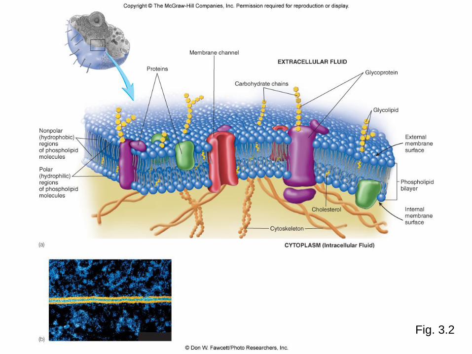
Fig. 3.2
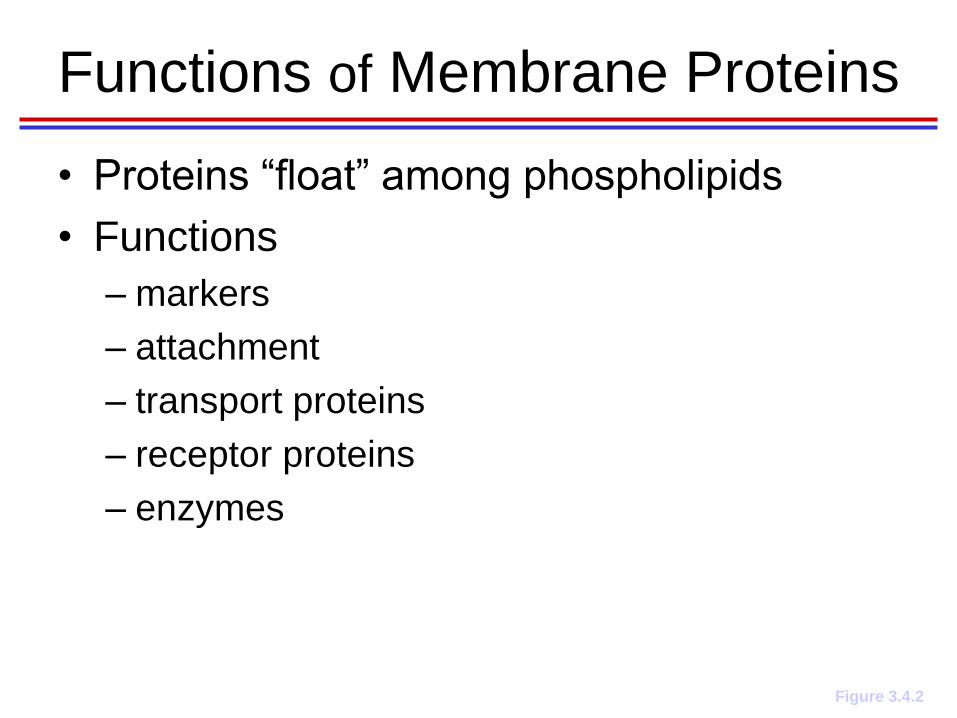
Functions of Membrane Proteins
Figure 3.4.2
• Proteins “float” among phospholipids
• Functions
– markers
– attachment
– transport proteins
– receptor proteins
– enzymes

Cell Membrane Transport

Movement Through Plasma Membrane
Ions & molecules move across by
1. diffusion
2. osmosis
3. mediated transport
4. vesicular transport

1. Diffusion
• movement of solute from area of higher conc. to
area of lower conc, w/in solvent
– at equilibrium: uniform distribution of molecules
• Terms--
– Solution: mixture, substances uniformly distributed
w/no clear boundary between substances
– Solute dissolves in a solvent to form a solution
– Concentration gradient: concentration difference
across a membrane or barrier

What Can Diffuse?
1. Lipid-soluble molec.
diffuses directly
through membrane
2. Most non-lipid-
soluble molecs and
ions do not diffuse
through
1.Some can pass
through membrane
channels or other
transport proteins

Diffusion

http://highered.mcgraw-hill.com/sites/0072495855/student_view0/chapter2/animation__how_diffusion_works.html

2. Osmosis
• diffusion of water across selectively
permeable membrane
– through a specific channel protein (aquaporin)
– or through lipid bilayer
• Types of Solutions
– Isosmotic: same conc solutes as reference
solution
– Hyperosmotic: greater conc solutes vs. ref
soln
– Hyposmotic: lesser conc solutes vs. ref soln

Fig. 3.5
Osmosis

Osmotic Concentration of Solutions
a) hypotonic solution with low solute conc
results: swelling of RBC placed into the solution.
Water enters cell by osmosis, RBC lyses (bursts).
b) isotonic solution with conc solutes = inside cells
Results: normal shaped RBC.
Water moves into and out of cell at same rate, but no net water movement.
c) hypertonic solution, high solute conc
Results: shrinkage (crenation) of RBC
Water moves by osmosis out of cell and into hypertonic solution.
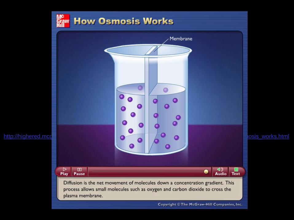
http://highered.mcgraw-hill.com/sites/0072495855/student_view0/chapter2/animation__how_osmosis_works.html

3. Mediated Transport
• transport proteins mediate (assist in)
movement of ions & molecules across
plasma memb
• Characteristics
1. Specificity: selectiveness
2. Competition: similar molecules/ions compete
for transport protein
3. Saturation: rate of transport cannot increase
because all ptns in use

Types of Transport Proteins
1. Channel proteins: form membrane ion channels
2. Carrier proteins: bind to ions/molecules & transport them 1. Uniport (facilitated diffusion) moves ion/molec down its conc
gradient
2. Symport moves 2 or more ions/molecs in same direction
3. Antiport moves 2 or more ions/molecs in opp directions
3. Protein Pumps: move ions/molecs against concentration gradient using ATP energy
4. Secondary active transport uses energy of 1 substance moving down its conc gradient to move another across p memb

http://highered.mcgraw-hill.com/olcweb/cgi/pluginpop.cgi?it=swf::535::535::/sites/dl/free/0072437316/120068/bio04.swf::Cotransport

Fig. 3.7
Facilitated
Diffusion
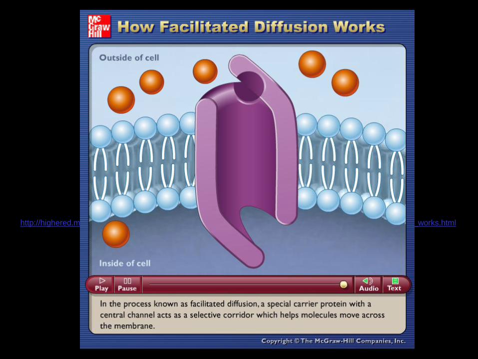
http://highered.mcgraw-hill.com/sites/0072495855/student_view0/chapter2/animation__how_facilitated_diffusion_works.html

Sodium-Potassium Pump 1. 3 Na+ & ATP bind to Na+-K+ pump (ATP-
powered).
2. ATP ADP + P , releases energy -- used to change shape of pump. P remains bound to pump.
3. Pump changes shape, 3 Na+ transported across membrane, OUT of cell.
4. Na+ diffuses away from pump.
5. 2 K+ bind to pump.
6. P is released from pump binding site.
7. Pump resumes original shape, transports 2 K+ across membrane INTO cell, K+ diffuses away from pump. Pump can again bind to Na+ & ATP.

http://highered.mcgraw-hill.com/sites/0072495855/student_view0/chapter2/animation__how_the_sodium_potassium_pump_works.html

Secondary Active Transport
• Symport of Na+ and Glucose 1. Na+-K+ pump maintains high conc. of Na+ outside the cell (vs.
inside).
2. Na+ move back into cell through carrier protein (symporter) that also moves glucose. Conc. gradient for Na+ provides energy required to move glucose against its conc gradient.
Fig. 3.9

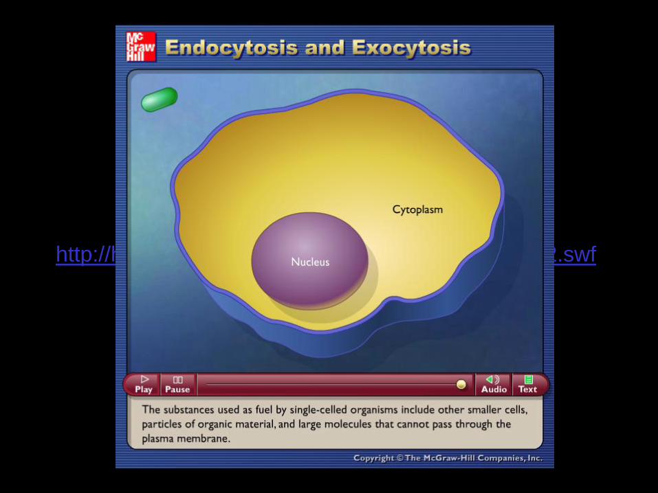
4. Vesicular Transport
• Transport of large particles & macromolecules
across p memb
– Endocytosis: movement of materials INTO
cells by vesicle formation
• Phagocytosis: movement of solid material
• Pinocytosis: small droplets of liquids and materials
• Receptor-mediated endocytosis: p memb receptors
attach to molecules, then into cells
– Exocytosis: secretion of materials OUT of cells
by vesicle formation

Phagocytosis
Fig. 3.10

http://highered.mcgraw-hill.com/sites/0072495855/student_view0/chapter2/animation__phagocytosis.html

Receptor-Mediated Endocytosis
1. Receptors in plasma membrane bind to molecules to be taken into cell
2. Receptors and bound molecules taken into cell as a vesicle begins to form
3. Vesicle fuses and separates from plasma membrane
Fig. 3.11
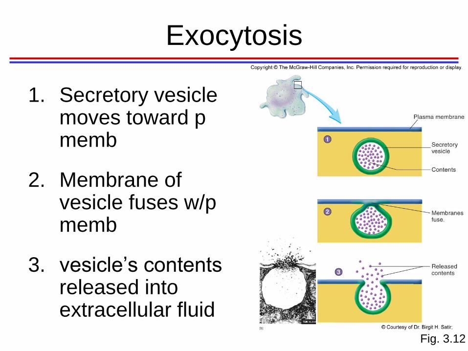
Exocytosis
1. Secretory vesicle moves toward p memb
2. Membrane of vesicle fuses w/p memb
3. vesicle’s contents released into extracellular fluid
Fig. 3.12
http://highered.mcgraw-hill.com/olc/dl/120068/bio02.swf
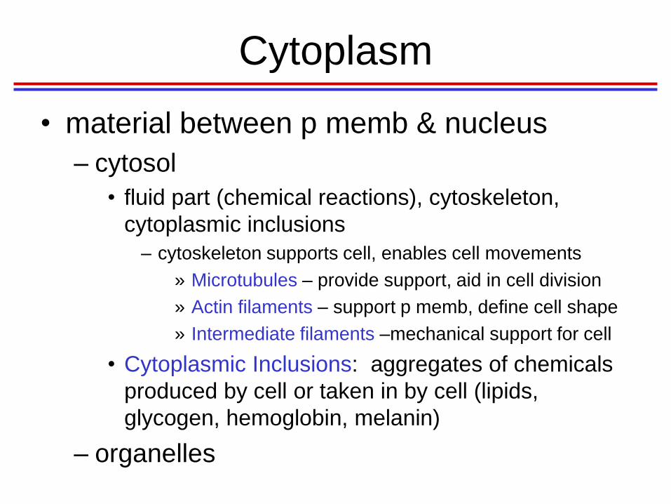
Cytoplasm
• material between p memb & nucleus
– cytosol
• fluid part (chemical reactions), cytoskeleton,
cytoplasmic inclusions
– cytoskeleton supports cell, enables cell movements
» Microtubules – provide support, aid in cell division
» Actin filaments – support p memb, define cell shape
» Intermediate filaments –mechanical support for cell
• Cytoplasmic Inclusions: aggregates of chemicals
produced by cell or taken in by cell (lipids,
glycogen, hemoglobin, melanin)
– organelles

Fig. 3.13
Cytoskeleton

Cytoplasmic Organelles
• Specialized structures w/specific functions
• Membranous
– Mitochondria, peroxisomes, lysosomes,
endoplasmic reticulum, Golgi apparatus
• Nonmembranous
– Centrioles, ribosomes
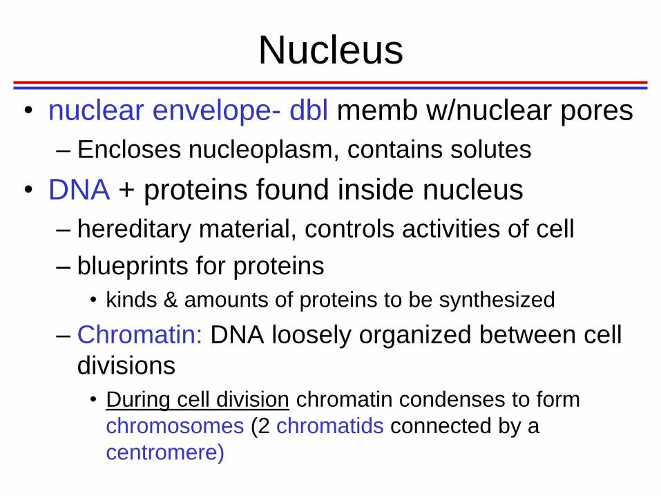
Nucleus
• nuclear envelope- dbl memb w/nuclear pores
– Encloses nucleoplasm, contains solutes
• DNA + proteins found inside nucleus
– hereditary material, controls activities of cell
– blueprints for proteins
• kinds & amounts of proteins to be synthesized
– Chromatin: DNA loosely organized between cell
divisions
• During cell division chromatin condenses to form
chromosomes (2 chromatids connected by a
centromere)

Fig. 3.14
Nucleus

Fig. 3.15
Chromosome
Structure
Nucleoli and Ribosomes
• Nucleoli: spherical bodies w/in nucleus
– RNA & proteins
– Produces ribosomal RNA (rRNA)
• Ribosomes: sites of protein synthesis
– Free ribosomes not attached to any organelles
• synthesize proteins used inside cell
– Attached ribosomes -rough endoplasmic reticulum (rER)
• produce proteins secreted from cell
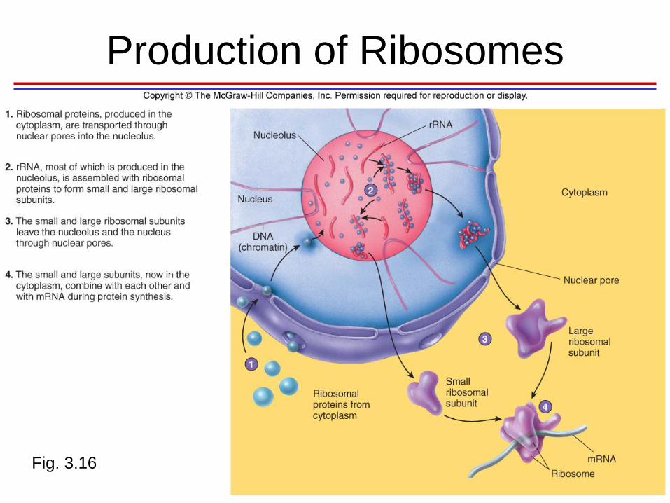
Fig. 3.16
Production of Ribosomes

Endoplasmic Reticulum (ER)
• Series of membranes, form sacs & tubules
• Extends from outer nuclear membrane into cytoplasm – Rough ER (RER)
• with ribosomes
• protein synthesis
– Smooth ER (SER) • No ribosomes
• lipid and carbohydrate synthesis – Liver: lipid and cholesterol metabolism, breakdown of
glycogen and along with the kidneys, detoxify drugs
– Testes: synthesis of steroid-based hormones
– Intestinal cells: absorption, synthesis, and transport of fats
– Skeletal and cardiac muscle: storage and release of calcium

Endoplasmic Reticulum (ER)
Fig. 3.17
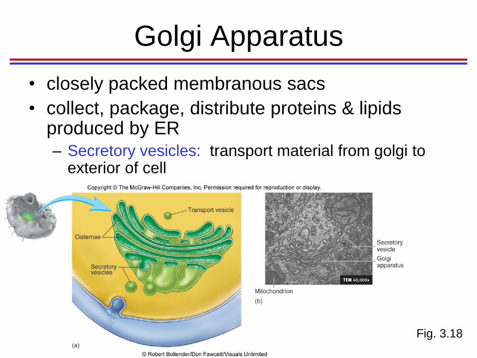
Golgi Apparatus
• closely packed membranous sacs
• collect, package, distribute proteins & lipids produced by ER – Secretory vesicles: transport material from golgi to
exterior of cell
Fig. 3.18

Function of the Golgi Apparatus 1. Some proteins produced at ribosomes on
RER and transferred into cisterna
2. proteins surrounded by vesicle from membrane of ER
3. transport vesicle moves from ER to Golgi, fuses with its membrane, releases proteins into its cisterna
4. Golgi concentrates and modifies proteins into glycoproteins or lipoproteins
5. proteins packaged into vesicles from membrane of Golgi
6. Some vesicles (lysosomes) contain enzymes used within cell
7. Secretory vesicles carry proteins to plasma memb, where proteins secreted from cell by exocytosis
8. Some vesicles contain proteins that become part of plasma memb
Fig. 3.19

Lysosomes
• Spherical membranous bags containing
digestive enzymes
– Digest ingested bacteria, viruses, and toxins
– Degrade nonfunctional organelles
– Breakdown glycogen, release thyroid hormone
– Breakdown non-useful tissue
– Breakdown bone to release Ca2+
– Secretory lysosomes in white blood cells, immune
cells, melanocytes

Action of Lysosomes
1. vesicle forms around material outside cell
2. vesicle pinched off from plasma memb and becomes separate vesicle inside cell
3. lysosome pinched off Golgi
4. lysosome fuses with vesicle Fig. 3.20
5. enzymes from lysosome mix with material in vesicle, enzymes digest material

http://highered.mcgraw-hill.com/sites/0072495855/student_view0/chapter2/animation__lysosomes.html

Peroxisomes
• Membranous sacs containing oxidases
and catalases
– Breakdown fatty acids, amino acids, and
hydrogen peroxide
– Detoxify harmful or toxic substances
– Neutralize dangerous free radicals
• Free radicals: highly reactive chemicals with
unpaired electrons (i.e., O2–)
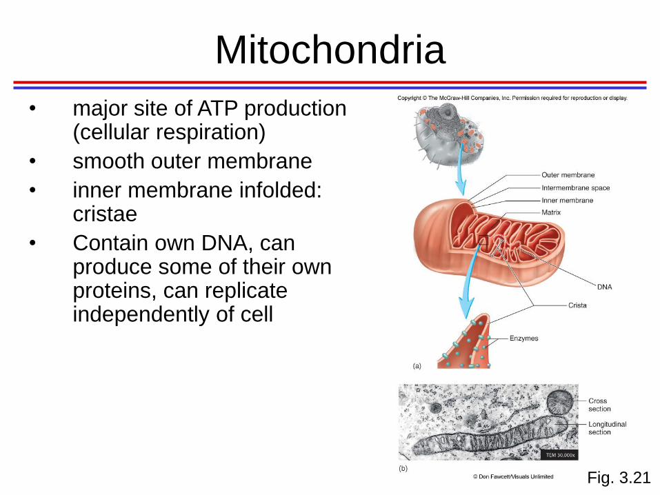
Mitochondria
Fig. 3.21
• major site of ATP production (cellular respiration)
• smooth outer membrane
• inner membrane infolded: cristae
• Contain own DNA, can produce some of their own proteins, can replicate independently of cell

Centrioles and Spindle Fibers
• Centrioles: cylindrical
organelles located in
centrosome
– 9 triplets of microtubules
– Centrosome: area of cytoplasm
• site of microtubule formation
– Microtubule “spindle fibers”
extend in all directions from
centrosome
• involved in cell division
– Form basis of cilia and flagella
Fig. 3.22

Cilia, Flagella, and Microvilli
• Cilia
– move substances over surface of cells
• Flagella
– much longer than cilia, propel sperm cells
• Microvilli
– increase surface area of cell, aid in absorption
and secretion

Part 3: Cellular Processes
• Protein Synthesis
– Transcription
– Translation
• Cell Division (Mitosis)

Protein Synthesis
• DNA: master blueprint for protein synth
• DNA controls enzyme production
• Cell activity regulated by enzymes (Proteins)
• Genes: segments of DNA w/instructions for a polypeptide chain
– Triplets of nucleotide bases
– Each triplet codes for 1 amino acid

Protein Synthesis
• 2 step process
– Transcription
• cell copies gene to make a particular protein:
messenger RNA (mRNA)
• mRNA travels from nucleus to ribosome where info
is translated into a protein
– Translation
• requires mRNA and transfer RNA (tRNA)
• tRNA brings amino acids to ribosome to synthesize
protein

Overview of Protein Synthesis
1. DNA contains info to produce proteins
2. Transcription of 1 DNA strand results in mRNA (complementary copy of info in DNA strand)
3. mRNA leaves nucleus and goes to a ribosome
4. Amino acids, building blocks of proteins, carried to ribosome by tRNAs
5. Translation: info contained in mRNA used to determine #, kinds, and arrangement of amino acids in polypeptide chain
Fig. 3.23
http://highered.mcgraw-hill.com/olcweb/cgi/pluginpop.cgi?it=swf::535::535::/sites/dl/free/0072437316/120077/micro06.swf::Protein%20Synthesis

Transcription
• Synthesis of RNA based on nucleotide
sequence in DNA
– Messenger RNA (mRNA) – carries genetic
info from DNA in nucleus to ribosomes in
cytoplasm
– Transfer RNAs (tRNAs) – bound to amino
acids, base pair with codons of mRNA at
ribosome to begin process of translation
– Ribosomal RNA (rRNA) – structural
component of ribosomes

Transcription
1. strands of DNA molecule separate from each other. 1 DNA strand serves as template for mRNA synthesis
2. Nucleotides that will form mRNA pair with DNA nucleotides according to base-pair rules. Sequence of n’tides in template DNA strand (purple) determines sequence of n’tides in mRNA (grey). RNA polymerase (enzyme, not shown) joins n’tides of mRNA together
3. As n’tides are added, an mRNA molecule is formed
Fig. 3.24

Transcription: RNA Polymerase
• enzyme that oversees synthesis of RNA
• Unwinds DNA template
• Adds complementary RNA nucleotides on
DNA template
• Joins these RNA n’tides together
• termination signal stops transcription

@ End of Transcription
• Posttranscriptional processing
– modifies mRNA before it leaves nucleus
– removes introns (non-coding) and
– splices exons (coding) together with enzymes
(spliceosomes)
– Functional mRNA consists only of exons
• Alternative splicing
– produces different combination of exons,
allowing 1 gene to produce >1 type of protein

http://highered.mcgraw-hill.com/sites/9834092339/student_view0/chapter25/transcription_factors.html

http://glencoe.mcgraw-hill.com/sites/9834092339/student_view0/chapter16/control_of_gene_expression_in_eukaryotes.html

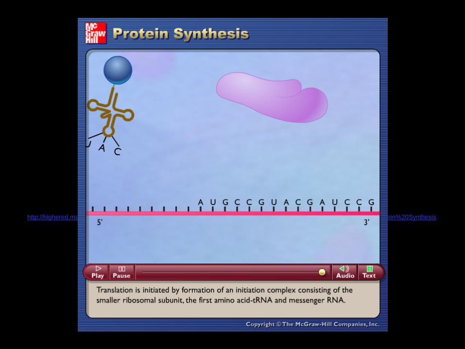
Translation
• Synthesis of proteins
– Codon: set of 3 mRNA n’tides, codes for 1 amino acid during translation
– Anticodon: set of 3 tRNA n’tides, complementary to codons of mRNA
• mRNA leaves nucleus through nuclear pores, to ribosomes
• tRNA carries amino acids, interacts at ribosome with mRNA.
• anticodons of tRNA bind to codons of mRNA, amino acids joined to form protein

Steps of Translation
1.Ribosome binds to mRNA-- has 2 binding sites for tRNA. **1st codon is ALWAYS “AUG” (start codon), which codes for methionine. Codon and anticodon bind. 2nd tRNA binding site is open.
2.2nd tRNA binds to mRNA @ 2nd site on ribosome.
3.An enzyme within ribosome catalyzes a synthesis reaction to form a peptide bond between amino acids. **Amino acids are now associated with only 1 of the tRNAs.
Fig. 3.25
Translation
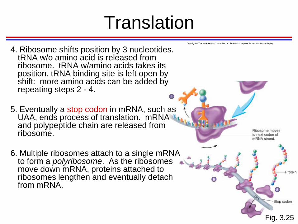
4. Ribosome shifts position by 3 nucleotides. tRNA w/o amino acid is released from ribosome. tRNA w/amino acids takes its position. tRNA binding site is left open by shift: more amino acids can be added by repeating steps 2 - 4.
5. Eventually a stop codon in mRNA, such as UAA, ends process of translation. mRNA and polypeptide chain are released from ribosome.
6. Multiple ribosomes attach to a single mRNA to form a polyribosome. As the ribosomes move down mRNA, proteins attached to ribosomes lengthen and eventually detach from mRNA.
Fig. 3.25

Information Transfer from DNA to RNA
• DNA triplets transcribed into mRNA
codons by RNA polymerase
• mRNA Codons base pair with tRNA
anticodons at ribosomes
• Amino acids are peptide bonded at
ribosomes to form polypeptide chains
• Start and stop codons initiate and end
translation

Study Guide • Optional
• All of Study Guide 1 and part of Study Guide 2—
chapter 4 only
• BONUS!

Sample Questions
• Which of the following is NOT true of a negative-feedback mechanism?
A. The change from the set point is made smaller
B. Most control mechanisms in the body are of this type
C. The deviation from normal is made larger
D. Blood pressure maintenance is an example of negative-feedback
• Which anatomical body region is NOT matched with its common name?
A. Gluteal – buttock B. Olecranon - point of shoulder
C. Femoral – thigh D. Pedal - foot
• An ionic bond is formed by the
A. Sharing of electrons between two atoms
B. Loss of electrons from two atoms
C. Attraction between cations and anions
D. Gain of electrons from two atoms
• Which organelle functions to break down fatty acids, amino acids and
hydrogen peroxide?
A. Proteasomes B. Centrioles C. Lysosomes D. Peroxisomes

Cell Division: The Cell’s Life Cycle

2 Types of Cell Division
• Mitosis produces new cells for growth and
tissue repair
• Meiosis produces gametes (sex cells)
– Sperm cells in males
– Oocytes (egg cells) in females

Cell Division: DNA
• Chromosomes
– Somatic cells: diploid # of chromosomes
– Gametes: haploid #
– Humans: diploid #: 46 (23 pairs),haploid #: 23
• 22 pairs autosomal chromosomes
• 1 pair sex chromosomes
– Females XX
– Males XY
• DNA replicates during interphase (time
between cell division)

Replication of DNA
1. strands of DNA separate
2. Each old strand (dark purple) functions as template on which new, complementary strand (light purple) is formed. Base-pairing determines sequence of n’tides in newly formed strands
3. 2 identical DNA molecules are produced
Fig. 3.26

Replication of a Chromosome
1. DNA of chromosome is dispersed as chromatin
2. DNA unwinds, each strand is replicated
3. During mitosis, chromatin from each replicated DNA strand condenses to form a chromatid. Chromatids are joined at centromere to form a single chromosome
4. Chromatids separate to form 2 new, identical chromosomes. They’ll unwind to form chromatin in nuclei of 2 daughter cells
Fig. 3.26

http://highered.mcgraw-
hill.com/olcweb/cgi/pluginpop.cgi?it=swf::535::535::/sites/dl/free/0072437316/120076/bio23.swf::How%20Nucleotides%20are%20Add
ed%20in%20DNA%20Replication
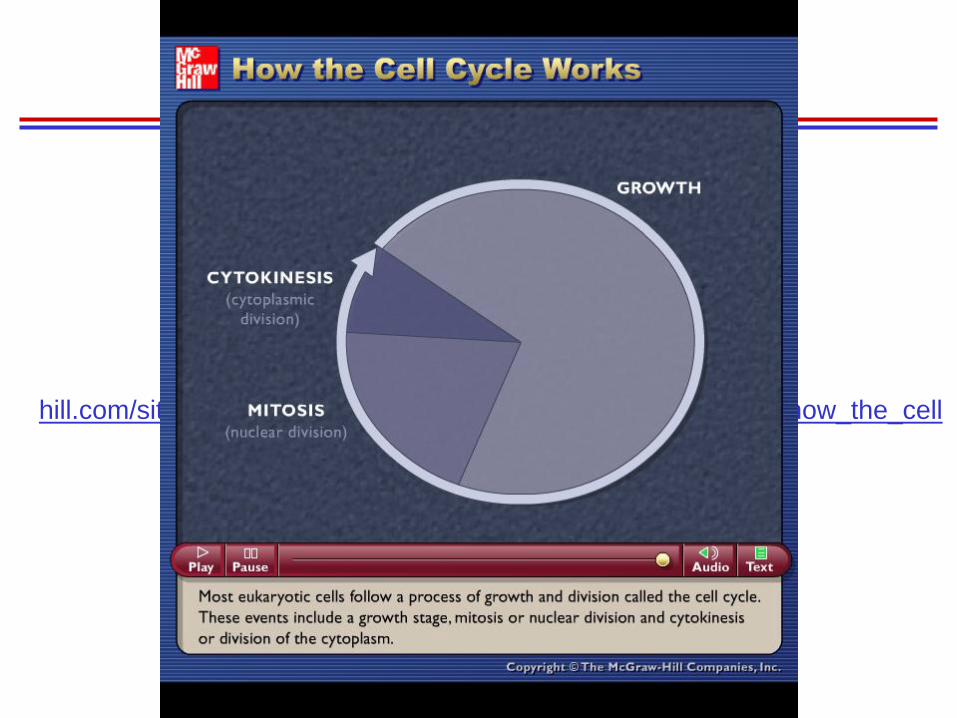
http://highered.mcgraw-
hill.com/sites/0072495855/student_view0/chapter2/animation__how_the_cell
_cycle_works.html

http://highered.mcgraw-
hill.com/sites/0072495855/student_view0/chapter2/animation__control_of_the_cell_cycl
e.html
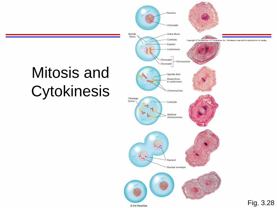
Mitosis and
Cytokinesis
Fig. 3.28

Interphase
Fig. 3.28
1. Interphase = time between cell divisions. DNA is thin threads of chromatin (in nucleus). DNA replication occurs. Organelles duplicate.

Prophase
Fig. 3.28
2. Prophase: chromatin condenses into chromosomes. Centrioles move to opposite ends of cell, nucleolus and nuclear envelope disappear. Microtubules (spindle fibers) form near centrioles, project in all directions toward equator and overlap with fibers from opposite centrioles.

Metaphase
Fig. 3.28
3. In metaphase, chromosomes align in center of cell in association with spindle fibers. Some spindle fibers are attached to kinetochores in centromere of each chromosome

Anaphase
Fig. 3.28
4. Anaphase: chromatids separate, each chromatid is then referred to as a chromosome. Chromo # is double, 2 identical sets of chromos. Chromos (b/c microtubules) move toward centrioles at each end of cell. Separation of chromatids is @ beginning of anaphase; @ end, chromos @ poles.

Telophase and Cytokinesis
Fig. 3.28
5. Telophase: each set of chromos @ ends. Unravel to become chromatin. nuclear envelope forms from endoplasmic reticulum. Nucleoli form, and cytokinesis forms 2 cells
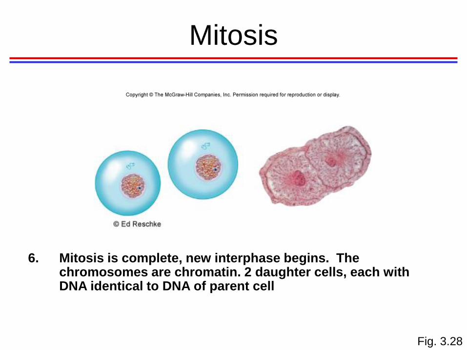
Mitosis
Fig. 3.28
6. Mitosis is complete, new interphase begins. The chromosomes are chromatin. 2 daughter cells, each with DNA identical to DNA of parent cell

Mitosis and Cytokinesis
Fig. 3.28
1. Interphase is the time between cell divisions. DNA is found as thin threads of chromatin in the nucleus. DNA replication occurs during interphase. Organelles, other than the nucleus, duplicate during interphase
2. In prophase, the chromatin condenses into chromosomes. The centrioles move to the opposite ends of the cell, and the nucleolus and the nuclear envelope disappear. Microtubules form near the centrioles and project in all directions. Spindle fibers, project toward an invisible line called the equator and overlap with fibers from opposite centrioles.
3. In metaphase, the chromosomes align in the center of the cell in association with the spindle fibers. Some spindle fibers are attached to kinetochores in the centromere of each chromosome
4. In anaphase, the chromatids separate, and each chromatid is then referred to as a chromosome. Thus, the chromosome number is double, and there are two identical sets of chromosomes. The chromosomes, assisted by the spindle fibers, move toward the centrioles at each end of the cell. Separation of the chromatids signals the beginning of anaphase, and, by the time anaphase has ended, the chromosomes have reached the poles
5. In telophase, migration of each set of chromosomes is complete. The chromosomes unravel to become less distinct chromatin threads. The nuclear envelope forms from the endoplasmic reticulum. The nucleoli form, and cytokinesis continues to form two cells
6. Mitosis is complete, and a new interphase begins. The chromosomes have unraveled to become chromatin. Cell division has produced two daughter cells, each with DNA that is identical to the DNA of the parent cell

http://highered.mcgraw-hill.com/sites/0072495855/student_view0/chapter2/animation__mitosis_and_cytokinesis.html
Lastly...Cell Differentiation!
• Process by which cells develop
specialized structures & functions
• All cells in individual’s body contain same
amount & type of DNA because resulted
from mitosis
• Differentiation results from selective
activation and inactivation of segments of
DNA in each different cell type