Embed Size (px)
Citation preview
Chapter 8 Modification of intestinal flora with multispecies probiotics reduces bacterial translocation and improves clinical course in a rat model of acute pancreatitis
L.P. van Minnen1
H.M. Timmerman1
F. Lutgendorff1
A. Verheem1
W. Harmsen2
S.R. Konstantinov4
H. Smidt4
M.R.Visser2
G.T. Rijkers3,5
H.G. Gooszen1
L.M.A. Akkermans1
Departments of 1Surgery, 2Medical Microbiology and 3Pediatric Immunology,
University Medical Center, Utrecht. 4Laboratory of Microbiology, Agrotechnology and Food Sciences Group, Wageningen
University, Wageningen. 5Department of Medical Microbiology and Immunology, St. Antonius Hospital,
Nieuwegein.
Surgery. In press
Chapter 8
150
Probiotics and bacterial translocation
151
ABSTRACT
Background
Infection of pancreatic necrosis by gut bacteria is a major cause of morbidity and
mortality in patients with severe acute pancreatitis. Use of prophylactic antibiotics
remains controversial.
Aim of this experiment was to assess if modification of intestinal flora with
specifically designed multispecies probiotics (Ecologic® 641) reduces bacterial
translocation or improves outcome in a rat model of acute pancreatitis.
Methods
Male Sprague-Dawley rats were allocated into three groups: 1) controls (sham
operated, no treatment), 2) pancreatitis and placebo or 3) pancreatitis and probi-
otics. Acute pancreatitis was induced by intraductal glycodeoxycholate and intra-
venous cerulein infusion. Daily probiotics or placebo were administered intragas-
trically, five days before until seven days after induction of pancreatitis. Tissue and
fluid samples were collected for microbiological and quantitative real-time PCR
analysis of bacterial translocation.
Results
Probiotics reduced duodenal bacterial overgrowth of potential pathogens (Log10
colony forming units (CFU)/g 5.0 ± 0.7 (placebo) vs. 3.5 ± 0.3 CFU/g (probiotics),
P < 0.05), resulting in reduced bacterial translocation to extra-intestinal sites, in-
cluding the pancreas (5.4 ± 1.0 CFU/g (placebo) vs. 3.1 ± 0.5 CFU/g (probiotics),
P < 0.05). Accordingly, health scores were better and late phase mortality was
reduced (27% (4/15, placebo) vs. 0% (0/13, probiotics), P < 0.05).
Conclusions
This experiment supports the hypothesis that modification of intestinal flora with
multispecies probiotics results in reduced bacterial translocation, morbidity and
mortality in the course of experimental acute pancreatitis.
INTRoDUCTIoN
Severe acute pancreatitis follows a biphasic clinical course. The early pro-
inflammatory phase is associated with systemic inflammatory response syndrome
(SIRS), (multiple) organ damage and early mortality (< 1 week). The late phase is
characterized by infectious complications following bacterial translocation of
intestinal bacteria and late mortality (> 3 weeks).1-4 Infectious complications are
frequently the cause of mortality in acute pancreatitis patients.1,5
The use of prophylactic antibiotics to prevent infectious complications remains a
topic of debate. A recent meta-analysis of six randomized controlled trials
concluded that prophylactic antibiotics do not prevent infection of pancreatic
necrosis or mortality in severe acute pancreatitis.6 Furthermore, increased
concerns regarding the widespread use of prophylactic antibiotics associated
complications (i.e. fungal infections or antibiotics resistance) have been reported.7-11
Prophylactic probiotics have been suggested as an alternative to the use of
prophylactic antibiotics.12,13 Beneficial effects of prophylactic probiotics for acute
pancreatitis have been reported in animal experiments and clinical trails.12,14 Most
studies on prophylactic probiotics however, have focused on a single probiotic
strain for a variety of medical disorders. A recent report however, has advocated
the use of specifically selected multiple probiotic strains.15 For optimal results,
probiotic strains should be selected to target known pathophysiological aspects
of the disorder addressed.16
Experimental and clinical studies have greatly increased the knowledge of the
pathophysiology of bacterial translocation during acute pancreatitis. Three major
steps in the sequence of bacterial translocation have been identified: 1) small
bowel bacterial overgrowth, 2) mucosal barrier failure and 3) pro-inflammatory
responses.17-23 These three phenomena occur early after the start of acute
pancreatitis and cumulate into bacterial translocation and infectious complications.
Based on these considerations, we have designed a mixture of six probiotic strains,
especially selected to reduce small bowel bacterial overgrowth and reduce pro-
inflammatory immune responses. (Timmerman et al. Manuscript submitted for
publication)
It is unknown if modification of the intestinal flora with such a multispecies probiotic
mixture reduces bacterial translocation and consequently, alter the course of
disease. Therefore, the aim of the present study was to assess if modification of
intestinal flora by a specifically designed, multispecies probiotic mixture changes
disease course using a well established rat model of acute pancreatitis.

Chapter 8
152
Probiotics and bacterial translocation
153
MATERIALS AND METHoDS
Animals
Male specific pathogen-free Sprague-Dawley rats, 250-350 grams (Harlan, Horst,
The Netherlands) were kept under constant housing conditions (temperature
(22°C), relative humidity (60%) and a 12-hour light/dark cycle) and had free access
to water and food (RMH 1110, Hope Farms, Woerden, The Netherlands) throughout
the experiment. Rats were allowed to adjust to these conditions for one week prior
to surgery. The experimental design, shown in Figure 1, was approved by the
institutional animal care committee of the University Medical Center, Utrecht, The
Netherlands. Rats were randomized (1: 2: 2 relative group size computer generated
randomization for the respective groups) between three experimental groups: 1)
control animals (gastric cannula, sham pancreatitis, no treatment), 2) acute
pancreatitis and placebo, and 3) acute pancreatitis and probiotics. In total, 10 rats
were included in the control group, 21 rats in the placebo group, and 17 rats in the
group given daily probiotics.
Probiotics and placebo
The study product (Ecologic® 641, Winclove Bio Industries, Amsterdam, The
Netherlands) consisted of viable and freeze-dried probiotic strains; 4 lactobacilli:
Lactobacillusacidophilus (W70),Lactobacilluscasei (W56),Lactobacillussalivarius
(W24),Lactococcuslactis (W58),and two bifidobacteria: Bifidobacteriumbifidum
(W23)and Bifidobacteriuminfantis (W52). The placebo product consisted of carrier
substance only (corn-starch). Probiotics and placebo were packed in identical
sachets and coded by the producer to guarantee blinding during the experiment.
Directly before administration of the doses, the products were reconstituted in
sterile water, for 15 minutes at 37°C. Single probiotics dose volume of 1.0 ml
contained a total of 5 x 10⁹ CFU bacteria. Probiotics or placebo were administered
intragastrically through a permanent gastric cannula once daily, starting five days
prior to induction of acute pancreatitis, and twice daily for six days after induction
of acute pancreatitis.
Surgical procedures
All surgical procedures were performed on a heated operating table under general
anaesthesia using a combination of 2% Isoflurane gas (flow: 0.5 l/min o2, 1.5 l/m
air) through a snout-mask and intramuscular 0.3 ml 10% buprenonorphine
(Temgesic, Reckitt Beckiser Healthcare Ltd., Hull, UK). All surgical procedures
were performed with sterile instruments under strict aseptic conditions. Throughout
the experiment random control swabs of the abdomen remained negative on
bacterial cultures, ensuring external contamination did not occur.
Gastric cannulation
At the start of the experiment, a permanent gastric cannula was fitted in all rats.
Under general anesthesia, a 20 cm silicone cannula (outer diameter 1.65 mm,
inner diameter 0.76 mm, Rubber, Amsterdam, The Netherlands) was tunneled
subcutaneously from the abdominal wall to the back, penetrating the skin between
the scapulae. A 1.5 cm midline laparotomy was made to insert the gastric end of
the cannula into the stomach through a puncture within a purse-string suture on
the greater curvature. The cannula was securely fixed and the abdomen was
closed in two layers. The dorsal end of the cannula was kept in place between the
scapulae using a rodent infusion jacket (Uno Zevenaar BV, Zevenaar, The
Netherlands). Animals in the probiotics and placebo groups were allowed to
recover for three days, prior to the start of daily probiotics or placebo
administrations.
Induction of acute pancreatitis
Five days after starting daily administration of placebo or probiotics, acute
pancreatitis was induced using the internationally well accepted model described
by Schmidt et al.24 Briefly, during midline relaparotomy the papilla of Vater was
Gastriccannula
-8 Days -5 Days Day 0 Day 7
Twice daily probiotics or placebo
Pancreatitis
Daily probiotics or placebo
Experimental design. Eight days prior to induction of acute pancreatitis, a permanent gastric cannula was fitted. Probiotics or placebo were administered intragastrically through a permanent gastric cannula once daily, starting 5 days prior to induction of acute pancreatitis, and twice daily from days 1-7 after induction of acute pancreatitis. Seven days after induction of pancreatitis, surviving rats were anesthetized to allow sterile removal of organ and blood samples. Control animals did not receive administra-tions through the gastric cannula and underwent a sham pancreatitis procedure only.
Figure 1
Chapter 8
154
Probiotics and bacterial translocation
155
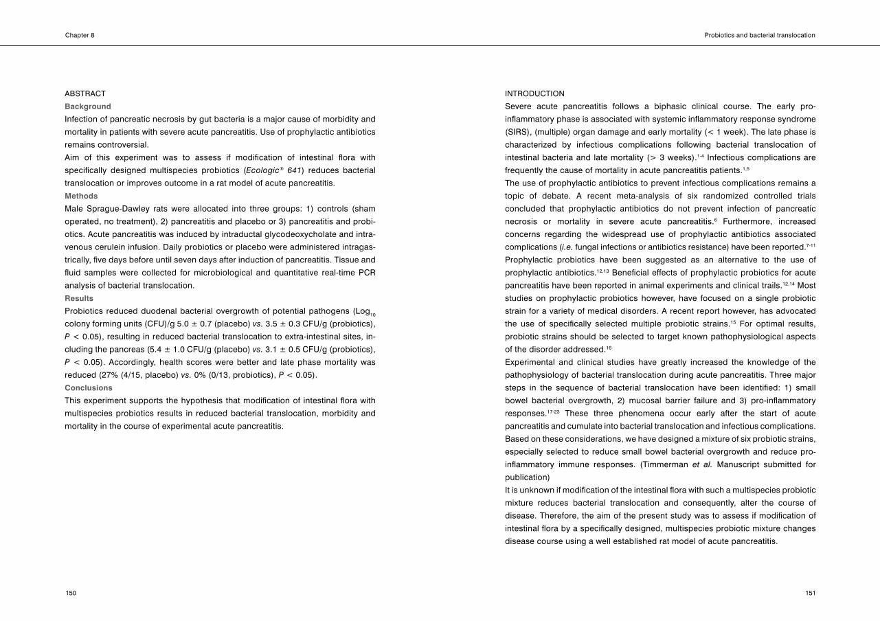
cannulated transduodenally using a 24G Abbocath®-T i.v. infusion cannula
(Abbott, Sligo, Republic of Ireland) (Figure 2). Before pressure monitored infusion
(MMS, Enschede, The Netherlands) of 0.5 ml sterilized glycodeoxycholic acid in
glycylglycine-NaoH-buffered solution (10 mmol/l, pH 8.0, 37°C, chemicals
obtained from Sigma-Aldrich Chemie BV, Zwijndrecht, The Netherlands), the
common bile duct was clamped and bile and pancreatic fluid were allowed to
drain through the cannula. No animals needed to be excluded for infusion
pressures exceeding 35 mmHg. Directly after infusion, hepato-duodenal bile flow
was restored by removal of the clamp. The puncture hole in the duodenum was
carefully closed using an 8.0 polyprolene serosal suture (Figure 2). After closure
of the abdomen in two layers, the right jugular vein was cannulated for continuous
postoperative intravenous infusion of cerulein (5 μg/kg/hr, for six hours). The
jugular vein cannula was fixed to the rodent infusion jacket and attached to a
swivel system to provide unrestricted mobility of the rat during infusion. During the
sham procedure in the control rats, the papilla of Vater was cannulated, the
common bile duct was clamped, but no glycodeoxycholic acid was infused. A
jugular vein cannula was fitted for six hours of intravenous saline infusion. After
acute pancreatitis induction or sham procedure, pain relief was provided for 48
hours by subcutaneous injections of 0.3 ml 10% buprenonorphine twice daily.
The clinical response of the rats after induction of acute pancreatitis was assessed
using a 0-6 points scoring system: Grooming: normal = 2 points, decreased = 1
point, none = 0 points. Mobility: normal = 2 points, decreased = 1 point, immobile
= 0 points. Painposture: none = 2 points, arching (convex back and retraction of
abdomen from floor) = 1 point, stretching (whole body is stretched out on floor,
spine is straight and horizontal) = 0 points. Aspects of this scoring system are well
recognized behavioral parameters expressing health or morbidity (including
abdominal / visceral pain).25,26 According to Dutch animal welfare laws and local
protocols of the animal ethics committee, daily assessments of these aspects are
mandatory to monitor animal welfare throughout the experimental protocol.
Indeed, two rats in the placebo group demonstrating signs of severe suffering and
poor clinical prognosis (low health scores) were terminated on day six and added
to the Kaplan-Meier statistic the same day.
Collection of tissue and fluid samples
on day seven, surviving rats were anesthetized to allow sterile removal of organ
and fluid samples. To avoid cross-contamination, samples were taken under strict
aseptic conditions in the following order: peritoneal fluid, blood (inferior vena
cava), mesenteric lymph nodes (MLN), liver, spleen, pancreas and duodenum.
After sample collection, rats were euthanized by blood loss. Samples were
collected for microbiological analysis and portion of each sample and a segment
of the ileum was snap frozen in liquid nitrogen and stored at -80°C for future
analysis. Another portion of pancreatic samples was analyzed histopathologically,
using standard hematoxilin and eosin (H&E) staining. Histopathological severity of
acute pancreatitis was assessed based on the acute pancreatitis scoring system
as previously described.27
Culture-based microbiological analysis for bacterial identification and
quantification
All organ samples were weighed and processed immediately for quantitative and
qualitative cultures of aerobic and anaerobic organisms. All organs were
homogenized in cysteine broth with a sterile blender and cultured in 10-fold
dilution series. The samples were cultured on bloodagar, MacConkey-agar (for
Gram-negative strains), Columbia Colistin Nalidixic Acid (CNA) agar (for
Schematic representation of the bile infusion model according to Schmidt etal.24 The duodenum is held aside with a cotton wool stick (*), pv: portal vein. After clamping of the common bile duct the papilla of Vater is cannulated transduodenally for bile salt infusion (left panel). Directly after infusion, hepato-duodenal bile flow was restored by removal of the clamp. The puncture hole in the duodenum was sutured (right panel).
Figure 2
Chapter 8
156
Probiotics and bacterial translocation
157
staphylococci and streptococci), Man-Rogosa-Sharpe-agar (for lactobacilli) and
Schaedler agar (for facultative anaerobic bacteria). The microorganisms were
identified using standard microbiological techniques. For analysis of organ
samples, cultured bacteria were subdivided in three groups: gram-positive cocci
(GPC), gram-positive rods (GPR) and gram-negative rods / anaerobes (GNR
+anear). Also, Hemolytic Streptococcus group B, Enterococcus spp., Staphy-
lococcus aureus, and Enterobacteriaceae such as Escherichia coli, Proteus
mirabilis and Morganella morganii were categorized as potential pathogens.
Bacterial counts are expressed as Log10 colony forming units per gram tissue
(CFU/g) ± standard error of the mean. Threshold detection level of bacterial growth
was >102 CFU/g.
DNA isolation and real-time PCR assay for total bacterial quantification
DNA was isolated from mesenteric lymph nodes and pancreas homogenates
using Fast DNA Spin Kit (Qbiogene, Inc, Carlsbad, CA, USA) as previously
described.28 Subsequently, total bacterial quantification was performed employing
16S rRNA gene-targeted primers, 968F (5’- AAC GCG AAG AAC CTT AC -3’) and
R1401 (5’-CGG TGT GTA CAA GAC CC-3’). Real-time PCR was done on an iCycler
IQ real-time detection system coupled to the iCycler optical system interface
software version 2.3 (Bio-Rad, Veenendaal, The Netherlands). The reaction mixture
(25 μl) consisted of 12.5 μl of IQ SYBR Green Supermix (Bio-Rad), 0.2 μM of each
primer set, and 5 μl of the template DNA. The PCR conditions for total bacterial
quantification were: 94 °C for 5 min, and 35 cycles of 94 °C for 30 sec, 56 °C for 20
sec, 68 °C for 40 sec.29 Serially diluted genomic DNA of selected bacterial isolates
was used as real-time PCR control for total bacteria quantification. PCR bacterial
counts are expressed as Log10 cells per gram tissue (Cells/g) ± standard error of
the mean.
Statistical analysis
Survival rates were analyzed with Kaplan-Meier analysis. Health scores and
incidence of positive bacterial cultures were compared between groups using the
non-parametric Mann-Whitney U test. Bacterial counts (cultures) and cell counts
(PCR) were analyzed using t-tests for relevant subgroups (SPSS 12.0 statistical
software, SPSS Benelux, Gorinchem, The Netherlands). Spearman’s rank
correlation coefficients were computed for linear correlation analyses. Results are
presented as mean ± standard error of the mean. Culture results are presented as
mean Log10 colony forming units (CFU)/gram tissue and quantitative real-time PCR
results as Log10 cells/gram tissue. Statistical significance was accepted when 2-
tailed P-values were below 0.05.
**
*
0 1 2 3 4 5 6 7
0
1
2
3
4
5
6
Placebo
Probiotics
Day
Hea
lthsc
ore
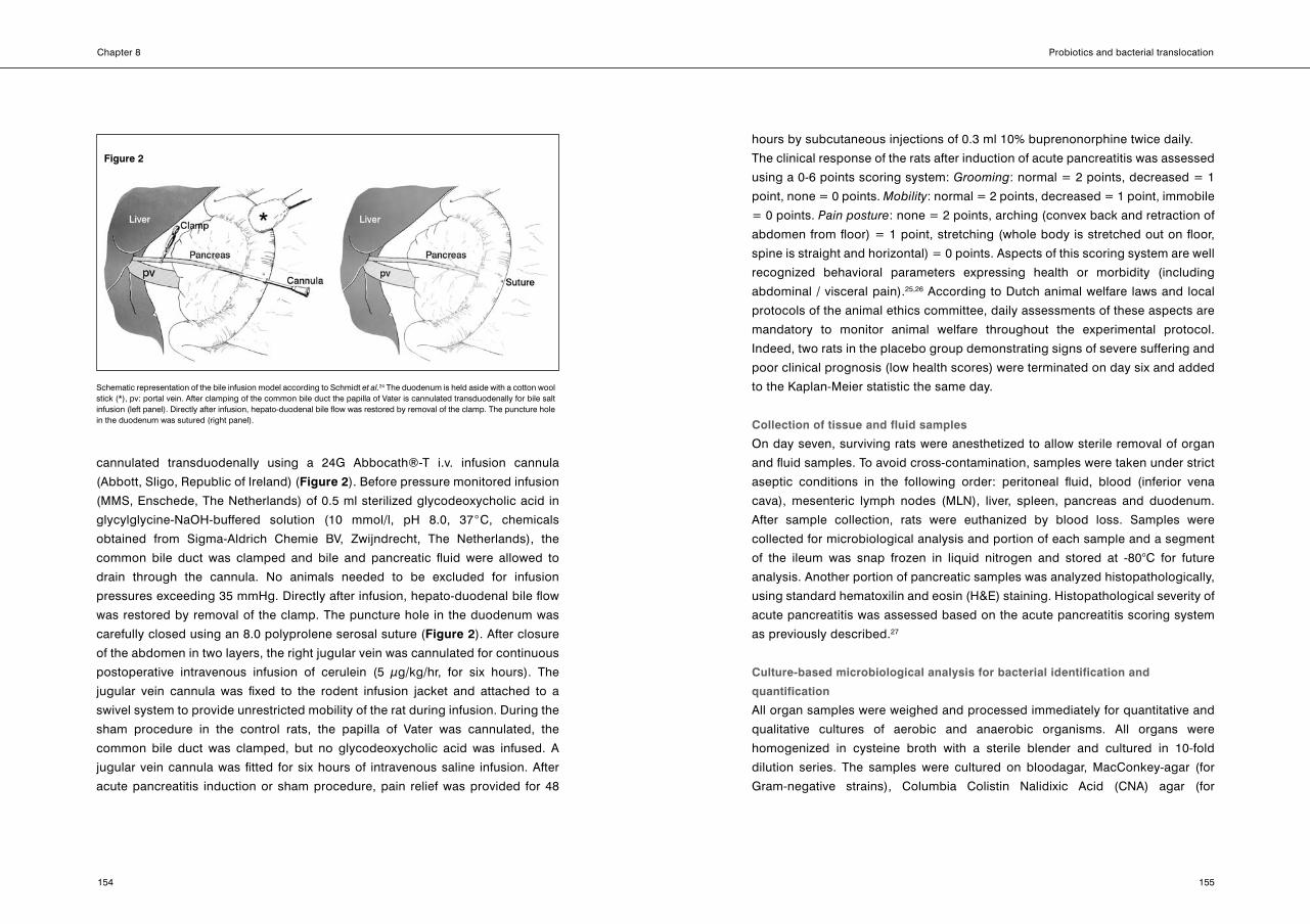
Median health scores after induction of acute pancreatitis, of placebo rats ( ) and rats of the probiotics group (•). Bars represent 25% - 75% interquartile range. Median health scores were improved by probiotics throughout days 1 to 7, with significant differ-ences on days 1, 2 and 3 (* P < 0.05). Health scores of all rats were invariably 6 (maximum score) from day -5 until induction of acute pancreatitis (data not shown). The solid (probiotics) and dashed (placebo) lines are fitted to demonstrate an interpretation of the biphasic course of acute pancreatitis.
Figure 3
Chapter 8
158
Probiotics and bacterial translocation
159
RESULTS
Morbidity and mortality
After the start of daily placebo or probiotic administrations, physical behavior of all
rats remained normal, resulting in maximal health scores from day -5 until day 0.
The clinical response of the rats after induction of experimental pancreatitis
followed a biphasic course. During the first 72 hours, the animals exhibited
decreased grooming or motility and to some extent behavior associated with pain,
despite analgesic administration during the first 48 hours. From days 3 to 5,
surviving animals apparently recovered, evidenced by near to normal physical
behavior. After day 5, rats deteriorated, resulting in a second decrease of health-
scores. Throughout days 1 to 7, median health scores of surviving rats were higher
for rats in the probiotics group compared to those in the placebo group, with
significant contrasts on days 1, 2 and 3 (median = 5 (range 3 – 6) vs. 4 (1 – 6),
P = 0.020; 5 (4 – 6) vs. 4.5 (2 – 6), P = 0.034 and 6 (3 – 6) vs. 3 (1 – 5), P < 0.001,
respectively). An interpretation of the biphasic course of acute pancreatitis is
visualized by the curves superimposed on the median health scores shown in Figure
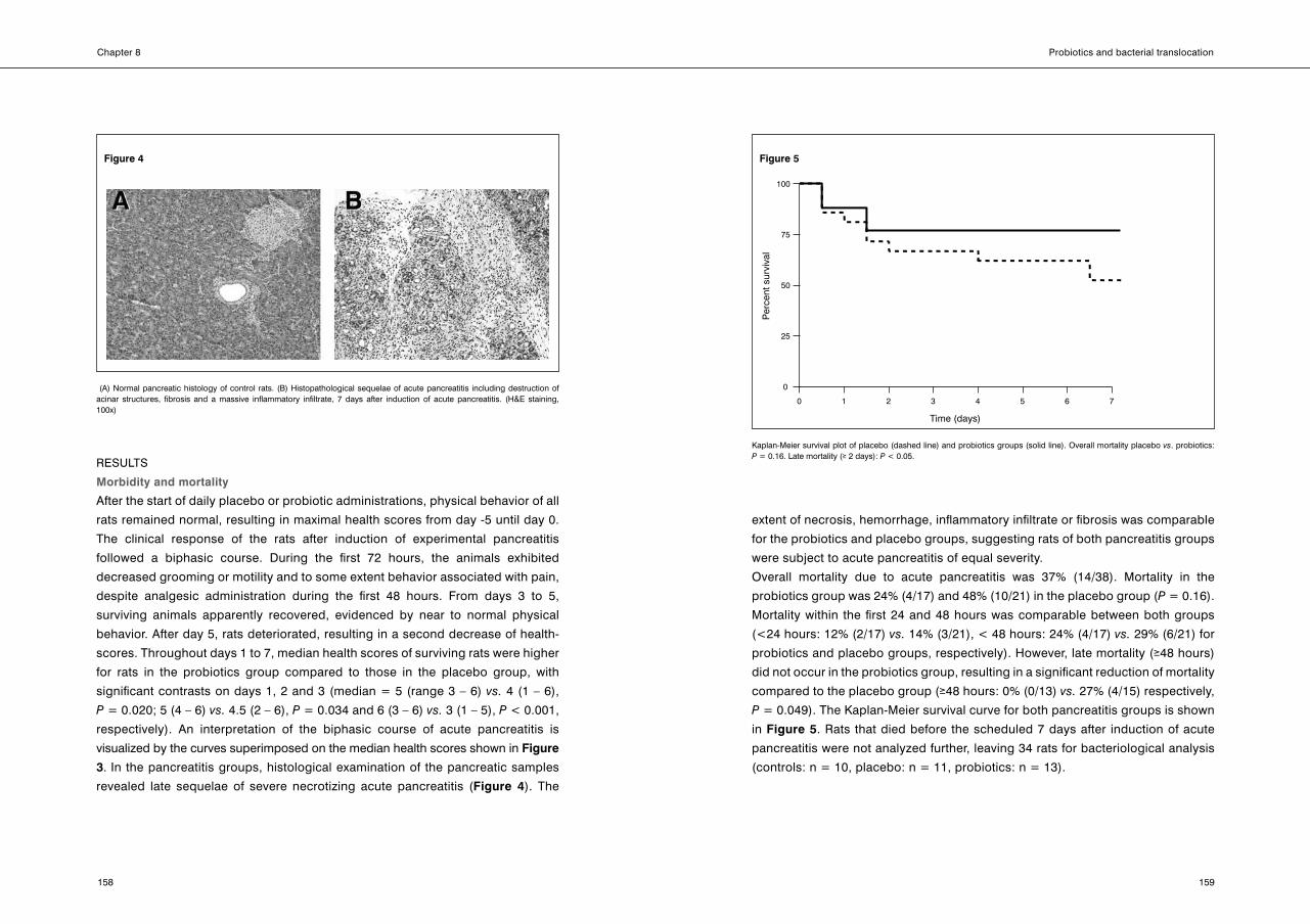
3. In the pancreatitis groups, histological examination of the pancreatic samples
revealed late sequelae of severe necrotizing acute pancreatitis (Figure 4). The
extent of necrosis, hemorrhage, inflammatory infiltrate or fibrosis was comparable
for the probiotics and placebo groups, suggesting rats of both pancreatitis groups
were subject to acute pancreatitis of equal severity.
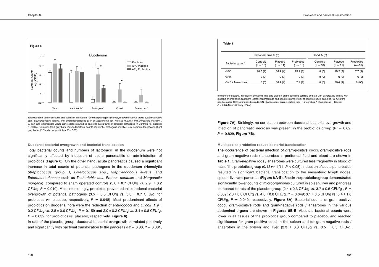
overall mortality due to acute pancreatitis was 37% (14/38). Mortality in the
probiotics group was 24% (4/17) and 48% (10/21) in the placebo group (P = 0.16).
Mortality within the first 24 and 48 hours was comparable between both groups
(<24 hours: 12% (2/17) vs. 14% (3/21), < 48 hours: 24% (4/17) vs. 29% (6/21) for
probiotics and placebo groups, respectively). However, late mortality (≥48 hours)
did not occur in the probiotics group, resulting in a significant reduction of mortality
compared to the placebo group (≥48 hours: 0% (0/13) vs. 27% (4/15) respectively,
P = 0.049). The Kaplan-Meier survival curve for both pancreatitis groups is shown
in Figure 5. Rats that died before the scheduled 7 days after induction of acute
pancreatitis were not analyzed further, leaving 34 rats for bacteriological analysis
(controls: n = 10, placebo: n = 11, probiotics: n = 13).
Figure 4
0 1 2 3 4 5 6 7
0
25
50
75
100
Time (days)
Per
cent
sur
viva
l
Kaplan-Meier survival plot of placebo (dashed line) and probiotics groups (solid line). overall mortality placebo vs. probiotics: P = 0.16. Late mortality (≥ 2 days): P< 0.05.
Figure 5
(A) Normal pancreatic histology of control rats. (B) Histopathological sequelae of acute pancreatitis including destruction of acinar structures, fibrosis and a massive inflammatory infiltrate, 7 days after induction of acute pancreatitis. (H&E staining, 100x)
Chapter 8
160
Probiotics and bacterial translocation
161
Duodenal bacterial overgrowth and bacterial translocation
Total bacterial counts and numbers of lactobacilli in the duodenum were not
significantly affected by induction of acute pancreatitis or administration of
probiotics (Figure 6). on the other hand, acute pancreatitis caused a significant
increase in total counts of potential pathogens in the duodenum (Hemolytic
Streptococcus group B, Enterococcus spp., Staphylococcus aureus, and
Enterobacteriaceae such as Escherichia coli, Proteus mirabilis and Morganella
morganii), compared to sham operated controls (5.0 ± 0.7 CFU/g vs. 2.9 ± 0.2
CFU/g, P = 0.010). Most interestingly, probiotics prevented this duodenal bacterial
overgrowth of potential pathogens (3.5 ± 0.3 CFU/g vs. 5.0 ± 0.7 CFU/g, for
probiotics vs. placebo, respectively, P = 0.048). Most predominant effects of
probiotics on duodenal flora were the reduction of enterococci and E.coli (1.9 ±
0.2 CFU/g vs. 2.8 ± 0.6 CFU/g, P = 0.159 and 2.0 ± 0.2 CFU/g vs. 3.4 ± 0.8 CFU/g,
P = 0.032, for probiotics vs. placebo, respectively. Figure 6).
In rats of the placebo group, duodenal bacterial overgrowth correlated positively
and significantly with bacterial translocation to the pancreas (R2 = 0.80, P = 0.001,
Figure 7A). Strikingly, no correlation between duodenal bacterial overgrowth and
infection of pancreatic necrosis was present in the probiotics group (R2 = 0.02,
P = 0.829, Figure 7B).
Multispecies probiotics reduce bacterial translocation
The occurrence of bacterial infection of gram-positive cocci, gram-positive rods
and gram-negative rods / anaerobes in peritoneal fluid and blood are shown in
Table 1. Gram-negative rods / anaerobes were cultured less frequently in blood of
rats of the probiotics group (0/13 vs. 4/11, P < 0.05). Induction of acute pancreatitis
resulted in significant bacterial translocation to the mesenteric lymph nodes,
spleen, liver and pancreas (Figure 8 A-E). Rats in the probiotics group demonstrated
significantly lower counts of microorganisms cultured in spleen, liver and pancreas
compared to rats of the placebo group (2.4 ± 0.3 CFU/g vs. 3.7 ± 0.5 CFU/g , P=
0.039; 2.8 ± 0.8 CFU/g vs. 4.6 ± 0.8 CFU/g, P= 0.049; 3.1 ± 0.5 CFU/g vs. 5.4 ± 1.0
CFU/g, P = 0.042; respectively. Figure 8A). Bacterial counts of gram-positive
cocci, gram-positive rods and gram-negative rods / anaerobes in the various
abdominal organs are shown in Figures 8B-E. Absolute bacterial counts were
lower in all tissues of the probiotics group compared to placebo, and reached
significance for gram-positive cocci in the spleen and for gram-negative rods /
anaerobes in the spleen and liver (2.3 ± 0.3 CFU/g vs. 3.5 ± 0.5 CFU/g,
Table 1
Peritoneal fluid % (n) Blood % (n)
Bacterial group1 Controls(n = 10)
Placebo(n = 11)
Probiotics(n = 13)
Controls(n = 10)
Placebo(n = 11)
Probiotics(n=13)
GPC 10.0 (1) 36.4 (4) 23.1 (3) 0 (0) 18.2 (2) 7.7 (1)
GPR 0 (0) 0 (0) 0 (0) 0 (0) 0 (0) 0 (0)
GNR+Anaerobes 0 (0) 36.4 (4) 7.7 (1) 0 (0) 36.4 (4) 0 (0*)
*
E. coli
*
†
Total Lactobacilli Pathogens1 Enterococci
3
4
5
6
7
2
Bac
teria
l cou
nts
log
10C
FU/g
ControlsAP / PlaceboAP / Probiotics
Duodenum
Total duodenal bacterial counts and counts of lactobacilli, 1potential pathogens (Hemolytic Streptococcus group B, Enterococcus spp., Staphylococcusaureus, and Enterobacteriaceae such as Escherichiacoli, Proteusmirabilis and Morganellamorganii), E. coli, and enterococci. Acute pancreatitis resulted in bacterial overgrowth of potential pathogens († Controls vs. placebo: P < 0.05). Probiotics (dark grey bars) reduced bacterial counts of potential pathogens, mainly E.coli, compared to placebo ( light grey bars). (* Placebo vs. probiotics: P < 0.05).
Figure 6
Incidence of bacterial infection of peritoneal fluid and blood in sham operated controls and rats with pancreatitis treated with placebo or probiotics. Numbers represent percentage and absolute numbers (n) of positive culture samples. 1GPC: gram-positive cocci, GPR: gram-positive rods, GNR+anaerobes: gram negative rods + anaerobes. * Probiotics vs. Placebo: P < 0.05 (Mann-Whitney U Test)

Chapter 8
162
Probiotics and bacterial translocation
163
R2 = 0.80P < 0.001
R2 = 0.02P < 0.892
A
B
AP/Placebo
AP/Probiotics
3 4 5 6 7 8 9 10
3
4
5
6
7
8
9
10
2
Pan
crea
s ba
cter
ial c
ount
s (lo
g10
CFU
/g)
Pan
crea
s ba
cter
ial c
ount
s (lo
g10
CFU
/g)
3
4
5
6
7
8
9
10
2
3 4 5 6 7 8 9 10 2
2
Duodenum bacterial counts (log10
CFU/g)
Correlation between duodenal bacterial counts and bacterial counts in the pancreas for rats in the placebo (panel A) and probiot-ics (panel B) group. In rats of the placebo group, there is a significant correlation between duodenal and pancreatic bacterial counts (P < 0.001), whereas in the probiotics treated group there is not (P > 0.05).
Figure 7
Controls
AP / Placebo
AP / Probiotics
A Total bacterial counts
†*
†
MLN Spleen Liver Pancreas
3
4
5
6
7
log
10 C
FU/g
2
*†
*†
B
D
Mesenteric lymph nodes
3
4
5
6
7
Liver
GPC GPR GNR+anaer
2
3
4
5
6
7
2
log
10 C
FU/g
log
10 C
FU/g
††
*†
†
C
E
Spleen
3
4
5
6
7
Pancreas
GPC GPR GNR+anaer
3
4
5
6
7
2
2
*†*†
†
(A) Total bacterial counts in mesenteric lymph nodes (MLN), spleen, liver and pancreas in control rats (white bars), placebo rats (light grey bars) and rats of the probiotics group (dark grey bars). (B-E) Bacterial counts of gram-positive cocci (GPC), gram-positive rods (GPR) and gram-negative rods/anaerobes (GNR+anaer) in mesenteric lymph nodes (MLN; B), spleen (C), liver (D) and pancreas (E). * P < 0.05: placebo vs. Probiotics, † P < 0.05 controls vs. placebo.
Figure 8
Chapter 8
164
Probiotics and bacterial translocation
165
P = 0.026; 2.0 ± 0.1 CFU/gvs. 3.0 ± 0.5 CFU/g, P = 0.032 ; 2.0 ± 0.1 CFU/gvs. 3.1
± 0.6 CFU/g, P = 0.022 ,respectively. Figures 8C-D).
Escherichiacoli and Enterococcus spp. were the most predominant bacteria found
in tissues of rats with acute pancreatitis. Administration of probiotics resulted in
significantly reduced bacterial growth of both E. coli and enterococci in the
mesenteric lymph nodes (1.9 ± 0.1 CFU/g vs. 2.6 ± 0.4 CFU/g, P = 0.045; 1.8 ±
0.05 CFU/gvs. 2.5 ± 0.3 CFU/g , P = 0.015, respectively). Pancreatic counts of E.
coli and enterococci were numerically reduced by probiotics, but failed to reach
significant differences (1.7 ± 0.01 CFU/g vs. 3.0 ± 0.7 CFU/g, P = 0.067; 1.7 ± 0.01
CFU/g vs. 3.0 ± 0.7 CFU/g, P = 0.060, respectively).
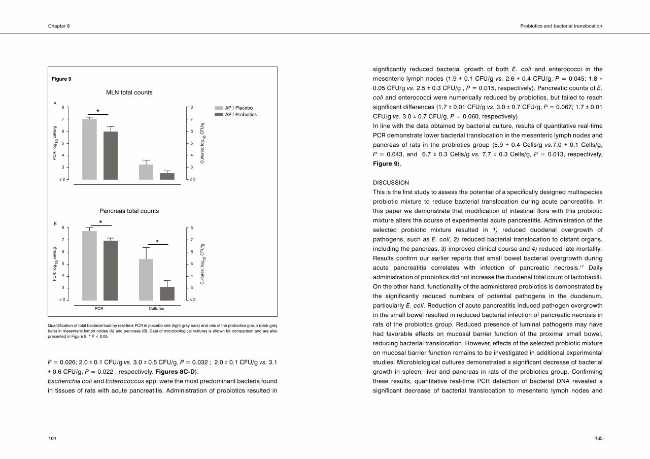
In line with the data obtained by bacterial culture, results of quantitative real-time
PCR demonstrate lower bacterial translocation in the mesenteric lymph nodes and
pancreas of rats in the probiotics group (5.9 ± 0.4 Cells/g vs.7.0 ± 0.1 Cells/g,
P = 0.043, and 6.7 ± 0.3 Cells/g vs. 7.7 ± 0.3 Cells/g, P = 0.013, respectively,
Figure 9).
DISCUSSIoN
This is the first study to assess the potential of a specifically designed multispecies
probiotic mixture to reduce bacterial translocation during acute pancreatitis. In
this paper we demonstrate that modification of intestinal flora with this probiotic
mixture alters the course of experimental acute pancreatitis. Administration of the
selected probiotic mixture resulted in 1) reduced duodenal overgrowth of
pathogens, such as E.coli, 2) reduced bacterial translocation to distant organs,
including the pancreas, 3) improved clinical course and 4) reduced late mortality.
Results confirm our earlier reports that small bowel bacterial overgrowth during
acute pancreatitis correlates with infection of pancreatic necrosis.17 Daily
administration of probiotics did not increase the duodenal total count of lactobacilli.
on the other hand, functionality of the administered probiotics is demonstrated by
the significantly reduced numbers of potential pathogens in the duodenum,
particularly E.coli. Reduction of acute pancreatitis induced pathogen overgrowth
in the small bowel resulted in reduced bacterial infection of pancreatic necrosis in
rats of the probiotics group. Reduced presence of luminal pathogens may have
had favorable effects on mucosal barrier function of the proximal small bowel,
reducing bacterial translocation. However, effects of the selected probiotic mixture
on mucosal barrier function remains to be investigated in additional experimental
studies. Microbiological cultures demonstrated a significant decrease of bacterial
growth in spleen, liver and pancreas in rats of the probiotics group. Confirming
these results, quantitative real-time PCR detection of bacterial DNA revealed a
significant decrease of bacterial translocation to mesenteric lymph nodes and
*
*
*
A
B
PC
R: l
og10
cel
ls/g
PC
R: l
og10
cel
ls/g
Cul
ture
s: lo
g10
CFU
/gC
ultu
res:
log
10 C
FU/g
MLN total counts
3
4
5
6
7
8
≤ 2
3
4
5
6
7
8
≤ 2
3
4
5
6
7
8
≤ 2
3
4
5
6
7
8
≤ 2
Pancreas total counts
PCR Cultures
AP / PlaceboAP / Probiotics
Quantification of total bacterial load by real-time PCR in placebo rats (light grey bars) and rats of the probiotics group (dark grey bars) in mesenteric lymph nodes (A) and pancreas (B). Data of microbiological cultures is shown for comparison and are also presented in Figure 8. * P < 0.05
Figure 9
Chapter 8
166
Probiotics and bacterial translocation
167
pancreas. Bacterial DNA is stable, and PCR based methods are highly sensitive
and specific to detect minimal amounts of bacterial DNA in serum of patients with
acute pancreatitis.30 This method detects not only viable, but also non-viable
translocated bacteria, probably killed by the host immune system. Therefore, total
bacterial load estimated by real-time PCR is higher than counts of viable
microorganisms only. Moreover, average reduction of pancreatic bacterial load by
probiotic prophylaxis was greater when analyzed by culture (> 2 Log) then by
PCR (< 1 Log). Thus, reduction of viable bacteria cultured from pancreatic necrosis
cannot be completely explained by an absolute reduction of bacterial translocation,
as indicated by quantitative real-time PCR. It could be suggested that probiotic
prophylaxis renders the immune system more capable to kill translocated bacteria
in distant organs. In follow-up studies we are currently addressing effects of enteral
probiotics on a wide panel of plasma cytokines to assess immune modulatory
potential in the early and late phase of experimental acute pancreatitis.
Rats in the probiotics group showed less stress- or pain-associated behavior,
demonstrated by objective improvement in the clinical course of experimental
pancreatitis. Albeit a biphasic course in clinical presentation could still be identified
in rats of the probiotics group, health scores were clearly improved compared to
placebo rats. Moreover, probiotic prophylaxis numerically reduced overall mortality
of acute pancreatitis, and a significant reduction was observed in late phase
mortality. In humans, infectious complications are held accountable for late phase
mortality.2,5 In line with these reports, reduced infectious complications in probiotic
treated rats were associated with reduced late phase mortality in the present
experiment. Probiotics did not affect histological severity, assessed seven days
after induction of acute pancreatitis. Early phase histological changes were not
assessed.
The experimental design of the present study aimed to assess the effect of gut
flora modulation by probiotics on the course of experimental acute pancreatitis,
using bacterial translocation as a major outcome parameter. Therefore, rats were
pretreated with the selected probiotics or placebo. In experimental acute
pancreatitis, timing of the start of treatment remains a challenging issue. The
course of acute pancreatitis in rats is approximately 3 to 6 times faster than in
man.31-33 This leaves only a small treatment-window between onset of disease and
occurrence of complications, potentially leading to false negative results if
treatment is started after induction of pancreatitis. For this reason treatment started
before induction of acute pancreatitis in many other studies.32,34,35 Also, for
probiotics in particular, assessment of their efficacy by pretreatment is an accepted
experimental method to provide proof of principle.36 We emphasize that results of
the present experiment do not necessarily reflect potential results if treatment is
started after the onset of acute pancreatitis in general, and potential clinical
success or validity in particular.
For a prophylactic strategy to be effective, it should intervene with the
pathophysiology of bacterial translocation during acute pancreatitis as early as
possible. The exact pathophysiology of bacterial translocation, infection of
pancreatic necrosis and the ensuing systemic effects is still not fully understood.
Yet, the sequence of some major pathophysiological aspects has been clarified.
Early after the onset of acute pancreatitis, neurohormonal effects result in reduced
small bowel motility.17 This causes stasis of luminal contents and small bowel
bacterial overgrowth with potential pathogens, including E.coliand Enterococcus
species. The abundant presence of luminal pathogens forms a challenge for the
mucosal barrier. Furthermore, pancreatitis associated reduced intestinal blood
flow results in mucosal ischemia and reperfusion damage.37-39 Luminal bacteria,
normally held at bay by the mucosal barrier, now have opportunity to penetrate
into the intestinal epithelium. Local intestinal inflammation follows, further
compromising mucosal barrier function. Pancreatitis and ensuing intestinal
inflammation both contribute to a systemic pro-inflammatory response (Systemic
Inflammatory Response Syndrome, SIRS), with damaging effects on distant
organs.40,41 If the systemic response is severe, multiple organ dysfunction syndrome
(MoDS) might follow.42,43 If the patient survives the early phase, counter regulatory
immunological pathways releasing anti-inflammatory cytokines result in a refractory
state characterised by immunosuppression.44,45 Persistent immunosuppression
will render the patient liable for infection of pancreatic necrosis. MoDS caused by
infectious complications is considered accountable for so-called late mortality or
“late septic death”.44,46
With this pathophysiology of local and systemic events during severe acute
pancreatitis in mind, six probiotic strains were selected for this study. Selection of
strains was based on their invitro antibacterial and immunomodulatory properties.
(Timmerman HM, etal., manuscript in preparation) Lactobacillusacidophilusand
Lactobacillussalivariuswere selected for their ability to suppress growth of E.coli
and enterococci. Bifidobacteriuminfantis also demonstrated antimicrobial effects.
Chapter 8
168
Probiotics and bacterial translocation
169
Lactococcuslactisand Bifidobacteriumbifidumdemonstrated immune-modulating
properties, including decreasing pro-inflammatory and increasing anti-inflammatory
immune-responses. Finally, Lactobacilluscasei demonstrated both antimicrobial
and immune-modulating properties.
In thorough reviews on the use of animal models of acute pancreatitis, the used
model was preferred to examine pathophysiology of bacterial translocation and
for testing treatment strategies.31 Resemblance to human acute pancreatitis with
regard to bacteriological results, reaction to treatment, and disease course are the
major advantages.24,31 The transduodenal approach to the biliopancreatic duct for
bile salt infusion is often suggested to be a major drawback of the model for its
potential to introduce bacteria in pancreatic tissue. However, results of the control
group in the present study once more confirm that transduodenal cannulation of
the biliopancreatic duct does not result in bacterial contamination of any concern
to study outcome. Because of its demonstrated value, the model has previously
been applied in many experiments testing the value of antibiotics during acute
pancreatitis.32,33,47,48
Experimentally, prophylactic antibiotics reduced overgrowth of E. coli and
enterococci in the small bowel, resulted in significantly reduced bacterial
translocation to distant organs, including the pancreas, and reduced mortality.4,32
Unfortunately, clinical results of prophylactic antibiotics were not as successful . In
a recent placebo-controlled double blinded clinical trial, Isenmann et al.
demonstrated that prophylactic antibiotics (ciprofloxacin/metronidazole) showed
no effect on bacterial infection of pancreatic necrosis or clinical outcome.49
A recent meta-analysis confirmed these findings.6 Furthermore, concerns on
prophylactic use of broad-spectrum antibiotics have been expressed, including
increased incidence of nosocomial infections with resistant bacteria or fungi.7-9,50-55
In this context, specifically selected multispecies probiotics as presented in this
study, may be a novel and potentially effective alternative. However, as was
demonstrated by the contrast between experimental and clinical results of
antibiotics in acute pancreatitis, the clinical value of specifically selected
multispecies probiotics remains to be proven. For this reason, the Dutch Acute
Pancreatitis Study Group embarked on a randomized double blind placebo-
controlled multicenter trial on prophylactic multispecies probiotics in patients with
predicted severe acute pancreatitis.56
In summary; modification of intestinal flora with multispecies probiotics, especially
designed to address pathophysiology of bacterial translocation, resulted in
reduced small bowel bacterial overgrowth, bacterial translocation to distant organs
and associated morbidity and late mortality in experimental acute pancreatitis.
ACKNoWLEDGEMENTS
The authors thank W. Renooij, PhD, of the Department of Surgery, University
Medical Center Utrecht, for creating Figure 2.
Chapter 8
170
Probiotics and bacterial translocation
171
References
1. Buchler MW, Gloor B, Muller CA, Friess H, Seiler CA, Uhl W. Acute necrotizing
pancreatitis: treatment strategy according to the status of infection. Ann Surg 2000;
232(5):619-626.
2. Widdison AL, Karanjia ND. Pancreatic infection complicating acute pancreatitis.
Br J Surg 1993; 80(2):148-154.
3. Isenmann R, Rau B, Zoellner U, Beger HG. Management of patients with extended
pancreatic necrosis. Pancreatology 2001; 1(1):63-68.
4. Beger HG, Rau B, Isenmann R, Schwarz M, Gansauge F, Poch B. Antibiotic prophylaxis
in severe acute pancreatitis. Pancreatology 2005; 5(1):10-19.
5. Beger HG, Rau B, Mayer J, Pralle U. Natural course of acute pancreatitis. World J Surg
1997; 21(2):130-135.
6. Mazaki T, Ishii Y, Takayama T. Meta-analysis of prophylactic antibiotic use in acute
necrotizing pancreatitis. Br J Surg 2006;93:674-684.
7. Hoerauf A, Hammer S, Muller-Myhsok B, Rupprecht H. Intra-abdominal Candida
infection during acute necrotizing pancreatitis has a high prevalence and is associated
with increased mortality. Crit Care Med 1998; 26(12):2010-2015.
8. Grewe M, Tsiotos GG, Luque de-Leon E, Sarr MG. Fungal infection in acute necrotizing
pancreatitis. J Am Coll Surg 1999; 188(4):408-414.
9. Isenmann R, Schwarz M, Rau B, Trautmann M, Schober W, Beger HG. Characteristics of
infection with Candida species in patients with necrotizing pancreatitis. World J Surg
2002; 26(3):372-376.
10. De Waele JJ, Vogelaers D, Blot S, Colardyn F. Fungal infections in patients with severe
acute pancreatitis and the use of prophylactic therapy. Clin Infect Dis 2003; 37(2):208-
213.
11. De Waele JJ, Vogelaers D, Hoste E, Blot S, Colardyn F. Emergence of antibiotic
resistance in infected pancreatic necrosis. Arch Surg 2004; 139(12):1371-1375.
12. olah A, Belagyi T, Issekutz A, Gamal ME, Bengmark S. Randomized clinical trial of
specific lactobacillus and fibre supplement to early enteral nutrition in patients with
acute pancreatitis. Br J Surg 2002; 89(9):1103-1107.
13. Besselink MG, Timmerman HM, van Minnen LP, Akkermans LM, Gooszen HG.
Prevention of Infectious Complications in Surgical Patients: Potential Role of Probiotics.
Dig Surg 2005; 22(4):234-244.
14. Mangiante G, Colucci G, Canepari P, Bassi C, Nicoli N, Casaril A et al. Lactobacillus
plantarum reduces infection of pancreatic necrosis in experimental acute pancreatitis.
Dig Surg 2001; 18(1):47-50.
15. Timmerman HM, Koning CJ, Mulder L, Rombouts FM, Beynen AC. Monostrain,
multistrain and multispecies probiotics--A comparison of functionality and efficacy. Int J
Food Microbiol 2004; 96(3):219-233.
16. Isolauri E, Rautava S, Kalliomaki M, Kirjavainen P, Salminen S. Role of probiotics in food
hypersensitivity. Curr opin Allergy Clin Immunol 2002; 2(3):263-271.
17. Van Felius ID, Akkermans LM, Bosscha K, Verheem A, Harmsen W, Visser MR et al.
Interdigestive small bowel motility and duodenal bacterial overgrowth in experimental
acute pancreatitis. Neurogastroenterol Motil 2003; 15(3):267-276.
18. Gautreaux MD, Deitch EA, Berg RD. T lymphocytes in host defense against bacterial
translocation from the gastrointestinal tract. Infect Immun
1994; 62(7):2874-2884.
19. Berg RD. Bacterial translocation from the gastrointestinal tract. Trends Microbiol
1995; 3(4):149-154.
20. Berg RD. Bacterial translocation from the gastrointestinal tract. Adv Exp Med Biol 1999;
473:11-30.
Chapter 8
172
Probiotics and bacterial translocation
173
21. Ammori BJ, Leeder PC, King RF, Barclay GR, Martin IG, Larvin M et al. Early increase in
intestinal permeability in patients with severe acute pancreatitis: correlation with
endotoxemia, organ failure, and mortality. J Gastrointest Surg 1999; 3(3):252-262.
22. Gloor B, Todd KE, Lane JS, Rigberg DA, Reber HA. Mechanism of increased lung injury
after acute pancreatitis in IL-10 knockout mice. J Surg Res 1998; 80(1):110-114.
23. Van Laethem JL, Eskinazi R, Louis H, Rickaert F, Robberecht P, Deviere J. Multisystemic
production of interleukin 10 limits the severity of acute pancreatitis in mice. Gut 1998;
43(3):408-413.
24. Schmidt J, Rattner DW, Lewandrowski K, Compton CC, Mandavilli U, Knoefel WT et al. A
better model of acute pancreatitis for evaluating therapy. Ann Surg 1992; 215(1):44-56.
25. Stam R, van Laar TJ, Wiegant VM. Physiological and behavioural responses to
duodenal pain in freely moving rats. Physiol Behav 2004; 81(1):163-169.
26. Houghton AK, Kadura S, Westlund KN. Dorsal column lesions reverse the reduction of
homecage activity in rats with pancreatitis. Neuroreport 1997;8:3795-3800.
27. van Minnen LP, Venneman NG, van Dijk JE, Verheem A, Gooszen HG, Akkermans LM,
van Erpecum KJ. Cholesterol crystals enhance and phospholipids protect against
pancreatitis induced by hydrophobic bile salts: a rat model study. Pancreas
2006;32:369-375.
28. Konstantinov SR, Awati A, Smidt H, Williams BA, Akkermans AD, de Vos WM. Specific
response of a novel and abundant Lactobacillus amylovorus-like phylotype to dietary
prebiotics in the guts of weaning piglets. Appl Environ Microbiol
2004; 70(7):3821-3830.
29. Nubel U, Engelen B, Felske A, Snaidr J, Wieshuber A, Amann RI et al. Sequence
heterogeneities of genes encoding 16S rRNAs in Paenibacillus polymyxa detected by
temperature gradient gel electrophoresis. J Bacteriol 1996; 178(19):5636-5643.
30. de Madaria E, Martinez J, Lozano B, Sempere L, Benlloch S, Such J et al. Detection
and identification of bacterial DNA in serum from patients with acute pancreatitis. Gut
2005; 54(9):1293-1297.
31. Foitzik T, Hotz HG, Eibl G, Buhr HJ. Experimental models of acute pancreatitis: are they
suitable for evaluating therapy? Int J Colorectal Dis 2000; 15(3):127-135.
32. Mithofer K, Fernandez-Del Castillo C, Ferraro MJ, Lewandrowski K, Rattner DW,
Warshaw AL. Antibiotic treatment improves survival in experimental acute necrotizing
pancreatitis. Gastroenterology 1996; 110(1):232-240.
33. Schwarz M, Thomsen J, Meyer H, Buchler MW, Beger HG. Frequency and time course
of pancreatic and extrapancreatic bacterial infection in experimental acute pancreatitis
in rats. Surgery 2000; 127(4):427-432.
34. Lange JF, van Gool J, Tytgat GN. The protective effect of a reduction in intestinal flora
on mortality of acute haemorrhagic pancreatitis in the rat Hepatogastroenterology
1987; 34(1):28-30.
35. Park SJ, Seo SW, Choi oS, Park CS. Alpha-lipoic acid protects against cholecystokinin-
induced acute pancreatitis in rats. World J Gastroenterol 2005; 11(31):4883-4885.
36. Zareie M, Johnson-Henry KC, Jury J, Yang PC, Ngan BY, McKay DM et al. Probiotics
prevent bacterial translocation and improve intestinal barrier function in rats following
chronic psychological stress. Gut 2006; Apr 25; [Epub ahead of print] .
37. Inoue K, Hirota M, Kimura Y, Kuwata K, ohmuraya M, ogawa M. Further evidence for
endothelin as an important mediator of pancreatic and intestinal ischemia in severe
acute pancreatitis. Pancreas 2003; 26(3):218-223.
38. Yasuda T, Takeyama Y, Ueda T, Hori Y, Nishikawa J, Kuroda Y. Nonocclusive visceral
ischemia associated with severe acute pancreatitis. Pancreas 2003; 26(1):95-97.
Chapter 8
174
Probiotics and bacterial translocation
175
39. Rahman SH, Ammori BJ, Holmfield J, Larvin M, McMahon MJ. Intestinal hypoperfusion
contributes to gut barrier failure in severe acute pancreatitis. J Gastrointest Surg 2003;
7(1):26-35.
40. McKay CJ, Imrie CW. The continuing challenge of early mortality in acute pancreatitis.
Br J Surg 2004; 91(10):1243-1244.
41. Tran DD, Cuesta MA, Schneider AJ, Wesdorp RI. Prevalence and prediction of multiple
organ system failure and mortality in acute pancreatitis. J Crit Care 1993; 8(3):145-153.
42. Bhatia M, Wong FL, Cao Y, Lau HY, Huang J, Puneet P et al. Pathophysiology of acute
pancreatitis. Pancreatology 2005; 5(2-3):132-144.
43. Bhatia M. Inflammatory response on the pancreatic acinar cell injury. Scand J Surg
2005; 94(2):97-102.
44. Gloor B, Muller CA, Worni M, Martignoni ME, Uhl W, Buchler MW. Late mortality in
patients with severe acute pancreatitis. Br J Surg 2001; 88(7):975-979.
45. Dugernier TL, Laterre PF, Wittebole x, Roeseler J, Latinne D, Reynaert MS et al.
Compartmentalization of the inflammatory response during acute pancreatitis:
correlation with local and systemic complications. Am J Respir Crit Care Med 2003;
168(2):148-157.
46. Wilson PG, Manji M, Neoptolemos JP. Acute pancreatitis as a model of sepsis. J
Antimicrob Chemother 1998; 41 Suppl A:51-63.
47. Foitzik T, Fernandez-Del Castillo C, Ferraro MJ, Mithofer K, Rattner DW, Warshaw AL.
Pathogenesis and prevention of early pancreatic infection in experimental acute
necrotizing pancreatitis. Ann Surg 1995; 222(2):179-185.
48. Gloor B, Worni M, Strobel o, Uhl W, Tcholakov o, Muller CA et al. Cefepime tissue
penetration in experimental acute pancreatitis. Pancreas 2003; 26(2):117-121.
49. Isenmann R, Runzi M, Kron M, Kahl S, Kraus D, Jung N et al. Prophylactic antibiotic
treatment in patients with predicted severe acute pancreatitis: a placebo-controlled,
double-blind trial. Gastroenterology 2004; 126(4):997-1004.
50. Clark NM, Patterson J, Lynch JP, III. Antimicrobial resistance among gram-negative
organisms in the intensive care unit. Curr opin Crit Care 2003; 9(5):413-423.
51. Sorberg M, Farra A, Ransjo U, Gardlund B, Rylander M, Settergren B et al. Different
trends in antibiotic resistance rates at a university teaching hospital. Clin Microbiol
Infect 2003; 9(5):388-396.
52. Murray BE. Vancomycin-resistant enterococcal infections. N Engl J Med 2000;
342(10):710-721.
53. Leavis HL, Willems RJ, Mascini EM, Vandenbroucke-Grauls CM, Bonten MJ.
[Vancomycin resistant enterococci in the Netherlands] Vancomycineresistente
enterokokken in Nederland. Ned Tijdschr Geneeskd 2004; 148(18):878-882.
54. Albrich WC, Angstwurm M, Bader L, Gartner R. Drug resistance in intensive care units.
Infection 1999; 27 Suppl 2:S19-S23.
55. Berrouane YF, Herwaldt LA, Pfaller MA. Trends in antifungal use and epidemiology of
nosocomial yeast infections in a university hospital. J Clin Microbiol 1999; 37(3):531-
537.
56. Besselink MGH, Timmerman HM, Buskens E, Nieuwenhuijs VB, Akkermans LMA,
Gooszen HG: Probiotic prophylaxis in patients with predicted severe acute pancreatitis
(PRoPATRIA): design and rationale of a blinded, placebo- controlled randomised
controlled trial. Dutch Acute Pancreatitis Study Group. BMC Surg 2004 Sep 29;4:12.
174


![ID# 08- 001 Sex: Male Breed: CD(SD) IGS [Sprague-Dawley] Investigator: Davis](https://img.dokumen.tips/doc/110x75/568150b7550346895dbed20a/id-08-001-sex-male-breed-cdsd-igs-sprague-dawley-investigator-davis.jpg)


