Embed Size (px)
Citation preview

Chapter 8Chapter 8
The Pelvis and ThighThe Pelvis and Thigh

IntroductionIntroduction
Pelvic girdle forms structural base of Pelvic girdle forms structural base of support between lower extremity and trunksupport between lower extremity and trunk
Hip articulation – strongest and most Hip articulation – strongest and most stable joint in the bodystable joint in the body– This benefit gained at the expense of ROMThis benefit gained at the expense of ROM
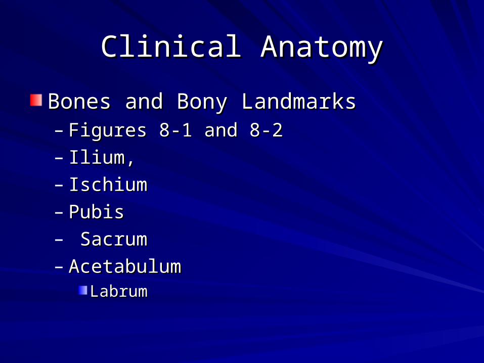
Clinical AnatomyClinical Anatomy
Bones and Bony LandmarksBones and Bony Landmarks– Figures 8-1 and 8-2Figures 8-1 and 8-2– Ilium, Ilium, – IschiumIschium– PubisPubis– SacrumSacrum– AcetabulumAcetabulum
LabrumLabrum
Clinical AnatomyClinical Anatomy
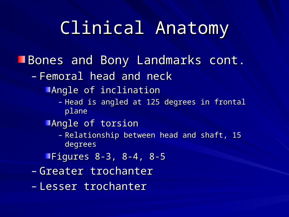
Bones and Bony Landmarks cont.Bones and Bony Landmarks cont.– Femoral head and neckFemoral head and neck
Angle of inclinationAngle of inclination– Head is angled at 125 degrees in frontal planeHead is angled at 125 degrees in frontal plane
Angle of torsionAngle of torsion– Relationship between head and shaft, 15 degreesRelationship between head and shaft, 15 degrees
Figures 8-3, 8-4, 8-5Figures 8-3, 8-4, 8-5
– Greater trochanterGreater trochanter– Lesser trochanterLesser trochanter

Clinical AnatomyClinical Anatomy
Articulations and Ligamentous SupportArticulations and Ligamentous Support– Pubic symphysisPubic symphysis
Fibrocartilaginous interpubic discFibrocartilaginous interpubic disc
Small degree of spreading, compression and Small degree of spreading, compression and rotation between halves of girdlerotation between halves of girdle
– Sacroiliac joint (SI joint)Sacroiliac joint (SI joint)Very study, limited ROMVery study, limited ROM
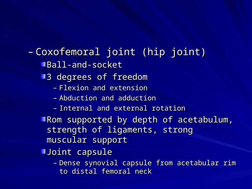
– Coxofemoral joint (hip joint)Coxofemoral joint (hip joint)Ball-and-socketBall-and-socket
3 degrees of freedom3 degrees of freedom– Flexion and extensionFlexion and extension– Abduction and adductionAbduction and adduction– Internal and external rotationInternal and external rotation
Rom supported by depth of acetabulum, strength Rom supported by depth of acetabulum, strength of ligaments, strong muscular supportof ligaments, strong muscular support
Joint capsuleJoint capsule– Dense synovial capsule from acetabular rim to distal Dense synovial capsule from acetabular rim to distal
femoral neckfemoral neck
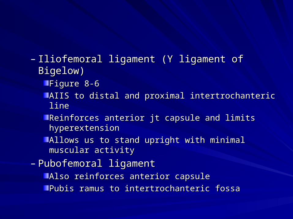
– Iliofemoral ligament (Y ligament of Bigelow)Iliofemoral ligament (Y ligament of Bigelow)Figure 8-6Figure 8-6
AIIS to distal and proximal intertrochanteric lineAIIS to distal and proximal intertrochanteric line
Reinforces anterior jt capsule and limits Reinforces anterior jt capsule and limits hyperextensionhyperextension
Allows us to stand upright with minimal muscular Allows us to stand upright with minimal muscular activityactivity
– Pubofemoral ligamentPubofemoral ligamentAlso reinforces anterior capsuleAlso reinforces anterior capsule
Pubis ramus to intertrochanteric fossaPubis ramus to intertrochanteric fossa

– Ligamentum teres (ligament of the head of the Ligamentum teres (ligament of the head of the femur)femur)
Conduit for medial and lateral circumflex arteriesConduit for medial and lateral circumflex arteries
Little function in stabilizing hipLittle function in stabilizing hip
Figure 8-7Figure 8-7
– Inguinal ligamentInguinal ligamentASIS to pubic symphysisASIS to pubic symphysis
Serves to contain soft tissues as they course Serves to contain soft tissues as they course anteriorly from trunk to lower extremityanteriorly from trunk to lower extremity
Superior border of femoral triangle Superior border of femoral triangle

Muscular AnatomyMuscular Anatomy
Table 8-1, pages 276-277Table 8-1, pages 276-277
Anterior MusculatureAnterior Musculature– QuadricepsQuadriceps– Iliopsoas groupIliopsoas group
Psoas major, psoas minor, iliacusPsoas major, psoas minor, iliacus
Primary hip flexors when knee extendedPrimary hip flexors when knee extended
Figure 8-8Figure 8-8
Medial MusculatureMedial Musculature– Adductor groupAdductor group– GracilisGracilis– Figure 8-9Figure 8-9

Lateral MusculatureLateral Musculature– Gluteus mediusGluteus medius– Tensor fascia lataeTensor fascia latae– Figure 8-10Figure 8-10– Trendelenburg’s gait patternTrendelenburg’s gait pattern– Intrinsic muscles form cuff around femoral Intrinsic muscles form cuff around femoral
head and externally rotate hiphead and externally rotate hipPiriformis, quadratus femoris, obturator internus, Piriformis, quadratus femoris, obturator internus, obturator externus, gemellus superior, gemellus obturator externus, gemellus superior, gemellus inferiorinferior
Figure 8-11Figure 8-11

Posterior MusculaturePosterior Musculature– Gluteus maximusGluteus maximus– hamstringshamstrings
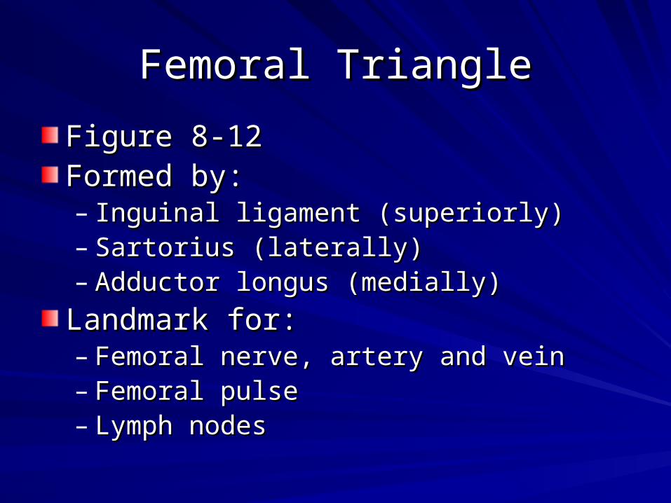
Femoral TriangleFemoral Triangle
Figure 8-12Figure 8-12Formed by:Formed by:– Inguinal ligament (superiorly)Inguinal ligament (superiorly)– Sartorius (laterally)Sartorius (laterally)– Adductor longus (medially)Adductor longus (medially)
Landmark for:Landmark for:– Femoral nerve, artery and veinFemoral nerve, artery and vein– Femoral pulseFemoral pulse– Lymph nodesLymph nodes

BursaeBursae
3 bursa to decrease friction between 3 bursa to decrease friction between gluteus maximus and adjacent bony gluteus maximus and adjacent bony structuresstructures– Trochanteric bursaTrochanteric bursa
Gluteus max – greater trochanterGluteus max – greater trochanter
– Gluteofemoral bursaGluteofemoral bursaGluteus max – vastus lateralisGluteus max – vastus lateralis
– Ischial bursaIschial bursaGluteus max – ischial tuberosityGluteus max – ischial tuberosity

Clinical Evaluation of Pelvis and Clinical Evaluation of Pelvis and ThighThigh
May necessitate evaluation of lower May necessitate evaluation of lower extremity, spinal column, and postureextremity, spinal column, and posture
Patient preparednessPatient preparedness
Clinician preparednessClinician preparedness
Gender issuesGender issues
Evaluation MapEvaluation Map– Page 280Page 280

HistoryHistory
Location of symptomsLocation of symptoms– Table 8-2, page 281Table 8-2, page 281
OnsetOnset
Training techniquesTraining techniques
Mechanism of injuryMechanism of injury
Prior medical conditionsPrior medical conditions– Legg-Calve-Perthes DiseaseLegg-Calve-Perthes Disease– Slipped capital femoral epiphysisSlipped capital femoral epiphysis

InspectionInspection
Most trauma to area cannot be visualizedMost trauma to area cannot be visualizedInspection of Hip AngulationsInspection of Hip Angulations– Angle of inclinationAngle of inclination
Relationship of femoral head and shaftRelationship of femoral head and shaftCoxa valgaCoxa valga
– Increase in angle, may lead to genu varum or lateral Increase in angle, may lead to genu varum or lateral patellapatella
Coxa varaCoxa vara– Decrease in angle, may lead to genu valgum or squinting Decrease in angle, may lead to genu valgum or squinting
patellapatella
Mechanical advantage of glut medius is reducedMechanical advantage of glut medius is reduced
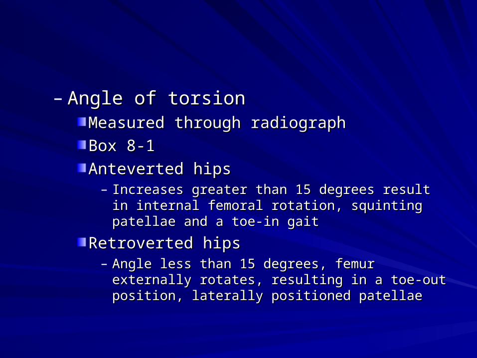
– Angle of torsionAngle of torsionMeasured through radiographMeasured through radiograph
Box 8-1Box 8-1
Anteverted hipsAnteverted hips– Increases greater than 15 degrees result in internal Increases greater than 15 degrees result in internal
femoral rotation, squinting patellae and a toe-in gaitfemoral rotation, squinting patellae and a toe-in gait
Retroverted hipsRetroverted hips– Angle less than 15 degrees, femur externally rotates, Angle less than 15 degrees, femur externally rotates,
resulting in a toe-out position, laterally positioned resulting in a toe-out position, laterally positioned patellaepatellae

Inspection of Medial StructuresInspection of Medial Structures– Adductor groupAdductor group
Inspection of Anterior StructuresInspection of Anterior Structures– Hip flexorsHip flexors
Inspection of Lateral StructuresInspection of Lateral Structures– Iliac crest (figure 8-13)Iliac crest (figure 8-13)– Nelaton’s lineNelaton’s line
ASIS to ischial tuberosityASIS to ischial tuberosityFigure 8-14Figure 8-14
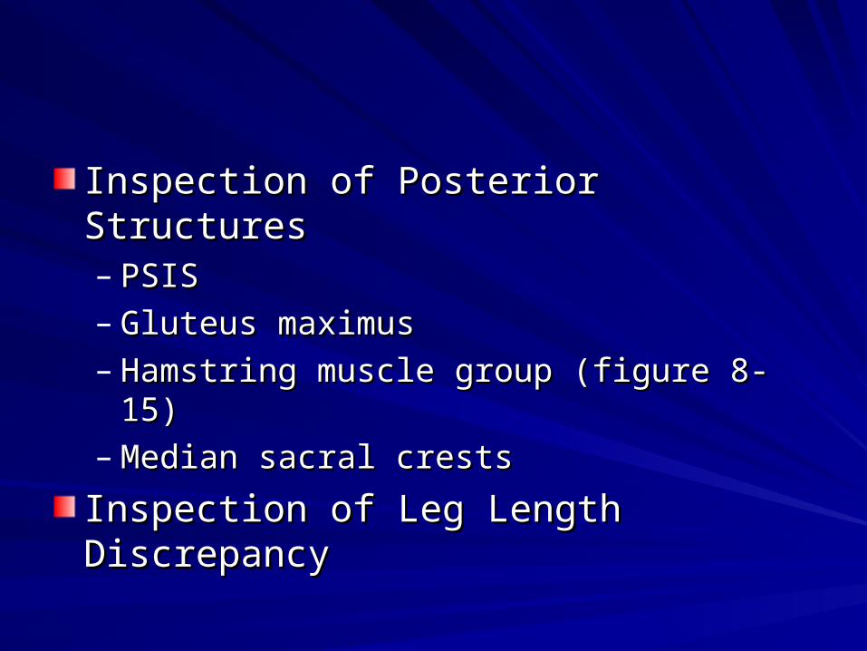
Inspection of Posterior StructuresInspection of Posterior Structures– PSISPSIS– Gluteus maximusGluteus maximus– Hamstring muscle group (figure 8-15)Hamstring muscle group (figure 8-15)– Median sacral crestsMedian sacral crests
Inspection of Leg Length DiscrepancyInspection of Leg Length Discrepancy

PalpationPalpation
Refer to list of Clinical ProficienciesRefer to list of Clinical Proficiencies
Utilize pages 283 - 285Utilize pages 283 - 285

Range of Motion TestingRange of Motion Testing
Limited by bony and soft tissue restraintsLimited by bony and soft tissue restraints
Position of kneePosition of knee– Flexed vs. extendedFlexed vs. extended
Table 8-3, page 286 (Muscle actions)Table 8-3, page 286 (Muscle actions)
Box 8-2, page 287 (Goniometry)Box 8-2, page 287 (Goniometry)

Active Range of MotionActive Range of Motion
Flexion and ExtensionFlexion and Extension– Figure 8-17Figure 8-17– 130-150 degrees (range, knee flexed)130-150 degrees (range, knee flexed)– Majority occurs during flexionMajority occurs during flexion– Extending knee limits hip flexionExtending knee limits hip flexion
Adduction and AbductionAdduction and Abduction– Figure 8-18Figure 8-18– Abduction – 45 degreesAbduction – 45 degrees– Adduction – 20-30 degrees Adduction – 20-30 degrees

Active Range of MotionActive Range of Motion
Internal and External RotationInternal and External Rotation– Figure 8-19Figure 8-19– ER – 40-50 degreesER – 40-50 degrees– IR – 45 degreesIR – 45 degrees– Hip flexed vs. extendedHip flexed vs. extended
Passive Range of MotionPassive Range of Motion
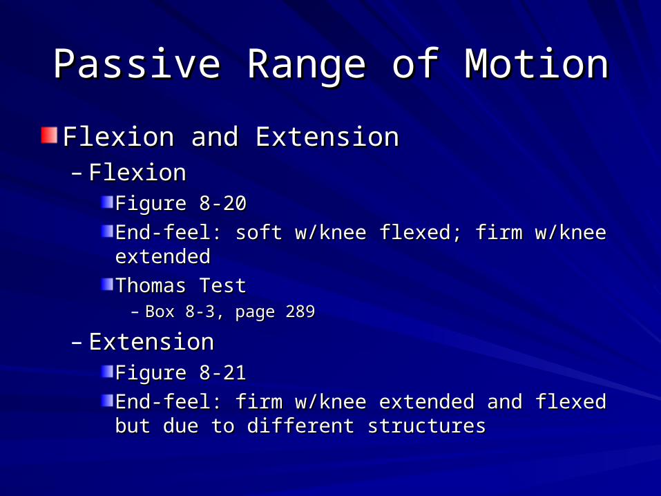
Flexion and ExtensionFlexion and Extension– FlexionFlexion
Figure 8-20Figure 8-20
End-feel: soft w/knee flexed; firm w/knee extendedEnd-feel: soft w/knee flexed; firm w/knee extended
Thomas TestThomas Test– Box 8-3, page 289Box 8-3, page 289
– ExtensionExtensionFigure 8-21Figure 8-21
End-feel: firm w/knee extended and flexed but due End-feel: firm w/knee extended and flexed but due to different structuresto different structures
Passive Range of MotionPassive Range of Motion
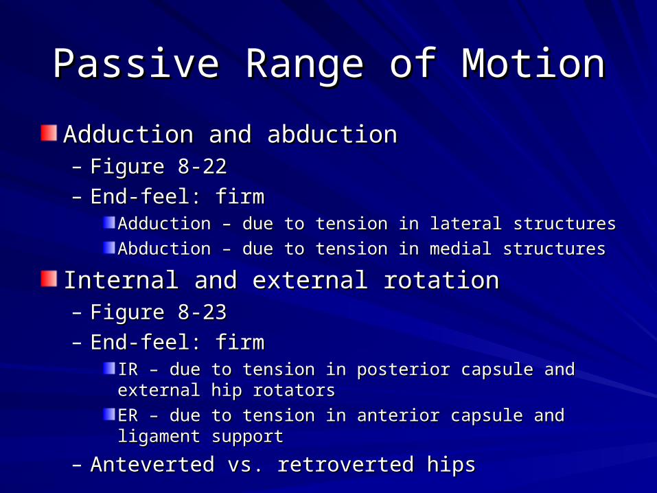
Adduction and abductionAdduction and abduction– Figure 8-22Figure 8-22– End-feel: firmEnd-feel: firm
Adduction – due to tension in lateral structuresAdduction – due to tension in lateral structures
Abduction – due to tension in medial structuresAbduction – due to tension in medial structures
Internal and external rotationInternal and external rotation– Figure 8-23Figure 8-23– End-feel: firmEnd-feel: firm
IR – due to tension in posterior capsule and external hip IR – due to tension in posterior capsule and external hip rotatorsrotators
ER – due to tension in anterior capsule and ligament supportER – due to tension in anterior capsule and ligament support
– Anteverted vs. retroverted hipsAnteverted vs. retroverted hips
Resisted Range of MotionResisted Range of Motion
Box 8-4, pages 291-292Box 8-4, pages 291-292
Trendelenburg’s Test for Gluteus Medius Trendelenburg’s Test for Gluteus Medius WeaknessWeakness– Box 8-5, page 293Box 8-5, page 293
Ligamentous TestingLigamentous Testing
No specific tests for hip ligamentsNo specific tests for hip ligaments
Dysfunction is determined through passive Dysfunction is determined through passive testing of movementtesting of movement– Hyperextension places iliofemoral, Hyperextension places iliofemoral,
pubofemoral, and ischiofemoral ligaments on pubofemoral, and ischiofemoral ligaments on stretchstretch

Neurologic TestingNeurologic Testing
Complete lower quarter screening should Complete lower quarter screening should be performedbe performed– Pathology involving femoral or sciatic nervePathology involving femoral or sciatic nerve
Piriformis SyndromePiriformis Syndrome– Impingement of sciatic nerve from spasm of Impingement of sciatic nerve from spasm of
piriformis musclepiriformis muscle
Pathologies and Related Special Pathologies and Related Special TestsTests
AcuteAcute– Contusions or strainsContusions or strains
ChronicChronic– Improper biomechanics from poor posture, leg Improper biomechanics from poor posture, leg
length discrepancies, overuse syndromeslength discrepancies, overuse syndromes
Injury to hip joint is rareInjury to hip joint is rare– Potential medical emergencyPotential medical emergency
Muscle StrainsMuscle Strains
Table 8-4, page 294Table 8-4, page 294
Occur secondary to dynamic overload Occur secondary to dynamic overload during eccentric muscle contractionduring eccentric muscle contraction
Commonly injured Commonly injured – Iliopsoas, quadriceps, adductors, hamstringsIliopsoas, quadriceps, adductors, hamstrings
Signs and SymptomsSigns and Symptoms

BursitisBursitis
Onset related to biomechanical factors, Onset related to biomechanical factors, congenital influences, or environmental congenital influences, or environmental conditions, such as prolonged periods of conditions, such as prolonged periods of sittingsitting
Septic infection may be a causeSeptic infection may be a cause

BursitisBursitis
Trochanteric BursitisTrochanteric Bursitis– Evaluative Findings - Table 8-5, page 295Evaluative Findings - Table 8-5, page 295– May result from a single blow or friction from May result from a single blow or friction from
IT bandIT band– History of training changes or increased Q History of training changes or increased Q
angle may be predisposing factorsangle may be predisposing factors– ““Snapping Hip” syndromeSnapping Hip” syndrome
BursitisBursitis
Ischial BursitisIschial Bursitis– Evaluative Findings - Table 8-6, page 296Evaluative Findings - Table 8-6, page 296– Movement of buttocks while patient is weight-Movement of buttocks while patient is weight-
bearing in seated position can irritate this bearing in seated position can irritate this bursabursa
Also irritated by prolonged sittingAlso irritated by prolonged sitting
– Need to rule out hamstring strain or avulsion Need to rule out hamstring strain or avulsion of its attachmentof its attachment
– Doughnut padding may helpDoughnut padding may help
BursitisBursitis
Iliopsoas BursitisIliopsoas Bursitis– Associated with rheumatoid arthritis or Associated with rheumatoid arthritis or
osteoarthritis of hiposteoarthritis of hip– Signs and symptomsSigns and symptoms
Pain in anterior hipPain in anterior hip
Palpable mass in groin or inguinal ligamentPalpable mass in groin or inguinal ligament
““snapping hip” syndromesnapping hip” syndrome
– Treatment includes strengthening hip rotatorsTreatment includes strengthening hip rotators

Degenerative Hip ChangesDegenerative Hip Changes
Due to age, repetitive trauma, acute trauma, or Due to age, repetitive trauma, acute trauma, or improper arrangements of hipimproper arrangements of hip– Degeneration of articular surfaces of femur and Degeneration of articular surfaces of femur and
acetabulumacetabulum– Arthritis, osteochondritis dissecans, acetabular labrum Arthritis, osteochondritis dissecans, acetabular labrum
tears, avascular necrosistears, avascular necrosis
Signs and symptomsSigns and symptoms– Pain, referred to low back, anterior thigh, kneePain, referred to low back, anterior thigh, knee– Loss of motion in all planes, decrease strengthLoss of motion in all planes, decrease strength– Hip Scouring, Box 8-6, page 297Hip Scouring, Box 8-6, page 297– Radiographic evaluationRadiographic evaluation
Piriformis SyndromePiriformis Syndrome
Sciatic nerve passes under or through the Sciatic nerve passes under or through the piriformis muscle as nerve travels across piriformis muscle as nerve travels across posterior pelvisposterior pelvisSpasm or hypertrophy of muscle places Spasm or hypertrophy of muscle places pressure on sciatic nervepressure on sciatic nerveSix times more common in womenSix times more common in womenRelatively undefined and confusingRelatively undefined and confusing– Mimics lumbar nerve root impingement and Mimics lumbar nerve root impingement and
intervertebral disk diseaseintervertebral disk disease

Piriformis SyndromePiriformis Syndrome
Evaluative FindingsEvaluative Findings– Table 8-7, page 298Table 8-7, page 298
Straight leg raise, passive hip internal Straight leg raise, passive hip internal rotation resisted external rotation with rotation resisted external rotation with patient seated, and resisted hip abduction patient seated, and resisted hip abduction may produce symptomsmay produce symptoms– Figure 8-24Figure 8-24
Treatment includes stretching and Treatment includes stretching and strengthening or surgical releasestrengthening or surgical release

On-Field Evaluation of Pelvis and On-Field Evaluation of Pelvis and Thigh InjuriesThigh Injuries
Trauma to coxofemoral joint is rareTrauma to coxofemoral joint is rare– Protection from paddingProtection from padding
More commonly, strains, contusions, More commonly, strains, contusions, sprains of SI jointsprains of SI joint
Note position of athleteNote position of athlete– If leg is moving, rule out dislocationIf leg is moving, rule out dislocation– Fixed, immobile awkward position may Fixed, immobile awkward position may
indicate dislocation indicate dislocation

On-Field Evaluation of Pelvis and On-Field Evaluation of Pelvis and Thigh InjuriesThigh Injuries
After ruling out dislocation or subluxation After ruling out dislocation or subluxation and femoral fracture – AROMand femoral fracture – AROM
Weight-bearing statusWeight-bearing status– Removal from fieldRemoval from field

Initial Evaluation and Management Initial Evaluation and Management of On-Field Injuriesof On-Field Injuries
Iliac Crest Contusion (hip pointer)Iliac Crest Contusion (hip pointer)– Evaluative Findings, Table 8-8, page 299Evaluative Findings, Table 8-8, page 299– Disproportionate amount of pain, swelling, Disproportionate amount of pain, swelling,
and loss of functionand loss of function– Recognition and immediate management of Recognition and immediate management of
pain reduces time lost due to injurypain reduces time lost due to injury– TreatmentTreatment
Ice, padding, reduced activity, crutches, if Ice, padding, reduced activity, crutches, if necessarynecessary

Initial Evaluation and Management Initial Evaluation and Management of On-Field Injuriesof On-Field Injuries
Quadriceps ContusionQuadriceps Contusion– As severity of impact increases, so does the As severity of impact increases, so does the
proportion of muscle fiber deathproportion of muscle fiber death– Can result in decreased force during knee Can result in decreased force during knee
extensionextensionAssociated pain and spasm may limit flexionAssociated pain and spasm may limit flexion
– Gross discoloration, painful to touch, Gross discoloration, painful to touch, intramuscular hematoma gives hardened feel, intramuscular hematoma gives hardened feel, increase in girth of muscleincrease in girth of muscle
Overtime, atrophy may occur Overtime, atrophy may occur

– Risk of myositis ossificans is increased when Risk of myositis ossificans is increased when effusion of knee joint occurseffusion of knee joint occurs
Figure 8-25Figure 8-25
– First 24 hours following injury are criticalFirst 24 hours following injury are critical– Pain during AROM, or weakness during MMT Pain during AROM, or weakness during MMT
= removal from activity= removal from activityIce applied in flexionIce applied in flexion
Maintaining ROM decrease possibility of myositis Maintaining ROM decrease possibility of myositis ossificans formationossificans formation
Figure 8-26Figure 8-26

Hip DislocationHip Dislocation– RareRare– Medical emergencyMedical emergency– Majority involve posterior displacement of Majority involve posterior displacement of
femoral headfemoral headFractures to femoral neck and acetabulum Fractures to femoral neck and acetabulum
– Most occur when hip is in flexion and Most occur when hip is in flexion and adduction and axial force is placed on femur, adduction and axial force is placed on femur, displacing it posteriorly and causing head to displacing it posteriorly and causing head to be driven through posterior capsulebe driven through posterior capsule
Signs and SymptomsSigns and Symptoms– Immediate pain within joint and buttocksImmediate pain within joint and buttocks– Sensation of “giving out”Sensation of “giving out”– Femur and lower leg positioned in internal Femur and lower leg positioned in internal
rotation and adductionrotation and adductionFigure 8-27Figure 8-27
– AROM is impossibleAROM is impossible– No attempt to reduceNo attempt to reduce– Sensory and vascular checkSensory and vascular check
Immediate immobilization and Immediate immobilization and transportation to emergency facilitytransportation to emergency facility
Reduction under anesthesiaReduction under anesthesia
Femoral FractureFemoral Fracture
Torsional or shear force to shaftTorsional or shear force to shaftRelatively rareRelatively rare– ““weak link” principleweak link” principle
Immediate loss of function, pain, Immediate loss of function, pain, deformity, easily recognizabledeformity, easily recognizableStress fractureStress fracture– Shaft and neck, difficult to diagnosisShaft and neck, difficult to diagnosis
Similar s/s to hip flexor strain or tendinitisSimilar s/s to hip flexor strain or tendinitis
– TreatmentTreatment