Embed Size (px)
DESCRIPTION
biosci
Citation preview

THE SPECIAL SENSES
THE SPECIAL SENSES
CHEMICAL SENSES: Smell and Taste
THE EAR: Hearing and Balance
THE EYE : Vision

OLFACTION: SENSE OF SMELL
Chemical sense
olfactory receptors are found in the roof of
the nasal cavity
sniffing intensifies the sense of smell
olfactory epithelium consists of:
olfactory receptors
supporting cells
basal stem cells

Anatomy of Olfaction
Olfactory receptors
first order neurons
bipolar neurons ( exposed tips are knobbed
dendrites)
site of olfactory transduction: olfactory
hairs

Supporting Cells
columnar epithelial cells
provides physical support, nourishment
& electrical stimulation for
olfactory receptors
help destroy chemicals

Basal Stem Cell
lie between bases of supporting cells
continuously undergo cell division


Olfactoy mucosa. Note the
thick layer of epithelium.
The underlying lamina
propria possesses many
glands

Olfactory system. 1. Olfactory bulb. 2. nasal cavity. 3.
Brain. 4. Olfactory epithelium 5. Vomeronasal organ. 6.
Ions. 7 Glomeruli. *. Axon. 9. To olfactory cortex

Physiology of Olfaction
1.odorants bind olfactory receptors that are
linked to G proteins
2.activation of the enzyme adenylate cyclase
3.Na+ channels open
4.inflow of Na+ ions
5.depolarization
6.generation and propagation of action
potential
7.nerve impulse

Odor Thresholds and Adaptation
low threshold
decreasing sensitivity

Olfactory Pathway
Olfactory Nerve (CN I)
Olfactory Bulbs
Olfactory Tract ( Brain)
Anosmias

NASAL POLYP

NASAL POLYP

Gustation : Sense of Taste
Chemical sense
4 classes of stimuli
sweet salty
sour bitter
gustation is thousands of times less
sensitive than olfaction
colds block olfaction not gustation

Anatomy of Gustation
Receptors for gustation are located in taste
buds
10,000 receptors are distributed in the
tongue, soft palate, pharynx & larynx
number of taste buds decline with age

The tongue is covered with dozens of pimple-like
projections called papillae. These grip and move food
when you chew. Around the sides of the papillae are about
10,000 microscopic taste buds. Different parts of the
tongue are sensitive to different flavours: sweet, salt, sour
and bitter

Taste buds consist of 3 kinds of cells:
• supporting cells
• 50 gustatory cells
• basal cells
gustatory hair
gustatory receptor
cell
supporting cell
fibers of CNs basal cell

Taste buds
Papillae
• circumvallate
• fungiform
• filiform - no taste buds


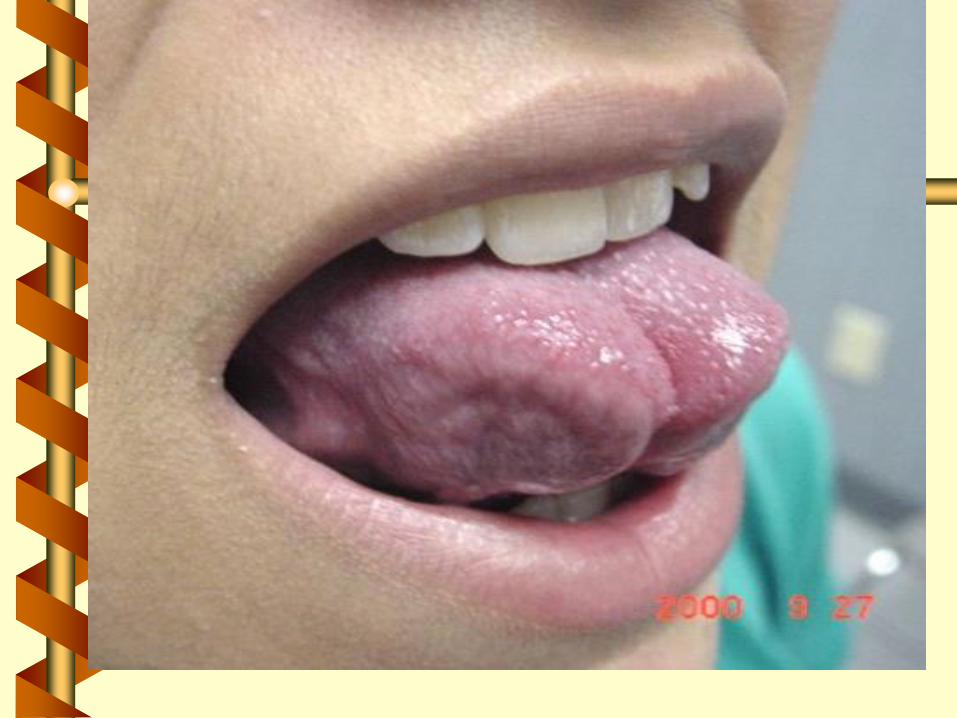

Tongue-tie is a condition caused by a short lingual
frenulum that prevents the tongue from
protruding. Occasionally, it could interfere with
breast feeding


The horizontally incised frenulum is now pulled
vertically, allowing the release of the tongue. The
incision is sutured vertically with absorbable
stitches.

Post-operative appearance of the tongue that
can now protrude down to the chin.


Physiology of Gustation
1. chemicals are dissolved in saliva
2. Contact with plasma membrane of
gustatory hair
3. receptor potential stimulates exocytosis of
neurotransmittercontaining synaptic
vesicles
4. Nerve impulse arise in the 1st order
neurons that synapse with gustatory
receptor cells

Taste Threshold and Adaptations
Bitter: lowest
sour: higher than that of bitter
sweet about the same but higher
salty than sour & bitter
complete adaptation occurs in 1-5 minutes
of continuous stimulation

First order gustatory fibers include 3
cranial nerves:
• Facial nerve: anterior 2/3 of tongue
• glossopharyngeal nerve: posterior 1/3 of
tongue
• vagus: throat & epiglottis
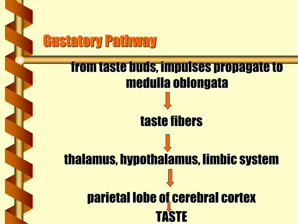
Gustatory Pathway
from taste buds, impulses propagate to
medulla oblongata
taste fibers
thalamus, hypothalamus, limbic system
parietal lobe of cerebral cortex
TASTE

The Eye and Vision
Accessory structures of the eye:
• eyelids
• eyelashes
• eyebrows
• lacrimal apparatus
• extrinsic eye muscles

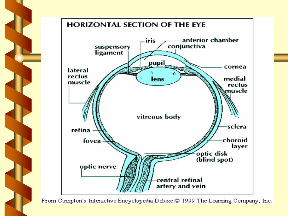
Anatomy of the Eyeball
Eyelids
• palpebrae
• palpebral fissure
• medial commissure
• lateral commissure
• lacrimal caruncle
• meibomian glands
• conjunctiva

Eyelashes and Eyebrows
• ciliary glands
• sty

Ciliary Body

Lacrimal apparatus
• lacrimal gland
• excretory lacrimal
ducts
• lacrimal puncta
• nasolacrimal duct

Extrinsic Eye Muscles
• superior rectus
• inferior rectus
• lateral rectus
• medial rectus
• superior oblique
• inferior oblique

Entrapment of the Inferior Rectus Muscle
of the Eye
in a Blow-out Fracture of the Orbit
Blunt trauma to the right eye, resulting in diplopia.
The right inferior rectus was caught between the
fragments of a blow-out fracture of the floor of the
orbit. This child was unable to move his right eyeball
up on upward gaze.

Tunics of the eye;
• Fibrous tunic; sclera
• Vascular tunic: choroid
• Retina: rods and cones

Uvea
• choroid
• ciliary body: ciliary body and ciliary muscles
• iris: colored portion of eyeball
• pupil: hole at the center of iris

Retina
• photoreceptors: rods and cones
• macula lutea
• fovea centralis
• blind spot

Fovea

Lens
cataract

Chambers of the eye
• anterior chamber: aqueous humor
• posterior chamber: vitreous humor
Intraocular pressure
Glaucoma


Refraction of Light Rays
Images on retina
• inverted
• right to left reversal
• brain coordinates visual images and
orientation of objects

Accomodation
Near point of Vision
Presbyopia

Light enters the front of the eye through the pupil and is
focused by the lens onto the retina. Rod cells on the retina
respond to the light and send a message through the optic
nerve fiber to the brain.

Emmetropia: Normal Vision


Refraction Abnormalities
Myopia: Near sightedness

Hyperopia: Farsightedness

Astigmatism
Nyctalopia
Binocular vision
Photopupillary reflex
Accomodation pupillary reflex

Physiology of Vision
Photopigments:
• rhodopsin (rods)
• opsin
• retinal

1. Light causes
4. cis-retinal isomerization of
binds to photopigments
opsin
trans
retinal
3. retinal isomerase
converts trans to
cis 2. Trans-retinal
separates from opsin
The Cyclic Bleaching & Regeneration of
Photopigments

Light Adaptation
• emerging from a darkened room, visual system
decreases its sensitivity
Dark Adaptation
• entering a darkened room, visual system
increases its sensitivity

The Visual Pathway
Cornea
pupil
iris
photoreceptors
ganglion cells
bipolar cells
thalamus
visual cortex ( occipital lobe)

Keratitis
Mydriasis
Nystagmus
Strabismus
The EAR : Hearing & Equilibrium
3 Principal Regions of the Ear:
• external ear
• middle ear
• internal ear

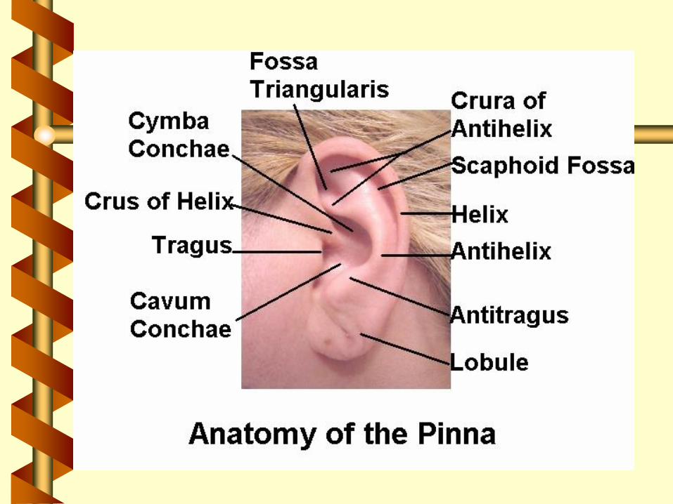
External Ear
auricle
pinna
external auditory canal
eardrum


Middle Ear
malleus
incus
stapes
oval window
round window
tensor tympani
muscles
stapedius
Eustachian tube

Inner Ear
2 main divisions
• bony labyrinth
– semicircular canal contain
(crista ampullaris & cupula) receptors for
– vestibule equilibrium
– cochlea: receptors for hearing
• membranous labyrinth
– K+
– utricle and saccule

Cochlea
– snail-shaped
spiral organ of Corti
– coiled sheet of epithelial cells
– 16,000 hair cells ( receptors for hearing)


The Nature of Sound Waves
sound waves
pitch
decibels
conduction deafness
sensorineural deafness

The Physiology of Hearing
Auricle
External auditory canal
Malleus
Incus
Stapes
Oval window
Scala vestibuli
Scala tympani
Round window
vestibular membrane
endolymph in cochlea
basilar membrane
stereocilia
generation of action
potential

Auditory Pathway
External auditory canal
tympanic membrane
auditory ossicles
oval window
cochlea
organ of Corti
Mechanisms of Equilibrium
Vestibular Apparatus
• arm for static equilibrium
• arm for dynamic equilibrium

utricle, saccule, and the three semicircular canals.
The saclike utricle and saccule sense the body's
relationship to gravity, or its static equilibrium.
A person knows that the body is right side up because
these structures relay messages about the body's position
to the brain. Both sacs are hollow. Hairlike nerve endings
are anchored into the inner surface of each structure. The
free ends of the nerve endings project into the hollow
space.
Tiny particles of limestone, known as otoliths, rest against
the bottom of each sac.
If the head moves, the otoliths change position. In shifting,
they pass over sensitive nerve endings. These send
immediate impulses to the brain. Notified of a change in
body position, the brain triggers the reflex mechanisms to
correct the position of the body .

Disorders of the Ear
Meniere’s disease
• due to increased amount of endolymph
• fluctuating hearing loss
• roaring tinnitus
• spinning or whirling vertigo
Otitis media
• acute infection of middle ear

PICTURE OF TYMPANIC MEMBRANE
PERFORATION
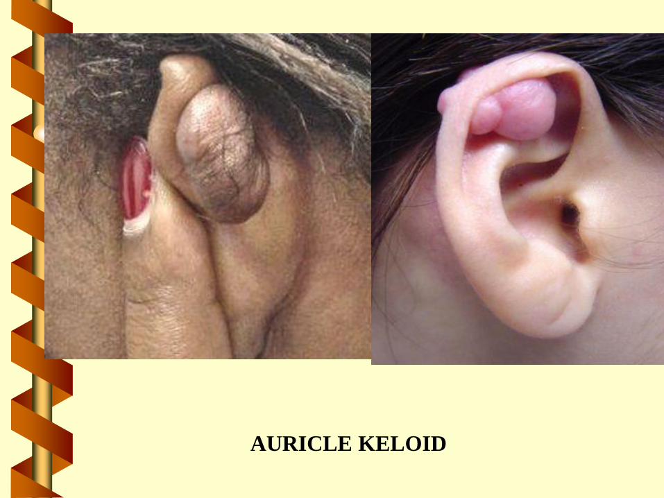
AURICLE KELOID

AURICULAR ABSCESS

Auricle Perichondritis


Static and Dynamic Equilibrium

vestibular apparatus –• functional components of the
membranous labyrinth involved in
the sensations of static and dynamic
equilibrium are a system of thin-
walled intercommunicating tubes
and ducts situated within the petrous
part of the temporal bone at the
base of the skull;
• there are five vestibular structures,
each containing a specialized
mechanoreceptor, a maculae, within
the utricle and saccule, and a cristae
within the ampullae of the superior,
horizontal, and posterior
semicircular canals.

vestibule –
The central cavity of the bony
labyrinth of the ear containing the
functional components of the
membranous labyrinth involved in
the sensations of static
equilibrium which are two
vestibular structures, each
containing a specialized
mechanoreceptor, a maculae,
within the utricle and saccule.

saccule - The smaller of the two
membranous sacs in the vestibule
of the inner ear; it contains a
specialized mechanoreceptor, a
maculae, for the detection of static
equilibrium.
.

utricle - The larger of the two
membranous sacs in the vestibule
of the inner ear; it contains a
specialized mechanoreceptor, a
maculae, for the detection of static
equilibrium.

static equilibrium - The special sense
which interprets the position of the
head permitting the CNS to maintain
stability and posture when the head
and body are not moving; it is detected
by mechanoreceptors in the vestibule
of the inner ear, the utricle and
saccule, which each contain a macula
with the receptors for static
equilibrium; when the head moves with
reference to gravity, the otolithic
membrane shifts and the
mechanoreceptors (hair cells) in the
macula detect this movement and send
the information along the vestibular
nerve to the brain for interpretation
("which way is up").

maculae - The specialized
mechanoreceptors within the
utricle and saccule for the
detection of static
equilibrium; they make use
of hair cells to detect
movements of the otolithic
membrane; the nerve
impulses thus generated are
transmitted along the
vestibular branch of cranial
nerve VIII to the CNS.


otolithic membrane - The gelatinous covering of macula of the utricle
and saccule of the vestibular apparatus which has many crystals of
calcium carbonate (otoconia or otoliths); their movements in response to
changes in the position of the head with reference to gravity stimulate the
hair cells to send nerve impulses to the CNS which are interpreted as
information about static equilibrium.

Dynamic Equilibrium
dynamic equilibrium –
The special sense which interprets balance when one is moving,
or at least the head is moving;
the semicircular canals contain the receptors for dynamic
equilibrium; within each semicircular canal is a complex
mechanoreceptor called a crista ampullaris which contains the
mechanoreceptors (Hair cells) for dynamic equilibrium;
when the perilymph in one of the semicircular canals moves, the
hair cells in the crista ampullaris are stimulated to send nerve
impulses to the brain;
this advises the brain of whether or not a person has their
balance during body movements or if their body is in motion,
e.g, riding in a car or turning one's head from side to side.

semicircular canals - The functional components of the membranous labyrinth, a series of three
interconnected perilymph-filled tubes with enlarged ends, involved in the sensations of dynamic
equilibrium; the contain the cristae ampullaris which detect acceleration in the three
perpendicular planes (superior, horizontal, and posterior); these accelerometers make use of hair
cells similar to those on the organ of Corti, but these hair cells detect movements of the fluid in the
canals caused by angular acceleration about an axis perpendicular to the plane of the canal; tiny
floating particles aid the process of stimulating the hair cells as they move with the fluid; the nerve
impulses thus generated are transmitted along the vestibular branch of cranial nerve eight to the
CNS.

ampulla - The dilation or expanded
end of each of the semicircular
canals of the vestibular apparatus
which contains the specialized
mechanoreceptor structure, the
crista, which detect acceleration in
the planes of the canal; these
accelerometers make use of hair
cells to detect movements of the
fluid in the canals caused by
angular acceleration about an axis
perpendicular to the plane of the
canal; the nerve impulses thus
generated are transmitted along the
vestibular branch of cranial nerve
eight to the CNS.

crista ampullaris - Within the
ampulla of each semicircular canal
is a complex mechanoreceptor
structure, the crista ampullaris; the
ampulla has a ridge covered by
neuroepithelium consisting of
sensory hair cells and supporting
cells; the hair cells attached to a
gelatinous mass, the cupula, which
rests on top of the crista ampularis;
when the perilymph in one of the
semicircular canals moves, the hair
cells in the crista ampullaris are
stimulated to send nerve impulses
to the brain; this advises the brain of
whether or not a person has their
balance during body movements or
if their body is in motion, e.g, riding
in a car.

vestibular nerve - The division of
the vestibulocochlear (eighth)
cranial nerve which conducts
sensory information regarding static
and dynamic equilibrium from the
vetibular apparatus of the inner ear
to the various centers of the CNS
which process and integrate that
information with visual and
proprioception.

THE SENSE OF TOUCH
The sense of touch is the name given to a network of nerve endings that reach just about every part of our body. These sensory nerve endings are located just below the skin and register light and heavy pressure on the skin and also differences in temperature. These nerve endings gather information and send it to the brain


![17 [chapter 17 the special senses]](https://img.dokumen.tips/doc/110x75/5a6496047f8b9a27568b6f5f/17-chapter-17-the-special-senses.jpg)