Embed Size (px)
Citation preview

Chapter 7Wrist & Hand Joints

Hand
Distal
Middle
BaseMetacarpals
Hamate
Capitate
TrapezoidPisiform
TriquetrumLunate
Scaphoid
ProximalPhalanges
Head
Shaft
Carpals Trapezum
Label

Joints
• Radioulnar (Pivot)– Supination and
pronation
• Radiocarpal (Condylodial)– Primarily articulates with
scaphoid, lunate, and triquetrum
– Movement and Range of Motion
• Flexion: 80°-90°• Extension: 70°-90°• Abduction: 15°-25°• Adduction: 25°-40°

Joints
• Intercarpal (Arthrodial)
• Intermetacarpal (Arthrodial)
• Metacarpophalangeal (Condylodial)– Movement and ROM
• Flexion: 90°• Extension: 0-40°
– Metacarpophalangeal of the thumb:
• 40º to 90º from full extension

Joints• Carpometacarpal of
the thumb: (seller)– Movement and ROM
• Flexion: 15º to 45º• Extension: 0º to
20º • Abduction: 50º to
70º• Opposition:
– movement of thumb across palmer aspect to oppose any or all of the phalanges
• Reposition: – movement of
thumb back to anatomical position
• Proximal Interphalangeal (ginglymus)– ROM:
• Flexion: 90º to 120º from full extension
• Distal Interphalangeal (ginglymus)– ROM:
• Flexion: 80º to 90º from full extension

Cutaneous Distribution

• Wrist Flexors– Flexor Carpi Radialis– Palmaris Longus– Flexor Carpi Ulnaris– Flexor Digitorum
Superficialis – Flexor Digitorum
Profundus– Flexor Pollicis Longus
Muscles
Label

Muscles
• Wrist Extensors– Extensor Carpi Radialis
Longus– Extensor Carpi Ulnaris– Extensor Carpi Radialis
Brevis– Extensor Digitorum– Extensor Indicis– Extensor Digiti Minimi– Abductor Pollicis Longus – Extensor Pollicis Brevis– Extensor Pollicis Longus
Not Shown?
Label

MusclesFlexor Carpi Radialis • Strengthening
• Stretching

MusclesPalmaris Longus • Strengthening
• Stretching

MusclesFlexor Carpi Ulnaris • Strengthening
• Stretching

MusclesExtensor Carpi Ulnaris • Strengthening
• Stretching

MusclesExtensor Carpi
Radialis Brevis
• Strengthening
• Stretching

MusclesExtensor Carpi
Radialis Longus
• Strengthening
• Stretching

Muscles
Flexor Digitorum Superficialis
• Strengthening
• Stretching

MusclesFlexor Digitorum
Profundus• Strengthening
• Stretching

MusclesExtensor Indicis • Strengthening
• Stretching

MusclesExtensor Digiti Minimi • Strengthening
• Stretching

MusclesExtensor Pollicus
Longus
• Strengthening
• Stretching

Muscles
Extensor Pollicus Brevis
• Strengthening
• Stretching

MusclesAbductor Pollicus
Longus• Strengthening
• Stretching

Thenar Muscles
Opponens Pollicis

Thenar Muscles
Abductor Pollicis Brevis

Thenar Muscles
Flexor Pollicis Brevis

Intermediate Muscles
Adductor Pollicis

Intermediate Muscles
Palmar Interossei
Drawn illustration

Intermediate Muscles
Dorsal Interossei
Drawn illustration
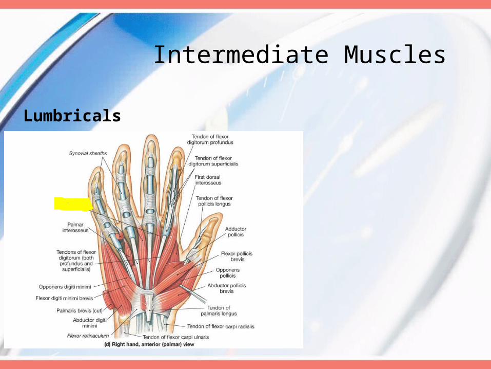
Intermediate Muscles
Lumbricals

Hypothenar Muscles
Opponens Digiti Minimi

Hypothenar Muscles
Abductor Digiti Minimi

Hypothenar Muscles
Flexor Digiti Minimi Brevis

Injuries of the Wrist
• Scaphoid Fracture • Carpel Tunnel• Claw Hand• Ape Hand

Carpal Tunnel Syndrome
1. Anatomya. Boundsb. Muscles/Tendonsc. Nerved. Other

Carpel Tunnel Syndrome
• Common Causes:– Pregnancy– Rheumatoid Arthritis – Diabetes– Wrist Trauma– Occupational (typing)

• Symptoms
• Tests:
Carpel Tunnel Syndrome
TINEL’S SIGN
STRIKE THE PATIENT’S WRIST AS SHOWN. A TINGLING SENSATION RADIATING DOWN THE WRIST TO THE HAND IN THE DISTIBUTION OF THE MEDIAN NERVE IS A POSITIVE SIGN.
Tinel’s Sign
Phalen Test
Thumb Abduction Test
PHALEN TEST
HAVE THE PATIENT HOLD THEIR WRISTS AS SHOWN FOR ONE MINUTE. NUMBNESS AND PARAESTHESIA IN THE DISTRIBUTION OF THE MEDIAN NERVE IS A POSITIVE TEST.
THUMB ABDUCTION TEST
TESTS THE STRENGTH OF THE ABDUCTOR POLLICIS BREVIS WHICH IS INERVATED BY THE MEDIAN NERVE.
HAVE THE PATIENT PLACE THEIR PALM UP WITH THEIR THUMB PERPENDICULAR TO IT.
APPLY DOWNWARD PRESSURE ON THE THUMB. WEAKNESS IS ASSOCIATED WITH CARPAL TUNNEL SYNDROME.
JAMA 2000;283:3110-3117MOSBY’S GUIDE TO THE PHYSICAL EXAMINATION 5TH ED.

Claw Hand
1. Description: Hyperextension at the MCP joint, and flexion at the IP joint
2. Etiology?
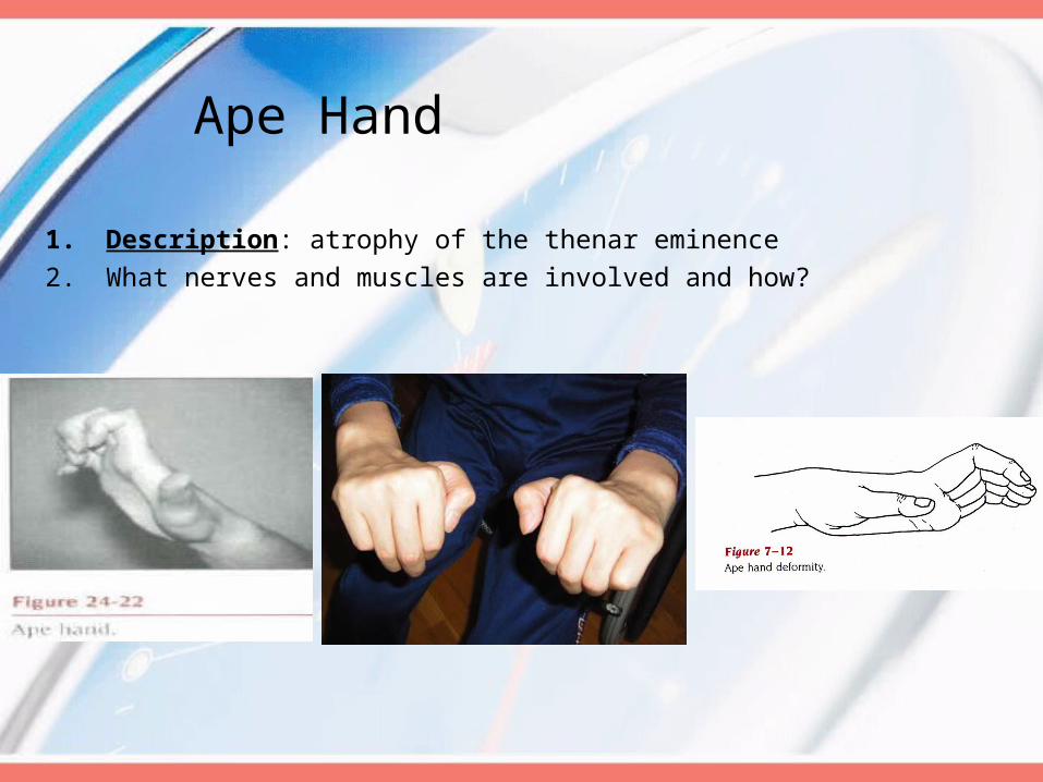
Ape Hand
1. Description: atrophy of the thenar eminence 2. What nerves and muscles are involved and how?