Embed Size (px)
Citation preview

Integrative Functions ofthe Nervous SystemCynthia J. Forehand, Ph.D.7
C H A P T E R
7
The central nervous system (CNS) receives sensorystimuli from the body and the outside world and
processes that information in neural networks or centers ofintegration to mediate an appropriate response or learnedexperience. Centers of integration are hierarchical in na-ture. In a caudal-to-rostral sequence, the more rostral it isplaced, the greater the complexity of the neural network.This chapter considers functions integrated within the di-encephalon and telencephalon, where emotionally moti-vated behavior, appetitive drive, consciousness, sleep, lan-guage, memory, and cognition are coordinated.
THE HYPOTHALAMUS
The hypothalamus coordinates autonomic reflexes of thebrainstem and spinal cord. It also activates the endocrineand somatic motor systems when responding to signalsgenerated either within the hypothalamus or brainstem orin higher centers, such as the limbic system, where theemotions and motivations are generated. The hypothala-mus can accomplish this by virtue of its unique location atthe interface between the limbic system and the endocrineand autonomic nervous systems.
As a major regulator of homeostasis, the hypothalamusreceives input about the internal environment of the body
via signals in the blood. In most of the brain, capillary en-dothelial cells are connected by tight junctions that preventsubstances in the blood from entering the brain. Thesetight junctions are part of the blood-brain barrier. Theblood-brain barrier is missing in several small regions of thebrain called circumventricular organs, which are adjacentto the fluid-filled ventricular spaces. Several circumventric-ular organs are in the hypothalamus. Capillaries in these re-gions, like those in other organs, are fenestrated (“leaky”),allowing the cells of hypothalamic nuclei to sample freely,from moment to moment, the composition of the blood.Neurons in the hypothalamus then initiate the mechanismsnecessary to maintain levels of constituents at a given setpoint, fixed within narrow limits by a specific hypothalamicnucleus. Homeostatic functions regulated by the hypothal-amus include body temperature, water and electrolyte bal-ance, and blood glucose levels.
The hypothalamus is the major regulator of endocrinefunction because of its connections with the pituitarygland, the master gland of the endocrine system. Theseconnections include direct neuronal innervation of the pos-terior pituitary lobe by specific hypothalamic nuclei and adirect hormonal connection between specific hypothala-mic nuclei and the anterior pituitary. Hypothalamic hor-mones, designated as releasing factors, reach the anterior
■ THE HYPOTHALAMUS
■ THE RETICULAR FORMATION
■ THE FOREBRAIN
C H A P T E R O U T L I N E
1. Homeostatic functions are regulated by the hypothalamus.2. Homeostatically regulated functions fluctuate in a daily
pattern.3. The reticular formation serves as the activating system of
the forebrain.4. Sleep occurs in stages that exhibit different EEG patterns.5. The limbic system receives distributed monoaminergic and
cholinergic innervation.
6. Limbic structures play a role in the brain’s reward system.7. The limbic system regulates aggression and sexual activ-
ity.8. Affective disorders and schizophrenia are disruptions in
limbic function.9. The cerebral cortex and hippocampus are involved in
learning and memory.10. Language is a lateralized function of association cortex.
K E Y C O N C E P T S
119

120 PART II NEUROPHYSIOLOGY
pituitary lobe by a portal system of capillaries. Releasingfactors then regulate the secretion of most hormones of theendocrine system.
The Hypothalamus Is Composed of
Anatomically Distinct Nuclei
The diencephalon includes the hypothalamus, thalamus,and subthalamus (Fig. 7.1). The rostral border of the hypo-thalamus is at the optic chiasm, and its caudal border is atthe mammillary body.
On the basal surface of the hypothalamus, exiting themedian eminence, the pituitary stalk contains the hypo-thalamo-hypophyseal portal blood vessels (see Fig. 32.3).Neurons within specific nuclei of the hypothalamus secretereleasing factors into these portal vessels. The releasing fac-tors are then transported to the anterior pituitary, wherethey stimulate secretion of hormones that are trophic toother glands of the endocrine system (see Chapter 32).
The pituitary stalk also contains the axons of magnocel-lular neurons whose cell bodies are located in the supraop-tic and paraventricular hypothalamic nuclei. These axonsform the hypothalamo-hypophyseal tract within the pitu-itary stalk and represent the efferent limbs of neuroen-docrine reflexes that lead to the secretion of the hormonesvasopressin and oxytocin into the blood. These hormonesare made in the magnocellular neurons and released bytheir axon terminals next to the blood vessels within theposterior pituitary.
The nuclei of the hypothalamus have ill-defined bound-aries, despite their customary depiction (Fig. 7.2). Many arenamed according to their anatomic location (e.g., anteriorhypothalamic nuclei, ventromedial nucleus) or for thestructures they lie next to (e.g., the periventricular nucleussurrounds the third ventricle, the suprachiasmatic nucleuslies above the optic chiasm).
The hypothalamus receives afferent inputs from all lev-els of the CNS. It makes reciprocal connections with thelimbic system via fiber tracts in the fornix. The hypothala-mus also makes extensive reciprocal connections with thebrainstem, including the reticular formation and themedullary centers of cardiovascular, respiratory, and gas-trointestinal regulation. Many of these connections travelwithin the medial forebrain bundle, which also connectsthe brainstem with the cerebral cortex.
Several major connections of the hypothalamus are one-way rather than reciprocal. One of these, the mammil-lothalamic tract, carries information from the mammillarybodies of the hypothalamus to the anterior nucleus of thethalamus, from where information is relayed to limbic re-gions of the cerebral cortex. A second one-way pathwaycarries visual information from the retina to the suprachi-asmatic nucleus of the hypothalamus via the optic nerve.Through this retinal input, the light cues of the day/nightcycle entrain or synchronize the “biological clock” of thebrain to the external clock. A third one-way connection isthe hypothalamo-hypophyseal tract from the supraopticand paraventricular nuclei to the posterior pituitary gland.The hypothalamus also projects directly to the spinal cordto activate sympathetic and parasympathetic preganglionicneurons (see Chapter 6).
Hypothalamic Nuclei Are Centers of
Physiological Regulation
The nuclei of the hypothalamus contain groups of neuronsthat regulate several important physiological functions:
1) Water and electrolyte balance in magnocellular cells ofthe supraoptic and paraventricular nuclei (see Chapter 32)
2) Secretion of hypothalamic releasing factors in thearcuate and periventricular nuclei and in parvocellular cellsof the paraventricular nucleus (see Chapters 32 and 33)
3) Temperature regulation in the anterior and posteriorhypothalamic nuclei (see Chapter 29)
4) Activation of the sympathetic nervous system andadrenal medullary hormone secretion in the dorsal and pos-terior hypothalamus (see Chapter 34)
5) Thirst and drinking regulation in the lateral hypo-thalamus (see Chapter 24)
6) Hunger, satiety, and the regulation of eating behav-ior in the arcuate nucleus, ventromedial nucleus, and lateralhypothalamic area
7) Regulation of sexual behavior in the anterior andpreoptic areas
8) Regulation of circadian rhythms in the suprachias-matic nucleus
The Hypothalamus Regulates Eating Behavior
Classically, the hypothalamus has been considered agrouping of regulatory centers governing homeostasis.With respect to eating, the ventromedial nucleus of the hy-pothalamus serves as a satiety center and the lateral hypo-thalamic area serves as a feeding center. Together, these ar-eas coordinate the processes that govern eating behaviorand the subjective perception of satiety. These hypothala-mic areas also influence the secretion of hormones, partic-
Cerebellum
Spinal cordMedullaoblongata
Pons
Midbrain
Mammillary body
Pituitary gland
Optic chiasm
Hypothalamus
Corpus callosumCerebral cortex
Subthalamus
Thalamus
A midsagittal section through the human
brain, showing the most prominent struc-
tures of the brainstem (gray), diencephalon (red), and fore-
brain (white). The cerebellum is also shown. (Modified fromKandel ER, Schwartz JH, Jessel TM. Principles of Neural Science.3rd Ed. New York: Elsevier, 1991.)
FIGURE 7.1

ularly from the thyroid gland, adrenal gland, and pancreaticislet cells, in response to changing metabolic demands.
Lesions in the ventromedial nucleus in experimental an-imals lead to morbid obesity as a result of unrestricted eat-ing (hyperphagia). Conversely, electrical stimulation ofthis area results in the cessation of eating (hypophagia).Destructive lesions in the lateral hypothalamic area lead tohypophagia, even in the face of starvation; electrical stim-ulation of this area initiates feeding activity, even when theanimal has already eaten.
The regulation of eating behavior is part of a complexpathway that regulates food intake, energy expenditure,and reproductive function in the face of changes in nutri-tional state. In general, the hypothalamus regulates caloricintake, utilization, and storage in a manner that tends tomaintain the body weight in adulthood. The presumptiveset point around which it attempts to stabilize body weight,however, is poorly defined or maintained, as it changesreadily with changes in physical activity, composition ofthe diet, emotional states, stress, pregnancy, and so on.
A key player in the regulation of body weight is the hor-mone leptin, which is released by white fat cells (adipocytes).As fat stores increase, plasma leptin levels increase; con-versely, as fat stores are depleted, leptin levels decrease. Cellsin the arcuate nucleus of the hypothalamus appear to be thesensors for leptin levels. Physiological responses to low leptinlevels (starvation) are initiated by the hypothalamus to in-crease food intake, decrease energy expenditure, decrease re-productive function, decrease body temperature, and increaseparasympathetic activity. Physiological responses to highleptin levels (obesity) are initiated by the hypothalamus todecrease food intake, increase energy expenditure, and in-crease sympathetic activity. Hypothalamic pathways involv-ing neuropeptide Y are important for the starvation response,while pathways involving the melanocyte-stimulating hor-mone are important for the obesity response.
In addition to long-term regulation of body weight, thehypothalamus also regulates eating behavior more acutely.Factors that limit the amount of food ingested during a sin-gle feeding episode originate in the gastrointestinal tractand influence the hypothalamic regulatory centers. Theseinclude sensory signals carried by the vagus nerve that sig-nify stomach filling and chemical signals giving rise to thesensation of satiety, including absorbed nutrients (glucose,certain amino acids, and fatty acids) and gastrointestinalhormones, especially cholecystokinin.
The Hypothalamus Controls the
Gonads and Sexual Activity
The anterior and preoptic hypothalamic areas are sites forregulating gonadotropic hormone secretion and sexual be-havior. Neurons in the preoptic area secrete gonadotropin-releasing hormone (GnRH), beginning at puberty, in re-sponse to signals that are not understood. These neuronscontain receptors for gonadal steroid hormones, testos-terone and/or estradiol, which regulate GnRH secretion ineither a cyclic (female) or a continual (male) pattern fol-lowing the onset of puberty.
At a critical period in fetal development, circulatingtestosterone secreted by the testes of a male fetus changesthe characteristics of cells in the preoptic area that are des-tined later in life to secrete GnRH. These cells, whichwould secrete GnRH cyclically at puberty, had they notbeen exposed to androgens prenatally, are transformed intocells that secrete GnRH continually at a homeostatically reg-ulated level. As a result, males exhibit a steady-state secre-tion rate for gonadotropic hormones and, consequently, fortestosterone (see Chapter 37).
In the absence of androgens in fetal blood during devel-opment, the preoptic area remains unchanged, so that atpuberty the GnRH-secreting cells begin to secrete in a
CHAPTER 7 Integrative Functions of the Nervous System 121
Posterior hypothalamic nucleus
Dorsomedial nucleus
Premammillary nucleus
Medial mammillary nucleus
Lateral mammillary nucleus
Mammillary body
Hypothalamo-hypophyseal tract
Posterior lobe
Third ventricle
Ventromedial nucleus
Anterior lobe
Portal hypophyseal vessel
Supraoptic nucleus
Anterior hypothalamic area
Paraventricular nucleus
Dorsal hypothalamic area
Preoptic area
Suprachiasmatic nucleus
Arcuate nucleus
Optic chiasm
Median eminence
Superior hypophyseal artery
Pituitary gland
The hypothalamus and its nuclei. The con-nections between the hypothalamus and the pi-
tuitary gland are also shown. (Modified from Ganong WF.
FIGURE 7.2 Review of Medical Physiology. 16th Ed. Norwalk, CT: Appleton& Lange, 1993.)

122 PART II NEUROPHYSIOLOGY
cyclic pattern. This pattern is reinforced and synchronizedthroughout female reproductive life by the cyclic feedbackof ovarian steroids, estradiol and progesterone, on secre-tion of GnRH by the hypothalamus during the menstrualcycle (see Chapter 38).
Steroid levels during prenatal and postnatal develop-ment are known to mediate differentiation of sexually di-morphic regions of the brain of most vertebrate species.Sexually dimorphic brain anatomy, behavior, and suscepti-bility to neurological and psychiatric illness are evident inhumans; however, with the exception of the GnRH-secret-ing cells, it has been difficult to definitively show a steroiddependency for sexually dimorphic differentiation in thehuman brain.
The Hypothalamus Contains the
“Biological Clock”
Many physiological functions, including body temperatureand sleep/wake cycles, vary throughout the day in a patternthat repeats itself daily. Others, such as the female men-strual cycle, repeat themselves approximately every 28days. Still others, such as reproductive function in seasonalbreeders, repeat annually. The hypothalamus is thought toplay a major role in regulating all of these biologicalrhythms. Furthermore, these rhythms appear to be endoge-nous (within the body) because they persist even in the ab-sence of time cues, such as day/night cycles for light anddark periods, lunar cycles for monthly rhythms, or changesin temperature and day length for seasonal change. Ac-cordingly, most organisms, including humans, are said topossess an endogenous timekeeper, a so-called biologicalclock that times the body’s regulated functions.
Most homeostatically regulated functions exhibit peaksand valleys of activity that recur approximately daily.These are called circadian rhythms or diurnal rhythms. Thecircadian rhythms of the body are driven by the suprachi-asmatic nucleus (SCN), a center in the hypothalamus thatserves as the brain’s biological clock. The SCN, which in-fluences many hypothalamic nuclei via its efferent connec-tions, has the properties of an oscillator whose spontaneousfiring patterns change dramatically during a day/night cy-cle. This diurnal cycle of activity is maintained in vitro andis an internal property of SCN cells. The molecular basis ofthe cellular rhythm is a series of transcriptional/transla-tional feedback loops. The genes involved in these loopsare apparently conserved from prokaryotes to humans. Animportant pathway influencing the SCN is the afferentretinohypothalamic tract of the optic nerve, which origi-nates in the retina and enters the brain through the opticchiasm and terminates in the SCN. This pathway is theprincipal means by which light signals from the outsideworld transmit the day/night rhythm to the brain’s internalclock, thereby entraining the endogenous oscillator to theexternal clock.
Figure 7.3 illustrates some of the circadian rhythms ofthe body. One of the most vivid is alertness, which peaks inthe afternoon and is lowest in the hours preceding and fol-lowing sleep. Another, body temperature, ranges approxi-mately 1�C (about 2�F) throughout the day, with the lowpoint occurring during sleep. Plasma levels of growth hor-
mone increase greatly during sleep, in keeping with thishormone’s metabolic role as a glucose-sparing agent duringthe nocturnal fast. Cortisol, on the other hand, has its high-est daily plasma level prior to arising in the morning. Themechanism by which the SCN can regulate diverse func-tions is related to its control of the production of melatoninby the pineal gland. Melatonin levels increase with de-creasing light as night ensues.
Other homeostatically regulated functions exhibit diur-nal patterns as well; when they are all in synchrony, theyfunction harmoniously and impart a feeling of well-being.When there is a disruption in rhythmic pattern, such as bysleep deprivation or when passing too rapidly through sev-eral time zones, the period required for reentrainment ofthe SCN to the new day/night pattern is characterized by afeeling of malaise and physiological distress. This is com-monly experienced as jet lag in travelers crossing severaltime zones or by workers changing from day shift to nightshift or from night shift to day shift. In such cases, the hy-pothalamus requires time to “reset its clock” before the reg-ular rhythms are restored and a feeling of well-being en-sues. The SCN uses the new pattern of light/darkness, asperceived in the retina, to entrain its firing rate to a patternconsistent with the external world. Resetting the clock may
Awake Awake
0
40
80
Ale
rtne
ss
96.8
98.6
100.4
Tem
pera
ture
(° F)
0
10
5
15
Gro
wth
hor
mon
e(n
g/m
L)
0
10
5
15
Cor
tisol
(µg/
100m
L)
6 12 18 24 6
Time of day (hours)
12 18 24
Sleep Sleep Sleep
Circadian rhythms in some homeostatically
regulated functions during two 24-hour pe-
riods. Alertness is measured on an arbitrary scale between sleepand most alert. (Modified from Coleman RM. Wide Awake at3:00 AM. New York: WH Freeman, 1986.)
FIGURE 7.3

be facilitated by the judicious use of exogenous melatoninand by altering exposure to light.
THE RETICULAR FORMATION
The brainstem contains anatomic groupings of cell bodiesclearly identified as the nuclei of cranial sensory and motornerves or as relay cells of ascending sensory or descendingmotor systems. The remaining cell groups of the brainstem,located in the central core, constitute a diffuse-appearingsystem of neurons with widely branching axons, known asthe reticular formation.
Neurons of the Reticular Formation Exert
Widespread Modulatory Influence in the CNS
As neurochemistry and cytochemical localization tech-niques improve, it is becoming increasingly clear that thereticular formation is not a diffuse, undefined system; itcontains highly organized clusters of transmitter-specificcell groups that influence functions in specific areas of theCNS. For example, the nuclei of monoaminergic neuronalsystems are located in well-defined cell groups throughoutthe reticular formation.
A unique characteristic of neurons of the reticular for-mation is their widespread system of axon collaterals,which make extensive synaptic contacts and, in some cases,travel over long distances in the CNS. A striking example isthe demonstration, using intracellular labeling of individualcells and their processes, that one axon branch descends allthe way into the spinal cord, while the collateral branchprojects rostrally all the way to the forebrain, making myr-iad synaptic contacts along both axonal pathways.
The Ascending Reticular Activating
System Mediates Consciousness and Arousal
Sensory neurons bring peripheral sensory information to theCNS via specific pathways that ascend and synapse with spe-cific nuclei of the thalamus, which, in turn, innervate primarysensory areas of the cerebral cortex. These pathways involvethree to four synapses, starting from a receptor that respondsto a specific sensory modality—such as touch, hearing, or vi-sion. Each modality has, in addition, a nonspecific form ofsensory transmission, in that axons of the ascending fiberssend collateral branches to cells of the reticular formation(Fig. 7.4). The latter, in turn, send their axons to the in-tralaminar nuclei of thalamus, which innervate wide areas ofthe cerebral cortex and limbic system. In the cerebral cortexand limbic system, the influence of the nonspecific projec-tions from the reticular formation is arousal of the organism.This series of connections from the reticular formationthrough the intralaminar nuclei of the thalamus and on to theforebrain is termed the ascending reticular activating system.
The reticular formation also houses the neuronal sys-tems that regulate sleep/wake cycles and consciousness. Soimportant is the ascending reticular activating system tothe state of arousal that a malfunction in the reticular for-mation, particularly the rostral portion, can lead to a loss ofconsciousness and coma.
An Electroencephalogram Records
Electrical Activity of the Brain’s Surface
The influence of the ascending reticular activating systemon the brain’s activity can be monitored via electroen-cephalography. The electroencephalograph is a sensitiverecording device for picking up the electrical activity of thebrain’s surface through electrodes placed on designated siteson the scalp. This noninvasive tool measures simultane-ously, via multiple leads, the electrical activity of the majorareas of the cerebral cortex. It is also the best diagnostic toolavailable for detecting abnormalities in electrical activity,such as in epilepsy, and for diagnosing sleep disorders.
The detected electrical activity reflects the extracellularrecording of the myriad postsynaptic potentials in corticalneurons underlying the electrode. The summated electricalpotentials recorded from moment to moment in each leadare influenced greatly by the input of sensory informationfrom the thalamus via specific and nonspecific projectionsto the cortical cells, as well as inputs that course laterallyfrom other regions of the cortex.
EEG Waves. The waves recorded on an electroen-cephalogram (EEG) are described in terms of frequency,which usually ranges from less than 1 to about 30 Hz, andamplitude or height of the wave, which usually ranges from20 to 100 �V. Since the waves are a summation of activityin a complex network of neuronal processes, they are highlyvariable. However, during various states of consciousness,EEG waves have certain characteristic patterns. At the high-est state of alertness, when sensory input is greatest, thewaves are of high frequency and low amplitude, as many
CHAPTER 7 Integrative Functions of the Nervous System 123
The brainstem reticular formation and retic-
ular activating system. Ascending sensorytracts send axon collateral fibers to the reticular formation. Thesegive rise to fibers synapsing in the intralaminar nuclei of the thala-mus. From there, these nonspecific thalamic projections influencewidespread areas of the cerebral cortex and limbic system.
FIGURE 7.4

124 PART II NEUROPHYSIOLOGY
units discharge asynchronously. At the opposite end of thealertness scale, when sensory input is at its lowest, in deepsleep, a synchronized EEG has the characteristics of low fre-quency and high amplitude. An absence of EEG activity isthe legal criterion for death in the United States.
EEG wave patterns are classified according to their fre-quency (Fig. 7.5). Alpha waves, a rhythm ranging from 8 to13 Hz, are observed when the person is awake but relaxedwith the eyes closed. When the eyes are open, the added vi-sual input to the cortex imparts a faster rhythm to the EEG,ranging from 13 to 30 Hz and designated beta waves. Theslowest waves recorded occur during sleep: theta waves at 4to 7 Hz and delta waves at 0.5 to 4 Hz, in deepest sleep.
Abnormal wave patterns are seen in epilepsy, a neuro-logical disorder of the brain characterized by spontaneousdischarges of electrical activity, resulting in abnormalitiesranging from momentary lapses of attention, to seizures ofvarying severity, to loss of consciousness if both brainhemispheres participate in the electrical abnormality. Thecharacteristic waveform signifying seizure activity is theappearance of spikes or sharp peaks, as abnormally largenumbers of units fire simultaneously. Examples of spike ac-tivity occurring singly and in a spike-and-wave pattern areshown in Figure 7.5.
Sleep and the EEG. Sleep is regulated by the reticularformation. The ascending reticular activating system is pe-riodically shut down by influences from other regions ofthe reticular formation. The EEG recorded during sleep re-veals a persistently changing pattern of wave amplitudesand frequencies, indicating that the brain remains continu-ally active even in the deepest stages of sleep. The EEG pat-tern recorded during sleep varies in a cyclic fashion that re-peats approximately every 90 minutes, starting from thetime of falling asleep to awakening 7 to 8 hours later (Fig.7.6). These cycles are associated with two different formsof sleep, which follow each other sequentially:
1. Slow-wave sleep: four stages of progressively deep-ening sleep (i.e., it becomes harder to wake the subject)
2. Rapid eye movement (REM) sleep: back-and-forthmovements of the eyes under closed lids, accompanied byautonomic excitation
EEG recordings of sleeping subjects in laboratory set-tings reveal that the brain’s electrical activity varies as the
Alpha (8–13 Hz)
(13–30 Hz)
(4–7 Hz)
(0.5–4 Hz)
Beta
Theta
Delta
Seizurespike
Spike-and-wave
1 sec100 µV
0�
�
Patterns of brain waves recorded on an
EEG. Wave patterns are designated alpha,beta, theta, or delta waves, based on frequency and relative ampli-tude. In epilepsy, abnormal spikes and large summated waves ap-pear as many neurons are activated simultaneously.
FIGURE 7.5
The brain wave patterns during a normal
sleep cycle. (See text for details.) (ModifiedFIGURE 7.6 from Kandel ER, Schwartz JH, Jessel TM. Principles of Neural
Science. 3rd Ed. New York: Elsevier, 1991.)

subject passes through cycles of slow-wave sleep, thenREM sleep, on through the night.
A normal sleep cycle begins with slow-wave sleep, fourstages of increasingly deep sleep during which the EEG be-comes progressively slower in frequency and higher in am-plitude. Stage 4 is reached at the end of about an hour,when delta waves are observed (see Fig. 7.6). The subjectthen passes through the same stages in reverse order, ap-proaching stage 1 by about 90 minutes, when a REM periodbegins, followed by a new cycle of slow-wave sleep. Slow-wave sleep is characterized by decreased heart rate andblood pressure, slow and regular breathing, and relaxedmuscle tone. Stages 3 and 4 occur only in the first few sleepcycles of the night. In contrast, REM periods increase in du-ration with each successive cycle, so that the last few cyclesconsist of approximately equal periods of REM sleep andstage 2 slow-wave sleep.
REM sleep is also known as paradoxical sleep, becauseof the seeming contradictions in its characteristics. First,the EEG exhibits unsynchronized, high-frequency, low-amplitude waves (i.e., a beta rhythm), which is more typi-cal of the awake state than sleep, yet the subject is as diffi-cult to arouse as when in stage 4 slow-wave sleep. Second,the autonomic nervous system is in a state of excitation;blood pressure and heart rate are increased and breathing isirregular. In males, autonomic excitation in REM sleep in-cludes penile erection. This reflex is used in diagnosing im-potence, to determine whether erectile failure is based on aneurological or a vascular defect (in which case, erectiondoes not accompany REM sleep).
When subjects are awakened during a REM period, theyusually report dreaming. Accordingly, it is customary to con-sider REM sleep as dream sleep. Another curious characteris-tic of REM sleep is that most voluntary muscles are tem-porarily paralyzed. Two exceptions, in addition to themuscles of respiration, include the extraocular muscles,which contract rhythmically to produce the rapid eye move-ments, and the muscles of the middle ear, which protect theinner ear (see Chapter 4). Muscle paralysis is caused by an ac-tive inhibition of motor neurons mediated by a group of neu-rons located close to the locus ceruleus in the brainstem.Many of us have experienced this muscle paralysis on wak-ing from a bad dream, feeling momentarily incapable of run-ning from danger. In certain sleep disorders in which skele-tal muscle contraction is not temporarily paralyzed in REMsleep, subjects act out dream sequences with disturbing re-sults, with no conscious awareness of this happening.
Sleep in humans varies with developmental stage. New-borns sleep approximately 16 hours per day, of whichabout 50% is spent in REM sleep. Normal adults sleep 7 to8 hours per day, of which about 25% is spent in REM sleep.The percentage of REM sleep declines further with age, to-gether with a loss of the ability to achieve stages 3 and 4 ofslow-wave sleep.
THE FOREBRAIN
The forebrain contains the cerebral cortex and the subcor-tical structures rostral to the diencephalon. The cortex, afew-millimeters-thick outer shell of the cerebrum, has a rich,multilayered array of neurons and their processes forming
columns perpendicular to the surface. The axons of corticalneurons give rise to descending fiber tracts and intrahemi-spheric and interhemispheric fiber tracts, which, togetherwith ascending axons coursing toward the cortex, make upthe prominent white matter underlying the outer corticalgray matter. A deep sagittal fissure divides the cortex into aright and left hemisphere, each of which receives sensoryinput from and sends its motor output to the opposite sideof the body. A set of commissures containing axonal fibersinterconnects the two hemispheres, so that processed neu-ral information from one side of the forebrain is transmittedto the opposite hemisphere. The largest of these commis-sures is the corpus callosum, which interconnects the majorportion of the hemispheric regions (Fig. 7.7).
Among the subcortical structures located in the fore-brain are the components of the limbic system, which reg-ulates emotional response, and the basal ganglia (caudate,putamen, and globus pallidus), which are essential for co-ordinating motor activity (see Chapter 5).
The Cerebral Cortex Is Functionally
Compartmentalized
In the human brain, the surface of the cerebral cortex is highlyconvoluted, with gyri (singular, gyrus) and sulci (singular, sul-cus), which are akin to hills and valleys, respectively. Deepsulci are also called fissures. Two deep fissures form promi-nent landmarks on the surface of the cortex; the central sul-cus divides the frontal lobe from the parietal lobe, and thesylvian fissure divides the parietal lobe from the temporallobe (Fig. 7.8). The occipital lobe has less prominent sulciseparating it from the parietal and temporal lobes.
Topographically, the cerebral cortex is divided into ar-eas of specialized functions, including the primary sensoryareas for vision (occipital cortex), hearing (temporal cor-tex), somatic sensation (postcentral gyrus), and primary
CHAPTER 7 Integrative Functions of the Nervous System 125
Left hemisphere Right hemisphere
Lateralventricle
Anteriorcommissure
Corpus callosumFrontal lobe
Basalganglia
Cerebralcortex
Sylvianfissure
Temporallobe
The cerebral hemispheres and some deep
structures in a coronal section through the
rostral forebrain. The corpus callosum is the major commissurethat interconnects the right and left hemispheres. The anteriorcommissure connects rostral components of the right and lefttemporal lobes. The cortex is an outer rim of gray matter (neu-ronal cell bodies and dendrites); deep to the cortex is white mat-ter (axonal projections) and then subcortical gray matter.
FIGURE 7.7

126 PART II NEUROPHYSIOLOGY
motor area (precentral gyrus) (see Chapters 4 and 5). Asshown in Figure 7.8, these well-defined areas comprise onlya small fraction of the surface of the cerebral cortex. Themajority of the remaining cortical area is known as associ-ation cortex, where the processing of neural information isperformed at the highest levels of which the organism is ca-pable; among vertebrates, the human cortex contains themost extensive association areas. The association areas arealso sites of long-term memory, and they control such hu-man functions as language acquisition, speech, musical abil-ity, mathematical ability, complex motor skills, abstractthought, symbolic thought, and other cognitive functions.
Association areas interconnect and integrate informa-tion from the primary sensory and motor areas via intra-hemispheric connections. The parietal-temporal-occipitalassociation cortex integrates neural information con-tributed by visual, auditory, and somatic sensory experi-ences. The prefrontal association cortex is extremely im-portant as the coordinator of emotionally motivatedbehaviors, by virtue of its connections with the limbic sys-tem. In addition, the prefrontal cortex receives neural inputfrom the other association areas and regulates motivatedbehaviors by direct input to the premotor area, whichserves as the association area of the motor cortex.
Sensory and motor functions are controlled by corticalstructures in the contralateral hemisphere (see Chapters 4and 5). Particular cognitive functions or components ofthese functions may be lateralized to one side of the brain(see Clinical Focus Box 7.1).
The Limbic System Is the Seat of the Emotions
The limbic system comprises large areas of the forebrainwhere the emotions are generated and the responses to
Primarymotor cortex
Primary somaticsensory cortex
Parietal-temporal-occipital association
cortex
Primaryvisual cortex
Sylvian fissurePrimary
auditory cortex
Prefrontalassociation cortex
Premotorcortex
Central sulcus
Frontallobe
Parietal lobe
Occipitallobe
Temporallobe
The four lobes of the cerebral cortex, con-
taining primary sensory and motor areas
and major association areas. The central sulcus and sylvian fis-sure are prominent landmarks used in defining the lobes of thecortex. Imaginary lines are drawn in to indicate the boundariesbetween the occipital, temporal, and parietal lobes. (Modifiedfrom Kandel ER, Schwartz JH, Jessel TM. Principles of NeuralScience. 3rd Ed. New York: Elsevier, 1991.)
FIGURE 7.8
emotional stimuli are coordinated. Understanding its func-tions is particularly challenging because it is a complex sys-tem of numerous and disparate elements, most of whichhave not been fully characterized. A compelling reason forstudying the limbic system is that the major psychiatric dis-orders—including bipolar disorder, major depression,schizophrenia, and dementia—involve malfunctions in thelimbic system.
Anatomy of the Limbic System. The limbic system com-prises specific areas of the cortex and subcortical structuresinterconnected via circuitous pathways that link the cere-brum with the diencephalon and brainstem (Fig. 7.9). Orig-inally the limbic system was considered to be restricted toa ring of structures surrounding the corpus callosum, in-cluding the olfactory system, the cingulate gyrus, parahip-pocampal gyrus, and hippocampus, together with the fibertracts that interconnect them with the diencephalic com-ponents of the limbic system, the hypothalamus and ante-rior thalamus. Current descriptions of the limbic systemalso include the amygdala (deep in the temporal lobe), nu-cleus accumbens (the limbic portion of the basal ganglia),septal nuclei (at the base of the forebrain), the prefrontalcortex (anterior and inferior components of the frontallobe) and the habenula (in the diencephalon).
Circuitous loops of fiber tracts interconnect the limbicstructures. The main circuit links the hippocampus to themammillary body of the hypothalamus by way of thefornix, the hypothalamus to the anterior thalamic nuclei viathe mammillothalamic tract, and the anterior thalamus tothe cingulate gyrus by widespread, anterior thalamic pro-jections (Fig. 7.10). To complete the circuit, the cingulategyrus connects with the hippocampus, to enter the circuitagain. Other structures of the limbic system form smallerloops within this major circuit, forming the basis for a widerange of emotional behaviors.
The fornix also connects the hippocampus to the base ofthe forebrain where the septal nuclei and nucleus accum-bens reside. Prefrontal cortex and other areas of associationcortex provide the limbic system with information based onprevious learning and currently perceived needs. Inputsfrom the brainstem provide visceral and somatic sensorysignals, including tactile, pressure, pain, and temperatureinformation from the skin and sexual organs and pain in-formation from the visceral organs.
At the caudal end of the limbic system, the brainstemhas reciprocal connections with the hypothalamus (see Fig.7.10). As noted above, all ascending sensory systems in thebrainstem send axon collaterals to the reticular formation,which, in turn, innervates the limbic system, particularly viamonoaminergic pathways. The reticular formation alsoforms the ascending reticular activating system, whichserves not only to arouse the cortex but also to impart anemotional tone to the sensory information transmittednonspecifically to the cerebral cortex.
Monoaminergic Innervation. Monoaminergic neuronsinnervate all parts of the CNS via widespread, divergentpathways starting from cell groups in the reticular forma-tion. The limbic system and basal ganglia are richly inner-vated by catecholaminergic (noradrenergic and dopamin-

ergic) and serotonergic nerve terminals emanating frombrainstem nuclei that contain relatively few cell bodiescompared to their extensive terminal projections. Fromneurochemical manipulation of monoaminergic neurons inthe limbic system, it is apparent that they play a major rolein determining emotional state.
Dopaminergic neurons are located in three major path-ways originating from cell groups in either the midbrain(the substantia nigra and ventral tegmental area) or the hy-pothalamus (Fig. 7.11). The nigrostriatal system consists ofneurons with cell bodies in the substantia nigra (pars com-pacta) and terminals in the neostriatum (caudate and puta-men) located in the basal ganglia. This dopaminergic path-way is essential for maintaining normal muscle tone andinitiating voluntary movements (see Chapter 5). Thetuberoinfundibular system of dopaminergic neurons is lo-cated entirely within the hypothalamus, with cell bodies inthe arcuate nucleus and periventricular nuclei and terminalsin the median eminence on the ventral surface of the hypo-thalamus. The tuberoinfundibular system is responsible for
the secretion of hypothalamic releasing factors into a por-tal system that carries them through the pituitary stalk intothe anterior pituitary lobe (see Chapter 32).
The mesolimbic/mesocortical system of dopaminergicneurons originates in the ventral tegmental area of the mid-brain region of the brainstem and innervates most struc-tures of the limbic system (olfactory tubercles, septal nu-clei, amygdala, nucleus accumbens) and limbic cortex(frontal and cingulate cortices). This dopaminergic systemplays an important role in motivation and drive. For exam-ple, dopaminergic sites in the limbic system, particularlythe more ventral structures such as the septal nuclei and nu-cleus accumbens, are associated with the brain’s rewardsystem. Drugs that increase dopaminergic transmission,such as cocaine, which inhibits dopamine reuptake, andamphetamine, which promotes dopamine release and in-hibits its reuptake, lead to repeated administration andabuse presumably because they stimulate the brain’s rewardsystem. The mesolimbic/mesocortical dopaminergic sys-tem is also the site of action of neuroleptic drugs, which
CHAPTER 7 Integrative Functions of the Nervous System 127
CLINICAL FOCUS BOX 7.1
The Split Brain
Patients with life-threatening, intractable epileptic seizureswere treated in the past by surgical commissurotomy orcutting of the corpus callosum (see Fig. 7.7). This proce-dure effectively cut off most of the neuronal communica-tion between the left and right hemispheres and vastly im-proved patient status because seizure activity no longerspread back and forth between the hemispheres.
There was a remarkable absence of overt signs of dis-ability following commissurotomy; patients retained theiroriginal motor and sensory functions, learning and mem-ory, personality, talents, emotional responding, and so on.This outcome was not unexpected because each hemi-sphere has bilateral representation of most known func-tions; moreover, those ascending (sensory) and descend-ing (motor) neuronal systems that crossed to the oppositeside were known to do so at levels lower than the corpuscallosum.
Notwithstanding this appearance of normalcy, follow-ing commissurotomy, patients were shown to be impairedto the extent that one hemisphere literally did not knowwhat the other was doing. It was further shown that eachhemisphere processes neuronal information differentlyfrom the other, and that some cerebral functions are con-fined exclusively to one hemisphere.
In an interesting series of studies by Nobel laureateRoger Sperry and colleagues, these patients with a so-called split-brain were subjected to psychophysiologicaltesting in which each disconnected hemisphere was ex-amined independently. Their findings confirmed what wasalready known: Sensory and motor functions are con-trolled by cortical structures in the contralateral hemi-sphere. For example, visual signals from the left visualfield were perceived in the right occipital lobe, and therewere contralateral controls for auditory, somatic sensory,and motor functions. (Note that the olfactory system is anexception, as odorant chemicals applied to one nostril areperceived in the olfactory lobe on the same side.) How-ever, the scientists were surprised to find that language
ability was controlled almost exclusively by the left hemi-sphere. Thus, if an object was presented to the left brainvia any of the sensory systems, the subject could readilyidentify it by the spoken word. However, if the object waspresented to the right hemisphere, the subject could notfind words to identify it. This was not due to an inability ofthe right hemisphere to perceive the object, as the subjectcould easily identify it among other choices by nonverbalmeans, such as feeling it while blindfolded. From theseand other tests it became clear that the right hemispherewas mute; it could not produce language.
In accordance with these findings, anatomic studiesshow that areas in the temporal lobe concerned with lan-guage ability, including Wernicke’s area, are anatomicallylarger in the left hemisphere than in the right in a majorityof humans, and this is seen even prenatally. Corroborativeevidence of language ability in the left hemisphere isshown in persons who have had a stroke, where aphasiasare most severe if the damage is on the left side of thebrain. Analysis of people who are deaf who communicatedby sign language prior to a stroke has shown that sign lan-guage is also a left-hemisphere function. These patientsshow the same kinds of grammatical and syntactical errorsin their signing following a left-hemisphere stroke as dospeakers.
In addition to language ability, the left hemisphere ex-cels in mathematical ability, symbolic thinking, and se-quential logic. The right hemisphere, on the other hand, ex-cels in visuospatial ability, such as three-dimensionalconstructions with blocks and drawing maps, and in musi-cal sense, artistic sense, and other higher functions thatcomputers seem less capable of emulating. The right brainexhibits some ability in language and calculation, but at thelevel of children ages 5 to 7. It has been postulated that bothsides of the brain are capable of all of these functions inearly childhood, but the larger size of the language area inthe left temporal lobe favors development of that side dur-ing language acquisition, resulting in nearly total special-ization for language on the left side for the rest of one’s life.

128 PART II NEUROPHYSIOLOGY
are used to treat schizophrenia (discussed later) and otherpsychotic conditions.
Noradrenergic neurons (containing norepinephrine)are located in cell groups in the medulla and pons (Fig.7.12). The medullary cell groups project to the spinal cord,where they influence cardiovascular regulation and otherautonomic functions. Cell groups in the pons include thelateral system, which innervates the basal forebrain and hy-
pothalamus, and the locus ceruleus, which sends efferentfibers to nearly all parts of the CNS.
Noradrenergic neurons innervate all parts of the limbicsystem and the cerebral cortex, where they play a majorrole in setting mood (sustained emotional state) and affect(the emotion itself; e.g., euphoria, depression, anxiety).Drugs that alter noradrenergic transmission have profoundeffects on mood and affect. For example, reserpine, whichdepletes brain norepinephrine (NE), induces a state of de-pression. Drugs that enhance NE availability, such asmonoamine oxidase inhibitors (MAOIs) and inhibitors ofreuptake, reverse this depression. Amphetamines and co-caine have effects on boosting noradrenergic transmissionsimilar to those described for dopaminergic transmission;they inhibit reuptake and/or promote the release of norepi-nephrine. Increased noradrenergic transmission results inan elevation of mood, which further contributes to the po-
Habenula
Longitudinal stria
Fornix
Stria medullaris
Stria terminalis
Mammillothalamic tractHippocampal formation
Anterior nucleus of thalamus
Corpus callosum
Cingulate gyrus
Amygdaloid complex
Olfactory bulb
Prefrontal cortex
Septal nuclei
Parahippocampal gyrusMammillary body
The cortical and subcortical
structures of the limbic system
extending from the cerebral cortex to the dien-
cephalon. The fiber tracts that interconnect thestructures of the limbic system are also shown.(Modified from Truex RC, Carpenter MB. Strongand Elwyn’s Human Neuroanatomy. 5th Ed. Balti-more: Williams & Wilkins, 1964.)
FIGURE 7.9
Motivationalprocessing
Prefrontalcortex
Associationcortex
Cingulategyrus
Memoryprocessing
Hippocampus
Fornix
Anteriorthalamicnuclei
Mammillarybody
Mammillothalamictract
Brainstem
Rest othypothalamus
The main circuit of the limbic system. FIGURE 7.10
MedullaPonsMidbrainVentral tegmental area
Hypothalamus
Tuberoinfundibularsystem
Mesolimbic/mesocortical
system
Frontalcortex
Cingulate gyrus
Basal ganglia
Thalamus
Nigrostriatalsystem
Substantianigra
The origins and projections of the three
major dopaminergic systems. (Modified fromHeimer L. The Human Brain and Spinal Cord. New York:Springer-Verlag, 1983.)
FIGURE 7.11
tential for abusing such drugs, despite the depression thatfollows when drug levels fall. Some of the unwanted conse-quences of cocaine or amphetamine-like drugs reflect theincreased noradrenergic transmission, in both the periph-ery and the CNS. This can result in a hypertensive crisis,myocardial infarction, or stroke, in addition to markedswings in affect, starting with euphoria and ending withprofound depression.
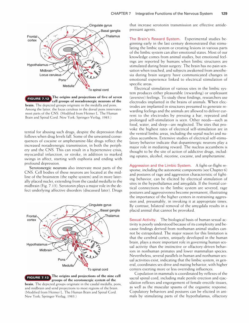
Serotonergic neurons also innervate most parts of theCNS. Cell bodies of these neurons are located at the mid-line of the brainstem (the raphe system) and in more later-ally placed nuclei, extending from the caudal medulla to themidbrain (Fig. 7.13). Serotonin plays a major role in the de-fect underlying affective disorders (discussed later). Drugs
that increase serotonin transmission are effective antide-pressant agents.
The Brain’s Reward System. Experimental studies be-ginning early in the last century demonstrated that stimu-lating the limbic system or creating lesions in various partsof the limbic system can alter emotional states. Most of ourknowledge comes from animal studies, but emotional feel-ings are reported by humans when limbic structures arestimulated during brain surgery. The brain has no pain sen-sation when touched, and subjects awakened from anesthe-sia during brain surgery have communicated changes inemotional experience linked to electrical stimulation ofspecific areas.
Electrical stimulation of various sites in the limbic sys-tem produces either pleasurable (rewarding) or unpleasant(aversive) feelings. To study these findings, researchers useelectrodes implanted in the brains of animals. When elec-trodes are implanted in structures presumed to generate re-warding feelings and the animals are allowed to deliver cur-rent to the electrodes by pressing a bar, repeated andprolonged self-stimulation is seen. Other needs—such asfood, water, and sleep—are neglected. The sites that pro-voke the highest rates of electrical self-stimulation are inthe ventral limbic areas, including the septal nuclei and nu-cleus accumbens. Extensive studies of electrical self-stimu-latory behavior indicate that dopaminergic neurons play amajor role in mediating reward. The nucleus accumbens isthought to be the site of action of addictive drugs, includ-ing opiates, alcohol, nicotine, cocaine, and amphetamine.
Aggression and the Limbic System. A fight-or-flight re-sponse, including the autonomic components (see Chapter 6)and postures of rage and aggression characteristic of fight-ing behavior, can be elicited by electrical stimulation ofsites in the hypothalamus and amygdala. If the frontal cor-tical connections to the limbic system are severed, ragepostures and aggressiveness become permanent, illustratingthe importance of the higher centers in restraining aggres-sion and, presumably, in invoking it at appropriate times.By contrast, bilateral removal of the amygdala results in aplacid animal that cannot be provoked.
Sexual Activity. The biological basis of human sexual ac-tivity is poorly understood because of its complexity and be-cause findings derived from nonhuman animal studies can-not be extrapolated. The major reason for this limitation isthat the cerebral cortex, uniquely developed in the humanbrain, plays a more important role in governing human sex-ual activity than the instinctive or olfactory-driven behav-iors in nonhuman primates and lower mammalian species.Nevertheless, several parallels in human and nonhuman sex-ual activities exist, indicating that the limbic system, in gen-eral, coordinates sex drive and mating behavior, with highercenters exerting more or less overriding influences.
Copulation in mammals is coordinated by reflexes of thesacral spinal cord, including male penile erection and ejac-ulation reflexes and engorgement of female erectile tissues,as well as the muscular spasms of the orgasmic response.Copulatory behaviors and postures can be elicited in ani-mals by stimulating parts of the hypothalamus, olfactory
CHAPTER 7 Integrative Functions of the Nervous System 129
Medulla
Pons
Midbrain
Hypothalamus
Frontalcortex
Cingulate gyrus
Basal ganglia
Thalamus
To spinal cord
Locus ceruleus
The origins and projections of five of seven
cell groups of noradrenergic neurons of the
brain. The depicted groups originate in the medulla and pons.Among the latter, the locus ceruleus in the dorsal pons innervatesmost parts of the CNS. (Modified from Heimer L. The HumanBrain and Spinal Cord. New York: Springer-Verlag, 1983.)
FIGURE 7.12
Medulla
Pons
Midbrain
Hypothalamus
Frontalcortex
Cingulate gyrus
Basal ganglia
Thalamus
To spinal cord
The origins and projections of the nine cell
groups of the serotonergic system of the
brain. The depicted groups originate in the caudal medulla, pons,and midbrain and send projections to most regions of the brain.(Modified from Heimer L. The Human Brain and Spinal Cord.New York: Springer-Verlag, 1983.)
FIGURE 7.13

130 PART II NEUROPHYSIOLOGY
system, and other limbic areas, resulting in mounting be-havior in males and lordosis (arching the back and raisingthe tail) in females. Ablation studies have shown that sexualbehavior also requires an intact connection of the limbicsystem with the frontal cortex.
Olfactory cues are important in initiating mating activityin seasonal breeders. Driven by the hypothalamus’ endoge-nous seasonal clock, the anterior and preoptic areas of thehypothalamus initiate hormonal control of the gonads.Hormonal release leads to the secretion of odorants(pheromones) by the female reproductive tract, signalingthe onset of estrus and sexual receptivity to the male. Theodorant cues are powerful stimulants, acting at extremelylow concentrations to initiate mating behavior in males. Theolfactory system, by virtue of its direct connections with thelimbic system, facilitates the coordination of behavioral, en-docrine, and autonomic responses involved in mating.
Although human and nonhuman primates are not sea-sonal breeders (mating can occur on a continual basis), ves-tiges of this pattern remain. These include the importanceof the olfactory and limbic systems and the role of the hy-pothalamus in cyclic changes in female ovarian functionand the continuous regulation of male testicular function.More important determinants of human sexual activity arethe higher cortical functions of learning and memory,which serve to either reinforce or suppress the signals thatinitiate sexual responding, including the sexual reflexes co-ordinated by the sacral spinal cord.
Psychiatric Disorders Involve the Limbic System
The major psychiatric disorders, including affective disor-ders and schizophrenia, are disabling diseases with a ge-netic predisposition and no known cure. The biologicalbasis for these disorders remains obscure, particularly therole of environmental influences on individuals with a ge-netic predisposition to developing a disorder. Alteredstates of the brain’s monoaminergic systems have been amajor focus as possible underlying factors, based on ex-tensive human studies in which neurochemical imbalancesin catecholamines, acetylcholine, and serotonin have beenobserved. Another reason for focusing on the monoamin-ergic systems is that the most effective drugs used in treat-ing psychiatric disorders are agents that alter monoamin-ergic transmission.
Affective Disorders. The affective disorders include ma-jor depression, which can be so profound as to provoke sui-cide, and bipolar disorder (or manic-depressive disorder),in which periods of profound depression are followed byperiods of mania, in a cyclic pattern. Biochemical studiesindicate that depressed patients show decreased use ofbrain NE. In manic periods, NE transmission increases.Whether in depression or in mania, all patients seem tohave decreased brain serotonergic transmission, suggestingthat serotonin may exert an underlying permissive role inabnormal mood swings, in contrast with norepinephrine,whose transmission, in a sense, titrates the mood fromhighest to lowest extremes.
The most effective treatments for depression, includingantidepressant drugs such as MAOIs and selective serotonin
reuptake inhibitors (SSRIs) and electroconvulsive therapy,have in common the ability to stimulate both noradrenergicand serotonergic neurons serving the limbic system. A ther-apeutic response to these treatments ensues only after treat-ment is repeated over time. Similarly, when treatment stops,symptoms may not reappear for several weeks. This time lagin treatment response is presumably due to alterations in thelong-term regulation of receptor and second messenger sys-tems in relevant regions of the brain.
The most effective long-term treatment for mania islithium, although antipsychotic (neuroleptic) drugs, whichblock dopamine receptors, are effective in the acute treat-ment of mania. The therapeutic actions of lithium remainunknown, but the drug has an important action on a recep-tor-mediated second messenger system. Lithium interfereswith regeneration of phosphatidylinositol in neuronalmembranes by blocking the hydrolysis of inositol-1-phos-phate. Depletion of phosphatidylinositol in the membranerenders it incapable of responding to receptors that use thissecond messenger system.
Schizophrenia. Schizophrenia is the collective name fora group of psychotic disorders that vary greatly in symp-toms among individuals. The features most commonly ob-served are thought disorder, inappropriate emotional re-sponse, and auditory hallucinations. While the biochemicalimbalance resulting in schizophrenia is poorly understood,the most troubling symptoms of schizophrenia are amelio-rated by neuroleptic drugs, which block dopamine recep-tors in the limbic system.
Current research is focused on finding the subtype ofdopamine receptor that mediates mesocortical/mesolimbicdopaminergic transmission but does not affect the nigrostri-atal system, which controls motor function (see Fig. 7.12). Sofar, neuroleptic drugs that block one pathway almost alwaysblock the other as well, leading to unwanted neurologicalside effects, including abnormal involuntary movements (tar-dive dyskinesia) after long-term treatment or parkinsonismin the short term. Similarly, some patients with Parkinson’sdisease who receive L-DOPA to augment dopaminergictransmission in the nigrostriatal pathway must be taken offthe medication because they develop psychosis.
Memory and Learning Require the
Cerebral Cortex and Limbic System
Memory and learning are inextricably linked because partof the learning process involves the assimilation of new in-formation and its commitment to memory. The most likelysites of learning in the human brain are the large associationareas of the cerebral cortex, in coordination with subcorti-cal structures deep in the temporal lobe, including the hip-pocampus and amygdala. The association areas draw onsensory information received from the primary visual, audi-tory, somatic sensory, and olfactory cortices and on emo-tional feelings transmitted via the limbic system. This in-formation is integrated with previously learned skills andstored memory, which presumably also reside in the asso-ciation areas.
The learning process itself is poorly understood, but itcan be studied experimentally at the synaptic level in iso-
lated slices of mammalian brain or in more simple inver-tebrate nervous systems. Synapses subjected to repeatedpresynaptic neuronal stimulation show changes in theexcitability of postsynaptic neurons. These changes in-clude the facilitation of neuronal firing, altered patternsof neurotransmitter release, second messenger formation,and, in intact organisms, evidence that learning occurred.The phenomenon of increased excitability and alteredchemical state on repeated synaptic stimulation is knownas long-term potentiation, a persistence beyond the ces-sation of electrical stimulation, as is expected of learningand memory. An early event in long-term potentiation isa series of protein phosphorylations induced by receptor-activated second messengers and leading to activation ofa host of intracellular proteins and altered excitability. Inaddition to biochemical changes in synaptic efficacy as-sociated with learning at the cellular level, structural al-terations occur. The number of connections between setsof neurons increases as a result of experience.
Much of our knowledge about human memory forma-tion and retrieval is based on studies of patients in whomstroke, brain injury, or surgery resulted in memory dis-orders. Such knowledge is then examined in more rigor-ous experiments in nonhuman primates capable of cog-nitive functions. From these combined approaches, weknow that the prefrontal cortex is essential for coordi-nating the formation of memory, starting from a learningexperience in the cerebral cortex, then processing theinformation and communicating it to the subcorticallimbic structures. The prefrontal cortex receives sensoryinput from the parietal, occipital, and temporal lobesand emotional input from the limbic system. Drawing onskills such as language and mathematical ability, the pre-frontal cortex integrates these inputs in light of previ-ously acquired learning. The prefrontal cortex can thusbe considered the site of working memory, where newexperiences are processed, as opposed to sites that con-solidate the memory and store it. The processed infor-mation is then transmitted to the hippocampus, where itis consolidated over several hours into a more permanentform that is stored in, and can be retrieved from, the as-sociation cortices.
Declarative and Procedural Memory. A remarkablefinding from studies of surgical patients who had bilateralresections of the medial temporal lobe is that there aretwo fundamentally different memory systems in the brain.Declarative memory refers to memory of events and factsand the ability to consciously access them. Patients withbilateral medial temporal lobectomies lose their ability toform any new declarative memories. However, they retaintheir ability to learn and remember new skills and proce-dures. This type of memory is called procedural memoryand involves several different regions of the brain, de-pending on the type of procedure. In contrast to declara-tive memory, structures in the medial temporal lobe arenot involved in procedural memory. Learning and re-membering new motor skills and habits requires the stria-tum, motor areas of the cortex, and the cerebellum. Emo-tional associations require the amygdala. Conditionedreflexes require the cerebellum.
An early demonstration of the dichotomy between de-clarative and procedural memory came from studies by Dr.Brenda Milner on a patient of Dr. Wilder Penfield in themid-1950s. This patient (H.M.) had received a bilateralmedial temporal lobectomy to treat severe epilepsy and,since that time, has been unable to form any new declara-tive memories. This deficit is called anterograde amnesia.Dr. Milner was quite surprised to learn that H.M. couldlearn a relatively difficult mirror-drawing task, in which(like anyone else) he got better with repeated trials and re-tained the skill over time. However, he could not remem-ber ever having done the task before.
Short-Term Memory. Declarative memory can be di-vided into that which can be recalled for only a brief period(seconds to minutes), and that which can be recalled forweeks to years. Newly acquired learning experiences can bereadily recalled for only a few minutes or more using short-term memory. An example of short-term memory is look-ing up a telephone number, repeating it mentally until youfinish dialing the number, then promptly forgetting it asyou focus your attention on starting the conversation.Short-term memory is a product of working memory; thedecision to process information further for permanent stor-age is based on judgment as to its importance or on whetherit is associated with a significant event or emotional state.An active process involving the hippocampus must be em-ployed to make a memory more permanent.
Long-Term Memory. The conversion of short-term tolong-term memory is facilitated by repetition, by addingmore than one sensory modality to learn the new experience(e.g., writing down a newly acquired fact at the same timeone hears it spoken) and, even more effective, by tying theexperience (through the limbic system) to a strong, mean-ingful emotional context. The role of the hippocampus inconsolidating the memory is reinforced by its participationin generating the emotional state with which the new expe-rience is associated. As determined by studying patientssuch as H.M., the most important regions of the medial tem-poral lobe for long-term declarative memory formation arethe hippocampus and parahippocampal cortex.
Once a long-term memory is formed, the hippocampusis not required for subsequent retrieval of the memory.Thus, H.M. showed no evidence of a loss of memories laiddown prior to surgery; this type of memory loss is known asretrograde amnesia. Nor was there loss of intellectual ca-pacity, mathematical skills, or other cognitive functions.An extreme example of H.M.’s memory loss is that Dr. Mil-ner, who worked with him for years, had to introduce her-self to her patient every time they met, even though hecould readily remember people and events that had oc-curred before his surgery.
Cholinergic Innervation. The primacy of the hip-pocampus and its connections with the base of the fore-brain for memory formation implicates acetylcholine as amajor transmitter in cognitive function and learning andmemory. The basal forebrain region contains prominentpopulations of cholinergic neurons that project to thehippocampus and to all regions of the cerebral cortex
CHAPTER 7 Integrative Functions of the Nervous System 131
132 PART II NEUROPHYSIOLOGY
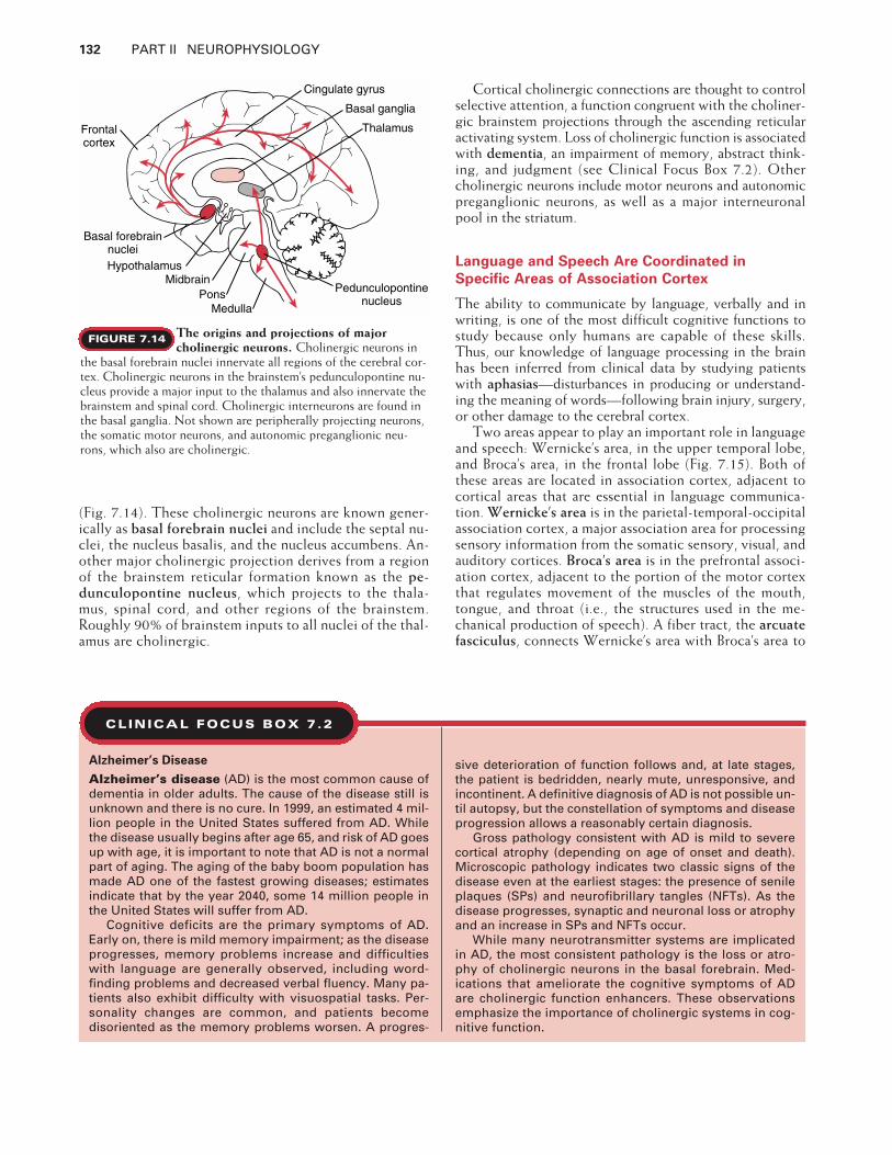
(Fig. 7.14). These cholinergic neurons are known gener-ically as basal forebrain nuclei and include the septal nu-clei, the nucleus basalis, and the nucleus accumbens. An-other major cholinergic projection derives from a regionof the brainstem reticular formation known as the pe-dunculopontine nucleus, which projects to the thala-mus, spinal cord, and other regions of the brainstem.Roughly 90% of brainstem inputs to all nuclei of the thal-amus are cholinergic.
Cortical cholinergic connections are thought to controlselective attention, a function congruent with the choliner-gic brainstem projections through the ascending reticularactivating system. Loss of cholinergic function is associatedwith dementia, an impairment of memory, abstract think-ing, and judgment (see Clinical Focus Box 7.2). Othercholinergic neurons include motor neurons and autonomicpreganglionic neurons, as well as a major interneuronalpool in the striatum.
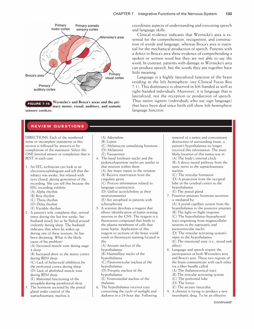
Language and Speech Are Coordinated in
Specific Areas of Association Cortex
The ability to communicate by language, verbally and inwriting, is one of the most difficult cognitive functions tostudy because only humans are capable of these skills.Thus, our knowledge of language processing in the brainhas been inferred from clinical data by studying patientswith aphasias—disturbances in producing or understand-ing the meaning of words—following brain injury, surgery,or other damage to the cerebral cortex.
Two areas appear to play an important role in languageand speech: Wernicke’s area, in the upper temporal lobe,and Broca’s area, in the frontal lobe (Fig. 7.15). Both ofthese areas are located in association cortex, adjacent tocortical areas that are essential in language communica-tion. Wernicke’s area is in the parietal-temporal-occipitalassociation cortex, a major association area for processingsensory information from the somatic sensory, visual, andauditory cortices. Broca’s area is in the prefrontal associ-ation cortex, adjacent to the portion of the motor cortexthat regulates movement of the muscles of the mouth,tongue, and throat (i.e., the structures used in the me-chanical production of speech). A fiber tract, the arcuatefasciculus, connects Wernicke’s area with Broca’s area to
MedullaPons
MidbrainHypothalamus
Frontalcortex
Cingulate gyrus
Basal ganglia
Thalamus
Pedunculopontinenucleus
Basal forebrainnuclei
The origins and projections of major
cholinergic neurons. Cholinergic neurons inthe basal forebrain nuclei innervate all regions of the cerebral cor-tex. Cholinergic neurons in the brainstem’s pedunculopontine nu-cleus provide a major input to the thalamus and also innervate thebrainstem and spinal cord. Cholinergic interneurons are found inthe basal ganglia. Not shown are peripherally projecting neurons,the somatic motor neurons, and autonomic preganglionic neu-rons, which also are cholinergic.
FIGURE 7.14
CLINICAL FOCUS BOX 7.2
Alzheimer’s Disease
Alzheimer’s disease (AD) is the most common cause ofdementia in older adults. The cause of the disease still isunknown and there is no cure. In 1999, an estimated 4 mil-lion people in the United States suffered from AD. Whilethe disease usually begins after age 65, and risk of AD goesup with age, it is important to note that AD is not a normalpart of aging. The aging of the baby boom population hasmade AD one of the fastest growing diseases; estimatesindicate that by the year 2040, some 14 million people inthe United States will suffer from AD.
Cognitive deficits are the primary symptoms of AD.Early on, there is mild memory impairment; as the diseaseprogresses, memory problems increase and difficultieswith language are generally observed, including word-finding problems and decreased verbal fluency. Many pa-tients also exhibit difficulty with visuospatial tasks. Per-sonality changes are common, and patients becomedisoriented as the memory problems worsen. A progres-
sive deterioration of function follows and, at late stages,the patient is bedridden, nearly mute, unresponsive, andincontinent. A definitive diagnosis of AD is not possible un-til autopsy, but the constellation of symptoms and diseaseprogression allows a reasonably certain diagnosis.
Gross pathology consistent with AD is mild to severecortical atrophy (depending on age of onset and death).Microscopic pathology indicates two classic signs of thedisease even at the earliest stages: the presence of senileplaques (SPs) and neurofibrillary tangles (NFTs). As thedisease progresses, synaptic and neuronal loss or atrophyand an increase in SPs and NFTs occur.
While many neurotransmitter systems are implicatedin AD, the most consistent pathology is the loss or atro-phy of cholinergic neurons in the basal forebrain. Med-ications that ameliorate the cognitive symptoms of ADare cholinergic function enhancers. These observationsemphasize the importance of cholinergic systems in cog-nitive function.
coordinate aspects of understanding and executing speechand language skills.
Clinical evidence indicates that Wernicke’s area is es-sential for the comprehension, recognition, and construc-tion of words and language, whereas Broca’s area is essen-tial for the mechanical production of speech. Patients witha defect in Broca’s area show evidence of comprehending aspoken or written word but they are not able to say theword. In contrast, patients with damage in Wernicke’s areacan produce speech, but the words they put together havelittle meaning.
Language is a highly lateralized function of the brainresiding in the left hemisphere (see Clinical Focus Box7.1). This dominance is observed in left-handed as well asright-handed individuals. Moreover, it is language that islateralized, not the reception or production of speech.Thus native signers (individuals who use sign language)that have been deaf since birth still show left-hemispherelanguage function.
CHAPTER 7 Integrative Functions of the Nervous System 133
Primarymotor cortex
Primary somaticsensory cortex
Primaryvisual cortex
Primaryauditory cortex
Wernicke's area
Broca's area
Wernicke’s and Broca’s areas and the pri-
mary motor, visual, auditory, and somatic
sensory cortices.
FIGURE 7.15
DIRECTIONS: Each of the numbereditems or incomplete statements in thissection is followed by answers or bycompletions of the statement. Select theONE lettered answer or completion that isBEST in each case.
1. An EEG technician can look at anelectroencephalogram and tell that thesubject was awake, but relaxed witheyes closed, during generation of therecording. She can tell this because theEEG recording exhibits(A) Alpha rhythm(B) Beta rhythm(C) Theta rhythm(D) Delta rhythm(E) Variable rhythm
2. A patient’s wife complains that, severaltimes during the last few weeks, herhusband struck her as he flailed aroundviolently during sleep. The husbandindicates that when he wakes upduring one of these sessions, he hasbeen dreaming. What is the likelycause of his problem?(A) Increased muscle tone during stage4 sleep(B) Increased drive to the motor cortexduring REM sleep(C) Lack of behavioral inhibition bythe prefrontal cortex during sleep(D) Lack of abolished muscle toneduring REM sleep(E) Abnormal functioning of theamygdala during paradoxical sleep
3. The hormone secreted by the pinealgland under control of thesuprachiasmatic nucleus is
(A) Adrenaline(B) Leptin(C) Melanocyte-stimulating hormone(D) Melatonin(E) Vasopressin
4. The basal forebrain nuclei and thepedunculopontine nuclei are similar inthat neurons within them(A) Are major inputs to the striatum(B) Receive innervation from thecingulate gyrus(C) Process information related tolanguage construction(D) Utilize acetylcholine as theirneurotransmitter(E) Are atrophied in patients withschizophrenia
5. A scientist develops a reagent thatallows identification of leptin-sensingneurons in the CNS. The reagent is afluorescent compound that binds tothe plasma membrane of cells thatsense leptin. Application of thisreagent to sections of the brain wouldresult in fluorescent staining located inthe(A) Arcuate nucleus of thehypothalamus(B) Mammillary nuclei of thehypothalamus(C) Paraventricular nucleus of thehypothalamus(D) Preoptic nucleus of thehypothalamus(E) Ventromedial nucleus of thethalamus
6. The hypothalamus receives cuesconcerning the cycle of sunlight anddarkness in a 24-hour day. Following
removal of a tumor and concomitantdestruction of surrounding tissue, apatient’s hypothalamus no longerreceived this information. The mostlikely location of this tumor was in(A) The body’s internal clock(B) A direct neural pathway from theoptic nerve to the suprachiasmaticnucleus(C) The reticular formation(D) A projection from the occipitallobe of the cerebral cortex to thehypothalamus(E) The pineal gland
7. Posterior pituitary hormone secretionis mediated by(A) A portal capillary system from thehypothalamus to the posterior pituitary(B) The fight-or-flight response(C) The hypothalamo-hypophysealtract originating from magnocellularneurons in the supraoptic andparaventricular nuclei(D) The reticular activating system’sinput to the hypothalamus(E) The emotional state (i.e., mood andaffect)
8. Language and speech require theparticipation of both Wernicke’s areaand Broca’s area. These two regions ofthe brain communicate with each othervia a fiber bundle called(A) The thalamocortical tract(B) The reticular activating system(C) The prefrontal lobe(D) The fornix(E) The arcuate fasciculus
9. A chemist is trying to produce a newneuroleptic drug. To be an effective
R E V I E W Q U E S T I O N S
(continued)

134 PART II NEUROPHYSIOLOGY
CASE STUDY FOR CHAPTER 4Dizziness
A 35-year-old man consulted his family physician be-cause of some recent episodes of what he described asdizziness. He was concerned that this complaint might berelated to a fall from a stepladder that had occurred theprevious month, although his symptoms did not beginimmediately after the incident. At the time of his visit tothe doctor, his symptoms are minimal, and he appears tobe in good general health. He states that the feeling ofdizziness, which also included sensations of nausea(without vomiting) and “ringing in the ears,” make himfeel as though his surroundings were spinning aroundhim. The episodes, which could last for several days at atime, are quite annoying and sufficiently severe to causehim concern for his safety on the job. When questioned,
he indicates that he also may not be hearing as well as heshould, but at other times he does not notice any hearingproblems. He further indicates that he may have had oc-casional dizzy spells before the ladder incident, but thatthey now appear to be much more frequent. The onlymedication he takes is aspirin for an occasionalheadache. He has no difficulty in following a moving fin-ger with his head held stationary, and on the day of thevisit he walks with a normal gait. He reports no light-headedness with moderate and continued exertion.
Gentle irrigation of his external ear canals with warmwater (at approximately 39�C) produces a feeling of dizzi-ness and nausea accompanied by nystagmus. The sub-jective sensations appeared to be the same for each ear.He is further evaluated with the Dix-Hallpike maneuver,and no sensations of vertigo are elicited during the posi-tional maneuvers. However, when he is rapidly rotated
neuroleptic, the new compound musttarget(A) Acetylcholine receptors(B) Dopamine receptors(C) Neuropeptide Y receptors(D) Norepinephrine receptors(E) Serotonin receptors
10.A patient suffered a stroke thatdestroyed the intralaminar nuclei ofthe thalamus. The location of thestroke was confirmed by magneticresonance imaging of the brain;however, an indication that the strokeaffected these nuclei was providedprior to imaging by an alteration inarousal in the patient. Which of thefollowing alterations in arousal is mostlikely following destruction of thesenuclei?(A) Loss of consciousness(B) Increased time spent in betarhythm(C) Increased attention to specificsensory inputs(D) Alterations in paradoxical, but notslow-wave sleep(E) Alteration in the period of thebiological clock
11.A blindfolded subject is asked toverbally identify a common objectpresented to her left hand. She is notallowed to touch the object with herright hand. Which of the followingstructures must be intact for her tocomplete this task?(A) The primary somatic sensorycortex on the left side of her brain(B) The primary visual cortex on theright side of her brain(C) The fornix(D) The corpus callosum(E) The hippocampus
12.A viral infection causes damage to bothhippocampi in a patient. This damagewould cause the patient to exhibitfunctional deficits in
(A) Recalling an old declarativememory(B) Recalling an old proceduralmemory(C) Forming a new short-term memory(D) Forming a new long-term memory(E) Forming a new procedural memory
13.An older gentleman is brought to theemergency department (ED) by hisdaughter. She had gone to his housefor lunch, which she did on a dailybasis. During her visit that day, shewas alarmed because his speech did notmake sense to her even though hetalked a lot and the words themselveswere clear. The physician in the EDinformed the daughter that her fatherhad most likely suffered a stroke thatdamaged(A) Broca’s area(B) The corpus callosum(C) The hippocampus(D) The arcuate fasciculus(E) Wernicke’s area
14.A woman agreed to visit her physicianbecause her husband was very worriedabout her behavior. She told thedoctor she felt great and that she wasgoing to run for governor of the statebecause she was smarter than thecurrent governor and people wouldimmediately agree to her plans. Herhusband said she had been sleepingvery little the last several days and hadspent several thousand dollars in ashopping spree the day before. Thiswas not typical behavior and hadsignificantly affected their ability tomeet their obligations for householdexpenses. The physician indicated adiagnosis of mania and started her on acourse of a drug that would decreaseneurotransmission in(A) Cholinergic pathways(B) Dopaminergic pathways(C) Glutaminergic pathways
(D) Noradrenergic pathways(E) Serotonergic pathways
15.Persons with mild cognitiveimpairments who smoke mayexperience a worsening of symptoms ifthey stop smoking. This worsening ofsymptoms is because nicotine acts asan agonist for receptors of a particularneurotransmitter. Thatneurotransmitter is(A) Acetylcholine(B) Dopamine(C) Neuropeptide Y(D) Nitric oxide(E) Serotonin
SUGGESTED READING
Bavelier D, Corina DP, Neville HJ. Brainand language: A perspective from signlanguage. Neuron 1998;21:275–278.
Cooke B, Hegstrom CD, Villenueve LS,Breedlove SM. Sexual differentiation ofthe vertebrate brain: Principles andmechanisms. Front Neuroendocr1998;19:323–362.
Dijk D-J, Duffy JF. Circadian regulation of human sleep and age-relatedchanges in its timing, consolidation andEEG characteristics. Ann Med1999;31:130–140.
Elmquist JK, Elias CF, Saper CB. From le-sions to leptin: Hypothalamic controlof food intake and body weight. Neu-ron 1999;22:221–232.
Gazzaniga MS. The split brain revisited.Sci Am 1998;279(1):50–55.
Kandel ER, Schwartz JH, Jessell TM. Prin-ciples of Neural Science. 4th Ed. NewYork: McGraw-Hill, 2000.
Milner B, Squire LR, Kandel ER. Cognitiveneuroscience and the study of memory.Neuron 1998;20:445–468.
Perry E, Walker M, Grace J, Perry R.Acetylcholine in mind: A neurotrans-mitter correlate of consciousness?Trends Neurosci 1999;22:273–280.
CASE STUDIES FOR PART II • • •
(continued)

in a swivel chair, he reports dizziness that was more se-vere than his usual symptoms. Rotation in the oppositedirection produced similar symptoms. His physician ad-vises him that there may be some appropriate specificmedications for his condition, but he would first like himto try a salt-restricted diet for the next 4 weeks. He alsoprescribes a mild diuretic.
Upon his return visit 4 weeks later, the patient reportsa gradual lessening of the frequency and duration of hisspells of dizziness and accompanying symptoms.
Questions
1. What features of this case would indicate that trauma fromthe stepladder incident was not the precipitating cause ofthe symptoms?
2. What factors would tend to rule out a diagnosis of benignparoxysmal positional vertigo?
3. Would the use of water at body temperature yield the samediagnostic information as warmer or cooler water?
4. Is the patient’s lack of light-headedness with moderate exer-cise relevant to the diagnosis of this problem?
5. Is it likely that the sensations produced by rapid rotation aremimicking those produced by his underlying disorder?
6. What is the purpose of the salt-restricted diet and diuretictherapy? Why was this tried before prescribing medicationfor his problem?
Answers to Case Study Questions for Chapter 4
1. Several features of this case suggest that trauma from thestepladder incident was not the precipitating cause. Thecaloric stimulation test and the rotation in the swivel chairindicate that his vestibular function is bilaterally symmetri-cal and of normal sensitivity. A defect arising from traumawould likely be localized to the injured side. His uncertaintyof the timing of the onset of the symptoms indicates thatthe problem may have preceded the accident (and, perhaps,led to it), and the lack of immediate appearance of symp-toms also tends to rule out trauma.
2. The relatively young age of the patient and the negativefindings from the Dix-Hallpike test argue against positionalvertigo and lend support to a tentative diagnosis ofMénière’s disease, as would the presence of tinnitus andthe fluctuating hearing loss. The patient’s positive responseto salt restriction and diuretic therapy is also indicative ofthis syndrome. (See answer to Question 6.)
3. The purpose of the application of water is to provide a ther-mal stimulus that will heat or cool the endolymph in thesemicircular canals and cause convection currents thatwould stimulate the ampullae. Use of water at body temper-ature would not produce this effect, and no symptomswould be elicited. Warmer or cooler water would each pro-duce symptoms of vertigo.
4. This observation tends to rule out cerebral ischemia as a re-sult of circulatory (vascular) or heart problems, factors thatwould also be more likely in an older patient.
5. The symptoms produced by the rotation are severe becauseof the simultaneous involvement of both sets of vestibularapparatus and the resulting heavy neural input, which islikely to be greater than that produced by his underlyingcondition.
6. The use of salt restriction and diuretics would reduce theoverall hydration state of his body and tend to reduce ab-normal pressure within the labyrinthine system. The use ofantimotion sickness drugs would interfere with the naturalneural compensation that would, it is hoped, reduce theseverity of the symptoms with time.
Reference
Drachman DA. A 69-year-old man with chronic dizziness. JAMA1998;280:2111–2118.
CASE STUDY FOR CHAPTER 5Upper Motor Neuron Lesion
A 50-year-old man comes for evaluation of persistent dif-ficulty using his right arm and leg. The patient was welluntil one month previously when he had abrupt onset ofweakness on the right side of his body while watching atelevision show. He was taken to the hospital by ambu-lance within one hour of onset of symptoms. The initialevaluation in the hospital emergency department showselevated blood pressure with values of 200 mm Hg sys-tolic and 150 mm Hg diastolic. The right arm and leg areseverely weak. Activity of the myotatic reflexes on theright side is very reduced in comparison with the leftside, where they are normal. Right side limb movementsare slightly improved by 12 hours after onset, but arestill moderately impaired on the fourth hospital day.
A magnetic resonance imaging study (MRI) of thebrain performed on the second day of hospitalizationshows a stroke involving the left cerebral hemisphere inthe region of the internal capsule. The blood pressure re-mains elevated, and medication to lower it is begun dur-ing the hospital stay. The patient is transferred to a reha-bilitation hospital on the fourth day for extensivephysical therapy to assist further recovery of neurologi-cal function.
At follow-up examination one month after onset ofthe stroke, the blood pressure remains normal on themedication that was started in the hospital. Neurologicalexamination demonstrates mild weakness of the rightarm and leg. There is still a slight but obvious delay be-tween asking the patient to move those limbs and themovement actually beginning. Passive movement of theright arm and leg by the physician provokes involuntarycontraction of the muscles in those limbs that seem tocounteract the attempted movement. Right side myotaticreflexes are very hyperactive compared with those ob-tained on the left. When the skin over the lateral plantararea of the right foot is stroked, the first toe extends in-voluntarily. When this maneuver is performed on theleft, the toes flex.
Questions
1. Explain the neurophysiology of the muscular weakness,slowness of movement initiation, increased muscle resist-ance to passive movement, and overactive myotatic re-flexes on the right side one month after stroke onset.
2. Explain why the toes extend on the right side and flex onthe left in response to plantar stimulation.
Answers to Case Study Questions for Chapter 5
1. The motor pathways that descend to the spinal cord fromhigher CNS levels initiate voluntary muscle action andalso regulate the sensitivity of the muscle stretch (my-otatic) reflex. Impairment of corticospinal tract input tothe alpha motor neuron pools results in weakness andslowness of initiation of voluntary movement. The corti-cospinal tract deficit also produces an increased sensitiv-ity of the spinal reflex pathways, resulting in overly vigor-ous muscle stretch reflexes. Muscle tone, the normalslight resistance to passive movement that is detectable ina relaxed muscle, becomes greatly increased and demon-strates a pattern that is called spasticity. Spastic tone ismost evident in the flexor muscles of the arm and the ex-tensor muscles of the leg.
2. The extensor movement of the first toe in response tostroking the plantar aspect of the foot, termed Babinskisign, is thought to occur because of modification of flexorwithdrawal reflexes secondary to the impaired input of the
CHAPTER 7 Integrative Functions of the Nervous System 135

136 PART II NEUROPHYSIOLOGY
corticospinal tract. The normal response is for the toes toflex when the plantar surface is stimulated.
The neurophysiological details of how the deficit incorticospinal input actually produces these commonlyencountered abnormalities in muscle tone and reflexpatterns are still not well understood. A current theory isthat the disturbance of central control reduces thethreshold of the stretch reflex but does not alter its gain.
References
Lance JW. The control of muscle tone, reflexes, and move-ment. Neurology 1980;30:1303–1313.Powers RK, Marder-Meyer J, Rymer WZ. Quantitative relationsbetween hypertonia and stretch reflex threshold in spastichemiparesis. Ann Neurol 1988;23:11–124.
CASE STUDY FOR CHAPTER 6Autonomic Dysfunction as a Result of CNS Disease
A 30-year-old patient came to the hospital emergencydepartment because of a terrible headache that beganseveral hours ago and did not improve. Previously hehad experienced only mild, infrequent tensionheadaches associated with stressful days. Because of theintensity of this new headache, he is treated with in-jectable analgesics and is admitted to the hospital forfurther observation.
During the next several hours, the patient’s level ofconsciousness declines to the point of responding onlyto painful stimuli. An emergency computed tomography(CT) scan of the brain demonstrates the presence ofblood diffusely in the subarachnoid space. The source ofthe blood is thought to be a ruptured cerebral arteryaneurysm.
During the next 24 hours, the patient’s ECG begins toshow abnormalities consisting of both tachycardia andchanges in the configuration of the waves suggestive ofa heart attack. The patient has no risk factors for prema-ture cardiac disease. A cardiology consultation is re-quested.
Questions
1. What is the explanation for the cardiac abnormalities in thissituation?
2. Describe two other scenarios in which there are prominentmanifestations of autonomic activation produced by abnor-malities in the central nervous system.
Answers to Case Study Questions for Chapter 6
1. The consulting cardiologist reviewed the situation andstated that the ECG abnormalities were all a result of sub-arachnoid blood and that an adrenergic antagonist medica-tion should be administered.
Blood released into the subarachnoid space by rup-ture of blood vessels or direct trauma to the brain canstimulate excessive activity of the sympathetic nervoussystem. Although a full explanation is still lacking, it ispostulated that the subarachnoid blood irritates the hy-pothalamus and autonomic regulatory areas in themedulla, resulting in excessive activation of the sympa-thetic pathways. This activation causes the secretion ofnorepinephrine from sympathetic nerve endings and epi-nephrine by the adrenal medulla. Direct stimulation ofthe sympathetic pathways that supply the heart can pro-duce the same ECG abnormalities in experimental ani-mals as were found in this patient. The heightened re-lease of norepinephrine and epinephrine stimulates thecardiac conducting system and may also produce directdamage of the myocardium. Treatment with medicationsthat attenuate the effects of sympathetic neurotransmit-ters can be lifesaving.
2. The Cushing response (described by famous neurosurgeonHarvey Cushing) consists of the development of hyperten-sion, bradycardia, and apnea in patients with increased in-tracranial pressure most often a result of tumors or other le-sions, such as hemorrhage, that compress the brain. Thepressure is transmitted downward to the brainstem and dis-torts the medulla, where the centers for blood pressure,heart rate, and respiratory drive originate. Correct interpre-tation of these abnormalities in vital signs permits begin-ning treatments that reduce intracranial pressure. These in-clude elevating the head of the bed, placing the patient onan artificial respirator, and then instituting hyperventilationto lower the blood PCO2 to produce cerebral vasoconstric-tion and giving mannitol to reduce the fluid content of thebrain temporarily.
Another autonomic reaction from the CNS that is uti-lized daily in hospitals is the response of fetal heart rateto compression of the head during labor. During uterinecontractions, the fetal head is temporarily compressed.As the fetal skull is still malleable because the bones ofthe cranium are not yet fused, the pressure of the con-traction is transmitted to the brain. The same mecha-nism of cardiac slowing as cited for the Cushing re-sponse is presumed to cause the temporary bradycardia.Slowing of greater than established normal limits indi-cates the fetus is suffering significant physiological dis-tress. Additional factors, such as umbilical cord com-pression, may also produce patterns of slowing outsideof the normal range.
Reference
Talman WT. The central nervous system and cardiovascularcontrol in health and disease. In: Low PA. Clinical AutonomicDisorders. 2nd Ed. Philadelphia: Lippincott-Raven, 1997
CASE STUDY FOR CHAPTER 7Stroke
A 67-year-old man was taken to see his physician by hiswife. For the preceding 2 days, the patient’s wife had no-ticed that he did not seem to make sense when he spoke.She also indicated that he seemed a little disorientedand did not respond appropriately to her questions. Hehas no obvious motor or somatic sensory deficits.
On examination, the physician concludes that theman had a stroke in a region of one of his cerebral hemi-spheres. As part of the diagnosis, the physician tests theman’s visual fields and notices a decreased awarenessof stimuli presented to one visual field.
Questions
1. Which side of the brain most likely suffered the stroke?2. Which regions of the hemisphere suffered the stroke?3. What information from the case history gives the answers
to questions 1 and 2?4. Which visual field is affected by the stroke?
Answers to Case Study Questions for Chapter 7
1. The stroke occurred on the left side of the brain.2. The stroke involved the superior posterior temporal lobe en-
compassing Wernicke’s area and the occipital lobe encom-passing the primary visual cortex.
3. Language deficits indicate involvement of the left hemi-sphere. The fluent but nonsensical speech indicates involve-ment of Wernicke’s area. The visual field deficit indicates aloss in the visual cortex. The lack of motor or somatic sen-sory deficits excludes the posterior frontal and anterior pari-etal lobes.
4. The right visual field would be affected, because visualfields are represented in the contralateral hemispheres.