Embed Size (px)
Citation preview

1
Chapter 06
Lecture and
Animation Outline
Copyright © The McGraw-Hill Companies, Inc. Permission required for reproduction or display.
See separate PowerPoint slides for all figures and
tables pre-inserted into PowerPoint without notes and
animations.
To run the animations you must be in Slideshow View. Use
the buttons on the animation to play, pause, and turn
audio/text on or off.
Please Note: Once you have used any of the animation
functions (such as Play or Pause), you must first click on the
slide’s background before you can advance to the next slide.

2
Chapter 6-Skeletal System
Functions
1. Support
2. Protect
3. Movement
4. Storage
5. Blood cell production

3
Components of Skeletal System
• Bone
• Cartilage:
reduce friction and model for bone formation
• Tendons:
attach bone to muscle
• Ligaments:
attach bone to bone
4
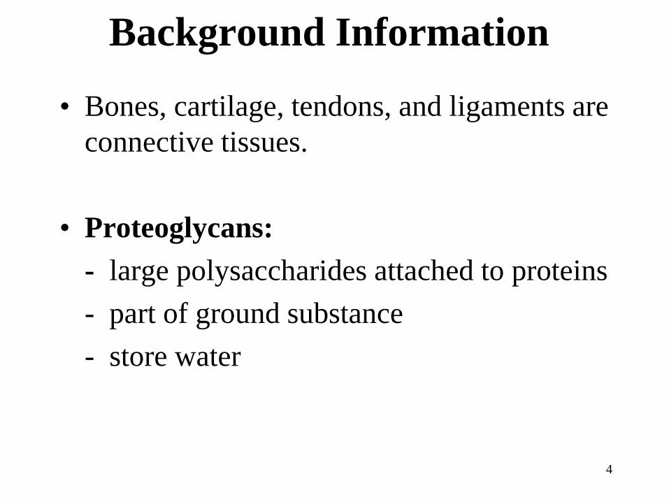
Background Information
• Bones, cartilage, tendons, and ligaments are
connective tissues.
• Proteoglycans:
- large polysaccharides attached to proteins
- part of ground substance
- store water
5
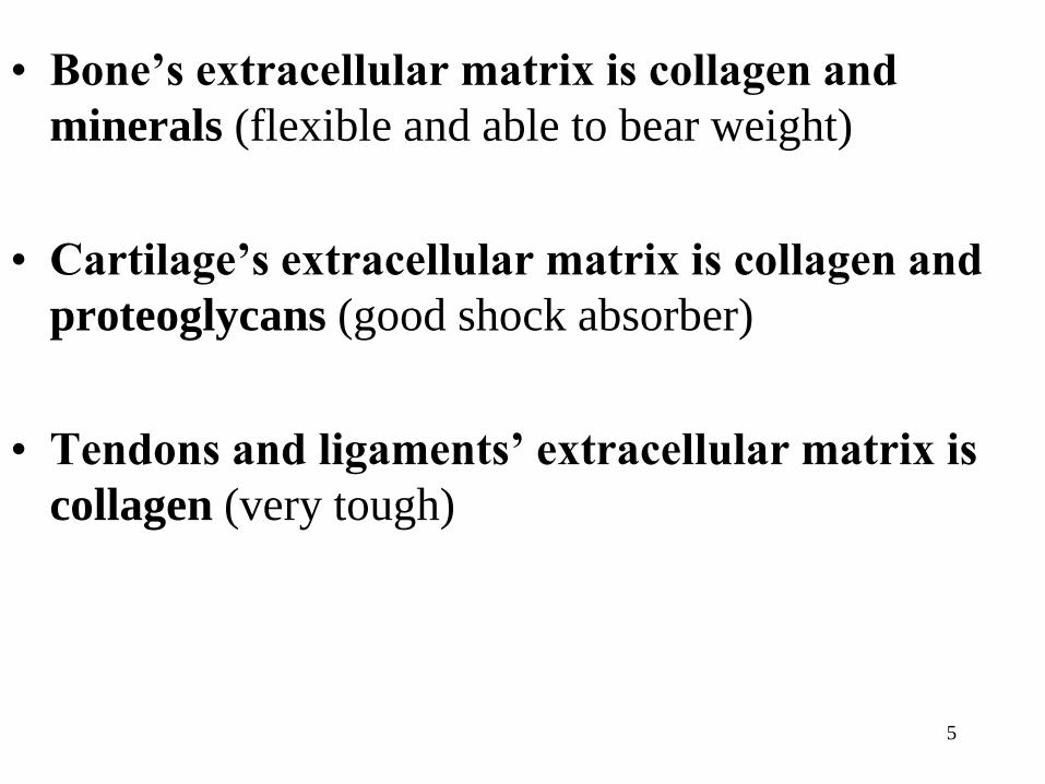
• Bone’s extracellular matrix is collagen and
minerals (flexible and able to bear weight)
• Cartilage’s extracellular matrix is collagen and
proteoglycans (good shock absorber)
• Tendons and ligaments’ extracellular matrix is
collagen (very tough)
6
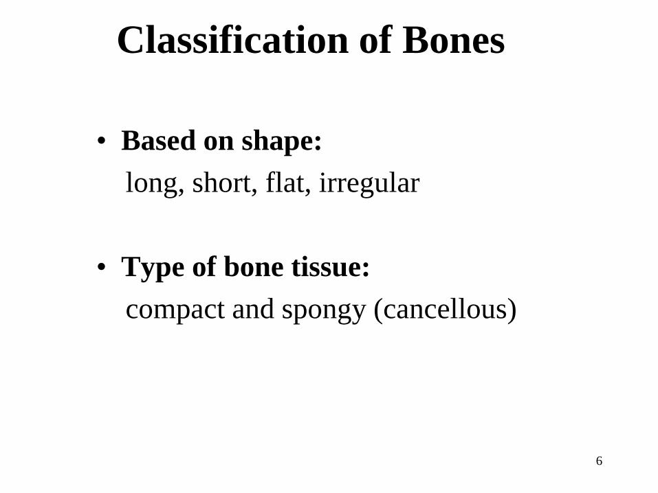
Classification of Bones
• Based on shape:
long, short, flat, irregular
• Type of bone tissue:
compact and spongy (cancellous)

7
Bone Shapes
• Long:
- Ex. Femur, tibia, fibula
• Short:
– Ex. Carpals, tarsals,
phlanges
• Flat:
– Ex. Ribs, sternum, skull
• Irregular:
– Ex. Vertebrae and facial
8
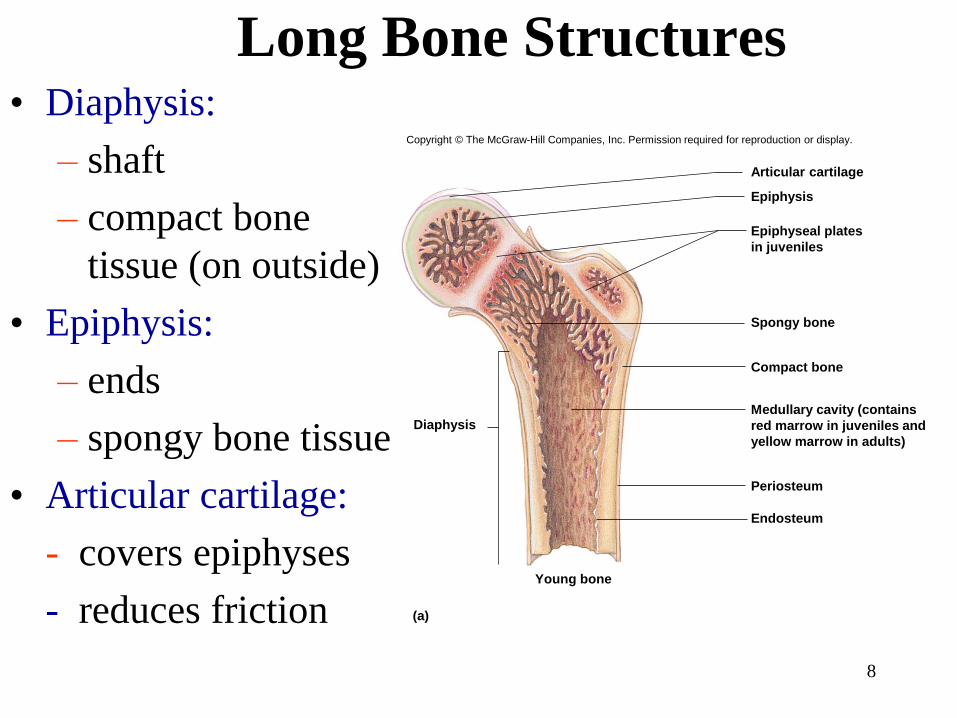
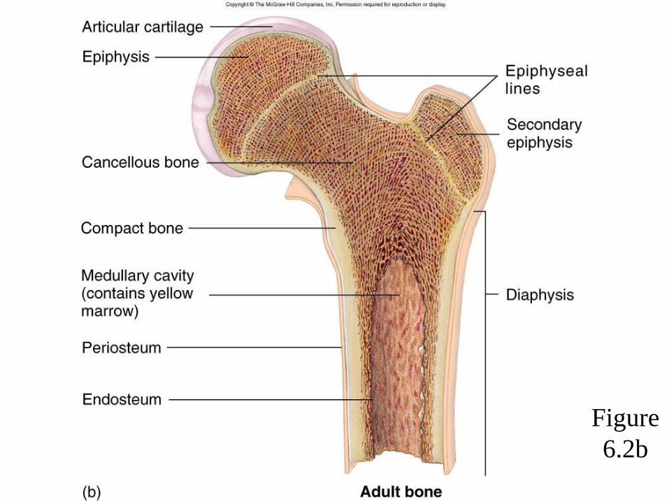
Long Bone Structures • Diaphysis:
– shaft
– compact bone
tissue (on outside)
• Epiphysis:
– ends
– spongy bone tissue
• Articular cartilage:
- covers epiphyses
- reduces friction
Endosteum
Periosteum
Medullary cavity (contains
red marrow in juveniles and
yellow marrow in adults)
Compact bone
Spongy bone
Epiphyseal plates
in juveniles
Epiphysis
Articular cartilage
Young bone
Diaphysis
(a)
Copyright © The McGraw-Hill Companies, Inc. Permission required for reproduction or display.

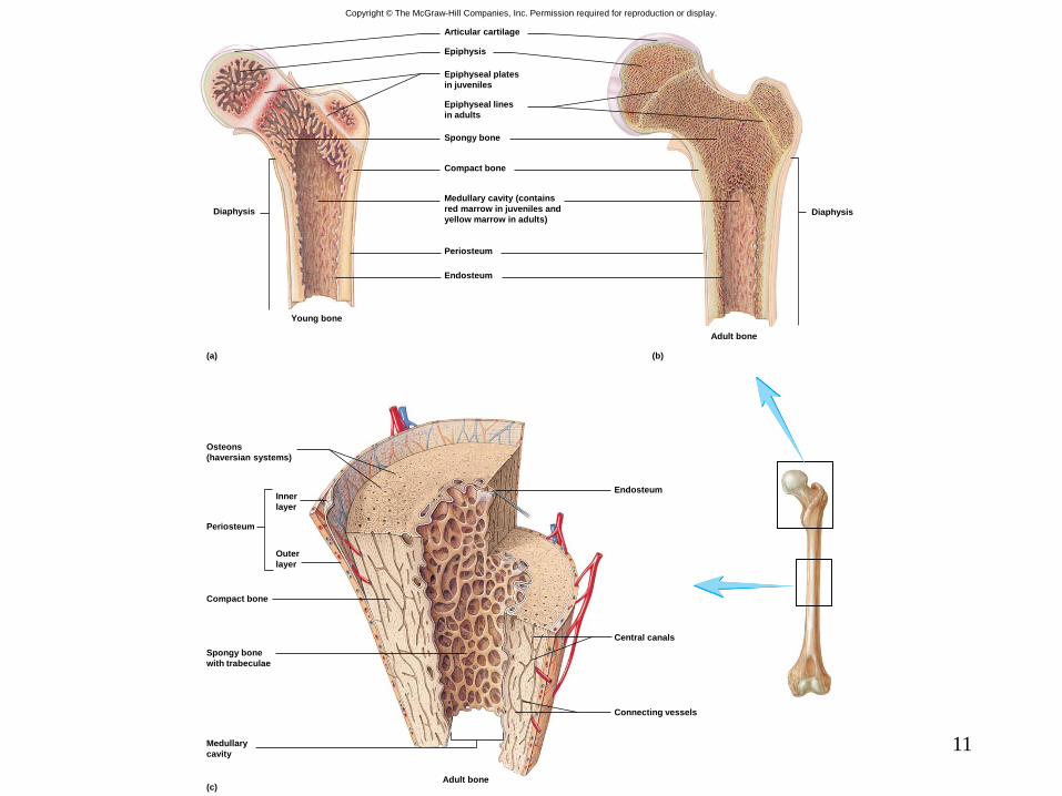
• Epiphyseal plate:
– site of growth
– between diaphysis
and epiphysis
• Medullary cavity:
– center of diaphysis
– red or yellow
marrow
Diaphysis
Adult bone
Endosteum
Periosteum
Medullary cavity (contains
red marrow in juveniles and
yellow marrow in adults)
Compact bone
Spongy bone
Epiphyseal lines
in adults
Epiphysis
Articular cartilage
(b)
Copyright © The McGraw-Hill Companies, Inc. Permission required for reproduction or display.

10
• Periosteum:
membrane around
bone’s outer surface
• Endosteum:
membrane that lines
medullary cavityEndosteum
Periosteum
Medullary cavity (contains
red marrow in juveniles and
yellow marrow in adults)
Compact bone
Spongy bone
Epiphyseal plates
in juveniles
Epiphysis
Articular cartilage
Young bone
Diaphysis
(a)
Copyright © The McGraw-Hill Companies, Inc. Permission required for reproduction or display.
11
Copyright © The McGraw-Hill Companies, Inc. Permission required for reproduction or display.
Diaphysis
Adult bone
Endosteum
Periosteum
Medullary cavity (contains
red marrow in juveniles and
yellow marrow in adults)
Compact bone
Spongy bone
Epiphyseal lines
in adults
Epiphyseal plates
in juveniles
Epiphysis
Articular cartilage
Young bone
Diaphysis
(a) (b)
Osteons
(haversian systems)
EndosteumInner
layer
Outer
layer
Periosteum
Compact bone
Central canals
Spongy bone
with trabeculae
Connecting vessels
Medullary
cavity
Adult bone(c)

12
Compact Bone Tissue
• Location:
outer part of diaphysis (long
bones) and thinner surfaces
of other bones
• Osteon:
- structural unit of compact
bone
- includes lamella, lacunae,
canaliculus, central canal,
osteocytes
• Lamella:
rings of bone matrix
Osteons
(haversian systems)
EndosteumInner
layer
Outer
layer
Periosteum
Compact bone
Central canals
Spongy bone
with trabeculae
Connecting vessels
Medullary
cavity
Adult bone(c)
Copyright © The McGraw-Hill Companies, Inc. Permission required for reproduction or display.

13
• Lacunae:
spaces between lamella
• Canaliculus:
- tiny canals
- transport nutrients and
remove waste
• Central canal:
- center of osteon
- contains blood vessels
Osteons
(haversian systems)
EndosteumInner
layer
Outer
layer
Periosteum
Compact bone
Central canals
Spongy bone
with trabeculae
Connecting vessels
Medullary
cavity
Adult bone(c)
Copyright © The McGraw-Hill Companies, Inc. Permission required for reproduction or display.

Copyright © The McGraw-Hill Companies, Inc. Permission required for reproduction or display.
Osteocytes in
lacunae
Canaliculi
Blood vessels
within a central
(Haversian) canal
Blood vessels
connecting to
a central canal
Blood vessel within
the periosteum
Periosteum
Lamellae
between osteons
Lamellae on
surface of boneOsteon
Blood vessel
connecting to
a central canal
between osteons
Lacunae
Canaliculi (b)(a)
Central canal
Concentric rings
of lamellae
Osteon
LM 400x
a: © Trent Stephens

15
• Cancellous bone
• Location: epiphyses of long bones and center of other bones
• Trabeculae: interconnecting rods, spaces contain marrow
• No osteons
Spongy Bone Tissue
16
Bone Cells
• Osteocytes:
maintain bone matrix
• Osteoblasts:
build bone
• Osteoclasts:
carve bone
17
Bone Formation
• Ossification:
process of bone formation
• Osteoblast’s role:
- build bone
- after an osteoblast becomes surrounded by bone
matrix it becomes an osteocyte
18
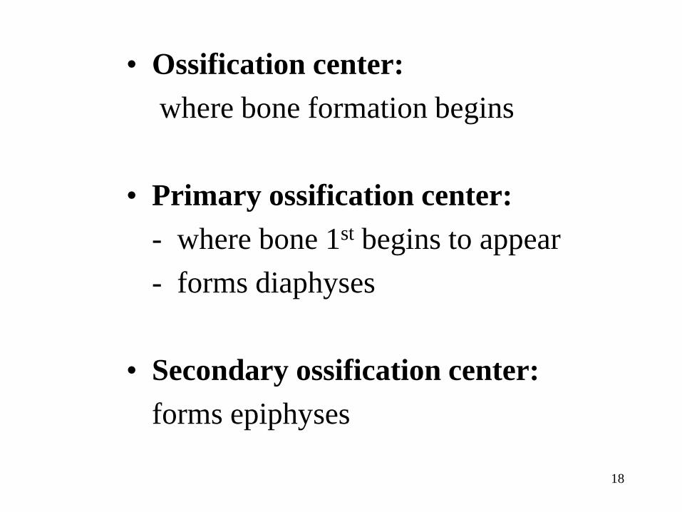
• Ossification center:
where bone formation begins
• Primary ossification center:
- where bone 1st begins to appear
- forms diaphyses
• Secondary ossification center:
forms epiphyses
19
Intramembranous Ossification
• Bone formation within connective tissue
membranes
• Osteoblasts build bone
• Ex. Skull bones


21
Endochondral Ossification
• Bone formation inside cartilage
• Cartilage models are replaced by bone
• Ex. All bones (except skull)
22
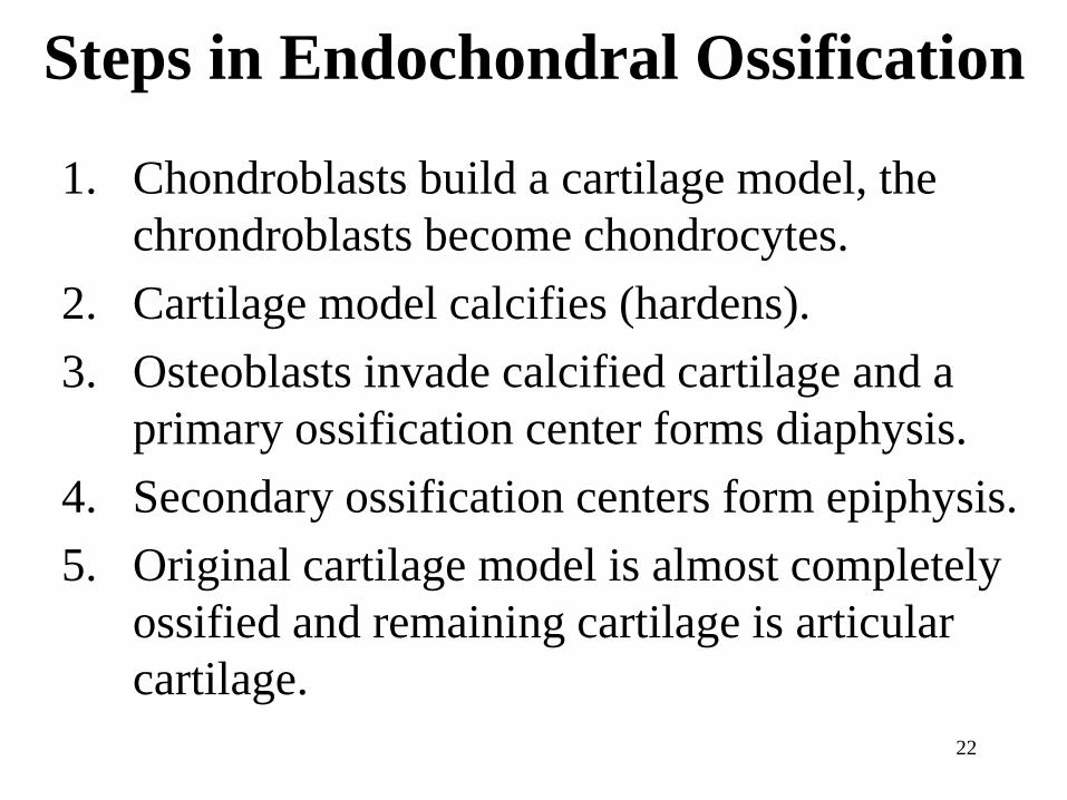
Steps in Endochondral Ossification
1. Chondroblasts build a cartilage model, the
chrondroblasts become chondrocytes.
2. Cartilage model calcifies (hardens).
3. Osteoblasts invade calcified cartilage and a
primary ossification center forms diaphysis.
4. Secondary ossification centers form epiphysis.
5. Original cartilage model is almost completely
ossified and remaining cartilage is articular
cartilage.

24
2
3
4
1
2
3
4
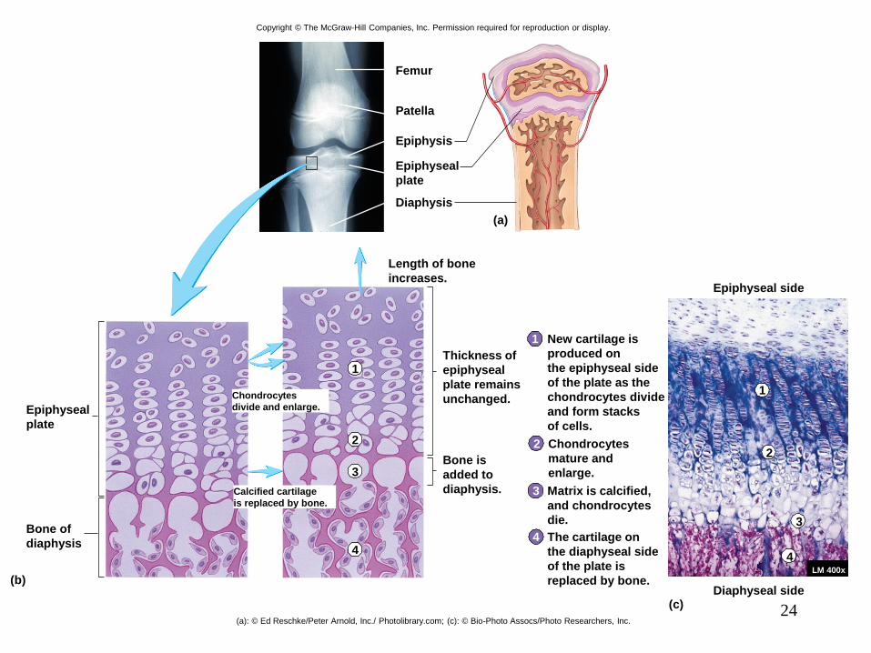
Calcified cartilage
is replaced by bone.
Chondrocytes
divide and enlarge.Epiphyseal
plate
Bone of
diaphysis
Length of bone
increases.
Thickness of
epiphyseal
plate remains
unchanged.
Bone is
added to
diaphysis.
The cartilage on
the diaphyseal side
of the plate is
replaced by bone.LM 400x
Matrix is calcified,
and chondrocytes
die.
Chondrocytes
mature and
enlarge.
New cartilage is
produced on
the epiphyseal side
of the plate as the
chondrocytes divide
and form stacks
of cells.
Epiphyseal side
Diaphyseal side
(a)
(b)
(c)
1
2
3
4
1
Diaphysis
Epiphyseal
plate
Epiphysis
Femur
Patella
Copyright © The McGraw-Hill Companies, Inc. Permission required for reproduction or display.
(a): © Ed Reschke/Peter Arnold, Inc./ Photolibrary.com; (c): © Bio-Photo Assocs/Photo Researchers, Inc.
25
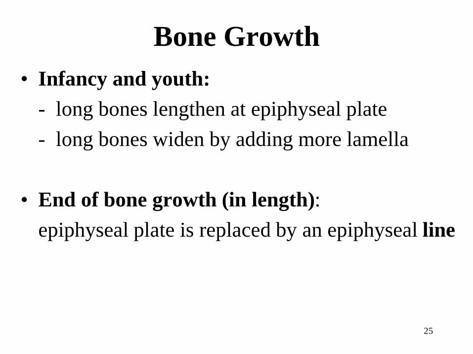
Bone Growth
• Infancy and youth:
- long bones lengthen at epiphyseal plate
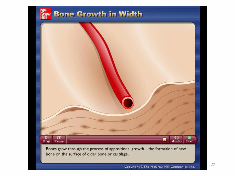
- long bones widen by adding more lamella
• End of bone growth (in length):
epiphyseal plate is replaced by an epiphyseal line
Figure
6.2b
27
Please note that due to differing
operating systems, some animations
will not appear until the presentation is
viewed in Presentation Mode (Slide
Show view). You may see blank slides
in the “Normal” or “Slide Sorter” views.
All animations will appear after viewing
in Presentation Mode and playing each
animation. Most animations will require
the latest version of the Flash Player,
which is available at
http://get.adobe.com/flashplayer.
Bone Remodeling
• What is it?
- removal of existing bone by osteoclasts and
deposition of new bone by osteoblasts
- occurs in all bones
- responsible for changes in bone shape, bone
repair, adjustment of bone to stress, and
calcium ion regulation
28
29
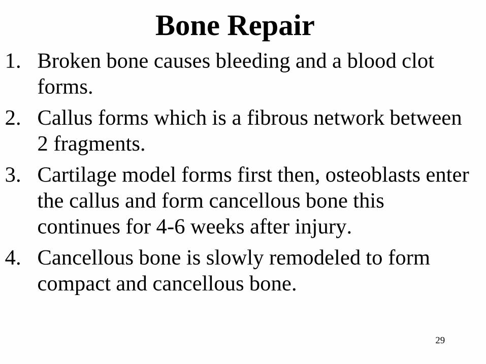
Bone Repair1. Broken bone causes bleeding and a blood clot
forms.
2. Callus forms which is a fibrous network between
2 fragments.
3. Cartilage model forms first then, osteoblasts enter
the callus and form cancellous bone this
continues for 4-6 weeks after injury.
4. Cancellous bone is slowly remodeled to form
compact and cancellous bone.


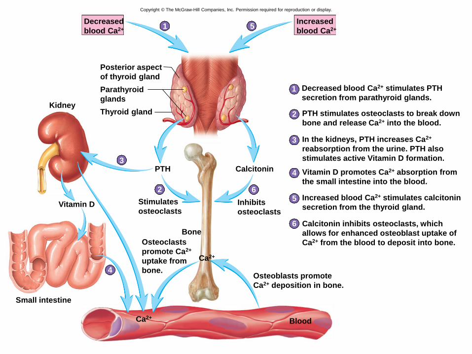
Bone and Calcium Homeostasis
• Bone is a major storage site for calcium
• Movement of calcium in and out of bone helps
determine blood levels of calcium
• Calcium moves into bone as osteoblasts build
new bone
• Calcium move out of bone as osteoclasts break
down bone
• Calcium homeostasis is maintained by
parathyroid hormone (PTH) and calcitonin
31
1
2
3
4
5
6
1
2
5
6
Blood
Small intestine
Osteoblasts promote
Ca2+ deposition in bone.
Vitamin D Stimulates
osteoclastsInhibits
osteoclasts
Calcitonin
Osteoclasts
promote Ca2+
uptake from
bone.
Increased
blood Ca2+
Decreased
blood Ca2+
Posterior aspect
of thyroid gland
Parathyroid
glands
Thyroid glandKidney
Ca2+
Bone
PTH
Ca2+
Decreased blood Ca2+ stimulates PTH
secretion from parathyroid glands.
PTH stimulates osteoclasts to break down
bone and release Ca2+ into the blood.
In the kidneys, PTH increases Ca2+
reabsorption from the urine. PTH also
stimulates active Vitamin D formation.
Vitamin D promotes Ca2+ absorption from
the small intestine into the blood.
Increased blood Ca2+ stimulates calcitonin
secretion from the thyroid gland.
Calcitonin inhibits osteoclasts, which
allows for enhanced osteoblast uptake of
Ca2+ from the blood to deposit into bone.
3
4
Copyright © The McGraw-Hill Companies, Inc. Permission required for reproduction or display.

33
Hematopoietic Tissue
• What is it?
tissue that makes blood cells
• Red marrow:
location of blood forming cells
• Yellow marrow:
mostly fat

34
• Location of hematopoietic tissue in newborns:
most bones (red marrow)
• Location of hematopoietic tissue in adults:
- red is replaced with yellow marrow
- red marrow is mainly in epiphyses of femur
and humerus
35
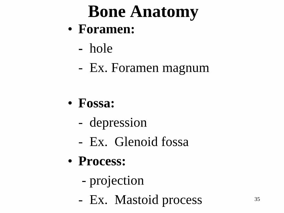
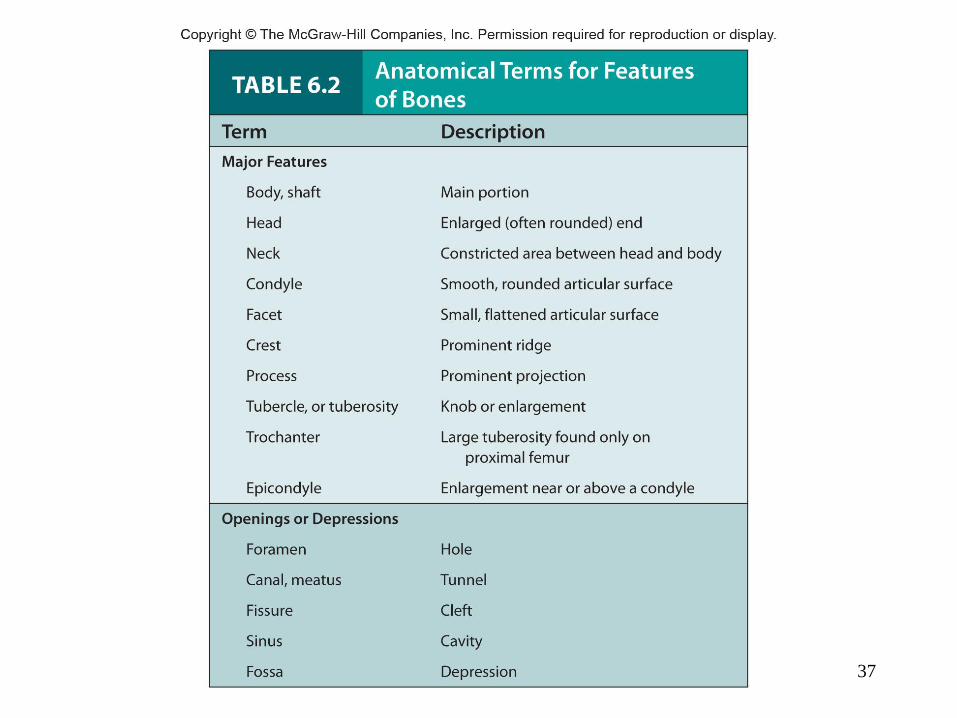
Bone Anatomy• Foramen:
- hole
- Ex. Foramen magnum
• Fossa:
- depression
- Ex. Glenoid fossa
• Process:
- projection
- Ex. Mastoid process

36
• Condyle:
- smooth, rounded end
- Ex. Occipital condyle
• Meatus:
- canal-like passageway
- Ex. External auditory meatus
• Tubercle:
- lump of bone
- Ex. Greater tubercle
37
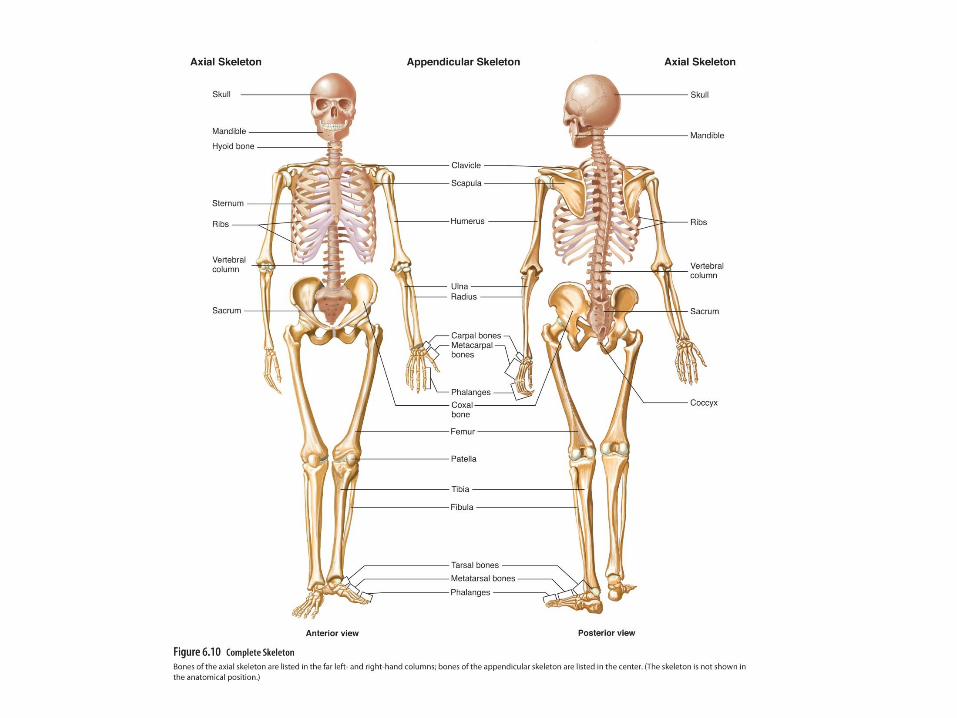
38
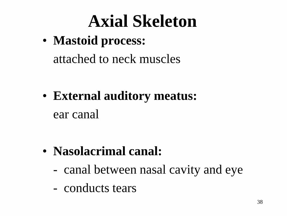
Axial Skeleton• Mastoid process:
attached to neck muscles
• External auditory meatus:
ear canal
• Nasolacrimal canal:
- canal between nasal cavity and eye
- conducts tears

39
• Styloid process:
attachment site for tongue
• Mandibular fossa:
depression where lower jaw and skull
meet
• Glenoid fossa:
where humerus meets scapula

40
• Hard palate:
roof of mouth
• Foramen magnum:
hole where spinal cord joins brainstem

41
• Zygomatic:
cheek bone
• Mandible:
lower jaw
• Maxilla:
upper jaw


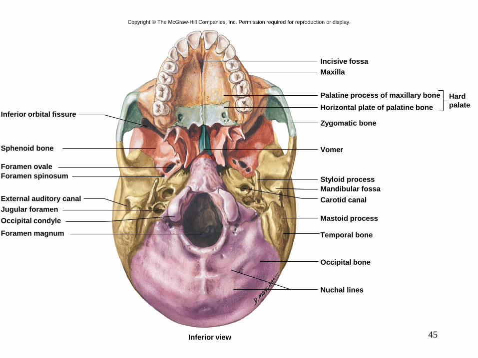
45
Copyright © The McGraw-Hill Companies, Inc. Permission required for reproduction or display.
Nuchal lines
Occipital bone
Temporal bone
Mastoid process
Carotid canal
Mandibular fossa
Styloid process
Hard
palate
Palatine process of maxillary bone
Horizontal plate of palatine bone
Zygomatic bone
Vomer
Incisive fossa
Maxilla
Inferior orbital fissure
Sphenoid bone
Foramen ovale
Foramen spinosum
External auditory canal
Jugular foramen
Occipital condyle
Foramen magnum
Inferior view
46
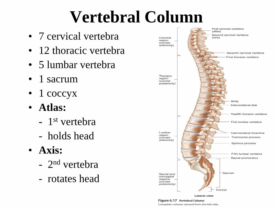
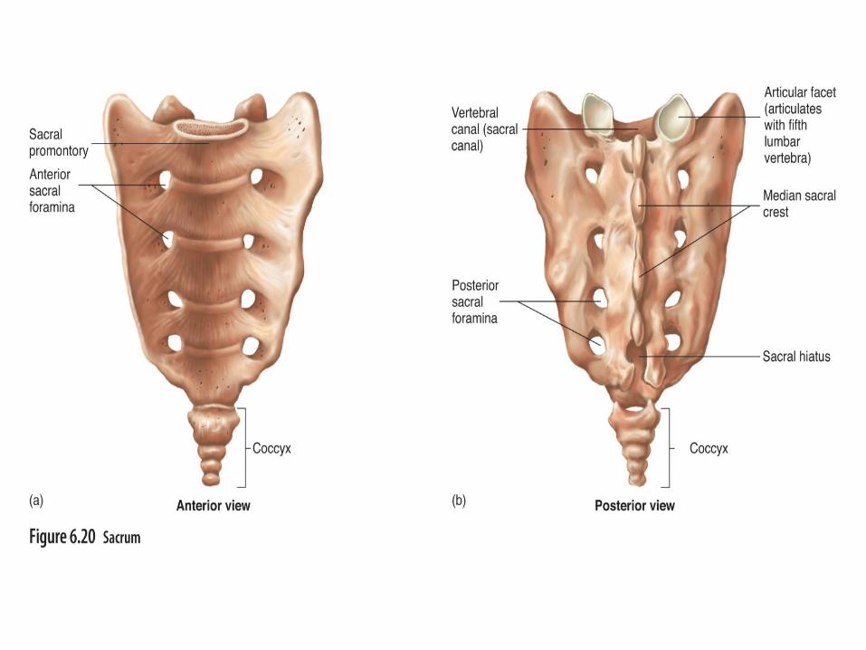
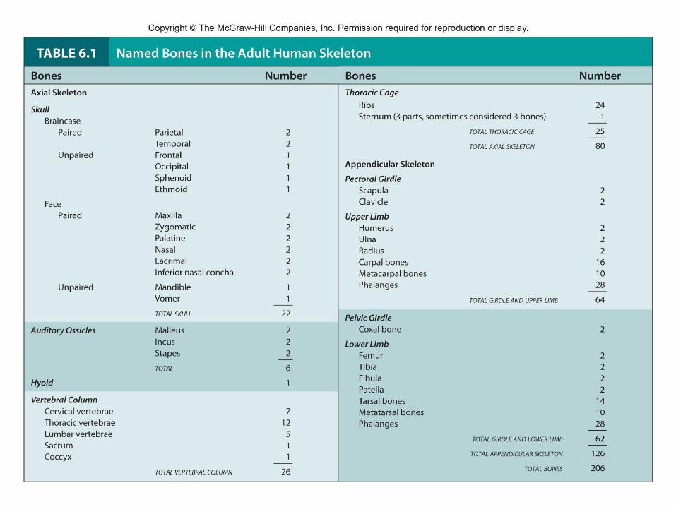
Vertebral Column• 7 cervical vertebra
• 12 thoracic vertebra
• 5 lumbar vertebra
• 1 sacrum
• 1 coccyx
• Atlas:
- 1st vertebra
- holds head
• Axis:
- 2nd vertebra
- rotates head

50
Functions of Vertebral Column
• Support
• Protect spinal cord
• Movement

51
Thoracic Cage• Protects vital organs
• 12 pair of ribs
• Sternum:
breastbone
• True ribs:
attach directly to sternum by cartilage
• False ribs:
attach indirectly to sternum by cartilage
• Floating ribs:
not attached to sternum

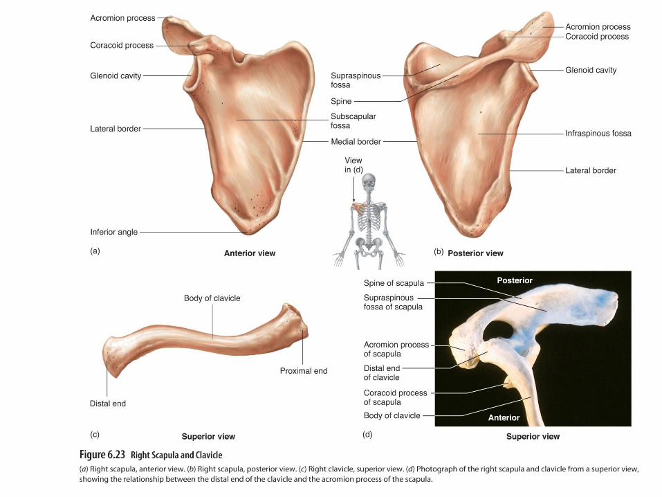
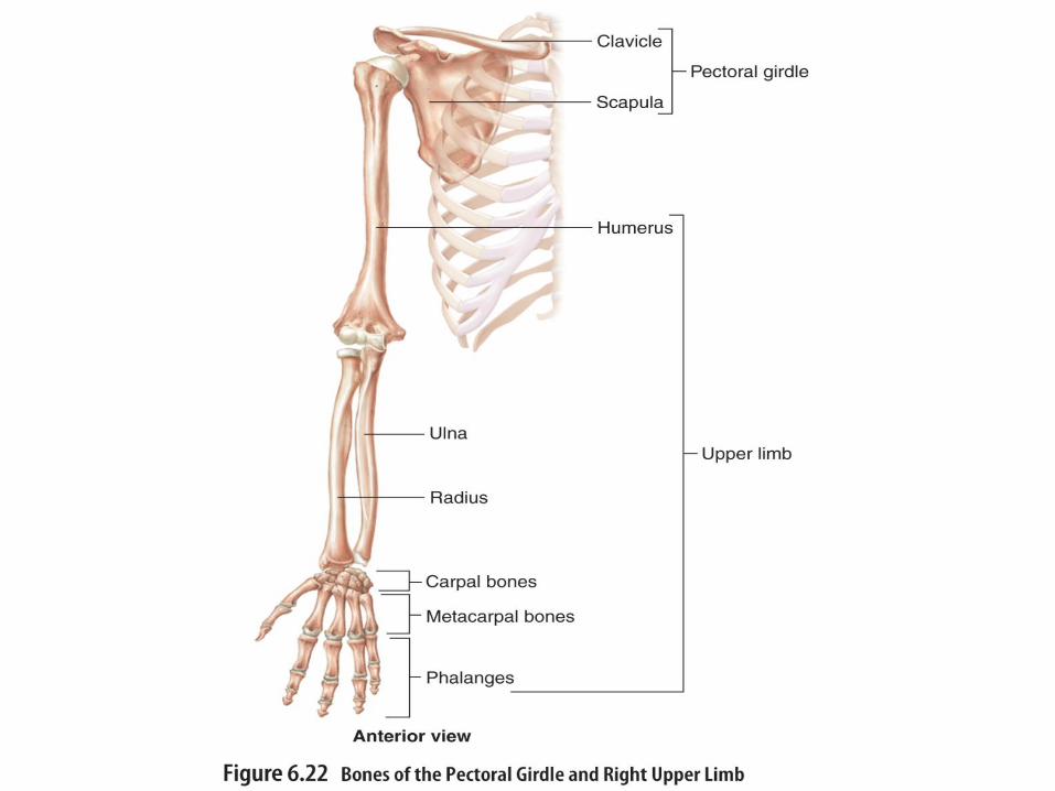
Pectoral Girdle
• Scapula:
shoulder blade
• Clavicle:
collar bone
53
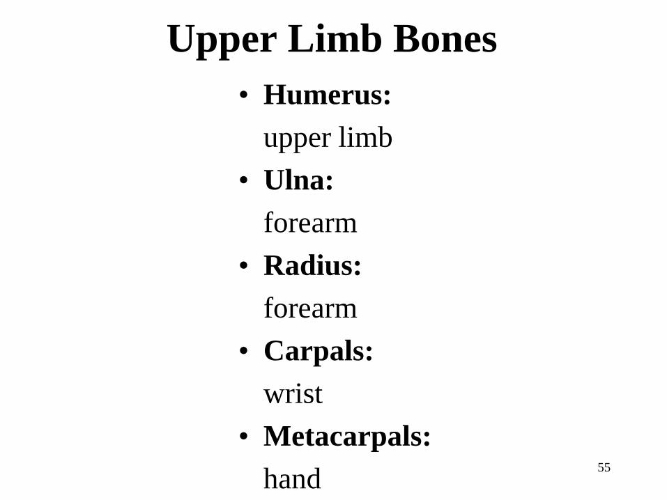
Upper Limb Bones
• Humerus:
upper limb
• Ulna:
forearm
• Radius:
forearm
• Carpals:
wrist
• Metacarpals:
hand55


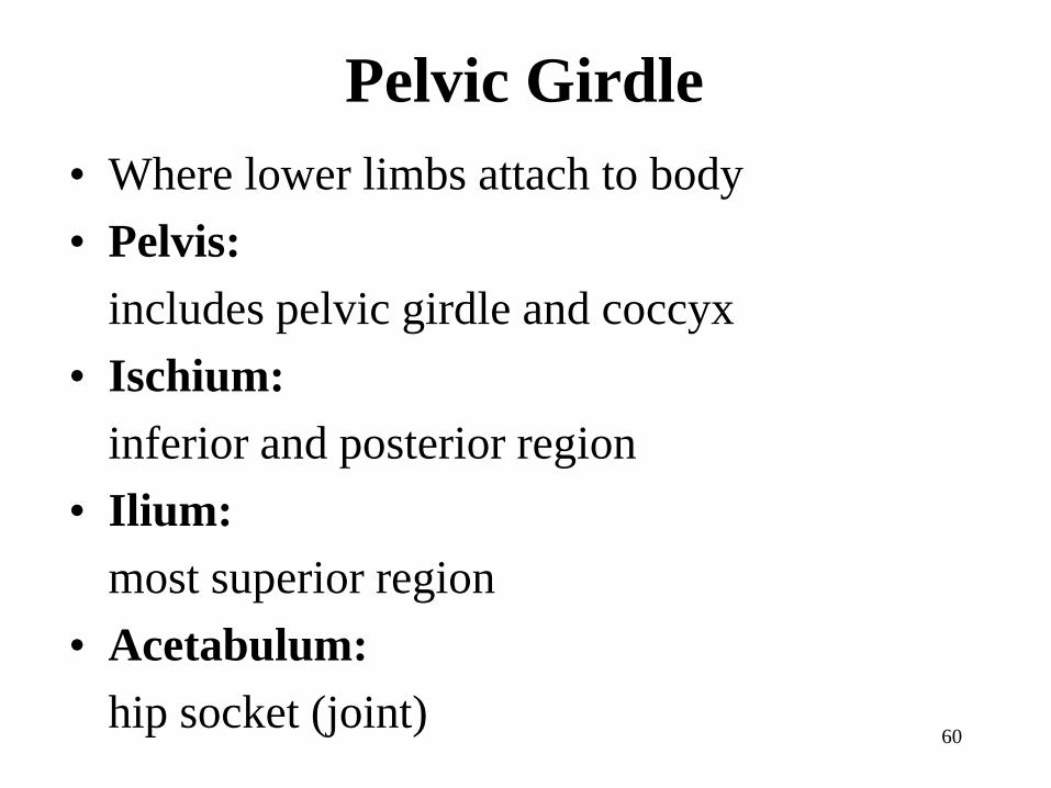
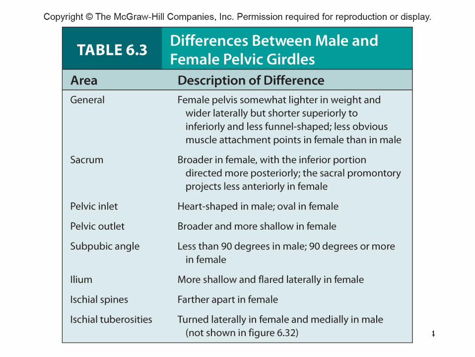
Pelvic Girdle
• Where lower limbs attach to body
• Pelvis:
includes pelvic girdle and coccyx
• Ischium:
inferior and posterior region
• Ilium:
most superior region
• Acetabulum:
hip socket (joint)60


Copyright © The McGraw-Hill Companies, Inc. Permission required for reproduction or display.
Medial viewLateral view
Ischial tuberosity
Ischium
Ischial spine
Greater
sciatic notch
Iliac crest
Ilium
Iliac fossa
Greater
sciatic notch
Ischial spine
Ischium
Articular surface
(area of
articulation
with sacrum)
Obturator foramen
Pubic symphysis
Pubis
Acetabulum
Pelvic brim
(a) (b)
64

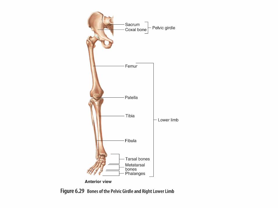
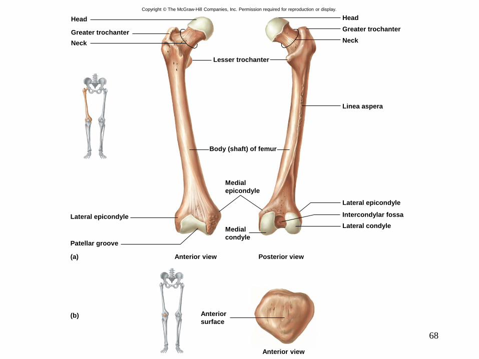
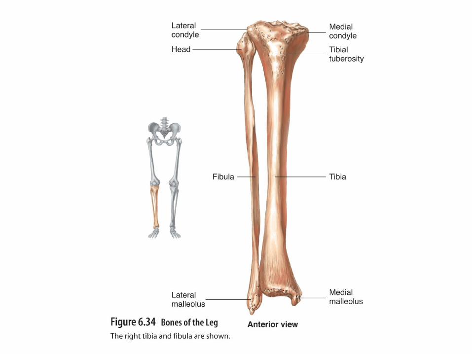
Lower Limb Bones
• Femur:
thigh
• Patella:
knee cap
• Tibia:
large lower leg
• Fibula:
small lower leg
65
• Tarsals:
ankle
• Metatarsals:
foot
• Phalanges:
toes and fingers
66
68
Copyright © The McGraw-Hill Companies, Inc. Permission required for reproduction or display.
Anterior view
Anterior
surface
Posterior viewAnterior view
Patellar groove
Lateral epicondyle
Lateral condyle
Intercondylar fossa
Lateral epicondyle
Linea aspera
Neck
Greater trochanter
Head
Neck
Greater trochanter
Head
Body (shaft) of femur
Medial
condyle
Medial
epicondyle
Lesser trochanter
(a)
(b)

71
72
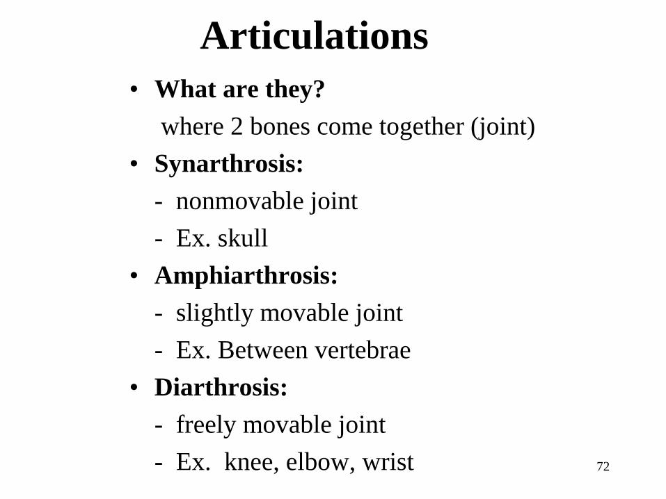
Articulations
• What are they?
where 2 bones come together (joint)
• Synarthrosis:
- nonmovable joint
- Ex. skull
• Amphiarthrosis:
- slightly movable joint
- Ex. Between vertebrae
• Diarthrosis:
- freely movable joint
- Ex. knee, elbow, wrist
73
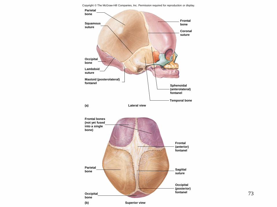
Copyright © The McGraw-Hill Companies, Inc. Permission required for reproduction or display.
Occipital
(posterior)
fontanel
Superior view
Occipital
bone
Parietal
bone
Frontal
(anterior)
fontanel
Frontal bones
(not yet fused
into a single
bone)
Sagittal
suture
Lateral view
Temporal bone
Sphenoidal
(anterolateral)
fontanel
Coronal
suture
Frontal
bone
Mastoid (posterolateral)
fontanel
Lambdoid
suture
Occipital
bone
Squamous
suture
Parietal
bone
(a)
(b)

74
Copyright © The McGraw-Hill Companies, Inc. Permission required for reproduction or display.
Bone
Blood vessel
Nerve
Articular
cartilage
Tendon
sheath
Tendon
BonePeriosteum
Outer layer
Inner layer
Joint cavity (filled
with synovial fluid)
Bursa
Joint
capsule
Synovial membrane
Fibrous part of joint
capsule
75
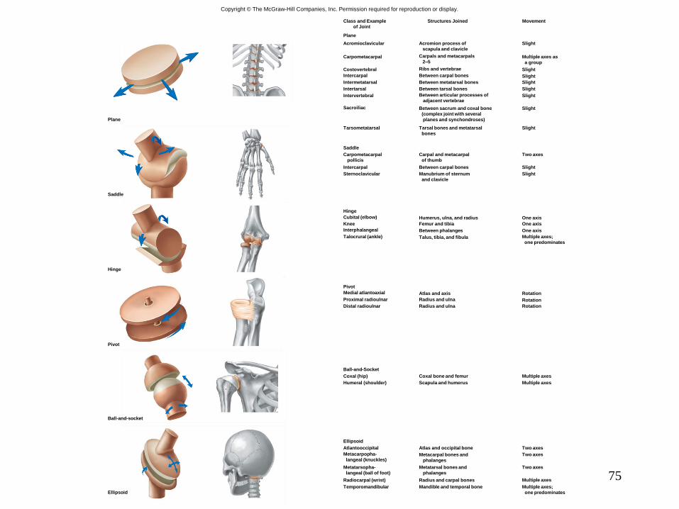
Copyright © The McGraw-Hill Companies, Inc. Permission required for reproduction or display.
Slight
Multiple axes as
a group
Slight
Slight
Slight
Slight
Slight
Slight
Slight
Acromion process of
scapula and clavicle
Carpals and metacarpals
2–5
Ribs and vertebrae
Between carpal bones
Between metatarsal bones
Between tarsal bones
Between articular processes of
adjacent vertebrae
Between sacrum and coxal bone
(complex joint with several
planes and synchondroses)
Tarsal bones and metatarsal
bones
Tarsometatarsal
Sacroiliac
Intervertebral
Intertarsal
Intermetatarsal
Intercarpal
Costovertebral
Carpometacarpal
Acromioclavicular
Plane
Class and Example
of Joint
MovementStructures Joined
Saddle
Two axes
Slight
Slight
Manubrium of sternum
and clavicle
Sternoclavicular
Intercarpal
Carpometacarpal
pollicis
Carpal and metacarpal
of thumb
Between carpal bones
Hinge
Cubital (elbow)
Knee
Talocrural (ankle)
Interphalangeal
Humerus, ulna, and radius
Femur and tibia
Talus, tibia, and fibula
Between phalanges
Multiple axes;
one predominates
One axis
One axis
One axis
Rotation
Rotation
RotationRadius and ulna
Atlas and axis
Radius and ulna
Distal radioulnar
Pivot
Medial atlantoaxial
Proximal radioulnar
Multiple axes
Multiple axesScapula and humerus
Coxal bone and femur
Ball-and-Socket
Humeral (shoulder)
Coxal (hip)
Two axes
Two axes
Two axes
Multiple axes
Multiple axes;
one predominates
Ellipsoid
Atlantooccipital
Metacarpopha-
langeal (knuckles)
Metatarsopha-
langeal (ball of foot)
Temporomandibular
Radiocarpal (wrist)
Atlas and occipital bone
Metacarpal bones and
phalanges
Metatarsal bones and
phalanges
Radius and carpal bones
Mandible and temporal boneEllipsoid
Ball-and-socket
Pivot
Hinge
Saddle
Plane
76
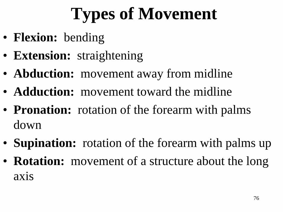
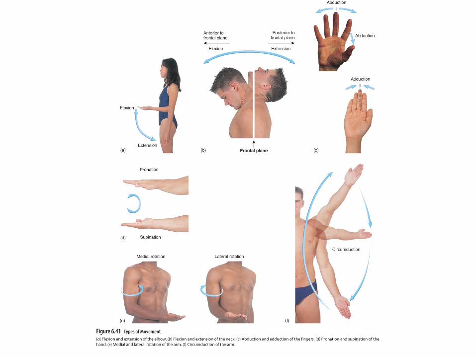
Types of Movement
• Flexion: bending
• Extension: straightening
• Abduction: movement away from midline
• Adduction: movement toward the midline
• Pronation: rotation of the forearm with palms
down
• Supination: rotation of the forearm with palms up
• Rotation: movement of a structure about the long
axis
Effects of Aging on the Skeletal
System and Joints
1. Decrease Collagen Production
2. Loss of Bone Density
3. Degenerative Changes
79

80
Please note that due to differing
operating systems, some animations
will not appear until the presentation is
viewed in Presentation Mode (Slide
Show view). You may see blank slides
in the “Normal” or “Slide Sorter” views.
All animations will appear after viewing
in Presentation Mode and playing each
animation. Most animations will require
the latest version of the Flash Player,
which is available at
http://get.adobe.com/flashplayer.