Embed Size (px)
Citation preview

8/3/2019 Chapter 6 - Neuroprotection as a Treatment for Nerve Agents Survivors - Pg. 221 - 242
http://slidepdf.com/reader/full/chapter-6-neuroprotection-as-a-treatment-for-nerve-agents-survivors-pg 1/22
221
Neuroprotection as a Treatment for Nerve Agent Survivors
Chapter 6
NEUROPROTECTION AS A TREATMENTFOR NERVE AGENT SURVIVORS
GERALD P.H. BALLOUGH, PHD*; JONATHAN NEWMARK, MD†; ERIC S. LEVINE, PHD‡; AND MARGARET G.
FILBERT, PHD§
INTRODUCTION
NEUROPATHOLOGY AND THE MECHANISM OF NERVE-AGENT–INDUCEDDAMAGE
SPECIFIC RELEVANCE OF NEUROPROTECTION TO NERVE AGENTSURVIVORS
NEUROPROTECTANTS WITH PROVEN EFFICACY AGAINST NERVE-AGENT– INDUCED SEIZURE-RELATED BRAIN DAMAGE
GangliosidesPoly(ADP-ribose) Polymerase InhibitorsRyanodine Receptor AntagonistsN -methyl-D-aspartate Receptor Antagonists
ADDITIONAL NEUROPROTECTIVE APPROACHES
Free Radical ScavengersMitochondrial Permeability Transition InhibitorsNeuroprotective Hypothermia
SUMMARY
* Professor of Biology, La Salle University, 1900 West Olney Avenue, Philadelphia, Pennsylvania 19141-1199† Colonel, US Army, Deputy Joint Program Executive Officer, Joint Program Executive Office for Chemical/Biological Defense, Skyline #2, Suite 1609,
5203 Leesburg Pike, Falls Church, Virginia 22041-3203‡ Assistant Program Manager, Science Applications International Corporation, 3465 Boxhill Corporate Center Drive, MS 23, Abingdon, Maryland
21009§ Special Assistant to the Commander, US Army Medical Research Institute of Chemical Defense, 3100 Ricketts Point Road, Aberdeen Proving Ground, Maryland 21010-5400

8/3/2019 Chapter 6 - Neuroprotection as a Treatment for Nerve Agents Survivors - Pg. 221 - 242
http://slidepdf.com/reader/full/chapter-6-neuroprotection-as-a-treatment-for-nerve-agents-survivors-pg 2/22
222
Medical Aspects of Chemical Warfare
Portions of this chapter appeared as: Filbert M, Levine E, Ballough G. Neuroprotection for nerve agent-induced brain damage by blocking delayed calcium overload: a review. Journal of Medical, Chemical, Biologi-cal, and Radiological Defense. 2005;3:1–21. Available at: http://jmedcbr.org/Issue_0301/Filbert/Filbert_1105.pdf. Accessed March 2007.

8/3/2019 Chapter 6 - Neuroprotection as a Treatment for Nerve Agents Survivors - Pg. 221 - 242
http://slidepdf.com/reader/full/chapter-6-neuroprotection-as-a-treatment-for-nerve-agents-survivors-pg 3/22
223
Neuroprotection as a Treatment for Nerve Agent Survivors
INTRODUCTION
Early use of an anticonvulsant does not guaranteethat seizures, once stopped, will not return. The recur-rence of seizures is often observed in animal studies inseveral species and is of concern in human exposures.
Although neuropathology is reduced in diazepam-treated animals, the incidence and degree of protectionafforded by diazepam is not complete.9,20–23 Moreover,switching the fielded anticonvulsant to another benzo-diazepine, such as midazolam or lorazepam, does notentirely solve the problem of refractory SE.
Seizures and SE are key causes of brain damage re-sulting from nerve agent poisoning, and their preven-tion or alleviation should be the primary objective.24–26 However, because of the refractory nature of seizuresand especially SE, prevention and alleviation becomeincreasingly difficult as more time elapses beforetherapy begins. Also, there is high probability that
seizures will return when anticonvulsants wear off.Therefore, it is reasonable to anticipate a high incidenceof brain damage connected to the increased survivalrate of nerve agent victims.
Casualties exhibiting seizures and SE can be an-ticipated not only from terrorist attacks but also from battlefield scenarios involving troops who were notin full protective ensemble at the time of the attack.27 In the confusion following a terrorist attack or on the battlefield, prompt treatment of nerve agent casualtiescan be expected to be problematic, and some victimsundergoing seizures may not receive anticonvulsantsinside the antiseizure therapeutic window. It is also
possible that some victims may undergo noncon-vulsive SE, a state of continuous seizures withoutobservable clinical movement.28 For these victims,treatment might be inadvertently delayed beyond thetherapeutic window. Under the Small Business Innova-tive Research Program, the US Army funds efforts tofield a far-forward, simple seizure detector to identifythese casualties.
This chapter presents a detailed overview of nerve-agent–induced neuropathology and explains the mech-anisms of action of candidate neuroprotectants that haveshown promise in various animal and human studies,especially those that have received US Food and DrugAdministration (FDA) approval for other indications.
Organophosphorus nerve agents are the principalchemical warfare agents known to produce braininjury. They block hydrolysis of the neurotransmitteracetylcholine by inhibiting the enzyme acetylcholin-
esterase, resulting in greatly increased postsynapticacetylcholine levels. This causes a spectrum of effects,including miosis, excess secretions, nausea, vomiting,and muscle fasciculations. At moderate to high doses,nerve agents also cause seizures and associated con-vulsions. If left untreated, seizures rapidly progress tostatus epilepticus (SE) and cause irreversible seizure-related brain damage (SRBD).1,2 The InternationalClassification of Epileptic Seizures defines SE as anyseizure lasting at least 30 minutes or intermittent sei-zures lasting longer than 30 minutes between whichthe patient does not regain consciousness.3,4
For over a decade acute therapy has effectively saved
those poisoned by nerve agents on the battlefield,5 afteraccidental exposures,6 and in terrorist attacks, as in the Japan subway attacks in 1994 and 1995. One lessonlearned from the 1995 Tokyo attack was that, lackingacute antidotal treatment, many survivors arrived athospitals in convulsive SE. The Tokyo experience illus-trates the necessity of acute antidotal therapy, such asthe regimen adopted by the US military. This regimenis aimed primarily at treating cholinergic crisis witha postexposure anticholinergic (atropine sulfate) andan oxime reactivator (2-pralidoxime [2-PAM Cl]). Inspecific intelligence-driven situations, pyridostigmine bromide (PB) pretreatment is added. Although these
medications greatly reduce morbidity and mortality,they do not always prevent seizures and brain damagein nerve agent casualties; therefore, the regimen nowincludes the anticonvulsant diazepam.2
Even with diazepam, however, the treatment regi-men has limitations. The decision to include diazepamwas based on animal data showing that it couldterminate nerve-agent–induced seizures and convul-sions and enhance survival when given in conjunctionwith the acute therapy described above.7–11 However,the therapeutic window for arresting seizures andSE with diazepam is less than an hour following on-set; after that, both are refractory to anticonvulsanttherapy.7,8,10–19
NEUROPATHOLOGY AND THE MECHANISM OF NERVE-AGENT–INDUCED DAMAGE
Although there is little neuropathological datafrom patients who have survived nerve agent attacks,abundant evidence is available from animal models,many of which involve persistent SE. The profound brain damage produced by nerve agents was first
described by Petras29; Lemercier et al30; and McLeodet al.31 Since then, numerous studies have greatly en-hanced the understanding of neuropathology resultingfrom nerve agent intoxication.23,32–38 These studies haveestablished that prolonged seizures and SE resulting

8/3/2019 Chapter 6 - Neuroprotection as a Treatment for Nerve Agents Survivors - Pg. 221 - 242
http://slidepdf.com/reader/full/chapter-6-neuroprotection-as-a-treatment-for-nerve-agents-survivors-pg 4/22
224
Medical Aspects of Chemical Warfare
from nerve agent exposure are directly responsiblefor the vast majority, if not all, of the neuropathologyproduced by these agents. The associated damage istypically bilaterally symmetrical and most severe intemporal lobe structures (ie, piriform and entorhinalcortices, hippocampus, and amygdala) as well as in
the thalamus.Brain damage resulting from agent-induced sei-zures is the result of the complex, multiphasic responseof individual neurons to numerous extracelluar andintracellular events. Following inhibition of acetyl-cholinesterase and accumulation of acetylcholine atcholinergic synapses, the hyperstimulation of cholin-ergic receptors on postsynaptic membranes triggersseizures.10,39,40 Subsequently, recruitment and excessiveactivation of the glutamatergic neurotransmitter sys-tem occurs. Glutamate, the most abundant excitatoryneurotransmitter in the brain, is responsible for sus-taining soman-induced seizures and promoting the
development of SE.1,24,41–44 Large pathological eleva-tions in the concentration of intracellular sodium and(especially) calcium are caused by excessive stimula-tion of ionotropic glutamate receptors, as is prolongeddepolarization of postsynaptic membranes. Thisinitiates a harmful cascade of pathological processes,most of which center around a prolonged increase inintracellular free calcium or delayed calcium overload,leading to excitotoxic cell death.1,24,45–47
Transient elevation in intracellular free calcium is aubiquitous signaling mechanism and regulator of in-tracellular processes, from cell growth and metabolismto cell death.48–50 Cytosolic free calcium is also a critical
neuronal mediator of learning and memory.51 How-ever, when normal homeostatic control of intracellularcalcium is lost and a sustained elevation occurs, thedelayed calcium overload triggers neuronal cell death by necrosis or apoptosis (a form of programmed celldeath).52–56 In neurons, the majority of calcium influxoccurs through N -methyl D-aspartate (NMDA) iono-tropic glutamate receptors as well as voltage-gated cal-cium channels (eg, L-type). Calcium influx also occurs,though to a lesser extent, through the other two classesof ionotropic glutamate receptors (alpha-amino-3-hydroxy-5-methylisoxazole-4-proprionic acid andkainate receptors).57 Excessive stimulation of NMDAreceptors is the first step in glutamate excitotoxicity.24,45
The release of intracellular stores is also responsiblefor increased cytosolic free calcium. The endoplasmicreticulum (ER) releases calcium following bindingof the second messenger, inositol triphosphate, toionotropic receptors located on the ER membrane.Calcium is released from the ER via ryanodine recep-tors. These ionotropic receptors are also located on theER membrane and open following binding of cytosolic
calcium; thus, cytosolic free calcium augments its ownconcentration by stimulating calcium release from theER.49 The ER plays a critical role in normal calciumhomeostasis. Excessive release or impaired uptake ofcalcium has been implicated in pathology resultingfrom calcium overload.49,52 Brain mitochondria are
important for calcium buffering as cytosolic concentra-tions rise, and their ability to sequester calcium is de-pendent on adenosine triphosphate (ATP).58 However,when calcium overload occurs, mitochondria undergoa permeability transition characterized by loss ofmitochondrial transmembrane potential, curtailmentof ATP synthesis, mitochondrial swelling, release ofstored calcium, and neuronal death by necrosis.59–62
The majority of soman-induced SRBD results fromglutamate excitotoxicity and the delayed calcium over-load that follows.1,24,42,43 Delayed calcium overload inneurons initiates a pathological sequence characterized by activation of several potentially damaging enzymes.
These include oxygenases, phospholipases, and nitricoxide synthase, which produce reactive oxygen spe-cies such as superoxide radical, hydrogen peroxide,hydroxyl radical, nitric oxide, and peroxynitrite.Neuronal injury induced by reactive oxygen speciesstems from direct damage to cell membranes, DNA,and intracellular proteins, and also induction of cyto-chrome C from mitochondria with subsequent caspaseactivation.62 Release of cytochrome C, caspase activa-tion, and DNA fragmentation are molecular hallmarksof apoptosis (Figure 6-1).56,62,63
Cysteine proteases called calpains are also activated by sustained elevations in intracellular free calcium.
Calpains degrade various intracellular proteins, in-cluding those of the cytoskeleton, membrane channels,and metabolic enzymes, and cause neuronal death bynecrosis.56,62,63 (Necrosis produces localized inflamma-tion, which exacerbates damage, while apoptosis isnot associated with inflammation.) The culminationof these events may result in cell death hours or daysafter the initial insult.53–55
Necrosis and apoptosis are not an either/or phe-nomena, that is, they are not completely distinct formsof cell death with no overlap; a necrosis versus apop-tosis dichotomy is a misleading over-simplification.64,65 Martin and colleagues proposed an “apoptosis-necrosiscontinuum,” reporting that dying neurons can exhibitintermediate forms between apoptosis and necrosis.66 Recently, Baille and colleagues confirmed that neuronalinjury, resulting from soman-induced seizures, exhibitsa large variety of hybrid forms between necrosis andapoptosis, but that the majority show more necroticfeatures.67 Whether soman-induced neuropathology ismostly necrotic, as it is in the piriform cortex of rats,38 or contains elements of apoptosis as first proposed
8/3/2019 Chapter 6 - Neuroprotection as a Treatment for Nerve Agents Survivors - Pg. 221 - 242
http://slidepdf.com/reader/full/chapter-6-neuroprotection-as-a-treatment-for-nerve-agents-survivors-pg 5/22
225
Neuroprotection as a Treatment for Nerve Agent Survivors
by Ballough et al in 1997 and definitively assessed by Baille et al is less important than the fact that bothforms of neuronal cell death are triggered by nerve-agent–induced seizures.38,67,68
Candidate drugs may alter the relative propor-tions of neurons undergoing death by necrosis versusapoptosis. Studies have reported that insufficient ATPavailability is an important determinant of whether
a cell that has been triggered to undergo apoptosisis instead forced to die by necrosis.55,69,70 Therefore, itis conceivable that a neuroprotectant candidate thatenhances ATP availability (for example, poly(ADP-ribose) polymerase [PARP] inhibitors) could suppressnecrosis while facilitating apoptosis. Neither possibil-ity should be excluded during pathological evaluationsof neuroprotectant candidates.
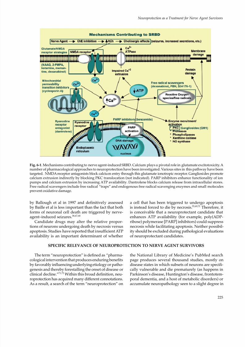
Fig. 6-1. Mechanisms contributing to nerve agent-induced SRBD. Calcium plays a pivotal role in glutamate excitotoxicity. Anumber of pharmacological approaches to neuroprotection have been investigated. Various sites in this pathway have beentargeted. NMDA receptor antagonists block calcium entry through this glutamate ionotropic receptor. Gangliosides promotecalcium extrusion indirectly by blocking PKC translocation (not indicated). PARP inhibitors enhance functionality of ion
pumps and calcium extrusion by increasing ATP availability. Dantrolene blocks calcium release from intracellular stores.Free radical scavengers include free radical “traps” and endogenous free radical scavenging enzymes and small moleculesprevent oxidative damage.
SPECIFIC RELEVANCE OF NEUROPROTECTION TO NERVE AGENT SURVIVORS
The term “neuroprotection” is defined as “pharma-cological intervention that produces enduring benefits by favorably influencing underlying etiology or patho-genesis and thereby forestalling the onset of disease orclinical decline.”71,72 Within this broad definition, neu-roprotection has acquired many different connotations.As a result, a search of the term “neuroprotection” on
the National Library of Medicine’s PubMed searchpage produces several thousand studies, mostly ondisease states in which subsets of neurons are specifi-cally vulnerable and die prematurely (as happens inParkinson’s disease, Huntington’s disease, frontotem-poral dementia, and a host of metabolic disorders) oraccumulate neuropathology seen to a slight degree in

8/3/2019 Chapter 6 - Neuroprotection as a Treatment for Nerve Agents Survivors - Pg. 221 - 242
http://slidepdf.com/reader/full/chapter-6-neuroprotection-as-a-treatment-for-nerve-agents-survivors-pg 6/22
226
Medical Aspects of Chemical Warfare
normal brains but in an accelerated fashion in somediseases (such as Alzheimer’s disease and trisomy 21).However, such interventions are unlikely to be relevantto the survivor of a single, brief nerve agent exposurethat has already caused sustained seizures and SE. Onthe other hand, research on neuroprotection following
stroke has provided valuable insights and clues thatdo apply to the nerve agent survivor.In this chapter, the term “neuroprotection” spe-
cifically refers to a putative intervention given over ashort period, ideally closely following the diagnosisof nerve agent exposure or before the acute toxicsyndrome of exposure has been adequately treated.The best neuroprotectant would have the longesttherapeutic window during which administrationwould be beneficial (even if the window is still only amatter of hours). At the same time, for logistical anddoctrinal reasons, the neuroprotection initiative doesnot extend to prophylactic treatments administered
to troops likely to experience nerve agent exposure(which would constitute a pretreatment, such as the bioscavenger initiative [see Chapter 7, Nerve AgentBioscavenger: Development of a New Approachto Protect Against Organophosphorus Exposure]).Therefore, in this chapter, neuroprotection refers onlyto postexposure treatment.
There are similarities between brain damage result-ing from nerve-agent–induced seizures and secondaryneuronal injury resulting from stroke.73,74 Although theimmediate aspect of stroke-related neuronal injury isnecrosis, which stems from anoxia or hypoxia, thereis a secondary component to stroke damage that takes
48 to 72 hours to become manifest. This componentaccounts for approximately 50% of the total damageresulting from the ischemic episode. Secondary strokeinjury involves brain tissue immediately surroundingthe necrotic core of primary injury (the penumbra).For the most part, glutamate excitotoxicity and ionicdestabilization, especially intracellular calcium, inducepenumbral damage.73–75 Thus, the similarities betweensecondary stroke damage and damage resulting fromnerve-agent–induced seizures become apparent: they both involve glutamate excitotoxicity, hinge on intra-
cellular calcium destabilization, and lead to necroticor apoptotic neuronal death. This similarity raises thepossibility that neuroprotectants being developed forstroke may be useful for nerve agent survivors. Neu-roprotective interventions in stroke models have beenshown to save neurons that otherwise would have died
via necrosis or apoptosis. There is hope, then, that atreatment can be found that can be administered afteragent exposure and that, although it may not haveany immediately discernible clinical effect, will pro-duce a significantly improved long-term neurologicaloutcome. Any of the many classes of compounds thathave been suggested as acute stroke neuroprotectantcandidates could be tried. This list is extensive; theInternet Stroke Center (http://www.strokecenter.org), maintained by Washington University,76 offersa continuously updated list of compounds that have been tried in clinical stroke trials.
The rationale for developing a protective agent,
especially one based on dissimilar clinical situationsthat give rise to similar neuronal pathology, assumesthat preventing neuronal loss will produce a superiorclinical outcome. In the case of stroke, this assumption isprobably warranted. In the case of nerve-agent–inducednerve cell damage, this assumption has never beentested directly, but it is consistent with a wide varietyof animal data in multiple models and species. The as-sumption that preventing brain damage will producesuperior behavioral outcome is even supported byLashley and Hebb’s studies in the early to mid 1900s.77 A neuroprotectant in this restricted sense should dem-onstrate that neurons that might have been lost are now
saved and that behavioral or neurological outcome isimproved. An ideal database to document such neu-roprotectants would include both neuropathologicalevidence of neuron survival and behavioral (in animals)or cognitive (in people) evidence that the neurologicoutcome is superior compared to subjects that did notreceive the neuroprotectant. Finally, the FDA must ap-prove use of the agent if it is a medication. (In clinicalmedicine, any FDA-approved medication can be usedoff-label by licensed physicians, but in military doctrine,specific on-label FDA approval is mandatory.)
NEUROPROTECTANTS WITH PROVEN EFFICACY AGAINST NERVE-AGENT–INDUCEDSIEZURE-RELATED BRAIN DAMAGE
This research comes from the consensus that nerve-agent–induced seizures and SE lead to the develop-ment of glutamate-mediated excitotoxicity, in whichdelayed calcium overload is the intracellular trigger ofthe final sequences leading to cell death.1,24,42,43,47,49,56,78–81 Classes of drugs that have been tested for their abilitiesto ameliorate nerve-agent–induced SRBD by specifi-
cally mitigating delayed calcium overload include thefollowing:
• NMDA receptor antagonists that block extra-cellular calcium influx;
• glycosphingolipids that reduce intracellularcalcium by blocking the translocation of

8/3/2019 Chapter 6 - Neuroprotection as a Treatment for Nerve Agents Survivors - Pg. 221 - 242
http://slidepdf.com/reader/full/chapter-6-neuroprotection-as-a-treatment-for-nerve-agents-survivors-pg 7/22
227
Neuroprotection as a Treatment for Nerve Agent Survivors
protein kinase C (PKC), thus enhancing thesodium-calcium exchange;
• ryanodine receptor antagonists that preventthe release of calcium from the ER; and
• PARP inhibitors that indirectly lower intra-cellular calcium by preventing ATP deple-
tion.82–89
Increased ATP availability facilitates calcium ef-flux by plasma membrane Ca2+ ATPase and calciumsequestration by the mitochondria, and indirectlyenhances sodium-calcium exchange by maintainingsodium-potassium-ATPase functionality.58
Gangliosides
Medications that target events subsequent to calci-um overload have been tested against soman-inducedSRBD in an effort to circumvent neurotoxicity associ-
ated with NMDA receptor antagonism and mitigateestablished delayed calcium overload. Intracerebro-ventricular infusion of GM1 monosialoganglioside (5mg/kg/day, for 5 days before and 27 h after somanexposure) in rats markedly reduced cross-sectionalareas of soman-induced temporal lobe necrosis (therewas an 85.9% lesion reduction in the piriform cortexand contiguous structures, compared with unpro-tected soman-positive controls).90 In this study, all ratswere pretreated with PB before soman exposure, andthen treated with atropine methylnitrate (AMN ) and2-pralidoxime (2-PAM). Considerable neuroprotec-tion was also obtained with the water-soluble GM1
monosialoganglioside derivative, WILD20. As anadjunct to HI-6 pretreatment and AMN posttreatment,WILD20 (2.5 mg/kg, intraperitoneal injection [IP])reduced volumetric temporal lobe necrosis by 75.2%.Neuroprotection by these two compounds occurred,and neither seizure intensity nor duration (assessedvia electroencephalography [EEG] monitoring) wasdiminished.
Gangliosides are sialic-acid–containing glycosphin-golipids that are natural constituents of cell mem- branes and are particularly abundant in neurons.91–93 The mechanism by which GM1 monosialogangliosideand WILD20 exert their neuroprotective effects in-volves inhibition of PKC translocation to the plasmamembrane.75, 82–86,94,95 PKC activation and translocationenhance glutamate excitotoxicity.96,97 Furthermore,PKC’s role in the excitotoxic process is to prolongNMDA receptor activation and possibly inhibit cal-cium extrusion mechanisms.82,75,98 In addition, WILD20is reported to reduce inflammation by its inhibitoryeffects on specific leukocytes (neutrophils).99 Despitethe promising results with gangliosides, further studies
have been discontinued because of concerns of possiblecontamination by prions associated with bovine spon-giform encephalopathy (mad cow disease).90,100
Poly(ADP-ribose) Polymerase Inhibitors
Recent studies indicate that PARP inhibition isneuroprotective following neuropathological insultsinvolving excitotoxicity, such as cerebral ischemiaand traumatic brain injury.101–108 PARP is an abundantnuclear enzyme that is activated by DNA strand breaks induced by reactive oxygen species.108,109 Withmoderate insults, it facilitates DNA repair by utiliz-ing cellular nicotinamide adenine dinucleotide toform poly(ADP-ribose). Excessive PARP activationleads to nicotinamide adenine dinucleotide depletion,metabolic inhibition via glycolysis block, ATP insuf-ficiency, and cell death by necrosis.104,109,110 Neuronsare especially vulnerable to metabolic insufficiency
resulting from PARP over-activation because glucoseis normally the only metabolic substrate and thedependency on glycolysis is exceptionally high.108 Inexcitotoxic models, over-activation of PARP is closelylinked to calcium-induced nitric oxide synthase activa-tion, which leads to the production of nitric oxide; thedetrimental effects of nitric oxide are mostly mediatedthrough peroxynitrite, which forms when nitric oxidereacts with superoxide.109,111,112
In 1999 Meier et al113 reported reduced lesion vol-umes and increased survival in soman-exposed ratsthat received the PARP inhibitor benzamide. Furtherinvestigation into the neuroprotective efficacy of PARP
inhibition warrants consideration, and subsequentstudies should include several new-generation PARPinhibitors that have shown increased usefulness, suchas ONO-1924H, DR2313, and FR247304.105,107,114
Ryanodine Receptor Antagonist
Dantrolene is another drug that has shown neuro-protective efficacy against soman-induced SRBD.88 Aryanodine receptor antagonist that prevents the releaseof calcium from the ER, dantrolene is FDA-approvedfor use in malignant hyperthermia. Although someneuroprotection is produced by diazepam alone (20mg/kg, intramuscular injection [IM], 40 min after sei-zure onset), this protection is significantly augmentedin the dorsal and lateral cortices of rats by coadminis-tration of dantrolene (10 mg/kg, intravenous [IV]).88 Administering the full dosage of dantrolene in a singleinjection is difficult because of insolubility problemsassociated with the medication. To overcome theseproblems and achieve the desired dantrolene dosage,four separate IV injections were performed between

8/3/2019 Chapter 6 - Neuroprotection as a Treatment for Nerve Agents Survivors - Pg. 221 - 242
http://slidepdf.com/reader/full/chapter-6-neuroprotection-as-a-treatment-for-nerve-agents-survivors-pg 8/22
228
Medical Aspects of Chemical Warfare
40 minutes and 8 hours after seizure onset, with atotal injection volume approximating 1 mL per rat. Aunique formulation of dantrolene (Lyotropic Thera-peutics, Inc, Ashland, Va) as a nanocrystal dispersionhas also been used to obviate solubility problems. Withthis formulation, it is possible to administer a much
higher dose of dantrolene in a much lower injectionvolume. This is critical because when dantrolene isadministered by IP injection, liver enzymes lower theconcentration of dantrolene reaching the brain. Thenanocrystal formulation of dantrolene minimizes theeffects of the liver enzymes.
Our results with the dantrolene nanocrystal formu-lation not only overcame the insolubility problems ofour previous dantrolene study, but corroborated andextended the results of that study. The nanocrystal studywas unable to demonstrate significant protection in thepiriform cortex, the most severely damaged region, butin this study the nanocrystal dispersion of dantrolene
(40 mg/kg, IP) plus diazepam (20 mg/kg, IM) reducedpiriform cortical necrosis by 15.6% more than diazepamalone (unpublished study by US Army Medical ResearchInstitute of Chemical Defense). In these experiments, allsoman-exposed rats also received HI-6 (125 mg/kg, IP, 30min after soman) and AMN (2 mg/kg, IM, < 1 min aftersoman) to protect against the peripheral effects of somanand ensure survival. Neuroprotection by dantrolene inthe above experiments occurred without changes in sei-zure intensity or duration, and dantrolene produced nodiscernible effects on the electrocorticographic profilesof soman-exposed subjects. These findings are consistentwith those of Frandsen and Schouosboe,115 who reported
that dantrolene prevented glutamate neurotoxicity by blocking release of calcium from intracellular stores.The results are also consistent with those of Niebauerand Gruenthal,87 who examined the protective effects ofdantrolene on hippocampal neuronal damage produced by SE in rats. In their study, dantrolene (10 mg/kg, IP)was administered either 30 or 140 minutes after the onsetof SE. Niebauer and Gruenthal reported that early admin-istration produced a significant reduction in neuronalinjury in all hippocampal subregions. When dantroleneadministration was delayed until 140 minutes after SEonset, some protection was still seen in hippocampal fieldCA3, but not the other subregions.87 Protection againstkainic-acid–induced apoptosis has also been reported.116
N -methyl-D-aspartate Receptor Antagonists
MK-801 (Dizocilpine)
The first NMDA receptor antagonist to showpromise as a putative neuroprotectant was MK-801(dizocilpine); however, it has been shown to have toxic
effects. When given in conjunction with PB, AMN,and 2-PAM, noncompetitive MK-801 was reportedto reduce nerve-agent–induced SRBD in the piriformcortex, amygdala, hippocampus, and thalamus.43 Asmentioned, these are among the most severely dam-aged brain regions in SRBD resulting from soman
exposure.29–32,35,37,38,90
In the Sparenborg study, MK-801(0.5, 1.0, or 5 mg/kg, IP) reduced brain damage anddiminished or arrested seizures in guinea pigs whenadministered as a pretreatment 30 minutes before so-man, and the effects were dose-dependent. The anti-convulsant profile of MK-801 against soman-inducedseizures was definitively characterized by Shih.11 Heshowed that the anticonvulsant effect of MK-801 is fourtimes greater than that of diazepam, but at doses of 1mg/kg or higher, MK-801 potentiated the lethal effectsof soman. Some concern arose about the use of NMDAantagonists when it was reported that MK-801 inducesneuronal degeneration in the posterior cingulate, retro-
splenial cortices, and other corticolimbic regions.117,118 This damage evidently occurs by disinhibition of mul-tiple converging excitatory pathways.119 Specifically,excessive blockage of glutamatergic pathways leadsto excessive stimulation of cholinergic function.120 Thisexplanation is supported by the findings that neuro-toxicity by MK-801 is augmented when cholinergicreceptors (ie, muscarinic) are activated.121
Memantine
Memantine is a noncompetitive NMDA receptorantagonist122 that has also been tested for its anti-
convulsant effects against soman-induced seizures.Studies have suggested that memantine’s pharma-cokinetics make it a safer candidate than MK-801.123,124 McLean et al125 reported that memantine alone (18 mg/kg, subcutaneous [SC]) blocked the onset of soman-induced seizures and was able to terminate seizureswhen administered 15 minutes after soman injection.These findings, however, are inconsistent with thoseof Shih et al17 who reported that memantine by itselfis completely ineffective as an anticonvulsant againstsoman-induced seizures. The latter authors pointedto a need for EEG monitoring when determining an-ticonvulsant efficacy and suggested that McLean et almay have mistaken diminished convulsive behavioras evidence of reduced seizure activity. Neither studyaddressed the possible neuroprotective effects of me-mantine (ie, reduced neuropathology independent ofanticonvulsant activity). On the other hand, Koplovitzet al126 observed a modest reduction in piriform corticaldamage following soman in rats treated with atropineand memantine, compared to those that received at-ropine alone. There were no differences between the

8/3/2019 Chapter 6 - Neuroprotection as a Treatment for Nerve Agents Survivors - Pg. 221 - 242
http://slidepdf.com/reader/full/chapter-6-neuroprotection-as-a-treatment-for-nerve-agents-survivors-pg 9/22
229
Neuroprotection as a Treatment for Nerve Agent Survivors
EEG power spectra of the two groups. Regardless ofthe above discrepancies, the neuroprotective benefit ofmemantine in other models of excitotoxicity is widelyaccepted.124,127 For example, in a rat model of stroke,memantine given 2 hours after the ischemic eventreduced brain damage by approximately 50%.128 In
addition, memantine is well tolerated and does notproduce neurotoxicity at therapeutic dosages. It wasrecently approved by the FDA for treating Alzheimer’sdisease.124
HU-211 (Dexanabinol)
The first real proof of concept of postexposureneuroprotection came from work with HU-211 (dex-anabinol), a nonpsychotropic analogue of tetrahydro-cannabinol, the active ingredient in marijuana. Filbertand colleagues129 showed that in rats exposed to highdoses of soman, dexanabinol protected neurons in the
piriform cortex (Figure 6-2) when given as late as 40minutes after the EEG-proven onset of seizures. Thedrug was not an anticonvulsant and had no effectupon the seizures, indicating that the results showeda true neuroprotective effect and not part of an anti-convulsant effect. HU-211 has been reported to inhibit
NMDA receptors, act as an antioxidant and free radicalscavenger, suppress nitrous oxide and tumor necrosisfactor-α generation, and stabilize calcium levels.130–132 HU-211 is generally well tolerated in humans.133
When HU-211 (25 mg/kg, IP) was administered 5minutes after the onset of soman-induced seizures,
in conjunction with HI-6 and AMN pretreatmentand posttreatment, respectively, temporal lobe lesionvolume/necrosis (assessed at 28 h after seizure onset)was reduced by 86%, compared with unprotectedsoman-positive controls (see Figure 6-2).134,135 HU-211had no effect on the strength or duration of seizureactivity, as determined by quantitative EEG analysis.Significant neuroprotection was also observed whenHU-211 administration was delayed 40 minutes afterseizure onset. Neuroprotection by HU-211 was mostevident in the piriform cortex and contiguous temporallobe structures, such as the amygdala, entorhinal, andperirhinal cortices, but did not extend to the thalamus.
Administration of HU-211 and diazepam 40 minutesafter seizure onset did not augment the neuroprotec-tion obtained with diazepam alone.
In analyzing the mechanisms of neuroprotection byHU-211 and diazepam, it is important to differentiate between protection obtained by anticonvulsant effects
Fig. 6-2. Dexanabinol (HU-211) protects against soman-induced neurological damage. Microtubule-associated protein 2(MAP-2) staining is neuron-specific. MAP-2 negative immunostaining indicates necrosis, except in areas of white matter.BL: basolateral amygdaloid nuclear groupDEn: dorsal endopiriform nucleusPir: piriform cortex.
8/3/2019 Chapter 6 - Neuroprotection as a Treatment for Nerve Agents Survivors - Pg. 221 - 242
http://slidepdf.com/reader/full/chapter-6-neuroprotection-as-a-treatment-for-nerve-agents-survivors-pg 10/22
230
Medical Aspects of Chemical Warfare
and that produced by interfering with delayed calciumoverload. In the above studies, HU-211 was protective,despite the continued presence of undiminished sei-zures and SE, whereas diazepam attenuated (withoutstopping) seizure intensity and thereby reduced the ini-tial insult. The anticonvulsant action of diazepam, via
agonistic modulation of γ-aminobutyric A (GABA[A])receptors, is well known. These mechanisms are non-overlapping, and neuroprotective effects should beadditive or synergistic. HU-211 is not approved forclinical use, and the company that owns the rights toit (Pharmos Ltd, Israel) is developing it as a possibleadjunctive therapeutic for head trauma.
Gacyclidine
Gacyclidine (GK-11) is another NMDA receptorantagonist that has shown considerable neuroprotec-tive efficacy. When GK-11 (0.01–0 .1 mg/kg, IV) was
given to rats 10 minutes after soman exposure (inconjunction with PB pretreatment, and AS, 2-PAM,and diazepam posttreatments, 1 min after soman in- jections), it completely blocked SRBD when assessed 3weeks after exposure.136 In a more realistic battlefieldscenario, GK-11 was administered 45 minutes after anexposure of 8 times the median lethal dose (LD50) of so-man in nonhuman primates. Animals also received PBpretreatment, followed by AS, 2-PAM, and diazepamposttreatments (1 min after soman exposure) equiva-lent to a single autoinjector of each in humans. When brain pathology was assessed 3 weeks after exposure,all three GK-11–treated primates showed little or no
evidence of pathology in the frontal and entorhinalcortices, amygdala, caudate nucleus, hippocampus,thalamus, midbrain, pons, medulla, and cerebellum,compared with the only surviving soman-treated ani-mal (1 of 3) that received AS, 2-PAM, and diazepam but not GK-11.137 In a study that approximates casualtymanagement following a terrorist attack, soman-in-toxicated (2 times the LD50) primates did not receivePB pretreatment and received delayed AS, 2-PAM,and diazepam treatments (one human-equivalent ofeach, as above) 30 minutes postexposure, followed byGK-11 (0.1 mg/kg, IV). In this study, the addition ofGK-11 restored normal EEG activity and completelyprevented neuropathology (assessed 5 weeks afterexposure), compared with subjects that received AS,2-PAM, or diazepam alone.138 GK-11 has a binding af-finity for NMDA receptors that is only one tenth that ofMK-801. In addition, it binds to non-NMDA receptorswhen interaction with NMDA receptors is prevented.For these reasons, GK-11 is considered substantiallyless neurotoxic than MK-801.139 It is currently beingevaluated in human clinical trials for a different neu-roprotective indication.139,140
Ketamine
Ketamine appears to be the most promising neu-roprotectant candidate to date,141,142 and it should beused in combination with a benzodiazepine, such asdiazepam. Ketamine is an FDA-approved anesthetic
that blocks neurotransmissions without depressingrespiratory and circulatory functions. Its actions aremediated by low-affinity binding to NMDA receptorchannels and prevention of calcium influx.142–145 Ket-amine is garnering considerable attention as a puta-tive neuroprotectant against ischemic brain injury,damage resulting from seizures and SE, irrespectiveof etiology, and SRBD specifically resulting fromnerve-agent–induced seizures.144–149 Fujikawa147re-ported remarkable neuroprotection in 21 of 24 brainregions in rats when 100 mg/kg of ketamine was ad-ministered (IP) 15 minutes after lithium-pilocarpine-induced SE onset. Similarly, 100 mg/kg of ketamine
(IP) prevented learning impairment in rats whenadministered immediately after lithium-pilocarpine-induced SE.150 Borris et al151 report that ketamine (58mg/kg, the effective dose in 50% of those taking it[ED50]) can control prolonged SE in rats when admin-istered 1 hour after onset. Cumulative evidence forthe beneficial effects of ketamine following SE onsethas led to its recommended use in humans when SEcannot be alleviated by conventional anticonvulsanttherapy.148
Based on its neuroprotective and anticonvulsantproperties, Mion et al145 recommend ketamine for vic-tims of nerve agent exposure. More recently, Dorandeu
et al149 reported that ketamine proved effective in stop-ping seizures, highly reducing SRBD, and improvingguinea pig survival when administered between 30minutes and 2 hours after soman poisoning. Increasingdosages of ketamine (ie, 10–60 mg/kg, IM) were re-quired as post-SE onset delay increased, and ketaminewas always administered with atropine sulfate (2–10mg/kg); in addition, guinea pigs received pyridostig-mine (26 mg/kg, IM) 30 minutes prior to soman andAMN (4 mg/kg, IM) within 1 minute following thesoman injection. Their study also provided compellingevidence of neuroprotection by ketamine at dosagesthat did not modify seizures (ie, 2–10 mg/kg), andsuggested combining ketamine and benzodiazepinetreatments when treatment is delayed 2 hours.
Results from the authors’ laboratory corroboratereports of neuroprotection by ketamine followingsoman-induced SE. The authors observed that neuro-protection was greatly augmented by administeringketamine plus diazepam, compared to diazepamalone. When soman-exposed (1.6 times the LD50) ratswere administered 20 mg/kg diazepam (IM) and 25mg/kg ketamine (IP), 40 minutes after seizure onset,

8/3/2019 Chapter 6 - Neuroprotection as a Treatment for Nerve Agents Survivors - Pg. 221 - 242
http://slidepdf.com/reader/full/chapter-6-neuroprotection-as-a-treatment-for-nerve-agents-survivors-pg 11/22
231
Neuroprotection as a Treatment for Nerve Agent Survivors
the mean cross-sectional area of temporal lobe necro-sis (ie, piriform cortex and surrounding structures)was reduced by 85.5% compared to soman-positivecontrols (P = 0.018). The mean reduction produced bydiazepam alone was only 39.9% and was not signifi-cant. In the lateral dorsal thalamus and surrounding
thalamic nuclei, diazepam plus ketamine reducedsevere damage by 91.4% compared to soman controls(P < 0.001). The reduction in lateral dorsal thalamusdamage by diazepam alone was only 27.4% and wasnot significant. Neuronal pathological assessments,using haematoxylin and eosin stain, confirmed thesequantitative findings. It is likely that reduced seizureintensities contributed to the observed neuroprotec-tion; however, this speculation is unconfirmed becauseEEGs were not obtained from these animals.
Taken together, the preponderance of evidenceindicates that ketamine is a viable neuroprotectant can-didate against nerve-agent–induced SRBD. However,
ketamine is not FDA approved for this purpose. Therehave been no human or nonhuman primate studies todetermine the optimal dose of ketamine to be used incombination with diazepam or other benzodiazepinesto alleviate nerve-agent–induced SE. On the otherhand, several case reports describe the effectivenessof ketamine, following benzodiazepine therapy, forrefractory human SE from different causes. Therefore,off-label use of ketamine, as adjunct neuroprotectivetherapy following nerve agent intoxication, should beundertaken with caution and consideration of the bestavailable evidence.
Because ketamine would be administered in con-
junction with diazepam, and because of an increasedrisk of respiratory insufficiency by the combined treat-ments (see below), it is important to review treatmentrecommendations for diazepam. The autoinjector is-sued by the US military contains 10 mg diazepam. Fora 70-kg (154-lb) individual, one autoinjector delivers adose (0.14 mg/kg, IM) consistent with the diazepamloading dosage (0.15 mg/kg, IV) recommended by therecent Belgian Consensus on SE.148 The autoinjectordose is also consistent with the diazepam dose (5–20mg/70 kg) recommended by Durham152 as initial treat-ment for SE, and is in agreement with the 20-mg diaz-epam dose (per rectum) recommended in “Treatmentof Status Epilepticus in Adults: Columbia UniversityProtocol,” as first line therapy when IV access is notavailable.153 The Belgian Consensus148 further recom-mends 4 to 8 mg per hour IV maintenance dosingwith diazepam. On the battlefield, medics and unitlifesavers are permitted to administer two additional10-mg dosages of diazepam. Overall there is regular-ity in the recommended use of diazepam in the initialtreatment of adult SE, regardless of cause. The mainadverse effects of diazepam, and benzodiazepines in
general, are respiratory depression, hypotension, anddecreased consciousness.148
For intractable SE, the Belgian Consensus advocatesan adult dosage of 50 to 100 mg ketamine as a followup to diazepam for its “theoretical neuroprotectiveeffects.”148 This dosage is consistent with Durham’s152
recommendation of 50 to 100 mg ketamine followed by 50 to 100 mg per hour, as a “second-line” treatmentfor refractory SE. Walker et al154 report successfullytreating an adult patient exhibiting “partial motor SE”with an anesthetic dosage of ketamine (ie, 100 mg/h).In a 13-year-old girl whose SE failed to respond to allstandard treatments, control of clinical and electro-graphic SE was obtained within 90 seconds followinga bolus injection (IV) of 2 mg/kg ketamine; control wasmaintained by continuous infusion of ketamine up toa maximum of 7.5 mg/kg per hour.155
Adverse effects of ketamine include a transientdecrease in respiratory rate with bolus administration
(ie, ≥ 2 mg/kg, IV), pulmonary secretions (controllablewith atropine), transient cardiovascular stimulationand possible tachycardia, intracranial hypertension(making it contraindicated for closed head injury),and undesired psychic effects.148,156 In field situations,ketamine is preferred above other anesthetics becauseit is relatively unlikely to cause respiratory depression.It is generally accepted that ketamine does not producesignificant ventilatory depression in humans.156
Ketamine may also produce neurotoxicity typicalof NMDA receptor antagonists. As mentioned above,NMDA receptor antagonists have been shown to causeneurotoxicity in the cingulate and retrosplenial cortices
as well as cerebellar Purkinje cells.117,118,157,158 A case ofpossible ketamine toxicity was seen in a 44-year-oldman treated for refractory SE.158 Control of his SE wasachieved with an initial bolus injection of 2 mg/kgketamine (IV, over 2 min), followed by a continuousinfusion of 2 mg/kg per hour. Infusion dosages wereprogressively increased until achieving a final dose of7.5 mg/kg per hour after 48 hours. Dosages were thentitrated down over the next 72 hours. The patient exhib-ited diffuse cerebellar and cerebral atrophy consistentwith animal models of NMDA antagonist-mediatedneurotoxicity.158 Studies have reported that the mecha-nism of this toxicity is indirectly mediated by exces-sive cholinergic stimulation,119–121 and supplementalatropine could have an ameliorative effect. In addition,GABAergic stimulation is reportedly protective againstthis specific form of neurotoxicity.119–121
However, high dosages of both diazepam and ket-amine could exacerbate respiratory distress alreadypresent in nerve agent casualties. Therefore, a conser-vative dose range for ketamine is advisable. In humans,a ketamine dose less than 1 mg/kg, IV, provides effec-tive analgesia against acute and chronic pain.146,156,159
8/3/2019 Chapter 6 - Neuroprotection as a Treatment for Nerve Agents Survivors - Pg. 221 - 242
http://slidepdf.com/reader/full/chapter-6-neuroprotection-as-a-treatment-for-nerve-agents-survivors-pg 12/22
232
Medical Aspects of Chemical Warfare
The anesthetic dose range in humans is 5 to 10 mg/kg, IV.146,159 For a nerve agent victim on the battlefield,a ketamine dosage below 2 mg/kg, IV, should provesafe in combination with the high dosages of diazepamthat are likely to be administered. While possibly nothigh enough to augment the anticonvulsant effects of
diazepam and arrest SE, anesthetic or subanestheticdosages of ketamine should provide considerable ad-ditional neuroprotection, compared to diazepam alone.Moreover, the ketamine dosage can be increased oncepatients reach a medical facility where intubation andventilation can be provided.
ADDITIONAL NEUROPROTECTIVE APPROACHES
Free Radical Scavengers
Damage produced by reactive oxygen species orfree radicals is a component of seizure and SE-relatedneurotoxicity,47,160,161 including damage resulting fromnerve agent poisoning.160 The liberation of catalytic ironfrom extravasated hemoglobin may generate reactiveoxygen species.160,161 Reactive oxygen species couldalso be generated by xanthine oxidase or impairedmitochondrial electron transport,161–163 offering the
hope that nerve-agent–induced neurotoxicity could bemitigated by antioxidants or free radical scavengers.
Nitrone-based free radical traps, such as alpha-phenyl-N-tert-butylnitrone (PBN), which react withreactive oxygen species, have proven to be neuro-protective following cholinesterase inhibition. Pre-treatment with PBN prevented seizures induced bydiisofluorophosphate, an organophosphonate andnerve agent simulant.164 Moreover, PBN (150 mg/kg,IP, 5 min after seizure onset) produced significant neu-roprotection in the piriform cortices and other corticalareas of rats following lithium pilocarpine-inducedSE.165 Unfortunately (and reminiscent of the findings
with HU-211 discussed above), thalamic damage waseither exacerbated or not diminished by PBN in thelatter study. Another report describes neuroprotec-tive effects by PBN 12 hours after ischemic insult.166 A pilot study of PBN did not show neuroprotectionagainst soman-induced injury.167 A new, centrally act-ing, nitrone-based free radical scavenger, S34176, hasshown superior neuroprotective properties comparedto PBN in stroke and other glutamate excitotoxicitymodels.168 S34176 may prove useful against nerve-agent–induced injury.
Mitochondrial Permeability Transition Inhibitors
As mentioned above, damaging stimuli can induceneuronal mitochondria to undergo permeability tran-sition, forming pores that allow the release of storedcalcium into the neuronal cytoplasm. This is accompa-nied by curtailment of ATP synthesis, mitochondrialswelling, exacerbation of calcium overload, and neu-ronal death.59–62 The assembly of mitochondrial transi-tion pores can be blocked by cyclosporin A, an FDA
-approved drug used in cancer chemotherapy. There isevidence that cyclosporin A and topiramate (anothertransition pore blocker) are neuroprotective in variousmodels of excitotoxic brain injury.169–174 Bauman andcolleagues169 found that cyclosporin A dramaticallyreduced brain injury in rats following seizures andSE induced by the organophosphate paraoxon. Thereis also evidence of neuroprotection by topiramate fol-lowing pilocarpine-induced seizures and SE.170
Neuroprotective Hypothermia
Total-body cooling is an effective nonpharmacologic
method of treating cerebrovascular disease. Severalstroke experts have advanced this approach as holdinggreat promise in reducing the amount of ischemic braindamage, and in 2004 the FDA approved a catheter forstroke and other specific uses that cools the blood ina penetrating artery. Less technologically complicatedapproaches to total-body cooling have been successfulin limited numbers of animal studies.175,176 Whether thisapproach would be practical in a battlefield situation,especially with mass casualties, is questionable, but itshould be kept in mind as a possibility.
SUMMARY
A variety of neuroprotective compounds have prov-en useful in alleviating brain damage caused by nerve-agent–induced seizures and SE. Of these, ketamine, me-mantine, and dantrolene have received FDA approvalfor other indications, and several other compounds arein clinical trials. Based on the evidence, ketamine, incombination with diazepam, is the top candidate andmost viable neuroprotectant for nerve agent survivors
exhibiting seizures and SE. A dantrolene and diazepamcombination is a viable possibility as well, though lessefficacious. In addition, free radical scavengers (eg,S34176) and transition pore blockers (eg, cyclosporinA) show great promise. It is conceivable that the bestpossible neuroprotective approach will be a “cocktail” oftwo or more agents that affect, in a synergistic fashion,different legs of the excitotoxic pathway.177
8/3/2019 Chapter 6 - Neuroprotection as a Treatment for Nerve Agents Survivors - Pg. 221 - 242
http://slidepdf.com/reader/full/chapter-6-neuroprotection-as-a-treatment-for-nerve-agents-survivors-pg 13/22
233
Neuroprotection as a Treatment for Nerve Agent Survivors
REFERENCES
1. Solberg Y, Belkin M. The role of excitotoxicity in organophosphorous nerve agents central poisoning. Trends PharmacolSci. 1997;18:183–185.
2. Shih TM, Duniho SM, McDonough JH. Control of nerve agent-induced seizures is critical for neuroprotection andsurvival. Toxicol Appl Pharm. 2003;188:69–80.
3. DeLorenzo RJ. Management of status epilepticus. Va Med Q. 1996;123:103–111.
4. DeLorenzo RJ, Hauser WA, Towne AR, et al. A prospective, population-based epidemiologic study of status epilepticusin Richmond, Virginia. Neurology. 1996;46:1029–1035.
5. Newmark J. The birth of nerve agent warfare: lessons from Syed Abbas Foroutan. Neurology. 2004;62:1590–1596.
6. Sidell FR. Nerve Agents. In: Sidell FR, Takafuji ET, Franz DR, eds. Medical Aspects of Chemical and Biological Warfare.In: Zajtchuk R, Bellamy RF, eds. Textbook of Military Medicine. Washington, DC: Department of the Army, Office of TheSurgeon General, Borden Institute; 1997: Chap 5.
7. Lipp JA. Effect of diazepam upon soman-induced seizure activity and convulsions. Electroencephalogr Clin Neurophysiol.
1972;32:557–560.
8. Lipp JA. Effect of benzodiazepine derivatives on soman-induced seizure activity and convulsions in the monkey. ArchInt Pharmacodyn Ther. 1973;202:244–251.
9. McDonough JH Jr, Jaax NK, Crowley RA, Mays MZ, Modrow HE. Atropine and/or diazepam therapy protects againstsoman-induced neural and cardiac pathology. Fundam Appl Toxicol. 1989;13:256–276.
10. McDonough JH Jr, Shih TM. Pharmacological modulation of soman-induced seizures.Neurosci Biobehav Rev. 1993;17:203–215.
11. Shih TM. Anticonvulsant effects of diazepam and MK-801 in soman poisoning. Epilepsy Res. 1990;7:105–116.
12. Shih TM, Koviak TA, Capacio BR. Anticonvulsants for poisoning by the organophosphorus compound soman: phar-
macological mechanisms. Neurosci Biobehav Rev. 1991;15:349–362.
13. Capacio BR, Shih TM. Anticonvulsant actions of anticholinergic drugs in soman poisoning. Epilepsia. 1991;32:604–615.
14. Philippens IH, Melchers BP, de Groot DM, Wolthuis OL. Behavioral performance, brain histology, and EEG se-quela after immediate combined atropine/diazepam treatment of soman-intoxicated rats. Pharmacol Biochem Behav.1992;42:711–719.
15. Sparenborg S, Brennecke LH, Beers ET. Pharmacological dissociation of the motor and electrical aspects of convulsivestatus epilepticus induced by the cholinesterase inhibitor soman. Epilepsy Res. 1993;14:95–103.
16. Harris LW, Gennings C, Carter WH, Anderson DR, Lennox WJ, Bowersox SL, Solana RP. Efficacy comparison of scopol-amine (SCP) and diazepam (DZ) against soman-induced lethality in guinea pigs. Drug Chem Toxicol. 1994;17:35–50.
17. Shih T, McDonough JH Jr, Koplovitz I. Anticonvulsants for soman-induced seizure activity. J Biomed Sci. 1999;6:86–96.
18. Lallement G, Renault F, Baubichon D, et al. Compared efficacy of diazepam or avizafone to prevent soman-inducedelectroencephalographic disturbances and neuropathology in primates: relationship to plasmatic benzodiazepinepharmacokinetics. Arch Toxicol. 2000;74:480–486.
19. McDonough JH Jr, Zoeffel LD, McMonagle J, Copeland TL, Smith CD, Shih TM. Anticonvulsant treatment of nerveagent seizures: anticholinergic versus diazepam in soman-intoxicated guinea pigs. Epilepsy Res. 2000;38:1–14.
8/3/2019 Chapter 6 - Neuroprotection as a Treatment for Nerve Agents Survivors - Pg. 221 - 242
http://slidepdf.com/reader/full/chapter-6-neuroprotection-as-a-treatment-for-nerve-agents-survivors-pg 14/22
234
Medical Aspects of Chemical Warfare
20. Clement JG, Broxup B. Efficacy of diazepam and avizafone against soman-induced neuropathology in brain of rats.Neurotoxicology. 1993;14:485–504.
21. Hayward IJ, Wall HG, Jaax NK, Wade JV, Marlow DD, Nold JB. Decreased brain pathology in organophosphate-ex-posed rhesus monkeys following benzodiazepine therapy. J Neurol Sci. 1990;98:99–106.
22. McDonough JH Jr, Dochterman LW, Smith CD, Shih TM. Protection against nerve agent-induced neuropathology, but not
cardiac pathology, is associated with the anticonvulsant action of drug treatment. Neurotoxicology. 1995;16:123–132.
23. Baze WB. Soman-induced morphological changes: an overview in the non-human primate. J Appl Toxicol. 1993;13:173–177.
24. Olney JW, de Gubareff T, Labruyere J. Seizure-related brain damage induced by cholinergic agents.Nature. 1983;301:520–522.
25. Sloviter RS. “Epileptic” brain damage in rats induced by sustained electrical stimulation of the perforant path. I. Acuteelectrophysiological and light microscopic studies. Brain Res Bull. 1983;10:675–697.
26. Meldrum BS. Concept of activity-induced cell death in epilepsy: historical and contemporary perspectives. ProgressBrain Res. 2002;135:3–11.
27. Fanzone JF, Levine ES, Hursh SR. Nerve Agent Bioscavenger Pretreatment Against Chemical Warfare Agents: Challenge,Casualty, and Intervention Modeling Support. Aberdeen Proving Ground, Md: US Army Medical Research and MaterielCommand. Interim Report. Contract No. DAMD17-98-D-0022; 2002.
28. DeLorenzo RJ, Waterhouse EJ, Towne AR, et al. Persistent nonconvulsive status epilepticus after the control of con-vulsive status epilepticus. Epilepsia. 1998;39:833–840.
29. Petras JM. Soman neurotoxicity. Fundam Appl Toxicol. 1981;1:242.
30. Lemercier G, Carpentier P, Sentenac-Roumanou H, Morelis P. Histological and histochemical changes in the centralnervous system of the rat poisoned by an irreversible anticholinesterase organophosphorus compound. Acta Neuro-
pathol (Berl). 1983;61:123–129.
31. McLeod CG Jr, Singer AW, Harrington DG. Acute neuropathology in soman poisoned rats. Neurotoxicology. 1984;5:53–57.
32. Carpentier P, Delamanche IS, Le Bert M, Blanchet G, Bouchaud C. Seizure-related opening of the blood-brain barrierinduced by soman: possible correlation with the acute neuropathology observed in poisoned rats. Neurotoxicology.1990;11:493–508.
33. Kadar T, Cohen G, Sahar R, Alkalai D, Shapira S. Long-term study of brain lesions following soman, in comparisonto DFP and metrazol poisoning. Hum Exp Toxicol. 1992; 11:517–523.
34. McDonough JH Jr, McLeod CG Jr, Nipwoda MT. Direct microinjection of soman or VX into the amygdala producesrepetitive limbic convulsions and neuropathology. Brain Res. 1987;435:123–137.
35. Pazdernik TL, Cross R, Giesler M, Nelson S, Samson F, McDonough J Jr. Delayed effects of soman: brain glucose use
and pathology. Neurotoxicology. 1985;6:61–70.
36. Petrali JP, Maxwell DM, Lenz DE, Mills KR. Effect of an anticholinesterase compound on the ultrastructure and func-tion of the rat blood-brain barrier: a review and experiment. J Submicrosc Cytol Pathol. 1991;23:331–338.
37. Petras JM. Neurology and neuropathology of soman-induced brain injury: an overview. J Exp Anal Behav. 1994;61:319–329.
38. Ballough GP, Martin LJ, Cann FJ, et al. Microtubule-associated protein 2 (MAP-2): a sensitive marker of seizure-related brain damage. J Neurosci Methods. 1995;61:23–32.

8/3/2019 Chapter 6 - Neuroprotection as a Treatment for Nerve Agents Survivors - Pg. 221 - 242
http://slidepdf.com/reader/full/chapter-6-neuroprotection-as-a-treatment-for-nerve-agents-survivors-pg 15/22
235
Neuroprotection as a Treatment for Nerve Agent Survivors
39. Lallement G, Carpentier P, Collet A, Baubichon D, Pernot-Marino I, Blanchet G. Extracellular acetylcholine changesin rat limbic structures during soman-induced seizures. Neurotoxicology. 1992;13:557–567.
40. Tonduli LS, Testylier G, Marino IP, Lallement G. Triggering of soman-induced seizures in rats: multiparametricanalysis with special correlation between enzymatic, neurochemical, and electrophysiological data. J Neurosci Res.1999;58:464–473.
41. Wade JV, Samson FE, Nelson SR, Pazdernik TL. Changes in extracellular amino acids during soman- and kainic acid-induced seizures. J Neurochem. 1987;49:645–650.
42. Braitman DJ, Sparenborg S. MK-801 protects against seizures induced by the cholinesterase inhibitor soman. BrainRes Bull. 1989;23:145–148.
43. Sparenborg S, Brennecke LH, Jaax NK, Braitman DJ. Dizocilpine (MK-801) arrests status epilepticus and prevents brain damage induced by soman. Neuropharmacology . 1992;31:357–368.
44. Fosbraey P, Wetherell JR, French MC. Neurotransmitter changes in guinea-pig brain regions following soman intoxica-tion. J Neurochem. 1990;54:72–79.
45. Choi DW. Calcium-mediated neurotoxicity: relationship to specific channel types and role in ischemic damage. TrendsNeurosci. 1988;11:465–469.
46. Shih TM, Capacio BR, Cook LA. Effects of anticholinergic-antiparkinsonian drugs on striatal neurotransmitter levelsof rats intoxicated with soman. Pharmacol Biochem Behav. 1993;44:615–622.
47. Fujikawa DG. Prolonged seizures and cellular injury: understanding the connection. Epilepsy Behav. 2005:7(suppl 3):S3–11.
48. Carafoli E. Calcium signaling: a tale for all seasons. Proc Natl Acad Sci U S A. 2002;99:1115–1122.
49. Verkhratsky A, Toescu EC. Endoplasmic reticulum Ca(2+) homeostasis and neuronal death. J Cell Mol Med. 2003;7:351–361.
50. Parekh AB. Store-operated Ca2+ entry: dynamic interplay between endoplasmic reticulum, mitochondria and plasma
membrane. J Physiol. 2003;547(pt 2):333–348.
51. Bliss TV, Collingridge GL. A synaptic model of memory: long-term potentiation in the hippocampus. Nature.1993;361:31–39.
52. Randall RD, Thayer SA. Glutamate-induced calcium transient triggers delayed calcium overload and neurotoxicityin rat hippocampal neurons. J Neurosci.1992;12:1882–1895.
53. Orrenius S, Burkitt MJ, Kass GE, Dypbukt JM, Nicotera P. Calcium ions and oxidative cell injury. Ann Neurol. 1992;32(suppl):S33–42.
54. Orrenius S, Nicotera P. The calcium ion and cell death. J Neural Transm Suppl. 1994;43:1–11.
55. Nicotera P. Molecular switches deciding the death of injured neurons. Toxicol Sci. 2003;74:4–9.
56. Nicholls DG. Mitochondrial dysfunction and glutamate excitotoxicity studied in primary neuronal cultures.Curr Mol Med. 2004;4:149–177.
57. Jayakar SS, Dikshit M. AMPA receptor regulation mechanisms: future target for safer neuroprotective drugs. Int JNeurosci. 2004;114:695–734.
58. Kulak W, Sobaniec W, Wojtal K, Czuczwar SJ. Calcium modulation in epilepsy. Pol J Pharmacol. 2004;56:29–41.
59. Duchen MR. Mitochondria and calcium: from cell signaling to cell death. J Physiology. 2000;529:57–68.

8/3/2019 Chapter 6 - Neuroprotection as a Treatment for Nerve Agents Survivors - Pg. 221 - 242
http://slidepdf.com/reader/full/chapter-6-neuroprotection-as-a-treatment-for-nerve-agents-survivors-pg 16/22
236
Medical Aspects of Chemical Warfare
60. Halestrap AP, McStay GP, Clarke SJ. The permeability transition pore complex: another view. Biochimie. 2002;84:153–166.
61. Chang LK, Putcha GV, Deshmukh M, Johnson EM Jr. Mitochondrial involvement in the point of no return in neuronalapoptosis. Biochimie. 2002;84:223–231.
62. Mattson MP. Excitotoxic and excitoprotective mechanisms: abundant targets for the prevention and treatment of
neurodegenerative disorders. Neuromolecular Med. 2003;3:65–94.
63. Hou ST, MacManus JP. Molecular mechanisms of cerebral ischemia-induced neuronal death. Intern Rev Cytol.2002;221:93–149.
64. Clarke PGH. Apoptosis versus necrosis--how valid a dichotomy for neurons: In: Koliatsos VE, Ratan RR, eds. CellDeath and Diseases of the Nervous System. Totowa, NJ: Humana Press Inc; 1999: 3–28.
65. Sloviter RS. Apoptosis: a guide for the perplexed. Trends Pharmacol Sci 2002;23:19–24.
66. Martin LJ, Al-Abdulla NA, Brambrink AM, Kirsch JR, Sieber FE, Portera-Cailliau C. Neurodegeneration in excitotox-icity, global cerebral ischemia, and target deprivation: a perspective on the contributions of apoptosis and necrosis.Brain Res Bull. 1998;46:281–309.
67. Baille V, Clarke PG, Brochier G, et al. Soman-induced convulsions: the neuropathology revisited. Toxicology. 2005;215:1–24.
68. Ballough GP, Forster JS, Makowski JP, Sordoni NC, Filbert MG. Soman-induced seizures produce neuronal apoptosis[abstract]. Abstr Soc Neurosci. 1997;23:1936.
69. Leist M, Single B, Castoldi AF, Kühnle S, Nicotera P. Intracellular adenosine triphosphate (ATP) concentration: a switchin the decision between apoptosis and necrosis. J Exp Med. 1997;185:1481–1486.
70. Nicotera P, Leist M, Ferrando-May E. Intracellular ATP, a switch in the decision between apoptosis and necrosis. ToxicolLett. 1998;102–103:139–142.
71. Shoulson I. Experimental therapeutics of neurodegenerative disorders: unmet needs.Science. 1998;282:1072–1074.
72. Schulz, JB. Neurodegeneration. In: Bahr M, ed. Neuroprotection: Models, Mechanisms, and Therapies. Weinheim, BadenWürttemberg, Germany: Wiley-VCH Verlag GmbH & Co; 2004: Chap 16.
73. Siesjo BK, Bengtsson F. Calcium fluxes, calcium antagonists, and calcium-related pathology in brain ischemia, hypo-glycemia, and spreading depression: a unifying hypothesis. J Cereb Blood Flow Metab. 1989;9:127–140.
74. Silver B, Weber J, Fisher M. Medical therapy for ischemic stroke. Clin Neuropharmacol. 1996;19:101–128.
75. Costa E, Armstrong DM, Guidotti A, et al. Gangliosides in the protection against glutamate excitotoxicity. Prog BrainRes. 1994;101:357–373.
76. Washington University. The Internet Stroke Center. Available at: www.strokecenter.org. Accessed March 21, 2007.
77. Orbach J. The Neuropsychological Theories of Lashley and Hebb: Contemporary Perspectives Fifty Years After Hebb’s TheOrganization of Behavior, Vanuxem Lectures and Selected Theoretical Papers of Lashley. Lanham, Md: University Press ofAmerica; 1998.
78. Choi DW. Ionic dependence of glutamate neurotoxicity. J Neurosci. 1987;7:369–379.
79. Lallement G, Carpentier P, Collet A, Pernot-Marino I, Baubichon D, Blanchet G. Effects of soman-induced seizures ondifferent extracellular amino acid levels and on glutamate uptake in rat hippocampus. Brain Res. 1991;563:234–240.
80. Shih TM, McDonough JH Jr. Neurochemical mechanisms in soman-induced seizures. J Appl Toxicol. 1997;17:255–264.

8/3/2019 Chapter 6 - Neuroprotection as a Treatment for Nerve Agents Survivors - Pg. 221 - 242
http://slidepdf.com/reader/full/chapter-6-neuroprotection-as-a-treatment-for-nerve-agents-survivors-pg 17/22
237
Neuroprotection as a Treatment for Nerve Agent Survivors
81. McDonough JH Jr, Shih TM. Neuropharmacological mechanisms of nerve agent-induced seizure and neuropathology.Neurosci Biobehav Rev. 1997;21:559–579.
82. Manev H, Guidotti A, Costa E. Protection by gangliosides against glutamate excitotoxicity. Adv Lipid Res. 1993;25:269–288.
83. Tubaro E, Santiangeli C, Cavallo G, et al. Effect of a new de-N-acetyl-lysoglycosphingolipid on chemically-induced
inflammatory bowel disease: possible mechanism of action. Naunyn Schmiedebergs Arch Pharmacol. 1993;348:670–678.
84. Otani S, Daniel H, Takita M, Crepel F. Long-term depression induced by postsynaptic group II metabotropic glutamatereceptors linked to phospholipase C and intracellular calcium rises in rat prefrontal cortex. J Neurosci. 2002;22:3434–3444.
85. Monnet FP, Morin-Surun MP, Leger J, Combettes L. Protein kinase C-dependent potentiation of intracellular calciuminflux by sigma1 receptor agonists in rat hippocampal neurons. J Pharmacol Exp Ther. 2003;307:705–312.
86. Chaban VV, Li J, Ennes HS, Nie J, Mayer EA, McRoberts JA. N-methyl-D-aspartate receptors enhance mechanicalresponses and voltage-dependent Ca2+ channels in rat dorsal root ganglia neurons through protein kinase C. Neuro-science. 2004;128:347–357.
87. Niebauer M, Gruenthal M. Neuroprotective effects of early vs. late administration of dantrolene in experimental statusepilepticus. Neuropharmacology . 1999;38:1343–1348.
88. Ballough GPH, Filbert MG. A Viable Neuroprotection Strategy Following Soman-Induced Status Epilepticus. AberdeenProving Ground, Md: US Army Medical Research Institute of Chemical Defense; 2003. USAMRICD Technical Report03-09, AD A443565.
89. Krause T, Gerbershagen MU, Fiege M, Weisshorn R, Wappler F. Dantrolene–a review of its pharmacology, therapeuticuse, and new developments. Anaesthesia. 2004;59:364–373.
90. Ballough GP, Cann FJ, Smith CD, Forster JS, Kling CE, Filbert MG. GM1 monosialoganglioside pretreatment protectsagainst soman-induced seizure-related brain damage. Mol Chem Neuropathol. 1998;34:1–23.
91. Ando S. Gangliosides in the nervous system. Neurochem Int. 1983;5:507–537.
92. Ledeen RW. Biology of gangliosides: neuritogenic and neuronotrophic properties. J Neurosci Res. 1984;12:147–159.
93. Yu RK, Goldenring JR, Kim JYH, DeLorenzo RJ. Gangliosides as differential modulators of membrane-bound proteinkinase systems. In: Tettamanti G, Ledeen RW, Sandhoff K, Nagai Y, Toffano G, eds. Fidia Research Series: Gangliosidesand Neuronal Plasticity. Vol 6. Padova, Italy: Liviana Press; 1986: 95–104.
94. Vaccarino F, Guidotti A, Costa E. Ganglioside inhibition of glutamate-mediated protein kinase C translocation inprimary cultures of cerebellar neurons. Proc Natl Acad Sci U S A. 1987;84:8707–8711.
95. Manev H, Favaron M, Vicini S, Guidotti A. Ganglioside-mediated protection from glutamate-induced neuronal death.Acta Neurobiol Exp (Wars). 1990;50:475–488.
96. Wagey R, Hu J, Pelech SL, Raymond LA, Krieger C. Modulation of NMDA-mediated excitotoxicity by protein kinase
C. J Neurochem. 2001;78:715–726.
97. Koponen S, Kurkinen K, Akerman KE, Mochly-Rosen D, Chan PH, Koistinaho J. Prevention of NMDA-induced deathof cortical neurons by inhibition of protein kinase Czeta. J Neurochem. 2003;86:442–450.
98. Zhang L, Rzigalinski BA, Ellis EF, Satin LS. Reduction of voltage-dependent Mg2+ blockade of NMDA current inmechanically injured neurons. Science. 1996;274:1921–1923.
99. Tubaro E, Croce C, Cavallo G, Belogi L, Guida G, Santiangeli C, Cifone MG, Santoni A, Mainiero F. In vitro and in vivoimpact of a new glycosphingolipid on neutrophils. Agents Actions. 1994;42:107–113.

8/3/2019 Chapter 6 - Neuroprotection as a Treatment for Nerve Agents Survivors - Pg. 221 - 242
http://slidepdf.com/reader/full/chapter-6-neuroprotection-as-a-treatment-for-nerve-agents-survivors-pg 18/22
238
Medical Aspects of Chemical Warfare
100. Mattei V, Garofalo T, Misasi R, Gizzi C, Mascellino MT, Dolo V, Pontieri GM, Sorice M, Pavan A. Association of cellularprion protein with gangliosides in plasma membrane microdomains of neural and lymphocytic cells. Neurochem Res.2002;27:743–749.
101. Eliasson MJ, Sampei K, Mandir AS, et al. Poly(ADP-ribose) polymerase gene disruption renders mice resistant tocerebral ischemia. Nat Med. 1997;3:1089–1095.
102. Mandir AS, Poitras MF, Berliner AR, et al. NMDA but not non-NMDA excitotoxicity is mediated by Poly(ADP-ribose)polymerase. J Neurosci. 2000;20:8005–8011.
103. Whalen MJ, Clark RS, Dixon CE, et al. Traumatic brain injury in mice deficient in poly-ADP(ribose) polymerase: apreliminary report. Acta Neurochir Suppl. 2000;76:61–64.
104. Abdelkarim GE, Gertz K, Harms C, et al. Protective effects of PJ34, a novel, potent inhibitor of poly(ADP-ribose)polymerase (PARP) in in vitro and in vivo models of stroke. Int J Mol Med. 2001;7:255–260.
105. Kamanaka Y, Kondo K, Ikeda Y, et al. Neuroprotective effects of ONO-1924H, an inhibitor of poly ADP-ribose poly-merase (PARP), on cytotoxicity of PC12 cells and ischemic cerebral damage. Life Sci. 2004;76:151–162.
106. Sharma SS, Munusamy S, Thiyagarajan M, Kaul CL. Neuroprotective effect of peroxynitrite decomposition catalystand poly(adenosine diphosphate-ribose) polymerase inhibitor alone and in combination in rats with focal cerebralischemia. J Neurosurg. 2004;101:669–675.
107. Nakajima H, Kakui N, Ohkuma K, Ishikawa M, Hasegawa T. A newly synthesized poly(ADP-ribose) polymeraseinhibitor, DR2313 [2-methyl-3,5,7,8-tetrahydrothiopyrano[4,3-d]-pyrimidine-4-one]: pharmacological profiles, neuro-protective effects, and therapeutic time window in cerebral ischemia in rats. J Pharmacol Exp Ther. 2005;312:472–481.
108. Ying W, Alano CC, Garnier P, Swanson RA. NAD+ as a metabolic link between DNA damage and cell death. J NeurosciRes. 2005;79:216–223.
109. Szabo C, Dawson VL. Role of poly(ADP-ribose) synthetase in inflammation and ischaemia-reperfusion. Trends Phar-macol Sci. 1998;19:287–298.
110. Virag L, Szabo C. The therapeutic potential of poly(ADP-ribose) polymerase inhibitors. Pharmacol Rev. 2002;54:375–
429.
111. Park EM, Cho S, Frys K, et al. Interaction between inducible nitric oxide synthase and poly(ADP-ribose) polymerasein focal ischemic brain injury. Stroke. 2004;35:2896–2901.
112. Wang H, Yu SW, Koh DW, et al. Apoptosis-inducing factor substitutes for caspase executioners in NMDA-triggeredexcitotoxic neuronal death. J Neurosci. 2004;24:10963–10973.
113. Meier HL, Ballough GP, Forster JS, Filbert MG. Benzamide, a poly(ADP-ribose) polymerase inhibitor, is neuroprotec-tive against soman-induced seizure-related brain damage. Ann N Y Acad Sci. 1999;890:330–335.
114. Iwashita A, Tojo N, Matsuura S, et al. A novel and potent poly(ADP-ribose) polymerase-1 inhibitor, FR247304 (5-chloro-2-[3-(4-phenyl-3,6-dihydro-1(2H)-pyridinyl)propyl]-4(3H)-quinazolinone), attenuates neuronal damage in vitro andin vivo models of cerebral ischemia. J Pharmacol Exp Ther. 2004;310:425–436.
115. Frandsen A, Schousboe A. Dantrolene prevents glutamate cytotoxicity and Ca2+ release from intracellular stores. JNeurochem. 1991;56:1075–1078.
116. Popescu BO, Oprica M, Sajin M, et al. Dantrolene protects neurons against kainic acid induced apoptosis in vitro andin vivo. J Cell Mol Med. 2002;6:555–569.
117. Olney JW, Labruyere J, Price MT. Pathological changes induced in cerebrocortical neurons by phencyclidine and relateddrugs. Science. 1989;244:1360–1362.

8/3/2019 Chapter 6 - Neuroprotection as a Treatment for Nerve Agents Survivors - Pg. 221 - 242
http://slidepdf.com/reader/full/chapter-6-neuroprotection-as-a-treatment-for-nerve-agents-survivors-pg 19/22
239
Neuroprotection as a Treatment for Nerve Agent Survivors
118. Fix AS, Horn JW, Wightman KA, et al. Neuronal vacuolization and necrosis induced by the noncompetitive N-methyl-D-aspartate (NMDA) antagonist MK(+)801 (dizocilpine maleate): a light and electron microscopic evaluation of therat retrosplenial cortex. Exp Neurol. 1993;123:204–215.
119. Corso TD, Sesma MA, Tenkova TI, et al. Multifocal brain damage induced by phencyclidine is augmented by pilocar-pine. Brain Res. 1997;752:1–14.
120. Olney JW, Labruyere J, Wang G, Wozniak DF, Price MT, Sesma MA. NMDA antagonist neurotoxicity: mechanism andprevention. Science. 1991;254:1515–1518.
121. Wozniak DF, Dikranian K, Ishimaru MJ, et al. Disseminated corticolimbic neuronal degeneration induced in rat brain by MK-801: potential relevance to Alzheimer’s disease. Neurobiol Dis. 1998;5:305–322.
122. Bormann J. Memantine is a potent blocker of N-methyl-D-aspartate (NMDA) receptor channels. Eur J Pharmacol.1989;166:591–592.
123. Chen HS, Pellegrini JW, Aggarwal SK, et al. Open-channel block of N-methyl-D-aspartate (NMDA) responses bymemantine: therapeutic advantage against NMDA receptor-mediated neurotoxicity. J Neurosci. 1992;12:4427–4436.
124. Lipton SA. Excitotoxicity. In: Bahr M, ed. Neuroprotection: Models, Mechanisms, and Therapies. Weinheim, Baden Würt-temberg, Germany: Wiley-VCH Verlag GmbH & Co; 2004: Chap 14.
125. McLean MJ, Gupta RC, Dettbarn WD, Wamil AW. Prophylactic and therapeutic efficacy of memantine against seizuresproduced by soman in the rat. Toxicol Appl Pharmacol. 1992;112:95–103.
126. Koplovitz I, Schulz S, Shutz M, Railer R, Smith F, Okerberg C, Filbert M. Memantine effects on soman-induced seizuresand seizure-related brain damage. Toxicol Meth 1997;7:227–239.AQ2
127. Parsons CG, Danysz W, Quack G. Memantine is a clinically well tolerated N-methyl-D-aspartate (NMDA) receptorantagonist--a review of preclinical data. Neuropharmacology. 1999;38:735–767.
128. Chen HS, Wang YF, Rayudu PV, et al. Neuroprotective concentrations of the N-methyl-D-aspartate open-channel blocker memantine are effective without cytoplasmic vacuolation following post-ischemic administration and do not block maze learning or long-term potentiation. Neuroscience. 1998;86:1121–1132.
129. Filbert MG, Forster JS, Smith CD, Ballough GP. Neuroprotective effects of HU-211 on brain damage resulting fromsoman-induced seizures. Ann N Y Acad Sci. 1999;890:505–514.
130. Shohami E, Novikov M, Mechoulam R. A nonpsychotropic cannabinoid, HU-211, has cerebroprotective effects afterclosed head injury in the rat. J Neurotrauma. 1993;10:109–119.
131. Biegon A, Joseph AB. Development of HU-211 as a neuroprotectant for ischemic brain damage. Neurol Res. 1995;17:275–280.
132. Lavie G, Teichner A, Shohami E, Ovadia H, Leker RR. Long term cerebroprotective effects of dexanabinol in a modelof focal cerebral ischemia. Brain Res. 2001;901:195–201.
133. Darlington CL. Dexanabinol: a novel cannabinoid with neuroprotective properties. IDrugs. 2003;6:976–979.
134. Kitagawa K, Matsumoto M, Niinobe M, et al. Microtubule-associated protein 2 as a sensitive marker for cerebralischemic damage—immunohistochemical investigation of dendritic damage. Neurosci 1989;31:401–411.
135. Gilland E, Bona E, Hagberg H. Temporal changes of regional glucose use, blood flow and microtubule-associated pro-tein 2 immunostaining after hypoxia ischemia in the immature rat brain. J Cereb Blood Flow Metab. 1998:18:222–228.
136. Lallement G, Mestries JC, Privat A, et al. GK 11: promising additional neuroprotective therapy for organophosphatepoisoning. Neurotoxicology. 1997;18:851–856.

8/3/2019 Chapter 6 - Neuroprotection as a Treatment for Nerve Agents Survivors - Pg. 221 - 242
http://slidepdf.com/reader/full/chapter-6-neuroprotection-as-a-treatment-for-nerve-agents-survivors-pg 20/22
240
Medical Aspects of Chemical Warfare
137. Lallement G, Clarencon D, Masqueliez C, et al. Nerve agent poisoning in primates: antilethal, anti-epileptic, andneuroprotective effects of GK-11. Arch Toxicol. 1998;72:84–92.
138. Lallement G, Clarencon D, Galonnier M, Baubichon D, Burckhart MF, Peoc’h M. Acute soman poisoning in primatesneither pretreated nor receiving immediate therapy: value of gacyclidine (GK-11) in delayed medical support. ArchToxicol. 1999;73:115–122.
139. Hirbec H, Gaviria M, Vignon J. Gacyclidine: a new neuroprotective agent acting at the N-methyl-D-aspartate receptor.CNS Drug Rev. 2001;7:172–198.
140. Lepeintre JF, D’Arbigny P, Mathe JF, et al. Neuroprotective effect of gacyclidine. A multicenter double-blind pilot trialin patients with acute traumatic brain injury. Neurochirurgie. 2004;50(Part 1):83–95.
141. Lallement G. Centre de Recherches du Service de Sante des Armees, La Tronche, France. Personal communicationwith Ballough G, June 2005.
142. Dorandeu F. Centre de Recherches du Service de Sante des Armees, La Tronche, France. Personal communication withBallough G, August 2006.
143. MacDonald JF, Nowak LM. Mechanisms of blockade of excitatory amino acid receptor channels. Trends Pharmacol Sci.1990;11:167–172.
144. Werner C, Reeker W, Engelhard K, Lu H, Kochs E. Ketamine racemate and S-(+)-ketamine. Cerebrovascular effectsand neuroprotection following focal ischemia. Anaesthesist. 1997;46(supp; 1):S55–S60.
145. Mion G, Tourtier JP, Petitjeans F, Dorandeu F, Lallement G, Ruttimann M. Neuroprotective and antiepileptic activitiesof ketamine in nerve agent poisoning. Anesthesiology. 2003;98:1517.
146. Ivani G, Vercellino C, Tonetti F. Ketamine: a new look to an old drug. Minerva Anestesiol. 2003;69:468–471.
147. Fujikawa DG. Neuroprotective effect of ketamine administration after status epilepticus onset.Epilepsia. 1995;36:186–195.
148. Van Rijckevorsel K, Boon P, Hauman H, et al. Standards of care for adults with convulsive status epilepticus: Belgian
consensus recommendations. Acta Neurol Belg. 2005;105:111–118.
149. Dorandeu F, Carpentier P, Baubichon D, et al. Efficacy of the ketamine-atropine combination in the delayed treatmentof soman-induced status epilepticus. Brain Res. 2005;1051:164–175.
150. Stewart LS, Persinger MA. Ketamine prevents learning impairment when administered immediately after statusepilepticus onset. Epilepsy Behav. 2001;2:585–591.
151. Borris DJ, Bertram EH, Kapur J. Ketamine controls prolonged status epilepticus. Epilepsy Res. 2000;42:117–122.
152. Durham D. Management of status epilepticus. Crit Care Resusc. 1999;1:344–353.
153. Robakis TK, Hirsch LJ. Literature review, case report, and expert discussion of prolonged refractory status epilepticus.Neurocrit Care. 2006;4:35–46.
154. Walker MC, Howard RS, Smith SJ, Miller DH, Shorvon SD, Hirsch NP. Diagnosis and treatment of status epilepticuson a neurological intensive care unit. QJM. 1996;89:913–920.
155. Sheth RD, Gidal BE. Refractory status epilepticus: response to ketamine. Neurology. 1998;51:1765–1766.
156. Annetta MG, Iemma D, Garisto C, Tafani C, Proietti R. Ketamine: new indications for an old drug. Curr Drug Targets.2005;6:789–794.
157. Nakki R, Koistinaho J, Sharp FR, Sagar SM. Cerebellar toxicity of phencyclidine. J Neurosci. 1995;15(pt 2):2097–2108.

8/3/2019 Chapter 6 - Neuroprotection as a Treatment for Nerve Agents Survivors - Pg. 221 - 242
http://slidepdf.com/reader/full/chapter-6-neuroprotection-as-a-treatment-for-nerve-agents-survivors-pg 21/22
241
Neuroprotection as a Treatment for Nerve Agent Survivors
158. Ubogu EE, Sagar SM, Lerner AJ, Maddux BN, Suarez JI, Werz MA. Ketamine for refractory status epilepticus: a caseof possible ketamine-induced neurotoxicity. Epilepsy Behav. 2003;4:70–75.
159. Wolff K, Winstock AR. Ketamine: from medicine to misuse. CNS Drugs. 2006;20:199–218.
160. Pazdernik TL, Emerson MR, Cross R, Nelson SR, Samson FE. Soman-induced seizures: limbic activity, oxidative stressand neuroprotective proteins. J Appl Toxicol. 2001:21(suppl 1):S87–S94.
161. Pagni CA, Zenga F. Posttraumatic epilepsy with special emphasis on prophylaxis and prevention. Acta Neurochir Suppl.2005;93:27–34.
162. Yokoi I, Toma J, Liu J, Kabuto H, Mori A. Adenosines scavenged hydroxyl radicals and prevented posttraumaticepilepsy. Free Radic Biol Med. 1995;19:473–479.
163. Davies KJ. Oxidative stress: the paradox of aerobic life. Biochem Soc Symp. 1995;61:1–31.
164. Zivin M, Milatovic D, Dettbarn WD. Nitrone spin trapping compound N-tert-butyl-alpha-phenylnitrone preventsseizures induced by anticholinesterases. Brain Res. 1999;850:63–72.
165. Peterson SL, Purvis RS, Griffith JW. Comparison of neuroprotective effects induced by alpha-phenyl-N-tert-butyl
nitrone (PBN) and N-tert-butyl-alpha-(2 sulfophenyl) nitrone (S-PBN) in lithium-pilocarpine status epilepticus. Neu-rotoxicology. 2005;26:969–979.
166. Cao X, Phillis JW. Alpha-Phenyl-tert-butyl-nitrone reduces cortical infarct and edema in rats subjected to focal ischemia.Brain Res. 1994;644:267–272.
167. Meyerhoff JL. Division of Neurosciences, Walter Reed Army Institute of Research, Silver Spring, Md 20910-7500, USA.Personal communication with Newmark J, March 2006.
168. Lockhart B, Roger A, Bonhomme N, Goldstein S, Lestage P. In vivo neuroprotective effects of the novel imidazolylnitrone free-radical scavenger (Z)-alpha-[2-thiazol-2-yl)imidazol-4-yl]-N-tert-butylnitrone (S34176). Eur J Pharmacol.2005;511:127–136.
169. Bauman RA, Yourick DL, Wessner K, et al. A model for studying neuroprotection from seizure-induced brain injury
resulting from exposure to organophosphate (OP) nerve agents. In: Proceedings of the NATO Task Group on Prophy-laxis and Therapy Against Chemical Agents; May 23–27, 2005. Hradec Kralove, Czech Republic.
170. Kudin AP, Debska-Vielhaber G, Vielhaber S, Elger CE, Kunz WS. The mechanism of neuroprotection by topiramatein an animal model of epilepsy. Epilepsia. 2004;45:1478–1487.
171. Setkowicz Z, Ciarach M, Guzik R, Janeczko K. Different effects of neuroprotectants FK-506 and cyclosporin A on sus-ceptibility to pilocarpine-induced seizures in rats with brain injured at different developmental stages. Epilepsy Res.2004;61:63–72.
172. Santos JB, Schauwecker PE. Protection provided by cyclosporin A against excitotoxic neuronal death is genotypedependent. Epilepsia. 2003;44:995–1002
173. Okonkwo DO, Melon DE, Pellicane AJ, et al. Dose-response of cyclosporin A in attenuating traumatic axonal injury
in rat. Neuroreport. 2003;14:463–466.
174. Friberg H, Wieloch T. Mitochondrial permeability transition in acute neurodegeneration. Biochimie. 2002;84:241–250.
175. De Keyser J, Uyttenboogaart M, Koch MW, et al. Neuroprotection in acute ischemic stroke. Acta Neurol Belg.2005;105:144–148.
176. Li J, Luan X, Lai Q, et al. Long-term neuroprotection induced by regional brain cooling with saline infusion into isch-emic territory in rats: a behavioral analysis. Neurol Res. 2004;26:677–683.

8/3/2019 Chapter 6 - Neuroprotection as a Treatment for Nerve Agents Survivors - Pg. 221 - 242
http://slidepdf.com/reader/full/chapter-6-neuroprotection-as-a-treatment-for-nerve-agents-survivors-pg 22/22
Medical Aspects of Chemical Warfare
177. Filbert M, Levine E, Ballough G. Neuroprotection for nerve agent-induced brain damage by blocking delayed calciumoverload: a review. Journal of Medical Chemical Biological and Radiological Defense. 2005;3:1–21. Available at http://jmed-cbr.org/Issue_0301/Filbert/Filbert_1105.pdf.