Embed Size (px)
DESCRIPTION
guyton
Citation preview
Chapter 45: Organization of the Nervous System, Basic Functions of Synapses, and Neurotransmitters
Guyton and Hall, Textbook of Medical Physiology, 12th edition

General Design of the Nervous System
• CNS Neuron: The Basic Functional Unit
Fig. 45.1

General Design of the Nervous System
• Sensory Part of the Nervous System-‐ Sensory Receptors
Fig. 45.2 Somatosensory axis of the nervous system

General Design of the Nervous System
• Sensory Part of the Nervous System-‐ Sensory Receptors
a. Information enters the CNS through peripheral
nerves and is conducted immediately to sensory areas in
1. The spinal cord at all levels 2. The reticular substance of the medulla, pons,
and mesencephalon 3. Cerebellum 4. Thalamus 5. Areas of the cerebral cortex

General Design of the Nervous System
• Motor Part of the Nervous System-‐ Effectors-‐ most important role of the nervous system is to control various bodily activities. This is achieved by controlling: a. Contraction of appropriate skeletal muscles b. Contraction of smooth muscles in internal organs c. Secretion of chemical substances by exocrine and
endocrine glands

General Design of the Nervous System
• Skeletal Motor Axis
Fig. 45.3 Skeletal motor nerve axis of the nervous system

General Design of the Nervous System
• Skeletal Motor Axis-‐ skeletal muscles can be controlled from many levels of the CNS
a. The spinal cord b. The reticular substance of the medulla, pons,
and mesencephalon c. The basal ganglia d. Cerebellum e. Motor cortex

General Design of the Nervous System
• Processing of Information-‐ “Integrative Function of the Nervous System
a. Channeling and processing of information
b. Approximately 99% of sensory information is filtered out and considered irrelevant and unimportant by the nervous system

General Design of the Nervous System
• Role of Synapses in Processing Information
a. Some synapses transmit info from one neuron to another with ease, and others with difficulty
b. Facilitatory and inhibitory signals from other areas of the nervous system can control synaptic transmission
c. Synapses perform a selective action, often blocking weak signals and allowing strong signals to pass but sometimes select and amplify certain weak signals

General Design of the Nervous System
• Storage of Information (Memory)
a. Information stored for future control of motor activities and for use in the thinking process is stored in the cerebral cortex
b. Facilitation-‐each time a synapse transfer info, the
synapses become more and more capable

Major Levels of CNS Func<on
• Spinal Cord Level
a. A conduit for information to travel from the periphery of the body to the brain and vice versa
b. Can cause walking movements
c. Withdrawal reflexes
d. Reflexes that stiffen the legs to support the body against gravity
e. Reflexes that control local blood vessels, G.I.
movements, and urinary excretion

Major Levels of CNS Func<on
• Lower Brain or Subcortical Level
a. Control of most of the “subconscious” activities
b. Arterial pressure and respiration
c. Control of equilibrium
d. Feeding reflexes
e. Many emotional patterns (anger, excitement, sexual response, reaction to pain and pleasure)

Major Levels of CNS Func<on
• Higher Brain or Cortical Level
a. Cerebral cortex is an extremely large memory storehouse
b. Never functions alone but in association with
lower centers of the nervous system c. Essential for most thought processes

CNS Synapses
• Types of Synapses
a. Chemical 1. Almost all of the synapses in the CNS 2. First neuron secretes a neurotransmitter 3. Neurotransmitter binds to receptors on the second neuron (excites, inhibits, or modifies its sensitivity

CNS Synapses (cont.)
• Types of Synapses
b. Electrical 1. Have direct open fluid channels that conduct
electricity from one cell to the next 2. Have gap junctions which allow the movement
of ions 3. Very few in the CNS but are the predominant
type in the periphery of the body (i.e. skeletal muscle and smooth muscle contraction)

CNS Synapses (cont.)
• “One-‐Way Conduction at Chemical Synapses
a. Always transmit signals in one direction (from the pre-‐synaptic neuron (releases neurotransmitter) to the post-‐synaptic neuron
b. Called the principle of one-‐way conduction
c. Allows signals to be directed toward specific goals

CNS Synapses (cont.)
• Physiologic Anatomy of the Synapse
Fig. 45.5 Typical anterior motor neuron, showing pre-‐synaptic terminals on the neuronal soma and dendrites

CNS Synapses (cont.)
• Physiologic Anatomy of the Synapse
a. Presynaptic terminals may be either stimulatory or inhibitory
b. (Fig. 45.5) Neurons in other parts of the spinal cord and brain differ from the anterior motor neuron in:
1. Size of the cell body 2. Length, number, and size of the dendrites 3. Length and size of the axon 4. The number of presynaptic terminals

CNS Synapses (cont.)
• Presynaptic Terminals
Fig. 45.6 Physiologic anatomy of the synapse

CNS Synapses (cont.)
• Neurotransmitter Release From the Presynaptic Terminal
a. The membrane of the presynaptic terminal contains large numbers of voltage gated Ca channels
b. When the membrane depolarizes, the channels open and Ca ions flow into the terminal
c. Quantity of transmitter released is directly related to the amount of Ca that enters
d. Ca binds with special proteins called release sites which open and allow the transmitter to diffuse into the synaptic cleft

CNS Synapses (cont.)
• Action of the Neurotransmitter
a. The postsynaptic membrane contains receptor proteins that have two components:
1. A binding part that protrudes outward and binds the
neurotransmitter, and 2. An ionophore part that passes through to the interior
of the postsynaptic neuron 3. The ionophore is either an ion channel or a second
messenger activator

CNS Synapses (cont.)
• Ion Channels-‐ two types
a. Cation-‐ most often allow Na ions to pass, but sometimes K, and Ca also; lined with negative charges which attract cations but repel anions; opened by excitatory transmitters
b. Anion-‐ when channels are large enough, Cl ions pass through (cations are hydrated and too large); opened by inhibitory transmitters
CNS Synapses (cont.)
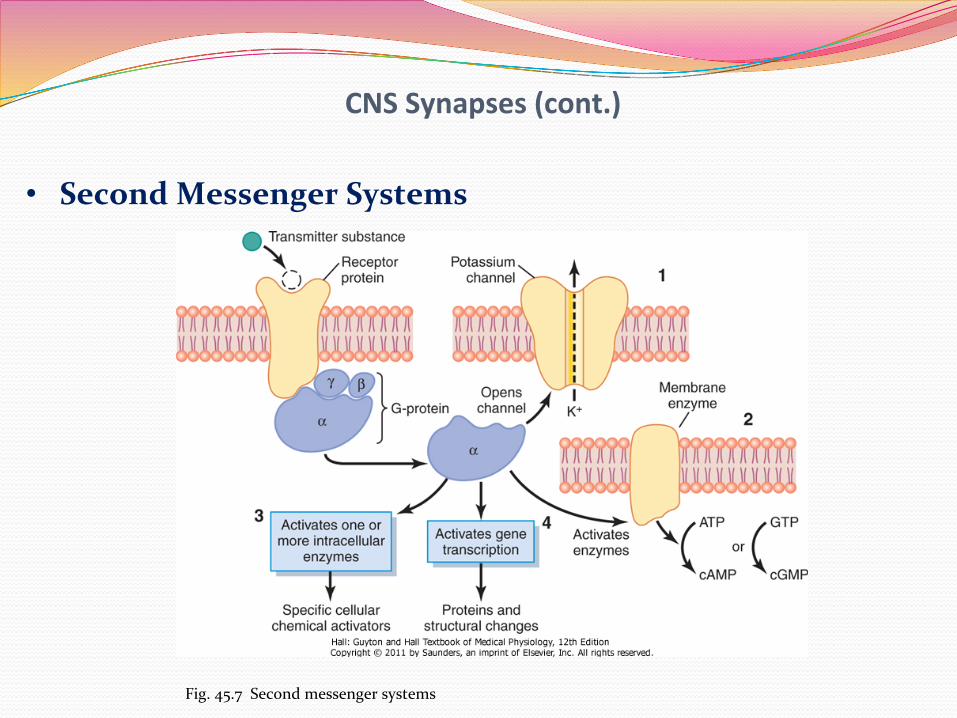
• Second Messenger Systems
Fig. 45.7 Second messenger systems

CNS Synapses (cont.)
• Second Messenger Systems-‐ the alpha component of the G protein performs one of four functions:
a. Opening specific ion channels through the post-‐
synaptic membrane b. Activation of cAMP or cGMP
c. Activation of one or more cellular enzymes
d. Activation of gene transcription

CNS Synapses (cont.)
• Excitatory Receptors in the Postsynaptic Membrane
a. In excitation: the opening of Na channels to allow large numbers of + electrical charges to flow to the interior. This raises the membrane potential toward threshold (most widely used method of excitation)
b. In excitation: depressed conduction through chloride
or potassium channels or both; decreases the diffusion of Cl to the inside or K to the outside which makes the membrane potential more positive
c. Metabolic changes to excite cell activity, increase
excitatory receptors or decrease inhibitory receptors
CNS Synapses (cont.)
• Inhibitory Receptors in the Postsynaptic Membrane
a. Opening of chloride channels allowing the rapid influx of ions which causes the membrane potential to become more negative, and therefore inhibitory
b. Increase in conductance of potassium ions out of the
neuron allowing positive ions to diffuse to the outside causing increased negativitiy, and therefore inhibitory
c. Activation of receptor enzymes that inhibit metabolic
functions or increase the number of inhibitory receptors or decrease the number of excitatory receptors

Types of NeurotransmiBers
• Small Molecule, Rapidly Acting Transmitters
Class I Class II: The Amines
Class III: Amino Acids
Class IV
Acetylcholine Norepinephrine GABA Nitric Oxide
Epinephrine Glycine
Dopamine Glutamate
Serotonin Aspartate
Histamine
Table 45.1
Types of NeurotransmiBers
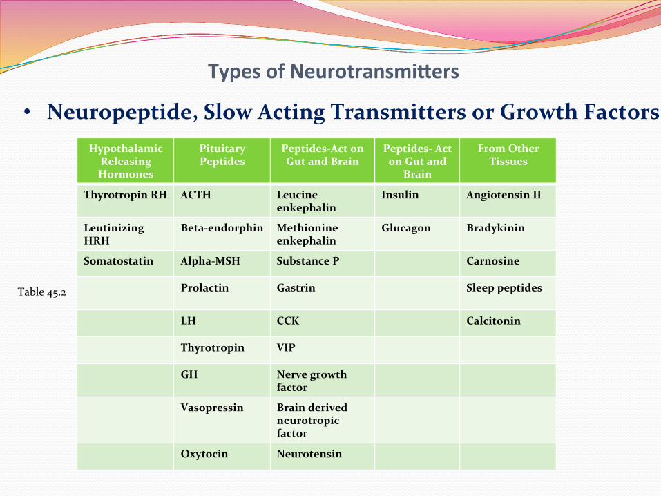
• Neuropeptide, Slow Acting Transmitters or Growth Factors Hypothalamic
Releasing Hormones
Pituitary Peptides
Peptides-‐Act on Gut and Brain
Peptides-‐ Act on Gut and
Brain
From Other Tissues
Thyrotropin RH ACTH Leucine enkephalin
Insulin Angiotensin II
Leutinizing HRH
Beta-‐endorphin Methionine enkephalin
Glucagon Bradykinin
Somatostatin Alpha-‐MSH Substance P Carnosine
Prolactin Gastrin
Sleep peptides
LH CCK Calcitonin
Thyrotropin VIP
GH Nerve growth factor
Vasopressin Brain derived neurotropic factor
Oxytocin Neurotensin
Table 45.2

Electrical Events During Excita<on
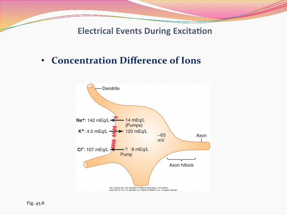
• Resting Membrane Potential (-‐65 mV for a spinal motor neuron)
Fig. 45.8
Electrical Events During Excita<on
• Concentration Difference of Ions
Fig. 45.8

Electrical Events During Excita<on
• Uniform Distribution of Electrical Potential Inside the Soma
• Effect of Synaptic Excitation on the Postsynaptic
Membrane—Excitatory Postsynaptic Potential

Electrical Events During Excita<on
Fig. 45.9 Three states of a neuron

Electrical Events During Excita<on
• Generation of APs in the Initial Segment
a. Axon hillock
b. The membrane has 7x the voltage gated Na channels as does the membrane of the soma
c. Threshold is about -‐45 mv (Fig. 45.9)

Electrical Events During Inhibi<on
• Effect of Inhibitory Synapses on the Postsynaptic Membrane—Inhibitory Postsynaptic Potential
a. Inhibitory synapses open mostly Cl channels
b. As the chloride ions enter, the membrane potential becomes more negative (toward -‐70 mV)
c. Opening K channels allows the positive ions to move
out; with the Cl, this causes a hyperpolarization d. Causes an IPSP (inhibitory postsynaptic potential)

Electrical Events During Inhibi<on
• Presynaptic Inhibition
a. Release of an inhibitory substance onto the outside of the presynaptic nerve fibrils (usually GABA)
b. Opens anion channels, allows Cl to diffuse inward
c. Negative charges cancel much of the excitatory effect
d. Occurs in many sensory pathways

Electrical Events During Inhibi<on
• Time Course of Postsynaptic Potentials
Fig. 45.10 EPSPs

Electrical Events During Inhibi<on
• Spatial Summation-‐ stimulation of many presynaptic terminals; the effects can summate until neuronal excitation occurs (Fig. 45.10)
• Temporal Summation-‐ successive discharges from
a single presynaptic terminal; if they occur rapidly enough, they also summate

Electrical Events During Inhibi<on
• Simultaneous Summation of IPSPs and EPSPs-‐ the two effects either completely or partially nullify each other
• Facilitation of Neurons
a. Occurs when the summated postsynaptic potential is excitatory but has not reached the threshold
b. Another excitatory signal can then excite the
membrane quite easily

Electrical Events During Inhibi<on
• Special Functions of Dendrites for Exciting Neurons
a. Large spatial field of excitation of the dendrites-‐ 80-‐95% of all presynaptic terminals of the anterior motor neuron terminate on dendrites
b. Most dendrites cannot transmit APs but they can
transmit signals by ion conduction of the fluids in cytoplasm

Electrical Events During Inhibi<on
• Decrement of Electrotonic Conduction in the Dendrites-‐ Greater Excitatory or Inhibitory Effect by Synapses Located Near the Soma
Fig. 45.11

Electrical Events During Inhibi<on
• Summation of Excitation and Inhibition in Dendrites
Fig. 45.11
Electrical Events During Inhibi<on
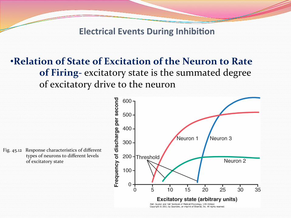
• Relation of State of Excitation of the Neuron to Rate of Firing-‐ excitatory state is the summated degree of excitatory drive to the neuron
Fig. 45.12 Response characteristics of different types of neurons to different levels of excitatory state

Special Characteris<cs of Synap<c Transmission
• Fatigue of Synaptic Transmission
• Effect of Acidosis or Alkalosis
• Effect of Hypoxia
• Effects of Drugs
• Synaptic Delay