Embed Size (px)
Citation preview

HISTORY
The innovative combination of selective alveolar periodontaldecortication surgery and augmentation grafting with ortho-dontic treatment results in an acceleration of orthodontictreatment, enhanced stability of orthodontic results, andlong-term improvement of the periodontium (Wilcko 2003).The purposes of this section are to describe the PeriodontallyAccelerated Osteogenic Orthodontics technique and to ex-plain how and why this advanced periodontal surgical tech-nique contributes to orthodontic treatment.
Decortication or corticotomy means simply the intentionalcutting or injury of cortical bone. While the bony jaws arecomposed of both cortical and medullary bone in variouscombinations depending on site and patient demographics,the periodontal surgery of interest in contemporary selectivealveolar decortication involves only surgical injury of alveolarcortical bone.
The application of corticotomy surgery to correct malocclu-sion was first described in 1892 by L. C. Bryan, but it wasHeinrich Köle in 1959 who reintroduced alveolar corticotomyto resolve malocclusion (Köle 1959). He combined interden-tal alveolar corticotomy surgery with a through-and-throughosteotomy apical to the teeth. Generson et al. in 1978 offereda modification of the Köle protocol and eliminated the subapi-cal osteotomy portion of the surgery.
In 2001, Wilcko et al. described selective alveolar decortica-tion with augmentation grafting combined with orthodontictreatment. They patented and trademarked their technique asPeriodontally Accelerated Osteogenic Orthodontics (PAOO).Full-thickness labial and lingual alveolar flaps were laid adja-cent to the teeth intended for movement, and selective decor-tication surgery was performed. Given that the surgery wasdone only in the area of desired tooth movement, it was thethickness of cortical bone that dictated where and how thecortical bone was injured. The depth of the decortication cutsbarely penetrated into medullary bone and bleeding was pro-moted; care was taken not to injure any tooth or encroach onthe periodontal ligament. An allograft of resorbable graftingmaterial plus antibiotic was applied directly over the bleeding
bone, and the surgical site was closed using Gore-Tex suturematerial. A frequently used grafting mixture was equal por-tions of demineralized freeze-dried bone (DFDBA) and bovinebone (Osteograf/N-300 or Bio-Oss) wetted with clindamycinphosphate (Cleocin) solution.
Wilcko et al. observed rapid orthodontics following PAOOand active treatment times of 6 to 8 months were common.They questioned Köle’s precept of “bony block” movementand offered an alternative hypothesis that rapid tooth move-ment resulted from marked but transient decalcification-recalcification of the alveolus. Frost, an orthopedic surgeon,had described a direct correlation between degree and prox-imity of bone trauma and intensity of physiological healing re-sponse, which he coined Regional Acceleratory Phenomena,or RAP (Frost 1983), and the decalcification-recalcificationdescribed by Wilcko et al. (2001, 2003) was consistent withRAP.
INDICATIONS
• Increased alveolar volume and enhanced periodontium(i.e., correction of dehiscences and fenestrations)
• Accelerated treatment (i.e., 3 to 4 times more rapid activeorthodontic treatment)
• Greater stability of clinical outcomes and less relapse
• Enhanced scope of malocclusion treatment (i.e., avoidingorthognathic surgery and extractions in selected cases)
• Enhancement of patient’s profile when indicated
• Rapid recovery of impacted teeth (i.e., canines)
The initial decortication protocol used by the Wilcko brothersdid not include augmentation alveolar grafting. ComputedTomography (CT) scans were taken on the first series of pa-tients to compare pretreatment status with immediate postorthodontics and at least 1-year retention conditions. The CTscans of the alveolus immediately after braces were removedshowed a paucity of alveolar bone in the areas where decor-tication surgery was performed. However, the retention im-ages demonstrated that a minimally adequate amount of
23
Chapter 4 The Contribution of Periodontics to Orthodontic Therapy
M. Thomas Wilcko, DMD, William M. Wilcko, DMD, MS,
M. Gabriela Marquez, DMD, MSD, and Donald J. Ferguson, DMD, MSD
Periodontally Accelerated Osteogenic Orthodontics (PAOO)

mineralized alveolar bone would return if the soft tissue peri-odontal envelope remained intact (Fig. 4.1).
Since 1996, the PAOO technique has included augmentationgrafting to increase alveolar volume (Fig. 4.2). Results of re-search showed no change in alveolar cortical bone thicknessat an average of 1.5 years following immediate post ortho-dontic treatment (Twaddle 2002). A recent study of attached
gingival levels in PAOO patients resulted in significantly moreattached gingiva evident at least 1 year after active orthodon-tic treatment. Increases in attached gingiva were consistentin the lower dental arch and inconsistent in the maxillary arch(Kacewicz 2007).
An additional benefit of augmentation bone grafting is the re-pair of alveolar cortical bone fenestrations and dehiscences
24 Practical Advanced Periodontal Surgery
Fig. 4.1. CT and surgery images of patient treated with selective alveolar decortication without augmentation grafting demonstrating lack of calcifiedcortical plate at post orthodontic treatment but return of cortical plate volume by 1 year retention. A: CT image at pre-treatment showing root promi-nences; B: view of labial cortical bone at the time of selective alveolar decortication surgery; C: CT image at immediate post orthodontic treatmentshowing lack of calcified labial cortex; D: CT Image at 1 year after active orthodontic treatment showing adequate but minimal calcified labial cortex.
A
B
C
D

Accelerated Osteogenic Orthodontics, Miniscrews 25
Fig. 4.2. CT and surgery images of patient treated with selective alveolar decortication and augmentation grafting demonstrating ample cortices postgrafting. A: CT image at pre-treatment showing root prominences; A-1: view of labial cortical bone at the time of selective alveolar decorticationsurgery; A-2: view of surgery area after grafting; B & C: CT images at post orthodontic treatment (B) and 2.5 years retention (C) showing increasedvolume of labial cortical bone.
A
B C
A1 A2

(Fig. 4.3). The grafting procedure is done without membranesbecause full-thickness flaps are used followed by primary clo-sure in a healthy periodontium. Under these circumstances,migration of the epithelial attachment is not likely to occur, andthe intact periosteum serves as a natural “membrane.”
PAOO has contributed greater stability of orthodontic clinicaloutcomes and less relapse. Stability of orthodontic clinicaloutcomes was analyzed by Nazarov in 2003 using theObjective Grading System (OGS) sanctioned by theAmerican Board of Orthodontics (ABO). While no differenceswere observed in nonextraction therapies at immediate posttreatment between PAOO and non-PAOO groups, three ofthe nine OGS variables (alignment, marginal ridges, and totalscore) were significantly better in the PAOO group at reten-tion. O’Hara (2005) compared nonextraction orthodontictreatment of moderate malocclusions and found no differ-ences in the OGS alignment variables at post treatment. Butat retention, alignment had improved in the PAOO group,whereas in the two non-PAOO groups, fixed and removableappliance retention, alignment had relapsed. In summary, im-mediate post orthodontic treatment results following nonex-traction therapy are statistically the same with or without
PAOO. However, during retention, the clinical outcomes ofPAOO patients improved and did not demonstrate relapse.
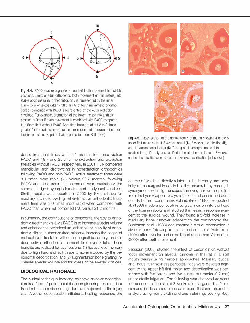
PAOO increases the scope of orthodontic tooth movementand the positions of the teeth after decortication and aug-mentation grafting are stable long-term. Sarver and Proffit(2005) offered guidelines as to the limits of central incisortooth movement in the adult patient with orthodontic treat-ment alone. Ferguson et al. (2006) suggested that these lim-its can be expanded 2-fold to 3-fold in all dimensions exceptretraction following PAOO (see Fig. 4.4) and that the stabilityof these positions is probably due to loss of tissue memoryfrom high turnover of the periodontium as well as increasedthickness of the alveolar cortices from the augmentationgrafting. Rothe et al. (2006) found that patients with thinnermandibular cortices were at increased risk for having dentalrelapse—hence the necessity for osseous grafting during thePAOO procedure.
Periodontics, through PAOO therapy, contributes to ortho-dontic treatment by reducing active orthodontic treatmenttime. Claims of rapid orthodontic treatment times were firstvalidated in research by Hajji in 2000; average active ortho-
26 Practical Advanced Periodontal Surgery
Fig. 4.3. Augmentation alveolar grafting repairs bony dehiscences and fenestrations. A: evidence of bony discrepancies after full thickness flap, B:decortication prior to grafting with dehiscences and fenestrations outlined; C-D: absence of dehiscences and fenestrations 7.5 years post PAOOdecortication and grafting.
BA
DC
dontic treatment times were 6.1 months for nonextractionPAOO and 18.7 and 26.6 for nonextraction and extractiontherapies without PAOO, respectively. In 2001, Fulk comparedmandibular arch decrowding in nonextraction orthodonticsfollowing PAOO and non-PAOO; active treatment times were3.1 times more rapid (6.6 versus 20.7 months) followingPAOO and post treatment outcomes were statistically thesame as judged by cephalometric and study cast variables.Similar results were reported in 2003 by Skountrianos formaxillary arch decrowding, wherein active orthodontic treat-ment time was 3.0 times more rapid when combined withPAOO than when not combined (7.0 versus 21.2 months).
In summary, the contributions of periodontal therapy to ortho-dontic treatment vis-à-vis PAOO is to increase alveolar volumeand enhance the periodontium, enhance the stability of ortho-dontic clinical outcomes (less relapse), increase the scope ofmalocclusion treatable without orthognathic surgery, and re-duce active orthodontic treatment time over 3-fold. Thesebenefits are realized for two reasons: (1) tissues lose memorydue to high hard and soft tissue turnover induced by the pe-riodontal decortication, and (2) augmentation bone grafting in-creases alveolar volume and thickness of the alveolar cortices.
BIOLOGICAL RATIONALE
The clinical technique involving selective alveolar decortica-tion is a form of periodontal tissue engineering resulting in atransient osteopenia and high turnover adjacent to the injurysite. Alveolar decortication initiates a healing response, the
degree of which is directly related to the intensity and prox-imity of the surgical insult. In healthy tissues, bony healing issynonymous with high osseous turnover, calcium depletionfrom the hydroxyappatite crystal lattice, and diminished bonedensity but not bone matrix volume (Frost 1983). Bogoch etal. (1993) made a penetrating surgical incision into the headof the tibia in rabbits and studied the healing response adja-cent to the surgical wound. They found a 5-fold increase inmedullary bone turnover adjacent to the corticotomy site.Buchanan et al. (1988) documented a similar observation inalveolar bone following tooth extraction, as did Yaffe et al.(1994) after alveolar periosteal flap elevation and Verna et al.(2000) after tooth movement.
Sebaoun (2005) studied the effect of decortication withouttooth movement on alveolar turnover in the rat in a splitmouth design using multiple approaches. Maxillary buccaland lingual full-thickness periosteal flaps were elevated adja-cent to the upper left first molar, and decortication was per-formed with five palatal and five buccal bur marks (0.2 mm)under sterile irrigation. The following was observed adjacentto the decortication site at 3 weeks after surgery: (1) a 2-foldincrease in decalcified trabecular bone (histomorphometricanalysis using hematoxylin and eosin staining; see Fig. 4.5),
Accelerated Osteogenic Orthodontics, Miniscrews 27
Fig. 4.4. PAOO enables a greater amount of tooth movement into stablepositions. Limits of adult orthodontic tooth movement (in millimeters) intostable positions using orthodontics only is represented by the innerblack-color envelope (after Proffit); limits of tooth movement for ortho-dontics combined with PAOO is represented by the outer red-colorenvelope. For example, protraction of the lower incisor into a stableposition is 9mm if tooth movement is combined with PAOO compared to a 5mm limit without PAOO. Note that limits are about 2 to 3 timesgreater for central incisor protraction, extrusion and intrusion but not forincisor retraction. (Reprinted with permission from Bell 2006)
Fig. 4.5. Cross section of the dentoalveolus of the rat showing 4 of the 5upper first molar roots at 3 weeks control (A), 3 weeks decortication (B),and 11 weeks decortication (C). Testing of histomorphometric dataresulted in significantly less calcified trabecular bone volume at 3 weekson the decortication side except for 7 weeks decortication (not shown).
A B
C

(2) a 1.5-fold increase in new trabecular bone formation (vitalstaining ad libitum), (3) a 4-fold increase is osteoclast count(osteoclast count after TRAP staining), and (4) a 2-fold in-crease in lamina dura apposition (vital stain injection series;see Fig. 4.6). These findings collectively indicate high tissueturnover immediately adjacent to the decortication site.
Pham-Nguyen (2006) studied the three-dimensional volumeof periodontal tissues surrounding the upper first molar inthe rat model following buccal and lingual selective decorti-cation. Using micro-CT technology, a significant decrease inalveolar mineralization was evident by 7 days post decorti-cation (Fig. 4.7).
Conceptually, increased tissue turnover (osteopenia) is a con-dition that favors rapid tooth movement. This tenet wasdemonstrated by Verna et al. (2000) using a rat model bymoving teeth after pharmacologically inducing high and lowbone turnover. They showed significantly greater tooth move-ment in the high turnover group compared with normal andlow bone turnover groups. In our laboratory rat studies(Pham-Nguyen 2006, Sebaoun 2005), surgical injury to thealveolus induced a dramatic increase in tissue turnover thatwas expressed both spatially and temporally. The effect ofalveolar decortication was localized to the area immediatelyadjacent to the injury. It is obvious that considerable medul-lary bone demineralization occurs immediately adjacent tothe decortication site. Although induced osteopenia is a tran-sient condition in the functionally normal alveolus, it was sur-mised that tooth movement perpetuates the decalicified con-dition of the trabecular bone (Fig. 4.8).
PERIODONTALLY ACCELERATEDOSTEOGENIC ORTHODONTICS IN THETREATMENT OF CROWDING
Using the PAOO technique, cases of moderate dental archcrowding are routinely completed in 4 to 6 months instead of
18 to 24 months of active orthodontic treatment, and the re-sults have been shown to be remarkably stable (Figs. 4.9 and4.10).
RAPID RECOVERY OF IMPACTED TEETH
Recovery of impacted teeth typically prolongs active ortho-dontic treatment time. Impacted tooth recovery combinedwith selective alveolar periodontal decortication surgery ac-celerates the recovery and reduces orthodontic treatmenttime. The purpose of this section is to describe an advancedperiodontal surgical technique of recovering impacted cus-pids that features the following: (1) complete exposure of theclinical crown, (2) clearing a path for recovery by way of alve-olar ostectomy, and (3) selective alveolar decortication or in-tramarrow penetrations surrounding the impacted tooth root.
With the exception of the third molars, the maxillary caninesare the most frequently encountered impaction, with an inci-dence of approximately 2% according to Ericson and Kurol(1987). The incidence of maxillary canine impaction is 3 timesgreater in females than in males and is often associated withmissing or peg-shaped laterals (Bec et al. 1981, Peck et al.1994). Maxillary permanent canines are impacted palatally
28 Practical Advanced Periodontal Surgery
Fig. 4.6. Amount of apposition of the lamina dura is shown in crosssection of the dentoalveolus of the rat at 4 weeks, the end of the firstseries of vital stain injections. Total width of apposition as revealed bythe three stains together was statistically greater on the decorticationside (A, 0.051mm) at 3 weeks than the control side (B, 0.037mm) at 3 weeks.
Fig. 4.7. Micro-CT analysis of osteopenia at 7 days following selectivealveolar decortication showing gross specimen #602 (A), control side (B)and decortication side (C).On the decortication side, osteopenia wasdemonstrated as the bone volume (BV) to total volume (TV) ratio de-creased to 45% (note the wide spaces surrounding the roots, C) com-pared to 57% on the control side (B). (Pham-Nguyen K, et al. 2006)
AA
B
B C
much more frequently than labially (85% palatal comparedwith 15% labial; Bass 1967, Hitchin 1956, Jacoby 1979).Rayne (1969) has pointed out that labially impacted caninesare often associated with inadequate arch space. Interceptivetreatment of impacted maxillary canines can often be suc-cessful in 10- to 13-year-olds depending on the degree of theimpaction (Jacobs 1992). The more severely impacted max-illary canines typically require combined surgical/orthodontictreatment (Bishara 1992).
Previous authors have indicated that details of the recoverytechnique depend on the type of impaction. When the crownis not covered by bone and there is a broad band of kera-tinized gingival tissue present, Kokich and Mathews (1993),Shiloah and Kopczyk (1978), and Jarjoura et al. (2002) havesuggested a surgical window (gingivectomy) to expose the
shallow impaction. In situations where there is only a nar-row zone of gingival attachment and a relatively shallow labialor mid-alveolar bony impaction, an open technique consist-ing of an apically repositioned flap is more suitable accordingto Wong-Lee and Wong (1985) and Vanarsdall and Corn(1977). When the bony impaction is deep in the vestibule,mid-alveolar, or impacted palatally, Vermette et al. (1965),McDonald and Yap (1986), and Smukler et al. (1987) havesuggested erupting the canine through the gingiva followingprimary surgical flap closure.
RECOVERY OF LABIALLY IMPACTEDCANINES
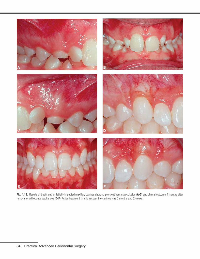
Recovery of labially impacted maxillary canines is illustrated inthe treatment of a girl aged 14 years 4 months using selec-
Accelerated Osteogenic Orthodontics, Miniscrews 29
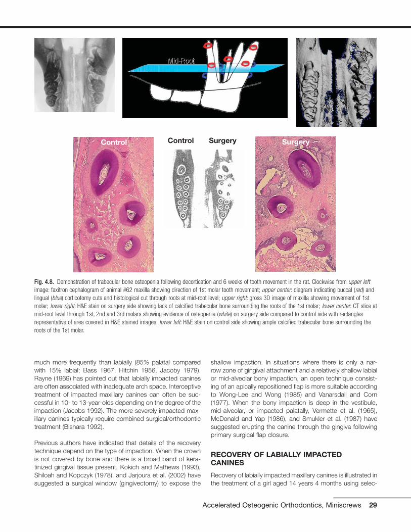
Fig. 4.8. Demonstration of trabecular bone osteopenia following decortication and 6 weeks of tooth movement in the rat. Clockwise from upper leftimage: faxitron cephalogram of animal #62 maxilla showing direction of 1st molar tooth movement; upper center: diagram indicating buccal (red) andlingual (blue) corticotomy cuts and histological cut through roots at mid-root level; upper right: gross 3D image of maxilla showing movement of 1stmolar; lower right: H&E stain on surgery side showing lack of calcified trabecular bone surrounding the roots of the 1st molar; lower center: CT slice atmid-root level through 1st, 2nd and 3rd molars showing evidence of osteopenia (white) on surgery side compared to control side with rectanglesrepresentative of area covered in H&E stained images; lower left: H&E stain on control side showing ample calcified trabecular bone surrounding theroots of the 1st molar.
Control Control Surgery Surgery

30 Practical Advanced Periodontal Surgery
Fig. 4.9. Adult female patient with moderate upper and lower dental arch malocclusion (A,B) treated by PAOO (A-1, A-2, B-1, B-2) showing immedi-ate post treatment outcome after 7 months of active orthodontic therapy (C,D).
A
B
C
D
A2
A1
B1
B2

Accelerated Osteogenic Orthodontics, Miniscrews 31
Fig. 4.10. Retention outcome at 2 years (A,B) and 9 years (C,D) after the completion of PAOO treatment. Improvement during retention and long termstability are likely due to high tissue turnover induced by decortication and increase in cortical bone thickness.
A B
C D

tive decortication technique (Figs. 4.11, 4.12, and 4.13). Afull-thickness labial flap, using a sulcular incision with verticalreleasing incisions mesial to the first premolars, was reflectedto reveal upper cuspid crowns labial to the upper permanentlateral incisors. No lingual flap was reflected. The upper pri-mary canines were extracted, and the soft tissue follicle andthin layer of bone surrounding cuspid crowns were removed.A path (trough) was cleared for the forced eruptions by re-moving the facial cortical plate between the cuspid crownsand extraction sockets including the facial plates over thesockets. In addition, numerous intramarrow penetrationswere made to stimulate alveolar bone turnover and to induce
32 Practical Advanced Periodontal Surgery
Fig. 4.11. Labially impacted maxillary canines showing pre-surgery (A),partial exposure of canine crowns after full-thickness flap (B), and boneremoval to expose canine crowns completely, intramarrow penetrations,and creation of a path by removing facial cortical plates of extractionsocket (C).
Fig. 4.12. Treatment for labially impacted maxillary canines showingviews of different cases at the time of surgery (A, D, G, B, E, H) and at10 weeks post surgery (C, F, I). An orthodontic bracket was placed in an optimal position on the exposed right canine (A) but on the lingualsurface of the crown on the left canine (G). At the time of soft tissueclosure, a vertical incision was made in the flap allowing exposure of theright canine bracket (B) but not the left canine (H) before suturing theflap to the original position (E). The canines were beginning to moverapidly by 10 weeks after the canine recovery surgery (C, F, I).
A
A
B
C
B
C

RAP or osteopenia. At least 1.5 mm of bone was left undis-turbed mesial to the first premolars and distal to lateral inci-sors. An orthodontic bracket was placed in an optimal posi-tion of the upper right permanent canine, but the upper leftpermanent canine was rotated, which necessitated placingthe bracket on the lingual surface. On the right side, the
bracket was exposed with a vertical incision in line with thebracket, and then suturing the flap back into the original po-sition. The upper left canine was covered by the flap and su-tured back in its original position. Because the upper left ca-nine was covered by the flap, a backup ligature wire was alsoused. In this case, a resorbable Vicryl suture was used.
Accelerated Osteogenic Orthodontics, Miniscrews 33
Fig. 4.12. (continued)
D
G
F
I
E
H
34 Practical Advanced Periodontal Surgery
Fig. 4.13. Results of treatment for labially impacted maxillary canines showing pre-treatment malocclusion (A-C) and clinical outcome 4 months afterremoval of orthodontic appliances (D-F). Active treatment time to recover the canines was 5 months and 2 weeks.
A B
C D
E F

RECOVERY OF DEEP PALATALLY IMPACTEDCANINES
Selective decortication is especially helpful in the recovery ofmaxillary permanent canines that are deeply impacted on thepalatal side and require a considerable amount of movementthrough alveolar medullary bone as illustrated with the treat-ment of a boy aged 14 years 6 months (Figs. 4.14 and 4.15).Decortication injury stimulates an osteopenic response thatreduces the mineral content of spongiosa, thereby reducingthe resistance to impacted canine movement.
Accelerated Osteogenic Orthodontics, Miniscrews 35
Fig. 4.14. Treatment for deep palatally impacted maxillary caninesshowing pre-treatment malocclusion (A), canine exposure, bracketingand elastic traction (B), flap repositioning and suturing (C), 3 monthspost surgery (D), and complete canine recovery (E). Active treatment timeto recover the canines was 6 months and 0 weeks.
A
C D
E
B

36 Practical Advanced Periodontal Surgery
Fig. 4.15. Treatment for deep palatally impacted maxillary canine showing pretreatment malocclusion (A) and radiograph (H), flap with verticalreleasing incision showing labial cortical plate (B), ostectomy removal of labial cortical and medullary bone to clear a movement path and exposecanine crown, and labial intramarrow penetrations (C), bracket placement and application of elastic traction (D), immediate postsurgical flap suturing(E) and radiograph (F) with radiograph (I), and 9 days after orthodontic appliance removal (G) with radiograph (K). Active treatment time to recover thecanine was 6 months 0 weeks.
A
B
F
C
D
E

Accelerated Osteogenic Orthodontics, Miniscrews 37
Fig. 4.15. (continued)
G
H
I
J
K

Both facial and lingual full-thickness flaps are used with dis-tal vertical releasing incisions for access to deep palatally im-pacted canines. Preference is given to placement of ortho-dontic brackets into an optimal positioned on the labialsurface of the canine crown for control and efficiency. Whendirect access to the labial surface and optimal bracket place-ment is not possible, the surface of choice for bracket place-ment is the distolabial surface of impacted canines. An ostec-tomy is required between the first premolar and the lateralincisor to gain access to the appropriate impacted caninesurface. The ostectomy not only permits optimal bracketplacement but also creates a space for the elastic chain fromthe bracket on the impacted canine to the brackets on theadjacent teeth already well positioned in the dental arch.Moreover, bone needs to be removed when crowns are ex-posed because the follicle surrounding the enamel is elimi-nated and bone resorption secondary to follicular activity islost. The ostectomy should eliminate all bone in the move-ment path of the crown during impacted canine recovery, butit is important to leave about 1.5 mm of bone on the proximalsurfaces of the adjacent teeth. Intramarrow penetrations arealso performed, especially in the bone that lies between theimpacted tooth and its eventual position in the dental arch.Intramarrow penetrations over the root prominence of the im-pacted tooth facing in the direction of movement stimulatemedullary osteopenia and reduce tooth movement resist-ance. After the bracket has been placed, a ligature wire is se-cured to the bracket of the impacted tooth as backup in theevent the elastic chain was to break. A bracket with a post orpower arm pointing apically is preferred to reduce the likeli-hood of the chain elastic slipping off the bracket. The full-thickness flaps are returned to their original positioning andsutured. In the case demonstrating recovery of deep palatallyimpacted canines (Figs. 4.14 and 4.15), nonresorbable 5-0 green braided polyester suture material was used, butthe type of suture material used is usually not relevant. Sutureremoval, when necessary, is recommended anytime after 1week of postoperative healing.
PERIODONTAL SURGICAL PROCEDURES FORORTHODONTIC ACCESS, AESTHETICS, ANDSTABILITY
Excessive gingival attachment serves no useful purpose be-fore, during, or after orthodontic treatment. There are severalreasons for excessive gingival attachment. Gingival excess canresult from systemic medications that produce undesirableside effects of gingival hyperplasia such as dilantin, nifedipine,and amlodipine or gingival swelling such as cyclosporine; thick,fibrotic gingival overgrowth can produce tooth movementand/or malocclusion. Excessive gingival attachment can resultfrom familial hereditary gingival hyperplasia or, more commonly,by delayed passive eruption of teeth (Fig. 4.16).
Surgical removal of gingiva is called gingivectomy, and gin-givoplasty refers to recontouring the gingival architecture
and/or changing the gingival shape. These simple periodon-tal surgical procedures can contribute meaningfully to ortho-dontic access, aesthetics, and treatment outcome stability.The fundamental reasons for gingivoplasty procedures are toprovide more optimal clinical crown appearances or accessand to change gingival shape. According to Sarver (2004),exposure of the clinical crowns that contribute most to theaesthetic smile, the maxillary central incisors, should be ap-proximately 80% width compared with height. Moreover, thegingival architecture for the anterior teeth should have certaincharacteristics. The gingival shape of the mandibular incisorsand the maxillary laterals should exhibit a symmetrical half-oval or half-circular shape, and the maxillary centrals and ca-nines should exhibit a gingival shape that is more elliptical.Thus, the gingival zenith (the most apical point of the gingivaltissue) should be located distal to the longitudinal axis of themaxillary centrals and canines, and the gingival zenith of themaxillary laterals and mandibular incisors should coincidewith their longitudinal axis (Fig. 4.17).
Excessive gingival attachment often makes optimal ortho-dontic bracket placement difficult. When the patient is readyfor orthodontic treatment with fixed appliances but access tothe clinical crown precludes optimal bracket position place-ment, a gingivoplasty procedure can be used to establish amore favorable crown-to-crestal height relationship by re-moval of excess gingiva. This is a frequent finding in youngpatients ready for orthodontic treatment in the late mixed orearly permanent dentition (Fig. 4.18).
Removal of excessive gingival tissues is useful during thecourse of orthodontic treatment to facilitate good oral hy-giene and to help in the consolidation of the dental arch
38 Practical Advanced Periodontal Surgery
Fig. 4.16. Delayed passive eruption of teeth can lead to excessive gingi-val attachment: normal attachment (left), moderate excess attachment(center), and severe excess attachment (right). Note that the tip of theperiodontal probe rests at the gingival crest demonstrating that clinicalcrown length deceases as gingiva excess increases.

Accelerated Osteogenic Orthodontics, Miniscrews 39
Fig. 4.17. Optimal maxillary incisor exposure for esthetics is 1.0 to 0.8crown height-to-width ratio and ideally, gingival zenith or most apicalpoint of the gingival tissue is distal to longitudinal crown axis. (After:Sarver MS, AJODO, 126:749-753, 2004.)
Fig. 4.18. Increasing access to upper incisor clinical crowns for bracketing on an 11 year old orthodontic patient: pre-treatment (A), 15 days postgingivoplasty (B), and post incisor bracketing (C).
A B
C
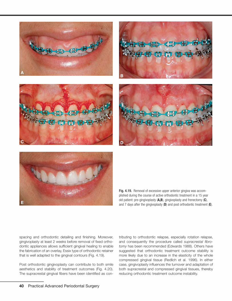
spacing and orthodontic detailing and finishing. Moreover,gingivoplasty at least 2 weeks before removal of fixed ortho-dontic appliances allows sufficient gingival healing to enablethe fabrication of an overlay, Essix type of orthodontic retainerthat is well adapted to the gingival contours (Fig. 4.19).
Post orthodontic gingivoplasty can contribute to both smileaesthetics and stability of treatment outcomes (Fig. 4.20).The supracrestal gingival fibers have been identified as con-
tributing to orthodontic relapse, especially rotation relapse,and consequently the procedure called supracrestal fibro-tomy has been recommended (Edwards 1988). Others havesuggested that orthodontic treatment outcome stability ismore likely due to an increase in the elasticity of the wholecompressed gingival tissue (Redlich et al. 1996). In eithercase, gingivoplasty influences the turnover and adaptation ofboth supracrestal and compressed gingival tissues, therebyreducing orthodontic treatment outcome instability.
40 Practical Advanced Periodontal Surgery
Fig. 4.19. Removal of excessive upper anterior gingiva was accom-plished during the course of active orthodontic treatment in a 15 yearold patient: pre-gingivoplasty (A,B), gingivoplasty and frenectomy (C),and 7 days after the gingivoplasty (D) and post orthodontic treatment (E).
AB
C D
E

REFERENCES Anholm, J.M., D.A. Crites, R. Hoff, and W.E. Rathbun, 1986.
Corticotomy facilitated orthodontics. Calif. Dental Assoc. J. 14:7–11.
Bass, T.B. 1967. Observations on the misplaced upper canine tooth.Dental Pract. Dental Rec. 18:25–30.
Bec, A., P. Smith, and R. Behar. 1981. The incidence of anomalous max-illary lateral incisors in relation to palatally displaced cuspids. AngleOrthod. 51:24–29.
Bishara, S.E. 1992. Impacted maxillary canines: a review. Am. J. Orthod.Dentofac. Orthop. 101:159–171.
Bogoch, E., N. Gschwend, B. Rahn, E. Moran, and S. Perren. 1993.Healing of cancellous bone osteotomy in rabbits, part I: regulation ofbone volume and the RAP in normal bone. J. Orthop. Res.11:285–291.
Buchanan, M., H.S. Sandhu, and C. Anderson. 1988. Changes in bonemineralization pattern: a response to local stimulus in maxilla andmandible of dogs. Histol. Histopathol. 3:331–336.
Edwards, J.G. 1988. A long-term prospective evaluation of the circum-ferential supracrestal fiberotomy in alleviating orthodontic relapse.Am. J. Orthod. Dentofac. Orthop. 93:380–387.
Accelerated Osteogenic Orthodontics, Miniscrews 41
Fig. 4.20. Effect of recontouring gingival architecture to improve the es-thetics an d stability of orthodontic treatment outcome in a 18 year oldmale patient; immediate post orthodontics (A,B), immediate post gin-givectomy (C) and 4 months post gingivectomy (D,E).E
A B
C D

Ericson, S., and J. Kurol. 1987. Radiographic examination of ectopicallyerupting maxillary canines. Am. J. Orthod. Dentofac. Orthop.91:483–492.
Ferguson, D.J., W.M. Wilcko, and M.T. Wilcko. 2006. Selective alveolardecortication for rapid surgical-orthodontic resolution of skeletal mal-occlusion. In W.E. Bell and C. Guerrero, editors. DistractionOsteogenesis of the Facial Skeleton. BC Decker, Hamilton, Ontario,Canada.
Frost, H.A. 1983. The regional acceleratory phenomena: a review. HenryFord Hosp. Med. J. 31:3–9.
Fulk, L.A. 2001. Lower arch decrowding comparing corticotomy-facilitated, midline distraction and conventional orthodontic tech-niques. Master’s degree thesis in orthodontics, Saint Louis University.
Gantes, B., E. Rathburn, and M. Anholm. 1990. Effects on the periodon-tium following corticotomy-facilitated orthodontics. J. Periodontol.61:234–238.
Generson, R.M., J.M. Porter, A. Zell, and G.T. Stratigos. 1978.Combined surgical and orthodontic management of anterior openbite using corticotomy. J. Oral Surg. 36:216–219.
Hajji, S.S. 2000. The influence of accelerated osteogenic response onmandibular decrowding. Master’s degree thesis in orthodontics, SaintLouis University.
Hitchin, A.D. 1956. The impacted maxillary canine. Br. Dental J.100:1–14.
Jacobs, S.G. 1992. Reducing the incidence of palatally impacted maxil-lary canines: a useful preventative interceptive orthodontic procedure:case report. Austral. Dental J. 37: 6–11.
Jacoby, H. 1979. The “Batista spring” system for impacted teeth. Am. J.Orthod. 75:43–51.
Jarjoura, K., P. Krespo, and J.B. Fine. 2002. Maxillary canine impactions:orthodontic and surgical management. Comp. Cont. Educ. Dent.23:23–38.
Kokich, V., and D.P. Mathews. 1993. Surgical and orthodontic manage-ment of impacted teeth. Dent. Clin. North Am. 37:181–204.
Köle, H. 1959. Surgical operations on the alveolar ridge to correct oc-clusal abnormalities. Oral Surg. Oral Med. Oral Pathol. 12:515–529.
Machado, I.M., D.J. Ferguson, W.M. Wilcko, and M.T. Wilcko. 2002.Reabsorcion radicular despues del tratamiento ortodoncico con o sincorticotomia alveolar. Rev. Venezuelana Orthod. 19:647–653.
McDonald, F., and W.L. Yap. 1986. Surgical exposure and application ofdirect traction of unerupted teeth. Am. J. Orthod. 89: 331–340.
Nazarov, A.D. 2003. Improved retention following corticotomy using BOObjective Grading System. Master’s degree thesis in orthodontics,Saint Louis University.
O’Hara, P. 2005. Orthodontic treatment and retention outcomes withand without PAOO and fixed retainers. Master’s thesis in orthodon-tics, Boston University.
Peck, S., and M. Kataja. 1994. The palatally displaced canine as a den-tal anomaly of genetic origin. Angle Orthod. 64:249–256.
Pham-Nguyen, K. 2006. Micro-CT analysis of osteopenia following se-lective alveolar decortication and tooth movement. Master’s degreethesis in orthodontics, Boston University.
Rayne, J. 1969. The unerupted maxillary canine. Dental Pract. DentalRec. 19:194–204.
Redlich, M., E. Rahamim, A. Gaft, and S. Shoshan. 1996. The responseof supra-alveolar gingival collagen to orthodontic rotation movementsin dogs. Am. J. Orthod. Dentofac. Orthop. 110:247–255.
Rothe, L.E. 2006. Trabecular and cortical bone as risk factors for ortho-dontic relapse. Am. J. Orthod. Dentofac. Orthop. 129:316.
Rothe, L.E., A.M. Bollen, R.M. Little, S.W. Herring, J.B. Chaison, C.S.K.Chen, and L.G. Hollender. 2006. Trabecular and cortical bone as riskfactors for orthodontic relapse. Am. J. Orthod. Dentofac. Orthop.130:476–484.
Sarver, D.M., and W.R. Proffit. 2005. Special considerations in diagnosisand treatment planning. In T.M. Graber, R.L. Vanarsdall, and K.W.L.Vig, editors. Orthodontics: Current Principles and Techniques.Elsevier, St. Louis, p. 15.
Sarver, D.M. 2004. Principles of cosmetic dentistry in orthodontics: part1. Shape and proportionality of anterior teeth. Am. J. Orthod.Dentofac. Orthop. 126:749–753.
Sebaoun, J.D. 2005. Trabecular bone modeling and RAP following se-lective alveolar decortication. Master’s degree thesis in orthodontics,Boston University.
Shiloah, J., and R. Kopczyk. 1978. Mucogingival considerations in sur-gical exposure of maxillary canines: report of case. ASDC J. Dent.Child. 45: 79–81.
Skountrianos, H.S. 2003. Maxillary arch decrowding and stability withand without corticotomy-facilitated orthodontics. Master’s degreethesis in Orthodontics, Saint Louis University.
Smukler, H., G. Castellucci, and H.M. Goldman. 1987. Surgical manage-ment of palatally impacted cuspids. Comp. Cont. Educ. Dent.8:10–77.
Suya, H. 1991. Corticotomy in orthodontics. In E. Hösl and A. Baldauf,editors. Mechanical and Biological Basics in Orthodontic Therapy.Hüthig, Heidelberg, pp. 207–226.
Twaddle, B.A., D.J., Ferguson, W.M., Wilcko, M.T., Wilcko, and C.-Y. Lin.2002. Dento-alveolar bone density changes following accelerated or-thodontics. J. Dental Res. 80:301.
Vanarsdall, R.L., and H. Corn. 1977. Soft tissue management of labiallypositioned unerupted teeth. Am. J. Orthod. 72:53–66.
Vermette, M.E., V.G. Kokich, and D.B. Kennedy. 1995. Uncovering labi-ally impacted teeth: apically positioned flap and closed eruption tech-nique, Angle Orthod. 65:23–32.
Verna, C., M. Dalstra, and B. Melsen. 2000. The rate and type of ortho-dontic tooth movement is influenced by bone turnover in the ratmodel. Eur. J. Orthod. 22:343–352.
Wilcko, W.M., D.J. Ferguson, J.E. Bouquot, and M.T. Wilcko. 2003.Rapid orthodontic decrowding with alveolar augmentation: case re-port. World J. Orthod. 4:197–205.
42 Practical Advanced Periodontal Surgery
HISTORY
Implants for use as orthodontic anchorage devices have re-cently become very popular because most are easy to placeand remove and inexpensive and can be directly or indirectlyloaded with biomechanical forces immediately after place-ment. Unlike the osseointegrated implants used for pros-thetic restorations, implants for orthodontic anchorage relyon mechanical retention and depend on the thickness of cor-tical bone for stability (Huja 2005). This screw-in type of im-plant is known by many names such as temporary anchor-age devise (TAD), mini-implant, microscrew, or miniscrew.
Implants for orthodontic anchorage were first described inthe literature in 1945 by Gainsforth and Higley and were usedto retract the canine tooth in the dog model. In 1983,Creekmore and Eklund reported placement of a screw devicein the anterior nasal spine area for use in the intrusion ofupper permanent incisors in the treatment of adult deep bitemalocclusion. Kanomi in 1997 provided detailed descriptionof loading microimplants less than 1.0 mm in diameter follow-ing 3 months of osseous integration. Since 2000, the dentalliterature has been inundated with articles describing new de-signs and the use of immediate-load implants as anchoragedevices for the treatment of malocclusion. In general, theyhave proved successful for intrusion and extrusion, protrac-tion, and retraction of individual teeth as well as groups ofteeth.
Miniscrew implants are the smallest among all fixed implants;they are the most versatile and adaptable for clinical use andare categorized as either non–bracket-head or bracket-head.The most commonly used implant for orthodontic anchorageis self-drilling with a nonbracket head. These self-drilling andself-tapping (a different cutting point for self-drilling) miniscrewshave emerged during the past 6 years as the most popular andfrequently used type of orthodontic anchorage implant, andthis type of miniscrew will be the focus of this chapter.
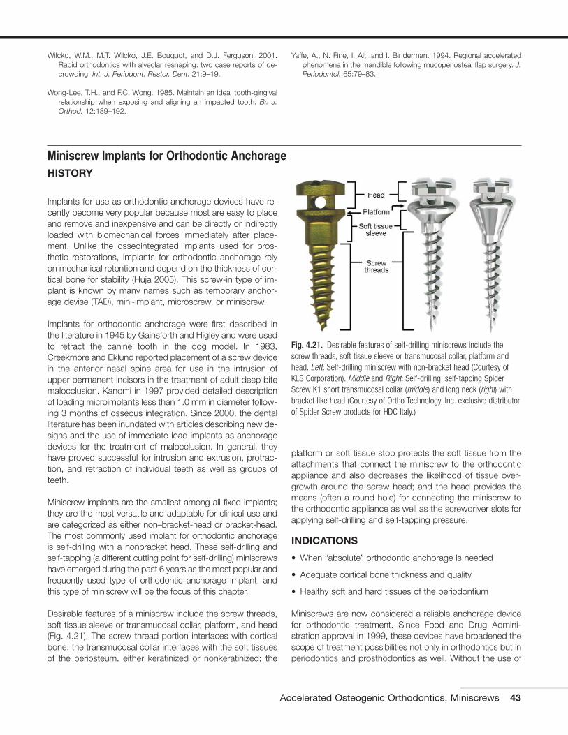
Desirable features of a miniscrew include the screw threads,soft tissue sleeve or transmucosal collar, platform, and head(Fig. 4.21). The screw thread portion interfaces with corticalbone; the transmucosal collar interfaces with the soft tissuesof the periosteum, either keratinized or nonkeratinized; the
platform or soft tissue stop protects the soft tissue from theattachments that connect the miniscrew to the orthodonticappliance and also decreases the likelihood of tissue over-growth around the screw head; and the head provides themeans (often a round hole) for connecting the miniscrew tothe orthodontic appliance as well as the screwdriver slots forapplying self-drilling and self-tapping pressure.
INDICATIONS
• When “absolute” orthodontic anchorage is needed
• Adequate cortical bone thickness and quality
• Healthy soft and hard tissues of the periodontium
Miniscrews are now considered a reliable anchorage devicefor orthodontic treatment. Since Food and Drug Admini-stration approval in 1999, these devices have broadened thescope of treatment possibilities not only in orthodontics but inperiodontics and prosthodontics as well. Without the use of
Accelerated Osteogenic Orthodontics, Miniscrews 43
Wilcko, W.M., M.T. Wilcko, J.E. Bouquot, and D.J. Ferguson. 2001.Rapid orthodontics with alveolar reshaping: two case reports of de-crowding. Int. J. Periodont. Restor. Dent. 21:9–19.
Wong-Lee, T.H., and F.C. Wong. 1985. Maintain an ideal tooth-gingivalrelationship when exposing and aligning an impacted tooth. Br. J.Orthod. 12:189–192.
Yaffe, A., N. Fine, I. Alt, and I. Binderman. 1994. Regional acceleratedphenomena in the mandible following mucoperiosteal flap surgery. J.Periodontol. 65:79–83.
Miniscrew Implants for Orthodontic Anchorage
Fig. 4.21. Desirable features of self-drilling miniscrews include thescrew threads, soft tissue sleeve or transmucosal collar, platform andhead. Left: Self-drilling miniscrew with non-bracket head (Courtesy ofKLS Corporation). Middle and Right: Self-drilling, self-tapping SpiderScrew K1 short transmucosal collar (middle) and long neck (right) withbracket like head (Courtesy of Ortho Technology, Inc. exclusive distributorof Spider Screw products for HDC Italy.)

implants, forces are generated using other structures as an-chorage such as teeth in order to produce desired orthodon-tic tooth movement. Teeth used as anchorage will inevitablyrespond with some movement because the anchor teeth aresurrounded by a periodontal ligament (PDL), and the PDL issensitive and responsive to changes in the environment.Miniscrews implanted into cortical bone are stable and do not
respond like teeth because the screw threads interface di-rectly with cortical bone as there is no mechanism like thePDL that enables movement of the implant.
Miniscrew implants are most appropriate when there is aneed for “absolute” anchorage. They should be inserted intokeratinized gingiva if possible and loaded immediately
44 Practical Advanced Periodontal Surgery
Fig. 4.22. Use of miniscrews as orthodontic anchorage to correct Class II malocclusion by retracting upper incisors and canines and to intrude upperleft first molar: pre-treatment (A-C), during treatment (D-F) and completed treatment (G-I). Note placement of miniscrew at junction of attached gin-giva and mucosa during treatment and use of palatal implant to assist with molar intrusion. Also note the interocclusal space created by intrusion ofthe upper left premolar and molar.
A B
C D
E
F

(Melsen and Costa 2000) or within the first 2 weeks of heal-ing (Liou et al. 2004). These devices are effective as anchor-age for moving teeth in three spatial planes such as retract-ing the upper incisors and canines in the treatment of ClassII malocclusion following first premolar extraction or intrusionof molars (Fig. 4.22) or protracting posterior teeth to closespace (Fig. 4.23).
Miniscrew stability depends on cortical bone thicknessbecause spongy bone contributes virtually nothing to im-plant stability (Melsen 2005). Alveolar bone is composed ofcompact (cortical) and spongy (trabecular or medullary)bone. Medullary bone is a highly adaptable tissue, and it re-sponds quickly to changes in the environment with model-ing or surface change, whereas cortical bone responds
Accelerated Osteogenic Orthodontics, Miniscrews 45
Fig. 4.22. (continued)
Fig. 4.23. Two uses of miniscrew as orthodontic anchorage to protract posterior teeth: Left: Miniscrew used to secure NiTi closed coil spring formolar protraction and elastic chain for premolar protraction. Right: Open coil spring placed mesial to molar allows protraction of the premolar only;steel ligature tie from miniscrew to molar prevents distal molar movement. Note the less desirable placement of the miniscrew below mucogingivaljunction (left) and more desirable placement of the miniscrew at junction of attached gingiva and mucosa (right).
G
I
H

slowly to stimuli with remodeling or internal change. Bonemodeling is an uncoupled activity of activation (A) followedby formation (F) or resorption (R). Because an activation canlead to either resorption (A-R) or formation (A-F), spongybone very quickly adapts to any stimuli or change in en-vironment. In contrast, compact bone undergoes boneremodeling which is a coupled sequence of activation (A)followed by resorption (R) followed by formation (F) or A-R-F sequence. The A-R-F sequence is otherwise knownas the process of secondary osteon formation: physiologi-cal activation (a stimulus) followed by bone resorption (cut-ting cone) followed by bone formation (filling cone).Remodeling (A-R-F) is a slow process that assures struc-tural integrity of the host skeleton (Roberts et al. 2004). The principal reason for miniscrew implant stability is attrib-uted to the slow bony turnover response of cortical bone(Fig. 4.24).
Miniscrew thread lengths vary from 4 mm to 12 mm withlonger thread lengths recommended for bicortical retention(Freudenthaler et al. 2001). In most circumstances, mini-screw thread length need not exceed the width of monocor-tical thickness. A miniscrew thread length of 6 mm should beadequate and a 4-mm thread length is optimal under mostcircumstances because monocortical alveolar bone thick-ness ranges between 2 and 3.5 mm on the facial or lingualmandible in the areas of the first and second molars(Masumoto et al. 2001) and 2 and 7 mm in the palate
(Bernhart et al. 2000, King et al. 2006). The soft tissue sleevewidth should be at least 2.5 mm, as this is approximately theaverage thickness of keratinized soft tissue of the periodon-tium (Costa et al. 2005). Some manufacturers provide shortand long transmucosal collars to accommodate variations ofsoft tissue thickness.
Some situations contraindicate use of miniscrews, accordingto Park et al. (2003). Miniscrews are contraindicated in thepresence of active infection such as untreated periodontaldisease and when there is inadequate bone quantity such asin severe alveolar bone loss. When bone quality is compro-mised, as in untreated osteoporosis or a history of systemicdrug use to counteract calcium depletion, miniscrews maybe contraindicated. Miniscrews should not be used whenthere is a limitation in blood supply, when patients are inca-pable of following home care instructions, and during themixed dentition if there is risk of permanently damaging toothbuds. The small size of the miniscrews for the most part pre-cludes any permanent tissue damage (Kanomi 1997); rootperforation is unlikely and any minor root damage will healuneventfully (Mah and Bergstrand 2005). No litigation con-cerning miniscrew use has yet been reported; however, caseselection should be carefully considered. An informed con-sent should be presented to the patient, fully explaining thebenefits and risks (including pain, bleeding, inflammation, im-plant fracture and implant mobility, and/or failure) of usingminiscrews.
46 Practical Advanced Periodontal Surgery
Fig. 4.24. Diagrams illustrating the differences in cortical and spongy bone response to a miniscrew. A: Compact or cortical bone undergoes remod-eling, a coupled sequence of activation then resorption then formation (A-R-F), whereas spongy bone undergoes modeling which is activation followedby either resorption or formation (A-R or A-F). B: Miniscrew implanted into slow turnover cortical bone and rapid reacting spongy bone; miniscrew sta-bility comes from cortical bone.
A B

ARMAMENTARIUM
• Self-drilling or self-tapping miniscrews
• Profound topical anesthetic or an injectable anesthetic
• Manual screwdriver or slow-speed handpiece with appro-priate screwdriver attachment
Technique
The overall soft and hard tissue periodontal condition of thepatient should be thoroughly evaluated and healthy beforeminiscrew placement and application of biomechanicalforces. Self-drilling miniscrews are easy to place and can beimplanted under local soft tissue anesthesia or with the useof a topical anesthetic alone (Graham 2006). A profoundtopical anesthetic is preferred because anesthetizing the
teeth alters the patient’s perception of increased resistanceto screwdriver pressure (suggesting contact with a root)and a change in sensitivity from pressure to sharp pain. Ifresistance is encountered during placement, the miniscrewshould be backed away and redirected or removed com-pletely and placed elsewhere. Melsen (2005) suggests atleast 1 month of healing if there is a desire to place a minis-crew at the identical location of a previous miniscrew. Whena miniscrew needs to be relocated immediately, she sug-gests at least a 5-mm distance from the initial implant site.Vital structures such as major blood vessels, nerves, maxil-lary sinus, and tooth roots need to be avoided during im-plant placement. The use of a guide or “jig” and a verticalbitewing as a reference point to determine the location ofinsertion can be useful in miniscrew placement (Figs. 4.25and 4.26).
Accelerated Osteogenic Orthodontics, Miniscrews 47
Fig. 4.25. Example of an implant guide or “jig” fabricated to assist in the placement of a miniscrew implant: Vacuum-type retainer fabricated on studycast incorporating wire adapted approximating the long axis of 1st molar and 2nd premolar (top left); transfer of guide to patient’s mouth (top right);radiographic image of patient with guide seated (bottom left); and transfer of guide back to study cast to mark position of miniscrew placement aftermeasuring distance between wires on the radiograph (bottom right).

Optimal placement of the miniscrew is into attached or kera-tinized gingiva or at the junction of attached gingiva and mu-cosa (Fig. 4.27). Keratinized gingiva is abundant on the hardpalate but usually limited on the buccal or labial alveolus. Anapically directed insertion is suggested when the band of at-tached gingiva is thin or the attached gingiva level is coronalto the mid-root.
Depending on design, some miniscrews require drilling a pilothole using a dental handpiece before insertion. Self-drillingand self-tapping miniscrews are placed without the assis-tance of a preliminary tap-hole and require only a screw-driver to turn the screw. Miniscrews can be inserted using aslow-speed handpiece attachment, but the use of a hand-manipulated screwdriver is more common. Whenever a man-ual screwdriver is used, irrigation is not necessary; however,a slow insertion speed should be used to minimize the heatgenerated in the bone. The type of equipment needed to in-sert the implant device depends on the screw diameter,screw design (cylindrical versus conical), location (palate ver-sus buccal-labial), and recipient’s bone density.
Where the miniscrew is to be inserted should be determinedbased on the biomechanical requirements and type of me-chanics as well as the root proximity. The location of mini-screw placement between the permanent second premolarand first molar at the level between mid-root to apex is fre-quently used in orthodontics because this location typicallyprovides adequate cortical bone thickness (Masumoto 2001)and is both convenient and strategic as a biomechanical an-chorage site. In general, the hard palate provides ample cor-
tical bone thickness, and all soft tissue is keratinized. Alveolarcortical bone is typically thinner in the maxilla than in themandible and thinner in females than in males, and the ca-nine fossa regions can be particularly thin and problematic forminiscrew retention (Costa et al. 2005).
48 Practical Advanced Periodontal Surgery
Fig. 4.26. Example of a guide fabricated with fixed orthodontic appliances in place. An arch wire segment placed in the brackets of the upper secondpremolar and first molar with loop extended toward vestibule (left). A bitewing radiograph shows the position of wire loop relative to the roots of thetwo teeth (right). The clinician will place the miniscrew mesio-apical to the height of the wire loop.
Fig. 4.27. Optimal placement of the miniscrew is into attached gingiva(AG) or at the junction of the mucosa and attached gingiva (MGJ). Theminiscrew should be directed apically during insertion if the band ofattached gingiva is thin.

Clinical success of the miniscrew is determined by implant sta-bility as long as it is being used as an anchorage device. Norandomized clinical trials have yet been published but manycase reports describe miniscrew failure rates (screw loss ormobility) ranging from 3% to 50% (Cheng et al. 2003, Deguchiet al. 2003, Melsen 2005B, Miyawaki et al. 2003). The stabil-ity depends on mechanical retention at the interfaces of thecortical bone and the screw threads; physical factors that in-fluence stability are screw diameter, thickness of cortical bone,and placement orientation (Miyawaki et al. 2003). There is a di-rect relationship between miniscrew diameter and stability butan inverse relationship between miniscrew diameter and easeof placement with screw thread diameters over 2.5 mm. Self-drilling miniscrews with diameters greater than 2.5 mm be-come difficult to place and screws less than 1.0 mm in diam-eter are at risk for implant fracture, increased instability and/orcomplete failure (loss). The optimal range of self-drilling mini-screw diameter is between 1.5 and 2.0 mm. Placement of aminiscrew perpendicular to cortical bone surface is acceptablewhen cortices are at least 2 mm thick, but when cortical boneis thinner, changing the orientation of the screw and insertingin a more apical direction allows greater screw thread to corti-cal bone contact and greater stability. Optimally, the miniscrewshould be placed into keratinized attached gingival or at leastat the boundary between attached gingival and unbound mu-cosal tissue. While some implant systems advocate place-ment in mucosa to favor distance and/or vector of force be-tween the miniscrew and the target teeth, inflammation andsoft tissue overgrowth are often problems. In those caseswhere miniscrew placement in mucosa cannot be avoided, asoft tissue punch prior to insertion is highly recommended toprevent the loose tissue from engaging the threads of thescrew. A postinsertion radiograph should be taken to confirmappropriate location of the miniscrew, and a 1-week follow-upvisit whenever possible is recommended to check for pain,peri-implantation soft tissue inflammation, and stability. Aftertreatment has been completed, removal of the miniscrew doesnot require anesthesia.
COMPLICATIONS
• Miniscrew mobility
• Peri-implantation inflammation
• Soft tissue injury
• Root damage
• Root resorption/bone loss
• Sinus perforation
• Implant fracture
Complications are less likely to occur with self-drilling or self-tapping miniscrews than with immediate load orthodontic an-chorage systems that require soft tissue incisions or surgicalflaps during placement. Complications that may arise during
self-drilling and/or self-tapping miniscrew placement and useinclude the following.
Miniscrew Mobility
Instability occurs when the interface between cortical boneand miniscrew is not sufficient to provide adequate supportfor the implant. The most common scenarios are when corti-cal bone is too thin or when there is sufficient thickness of thealveolar cortex, but soft tissue thickness is excessive, theminiscrew is too short, and screw threads do not sufficientlyimplant into bone. Failing to load the miniscrew within 2weeks of placement promotes epithelial proliferation in the in-terface of the bone and miniscrew and is another reason forexcessive implant mobility. Miniscrews often become loose in patients with compromised bone quality resulting fromsome systemic bone conditions or the therapeutic effects ofosteoporosis/osteopenia treatment. The miniscrew should beadequate if it is stable enough to withstand the loadingforces; however, if mobility is excessive, then the miniscrewshould be removed.
Peri-implantation Inflammation
Placing miniscrews into alveolar mucosa introduces the riskof soft tissue inflammation and overgrowth with the mini-screw head buried and out of reach. The risk of soft tissue in-flammation can be reduced by prescribing and using cloro-hexidine rinses for 7 days starting the day of the miniscrewinsertion into alveolar mucosa. When miniscrews are used,home care instruction should be carefully explained to thepatient and monitored throughout the treatment, especiallywhen miniscrews are placed into nonkeratinized periodontalsoft tissues.
Soft Tissue Injury
Soft tissue injury may result during placement of a miniscrewinto alveolar mucosa if a soft tissue punch is not used beforeinsertion because the loose mucosa easily engages thethreads of the screw. Attachments from the fixed orthodonticappliance to the head of the screw can injure soft tissue; thisproblem can be minimized by using screws designed withsoft tissue stops or platforms.
Root Damage
Minor root damage usually heals with no consequence (Mahand Bergstrand 2005), especially if the damaged PDL areadoes not exceed 2 mm (Andreasen and Kristerson 1981). Ifthe damage is greater than 2 mm, ankylosis and/or external/internal root resorption may result. In these situations de-velop, close radiographic monitoring is recommended.
Root Resorption/Bone Loss
Root resorption can be the result of excessive force, espe-cially during intrusion. Park et al. (2003) used between 200g
Accelerated Osteogenic Orthodontics, Miniscrews 49
and 300g force to intrude posterior teeth without noticeableroot resorption or vitality problems. Melsen et al. (1988) re-ported that following tooth intrusion, periodontal tissue wasrecovered by new attachment only when good hygiene wasenforced. Intrusion under unhealthy conditions can precipi-tate bone loss; therefore, a 3-month recall regimen is recom-mended for all patients undergoing intrusion movement.
Sinus Perforation
Sinus perforation can be easily avoided by close inspectionof the patient’s radiographs. Pneumatization of the sinus canincrease the likelihood of perforation. If the communicationdoes not exceed 2 mm, no further action needs to be taken.
Implant Fracture
The greater the miniscrew diameter, the less likely it is that theimplant will fracture or break. Miniscrews with diameters ex-ceeding 1.5 mm are easy to place, and the likelihood ofscrew breakage is minimal. A broken miniscrew should be re-moved unless the retrieval effort will result in an excessiveamount of tissue removal.
Complications in the placement and use of self-drilling and self-tapping miniscrews are infrequent. As with other forms of ther-apy, complications are minimized when miniscrews are placed,used, and monitored with careful and continuous attention tothe patient’s physical anatomy, health, and well-being.
REFERENCESAndreasen, J.O., and Kristerson, L. 1981. The effect of limited drying or
removal of the periodontal ligament: Periodontal healing after replan-tation of mature permanent incisors in monkeys. Acta Odontol.Scand. 29:1–13.
Bernhart, T., A. Völlgruber, A. Gahleitner, O. Dortbudak, and R. Haas.2000. Alternative to the median region of the palate for placement ofan orthodontic implant. Clin. Oral Implant Res. 11:595–601.
Cheng, H.C., E. Yen, L.S. Chen, and S.Y. Lee. 2003. The analysis of fail-ure using Orthoanchor K1 mini-implant system as orthodontic an-chorage. J. Dent. Res. 82(Spec Issue B):B-214:1624.
Costa, A., G. Pasta, and G. Bergamaschi. 2005. Intraoral hard and softtissue depths for temporary anchorage devices. Semin. Orthod.11:10–15.
Creekmore, T.D., and M.K. Eklund. 1983. The possibility of skeletal an-chorage. J. Clin. Orthod. 17:266–269.
Deguchi, T., T. Takano-Yamamoto, R. Kanomi, J.K. Hartsfield, Jr., W.E.Roberts, and L.P. Garetto. 2003. The use of small titanium screws fororthodontic anchorage. J. Dent. Res. 82:377–381.
Freudenthaler, J.W., R. Haas, and H.P. Bantleon. 2001. Bicortical tita-nium screws for critical orthodontic anchorage in the mandible: a pre-liminary report on clinical applications. Clin. Oral Implants Res.12:358–363.
Gainsforth, B.L., and L.B. Higley. 1945. A study of orthodontic anchor-age possibilities in basal bone. Am. J. Orthod. Oral Surg.31:406–416.
Graham, J.W. 2006. Profound, needle-free anesthesia in orthodontics. J.Clin. Orthod. 40:723–724.
Huja, S.S. 2005. Biologic parameters that determine success of screwsused in orthodontics to supplement anchorage. In J.J. McNamara,editor. Implants in Orthodontics. Ann Arbor, MI.
Kanomi, R. 1997. Mini-implant for orthodontic anchorage. J. Clin.Orthod. 31:763–767.
King, K.S., E.W. Lam, M.G. Faulkner, G. Heo, and P.W. Major. 2006.Predictive factors of vertical bone depth in the paramedian palate ofadolescents. Angle Orthod. 76:743–749.
Liou, E.J., B.C. Pai, and J.C. Lin. 2004. Do miniscrews remain station-ary under orthodontic forces? Am. J. Orthod. Dentofac. Orthop.126:42–47.
Mah, J., and F. Bergstrand. 2005. Temporary anchorage devices: a sta-tus report. J. Clin. Orthod. 39:132–136.
Masumoto, T., I. Hayashi, A. Kawamura, K. Tanaka, and K. Kasai. 2001.Relationship among facial type, buccolingual molar inclination, andcortical bone thickness on the mandible. Eur. J. Orthod. 23:15–23.
Melsen, B. 2005. Intraoral extradental anchorage. In J.A. McNamara,editor. Implants in Orthodontics. Ann Arbor, MI.
Melsen, B., N. Agerbaek, J. Eriksen, and S. Terp. 1988. New attachmentthrough periodontal treatment and orthodontic intrusion. Am. J.Orthod. Dentofac. Orthop. 94:104–116.
Melsen, B., and A. Costa. 2000. Immediate loading of implants used fororthodontic anchorage. Clin. Orthod. Res. 3:23–28.
Melsen, B., and C. Verna. 2005. Miniscrew implants: the Aarhus Anchor-age System. Semin. Orthod. 11:24–31.
Miyawaki, S., I. Koyama, M. Inoue, K. Mishima, T. Sugahara, and T.Takano-Yamamoto. 2003. Factors associated with the stability of ti-tanium screws placed in the posterior region for orthodontic anchor-age. Am. J. Orthod. Dentofac. Orthop. 124:373–378.
Park, Y.C., S.Y. Lee, D.H. Kim, and S.H. Jee. 2003. Intrusion of poste-rior teeth using mini-screw implants. Am. J. Orthod. Dentofac.Orthop. 123:690–694.
Roberts, W.E., A. Huja, and J.A. Roberts. 2004. Bone modeling: biome-chanics, molecular mechanics and clinical perspectives. Semin.Orthod. 10:123–161.
50 Practical Advanced Periodontal Surgery