Embed Size (px)
Citation preview

CH
AP
TE
R 3
5
C H A P T E R 3 5Protists are a diverse group of eukaryotes x
Where did eukaryotic cells come from? xxxxOrigin of the nucleus xThe endomembrane system: extension of the nuclear envelope xMitochondria and plastids arose by endosymbiosis xCilia and flagella: extensions of the cytoskeleton x
Are simple protists ancient eukaryotes? x
Sponge-like protists x‘Collar’ flagellates: choanoflagellates x
Slime moulds xCellular slime moulds x
Acellular slime moulds: myxomycetes x
Parasitic flagellates that contaminatewater supplies: diplomonads x
Symbionts and parasites: parabasalids x
Amoebae xRhizopods are amoebae that can alter their shape xActinopods are radially symmetrical unicells x
Protists with plastids x
Protists with primary plastids: the ‘green lineage’ xMissing links in endosymbiosis: glaucophytes xRed algae: rhodophytes xGreen algae: chlorophytes x
Protistan pirates with second-handplastids xChromist protists: the ‘brown lineage’ xFlagellates with second-hand plastids:cryptomonads xGolden flagellates: chrysophytes xChalk comes from dead algae: haptophytes xAlgae in glass houses: diatoms xBrown algae: phaeophytes xWater moulds and downy mildews: oomycetes x
Alveolates: dinoflagellates, ciliates and parasites xDinoflagellates: whirling algae xSmall but deadly: apicocomplexans xCiliates: eukaryotes with two different nuclei x
Euglenoids and kinetoplasts xEuglenoid flagellates xFlagellate parasites: kinetoplasts x
Cercozoa and forams xAmoebae with second-hand chloroplasts:chlorarachniophytes xMore chalky protists: forams x
The protists include a weird and wonderful potpourri of
eukaryotic organisms that few people ever see. Most protists
are single-celled organisms (unicellular) and live in aquatic habitats.
There are at least 100 000 species and new ones are being
discovered continually. Photosynthetic protists are major primary
producers in lakes, rivers and oceans, and during photosynthesis
they release into the atmosphere at least 30% of the planet’s oxygen.
Herbivorous protists are the link in food chains between algal
primary producers and larger animal consumers, such as fishes and
The protistsinvertebrates. Parasitic protists are responsible for serious human diseases, such as malaria, sleeping
sickness and certain types of dysentery. Protists also parasitise other animals and plants, causing
agricultural losses.
The classification of protists is undergoing major changes as their relationships are still being
discovered. Some groups that were traditionally classified as ‘orders’ are now treated at a higher level—as
new ‘phyla’. The protists are polyphyletic, including a number of major lines of evolution; various types
that were once classified together (such as the ‘algae’) are now known to be only distantly related. Thus,
in this chapter we will not use formal taxonomic names for the different groups until protistologists agree
on a new system of classification.
www.mhhe.com/au/knox3e T H E P R O T I S T S C H A P T E R 3 5 823For fur ther read ing and rev is ion
cryp
tom
onad
s
chry
soph
ytes
phae
ophy
tes
diat
oms
oom
ycet
es
eugl
enoi
ds
kine
topl
asts
cilia
tes
dino
flage
llate
s
apic
ompl
exan
s
cerc
hozo
ans
fora
ms
fossils,450 mya
Precambrian fossils,700 mya
fossils,540 mya
Cambrianfossils,
540 mya
The ‘green lineage’ Chromists—the ‘brown lineage’ Alveolates
chromalveolates
primary plastid
secondary plastid
mya = million years ago
Key
hapt
ophy
tes
FUN
GI
choa
nofla
gella
tes
AN
IMA
LS
dipl
omon
ads
para
basi
lids
amoe
bae
actin
opod
s, r
hizo
pods
myc
etoz
oa, s
lime
mou
lds
glau
coph
ytes
red
alga
e
gree
n al
gae
LAN
D P
LAN
TS
fossils,600 mya
Precambrian fossils,700 mya
Precambrian fossils,900 mya
PrecambrianEukarya 1.5 billion years
fossils, 590 mya
Fig. 35.1 Current view of thephylogeny of eukaryotes (super kingdomEukarya). Everything other than the threekingdoms fungi, animals and plants areprotists. Several protist lineages arenearest relatives to these more familiareukaryotic kingdoms. Other protistlineages form large groups, such as thechromists and the alveolates. How theseprotist groups relate to one another is notyet clear so the tree has a comb-likeappearance to reflect this lack ofunderstanding of the branching orders.Some groups are recorded in the fossilrecord and their ages are shown. Thelineages that have plastids (e.g.photosynthetic chloroplasts or remnantnon-photosynthetic organelles) areindicated on the tree.
CHAPTER 35/2ND 13/4/04 8:48 PM Page 822

proteins into its lumen. These protrusions could then havebecome elaborated into the Golgi apparatus and other compo-nents of the endomembrane network of eukaryotic cells.
The nuclear membrane and endomembrane system of eukaryotesprobably evolved from a prokaryote where invaginations of thebacterial cell membrane enveloped the nucleoid.
Mitochondria and plastids arose byendosymbiosis
Mitochondria and plastids of eukaryotes arose by an extraordi-nary process known as endosymbiosis, which refers to anorganism living inside another (‘endo’, inside, ‘symbiosis’, livingtogether). Plastids are sometimes referred to as chloroplasts, butchloroplast actually means ‘green plastid’ and the term shouldreally be reserved for plastids occurring in plants and greenalgae. In this chapter you will be introduced to a range of plas-tids that are red, brown, gold and even colourless, so we use thegeneric term plastid unless we are talking about a green plastid.Plastids and mitochondria have long been recognised as havinga degree of autonomy within the cell. They divide before the restof the cell by fission, just like bacteria (Chapter 7). This lednineteenth century microscopists to remark that plastids werereminiscent of cyanobacterial cells living inside plant cells. Theorganelles also have membranes separating them from the maincell compartment. These ideas of endosymbiosis did not achievemuch acceptance, though, until researchers in the 1960s dis-covered that plastids and mitochondria contain DNA. With therevelation that the DNA in plastids and mitochondria are cir-cular chromosomes (Chapter 9) and that the organelle geneswere typically prokaryotic, the endosymbiotic theory of the ori-gin of these organelles gained almost universal acceptance.
In fact, the more we look at plastids and mitochondria, themore convincing is the argument. Plastids and mitochondriahave 70 S ribosomes that contain ribosomal RNAs (rRNAs;Chapter 10) with nucleotide sequences similar to bacteria. Likebacterial ribosomes, ribosomes of plastids and mitochondriaare sensitive to the antibacterial compounds such as chloram-phenicol but insensitive to cycloheximide, which stops RNAtranslation, and thus protein synthesis, in eukaryotic cytoplasmicribosomes. Phylogenetic trees based on nucleotide sequences ofrRNAs actually group mitochondria and plastids with bacteria,not with eukaryotes. Plastids derive from cyanobacteria andmitochondria are descended from purple bacteria.
Interestingly, the circular chromosomes of plastids and mito-chondria are considerably smaller than those of their bacterialcounterparts. In fact, they are so small that their DNA can onlyencode a minor fraction of the proteins needed in the organelle.The remaining proteins (which number in the hundreds) areencoded by nuclear genes. Messenger RNAs (mRNAs) fromthese nuclear genes are translated on 80 S ribosomes in thecytoplasm and the proteins are translocated into the plastid or
www.mhhe.com/au/knox3e T H E P R O T I S T S C H A P T E R 3 5 825
CH
AP
TE
R 3
5
824 PA RT 5 EVO LU T I O N A N D B I O D I V E R S I T Y For fur ther read ing and rev is ion
Protists are diverse. Comparing two protistan phyla is likecomparing elephants with mushrooms or eels with tomatoes.In the past, protists were grouped together based on their formof nutrition—whether they were autotrophic (able to producefood by photosynthesis) or heterotrophic (consumers oforganic substances or other organisms). Photosynthetic protistswere known as algae, protists that ate smaller organisms wereknown as protozoa (simple animals), and some protists thatabsorbed small food molecules from the environment wereconsidered to be fungi.
It is now obvious that this system was far too simplistic.Numerous photosynthetic protists, for example, swim aboutlike animals and even capture smaller cells and eat them. Theseorganisms are both animal-like and plant-like and cannot beclassified on the basis of nutrition. A more natural classificationbased on morphological, biochemical and molecular features,particularly gene sequences, is now emerging. Most of thenewly recognised natural groups include organisms with vari-ous modes of nutrition. Alveolates (p. 000), for example, havephotosynthetic, parasitic and predatory members, but all areclose relatives based on comparison of the fine structural detailsof their cells and their DNA sequences.
From the phylogenetic tree in Figure 35.1, you can see thatprotists are not a monophyletic group (see Chapter 30). For along time all protists have been collectively grouped into king-dom Protista. However, it is now abundantly clear that thereis no such kingdom, and many of its members are more close-ly related to other kingdoms than to each other. Green algae,for example, are the closest relatives of land plants (Chapter 36)and choanoflagellates are an early offshoot on the way to ani-mals (Chapter 38). So why do we still put most protiststogether in one chapter as though they were one evolutionarylineage? The answer is partly historical and partly practical.There are still groups of unicellular eukaryotes of unknownevolutionary relationships, some not even named. For conven-ience, these organisms are temporarily grouped together underthe banner of protists. The study of protists is at an excitingperiod with new insights made daily and revolutionarychanges sweeping through the discipline of protist research.
Protists may be photosynthetic, parasitic, predatory or absorbsmall food molecules from the environment. Relationships amongthem are still unclear but they are a diverse range of eukaryoticcell types, and the kingdom Protista is polyphyletic.
nucleoid (DNA)
prokaryote
endoplasmic reticulum
nuclear envelope
No extantdescendantsknown
aerobicbacterialendosymbiont
Fungi
Animals
mitochondrion
photosyntheticbacterialendosymbiont
chloroplast
Green algae
Plants
nucleated cell
Fig. 35.2 The evolution of eukaryotic cells. The origin of the nucleus andendomembrane system. Mitochondria originated from an aerobic bacterialendosymbiont. Chloroplasts originated from a photosynthetic bacterialendosymbiont.
The oldest fossils of eukaryotic organisms do not appear untilabout 1.4 billion years ago. Since fossils of prokaryotes areolder (3.5 billion years ago), it is generally thought that eukary-otes evolved from prokaryotic organisms.
As we have seen in earlier chapters, prokaryotic andeukaryotic cells share many cellular processes but the inter-nal layout of their cells is different. Prokaryotic cells areessentially one single compartment, whereas eukaryotic cellscontain several membrane-bound subcompartments. Howdid these subcompartments originate? The answer turns outto be quite a surprise.
Origin of the nucleus
The eukaryotic nucleus differs from the prokaryotic nucleoidin numerous respects. Two major distinctions are the nuclearenvelope and the multiple linear chromosomes of eukaryotes(Chapter 7). Prokaryotes lack a nuclear envelope and usuallyhave a single circular chromosome. Transformation from a cir-cular chromosome to linear chromosomes might have arisenfrom a break in the circle and duplication of the linear chro-mosome to give multiple copies.
The origin of the nuclear envelope can be explained bythe accumulation of vesicles resulting from the infolding
(invagination) of the cell membrane around the prokaryoticnucleoid. If the vesicles flatten around the nucleoid, as shownin Figure 35.2, they form a rudimentary double envelope com-plete with gaps or nuclear pores. Such accumulations of mem-brane vesicles around the nucleoid are known to occur incertain cyanobacteria (Chapter 33).
The endomembrane system: extension ofthe nuclear envelope
The endomembrane system of eukaryotes forms a conduitfrom the nuclear envelope to various subcellular compartmentsand also to the exterior of the cell via the plasma membrane. Itprobably evolved as a means of sorting and transporting pro-teins and glycoproteins in large eukaryotic cells. Indeed, theevolution of the endomembrane system may have allowed theenlargement of cell size so characteristic of eukaryotes. Theendoplasmic reticulum probably developed from protrusionsof the nuclear envelope, to which it still remains attached(Fig. 35.2). Interestingly, the plasma membrane of prokaryotesbears ribosomes for secretion of proteins. Internalisation of aribosome-bearing membrane, such as this, could form a rudi-mentary rough endoplasmic reticulum that could secrete
Protists are a diverse group of eukaryotes
Where did eukaryotic cells come from?
CHAPTER 35/2ND 13/4/04 8:48 PM Page 824

reversion from a complex state to a more simple cell organisa-tion. For example, several protists that lack mitochondria, suchas microsporidia, diplomonads and trichomonads, were proposedto have diverged from the eukaryotic lineage prior to mitochon-drial acquisition by endosymbiosis. However, detective work byprotistologists has shown that most of these organisms have acryptic mitochondrion. Some may have lost the mitochondrionbut some molecular footprints, in the form of mitochondrialgenes transferred to the nucleus, assure us that these are second-ary losses rather than signs of a pre-mitochondrial existence.
These discoveries have caused biologists to revise their models ofearly eukaryotes and the phylogenetic relationships of variousprotistan groups (Fig. 35.1). Thus, the earliest lineages of eukary-otes are probably extinct, and it is unlikely that small, ephemeralorganisms will have left much trace in the fossil record. We maynever know exactly how eukaryotes arose.
Primitive eukaryotes may no longer exist. Simple protists lackingmajor eukaryotic organelles are now recognised as having lostorganelles and reverted to a simpler structure.
www.mhhe.com/au/knox3e T H E P R O T I S T S C H A P T E R 3 5 827
CH
AP
TE
R 3
5
mitochondrion. This was initially rather puzzling but it is nowbelieved that many of the endosymbiont’s genes moved fromthe organelle’s chromosome into the nucleus of the host. Exactlywhy this should have occurred remains a matter of vigorousdebate but it certainly serves to ‘hobble’ the endosymbiont bymaking it absolutely dependent on the host for its survival. Wecan think of this in terms of the host confiscating some of theendosymbiont’s genes as a means of enslaving it.
One common feature of plastids and mitochondria is thepresence of a double membrane. The two membranes mostprobably derive from the two membranes that surroundGram-negative bacteria (Chapter 33). The host plasma mem-brane (food vacuole) that surrounded the endosymbiont dur-ing engulfment has apparently been lost.
An endosymbiotic origin of eukaryotic organelles meansthat the evolutionary tree (Fig. 35.3) actually has two graftsjoining the prokaryotic line of descent to the eukaryotic linein at least two places: one for the mitochondrion of all eukary-otes and a second for the plastid of plants.
Plastids and mitochondria are derived from endosymbiotic bacteriathat have become organelles in eukaryotic cells.
Cilia and flagella: extensions of thecytoskeleton
Cilia or flagella, fine projections of cells for motility, occur inmost eukaryotic organisms. Although they are referred to by twonames (cilia in animals and certain protists; flagella in plants,sperm—including plant, animal and protist sperm, algae andflagellates), the two organelles are homologous, derived from acommon ancestral structure (Fig. 35.4). However, bacterial fla-gella should not be confused with eukaryotic flagella as they arefundamentally different in both chemical composition andstructure (Chapter 3) and are not homologous structures. Theyare a case of convergent evolution: two similar solutions to theone problem—how to get around in a liquid medium.
So where did eukaryotic cilia and flagella come from? Thisis presently one of the most contentious questions in evolu-tionary cell biology. One school of biologists suggests that ciliaor flagella arose as extensions of the cytoskeleton. A secondschool suggests that flagella or cilia are derived by endosym-biosis, one organism living inside another, in this case a
spirochaete bacterium living within a eukaryotic cell. Somecontroversial experimental work suggests that cilia and flagel-la contain DNA just as chloroplasts and mitochondria do.This work needs to be substantiated because it would supportthe notion that cilia and flagella were originally organisms intheir own right. A major component of cilia and flagella istubulin protein. Recent studies of protein structure demon-strate that tubulin probably evolved from a bacterial proteinknown as FtsZ, which has a key role in bacterial cell division.This exciting insight tells us that, contrary to previous dogma,prokaryotes do indeed have the rudiments of a cytoskeleton. Afilament-forming protein similar to actin (Chapter 3) has alsobeen discovered in prokaryotes very recently.
826 PA RT 5 EVO LU T I O N A N D B I O D I V E R S I T Y For fur ther read ing and rev is ion
To understand our own origins we would like to know what thefirst eukaryotic cell was like. This cell, which existed more thanone billion years ago, would presumably have been rather simple,fairly small, and might have lacked most of the structures cur-rently recognised as hallmarks of eukaryotes. Do such cells still
exist today? Probably not, but if they do we’d call them protists.Although a number of protists that fit the above description havebeen regarded as potentially primitive examples of eukaryotes, ithas recently emerged that these organisms, which were oftenknown as the Archaezoa (oldest animals), have, in fact, undergone
Fig. 35.3 Evolutionary tree showing the descent of the Bacteria and Archaea,animals, fungi and plants. Grafts joining lines of descent are formed by eukaryoticcells engulfing Bacteria (see Fig. 35.2), once for the origin of mitochondria, asecond time for the origin of chloroplasts. Animal and fungal cells are chimaeras(derived from cells of two different organisms) of two evolutionary lineages andplant cells are chimaeras of three lineages.
Bacteria
Archaeaoriginofmitochondrion
origin of greenchloroplast
Green algaeand plants
Fungi
Animals
purple bacterialendosymbiont
cyanobacterialendosymbiont
Eukarya
Fig. 35.4 Flagella or cilia? With the invention of the electron microscope it wasdiscovered that cilia and flagella are essentially identical and differ only in length.
anchorin cell
mobile shaft
Protists are classified as ten major groups (Table 35.1), repre-senting the main evolutionary lineages shown in Figure 35.1.
‘Collar’ flagellates: choanoflagellates
Choanoflagellates are free-living, usually unicellular het-erotrophs found in marine, brackish-water and freshwaterenvironments. Although tiny, they are of immense importance asmajor grazers of phytoplankton and thus a key link in aquaticfood chains. The cell has a single flagellum, which is surroundedby a ring of microvilli, tiny finger-like extensions that form a col-lar (Fig. 35.5 overleaf ). If the choanoflagellate is sessile (attachedto a substrate by a stalk), the flagellar beat draws water throughthe collar, where any small bacterial cells or detritus particles arecaptured and ingested. Some choanoflagellates swim freely, usingthe flagellum to push them through the water. Cells are small(less than 10 µm) but they are often surrounded by a basket-shaped structure, the lorica. The choanoflagellate lorica iscomposed of several silica strips cemented together and sur-rounded by a membranous web. Reproduction is asexual and theparent cell releases a smaller juvenile cell. In some forms the juve-nile cell inherits the silica strips from the parent lorica and uses
them to commence construc-tion of its own lorica.
Collar cells (choanocytes) ofsponges (Fig. 38.6) bear a strikingresemblance to choanoflagellatesand DNA data show that they arerelated. Thus, these protists share acommon ancestor with spongesStudies of signal transduction genes ofchoanoflagellates confirm that they arean early line of evolution leading to animals.
Choanoflagellates are marine protists that eat bacteria and detritalparticles. They resemble sponge collar cells, and choanoflagellatesand sponges are close relatives. Animals evolved from achoanoflagellate-like ancestor.
Are simple protists ancient eukaryotes?
nucleus
flagellum
collar
Fig. 35.5 Choanoflagellate cellshowing the collar of microvilli aroundthe flagellum. These cells closelyresemble sponge cells (see Chapter 38).
Slime moulds are amoeboid protists that produce fruitingbodies, sorocarps, as part of their life history. They were oftenclassified with fungi because they absorb nutrients directlyfrom the environment but this is their only similarity to fungi.The term slime mould refers to the habit of the most conspic-uous part of the life cycle, which is a small slimy mass.
Cellular slime moulds
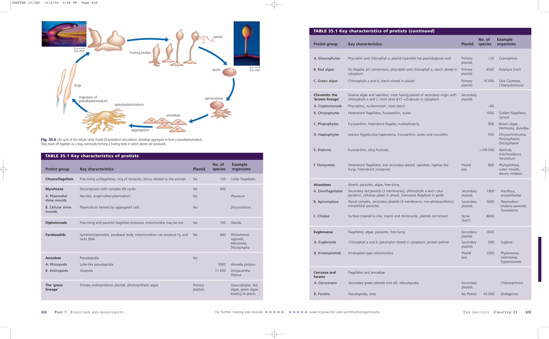
You could perhaps mistake a cellular slime mould for a minuteslug if you found one creeping across the forest floor. The‘slug’, or pseudoplasmodium, is a mass of amoebae that have
aggregated to form a single travelling colony. The amoebae,which are normally free-living individuals that prey on bacte-ria, congregate when their food supply runs short and moveoff collectively as a ‘slug’. Having found a suitable location,the slug differentiates into a fruiting body that producesnumerous spores (Fig. 35.6). Spores are released and eventual-ly produce amoebae, completing the life cycle.
Cellular slime moulds inhabit damp places in forests and gar-dens, where they are usually found on rotting plant material oranimal dung. Slime mould amoebae are often referred to asmyxamoebae (slime amoebae) to distinguish them from nor-mal amoebae. Most cellular slime moulds do not have flagella.
Sponge-like protists
Slime moulds
CHAPTER 35/2ND 13/4/04 8:48 PM Page 826
www.mcgraw-hi l l . com.au/mhha/b io logy/knox3e828 PA RT 5 EVO LU T I O N A N D B I O D I V E R S I T Y For fur ther read ing and rev is ion
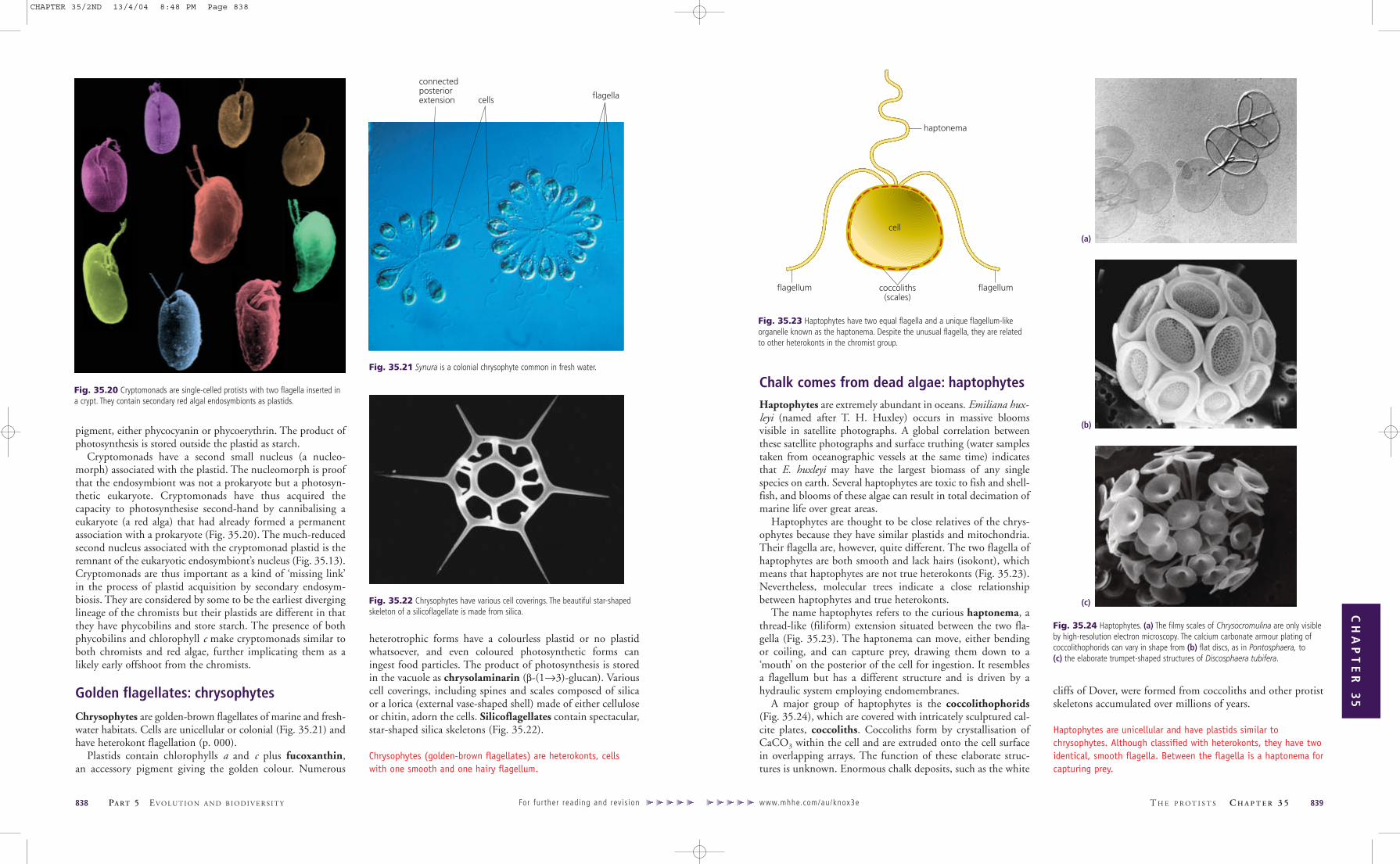
TABLE 35.1 Key characteristics of protists (continued)
Alveolates
Secondaryplastids
5000 Plasmodium(malaria parasite),Toxoplasma
B. Apicomplexa
None(lost?)
8000C. Ciliates
Secondaryplastids
1900 Noctiluca,zooxanthellae
A. Dinoflagellates
Key characteristics PlastidNo. ofspecies
Example organismsProtist group
Flagellates and amoebaeCercozoa andforams
Flagellates, algae, parasites, free-living Secondaryplastids
1600Euglenozoa
Chlorophyll a and b, paramylon stored in cytoplasm, protein pellicle Secondaryplastids
600 EuglenaA. Euglenoids
Kinetoplast-type mitochondria Plastidlost
1000 Phytomonas,Leishmania,trypanosomes
B. Kinetoplastids
Secondary green plastids (not all), reticulopodia Secondaryplastids
ChlorarachnionA. Cercozoans
Pseudopodia, tests No Plastid 45 000 GlobigerinaB. Forams
fruiting bodies
spores
spore
germination
amoebae
aggregation
pseudoplasmodium
migration ofpseudoplasmodium
slugs
0.5 mm
0.5 mm
Heterokont flagellates, lost secondary plastid, saprobes, hyphae likefungi, heterokont zoospores
Plastidlost
800 Phytophthora,water moulds,downy mildews
F. Oomycetes
Fucoxanthin, silica frustules >100 000 Navicula,Arachnoidiscus,Triceratium
E. Diatoms
Phycobilins, nucleomorph, store starch ~60A. Cryptomonads
Heterokont flagellates, fucoxanthin, scales 1000 Golden flagellates,Synura
B. Chrysophytes
Fucoxanthin, heterokont flagella, multicellularity 900 Brown algae,Hormosira, Durvillea
C. Phaeophytes
Isokont flagella plus haptonema, fucoxanthin, scales and coccoliths
Alveoli, parasites, algae, free-living
Secondary red plastids (3 membranes), chlorophylls a and c plusperidinin, cellulose plates in alveoli, transverse flagellum in girdle
Apical complex, secondary plastids (4 membranes, non-photosynthetic),intracellular parasites
Surface covered in cilia, macro and micronuclei, plastids not known
500 Chrysochromulina,Pontosphaera,Discosphaera
D. Haptophytes
No flagella, pit connections, phycobilin and chlorophyll a, starch stored incytoplasm
Primaryplastids
4500 Porphyra (nori)B. Red algae
Phycobilin and chlorophyll a, plastid (cyanelle) has peptidoglycan wall Primaryplastids
~20 CyanophoraA. Glaucophytes
Diverse algae and saprobes, most having plastid of secondary origin withchlorophylls a and c, most store β-(1→3)-glucan in cytoplasm
Secondaryplastids
Chromists: the‘brown lineage’
Chlorophylls a and b, starch stored in plastid Primaryplastids
16 000 Ulva Cauterpa,Chlamydomonas
C. Green algae
Fig. 35.6 Life cycle of the cellular slime mould Dictyostelium discoideum. Amoebae aggregate to form a pseudoplasmodium.They move off together as a slug, eventually forming a fruiting body in which spores are produced.
T H E P R O T I S T S C H A P T E R 3 5 829
TABLE 35.1 Key characteristics of protists
Decomposers with complex life cycles No 900Mycetozoa
Free-living and parasitic flagellate protozoa, mitochondria may be lost No 100 GiardiaDiplomonads
Symbionts/parasites, parabasal body, mitochondrion can produce H2 andlacks DNA
No 400 Trichomonasvaginalis,Mixotricha,Triconympha
Parabasalids
Free-living uniflagellates, ring of tentacles, lorica; related to the animals. No 120 Collar flagellatesChoanoflagellate
Axopods 11 650 Dictyacantha,Trizona
B. Actinopods
Lobe-like pseudopodia 5000 Amoeba proteusA. Rhizopods
Key characteristics PlastidNo. ofspecies
Example organismsProtist group
Pseudopodia NoAmoebae
Primary endosymbiosis plastids, photosynthetic algae Primaryplastids
Glaucophytes, redalgae, green algae,leading to plants
The ‘green lineage’
Net-like, single-celled plasmodium No PhysarumA. Plasmodialslime moulds
Plasmodium formed by aggregated cells No DictyosteliumB. Cellular slimemoulds
0.5 mm
0.5 mm
CHAPTER 35/2ND 13/4/04 8:48 PM Page 828

Diplomonads are unicellular, heterotrophic flagellates. Thename diplomonad refers to the presence of two nuclei, each ofwhich is associated with a pair of flagella. Diplomonads inhab-it the gut of various animals, where they attach by a sucker-like,ventral disc. They lack obvious mitochondria and are restrictedto an anaerobic environment.
Giardia, an intestinal parasite causing severe dysentery, isthe best known diplomonad (Fig. 35.9). It is one of the firstprotists on record, accurately described by van Leeuwenhoekin 1681 from his own diarrhoeic stools. Giardia caused amajor health scare in Australia in 1998 when it was discoveredin Sydney drinking water reservoirs.
www.mhhe.com/au/knox3e T H E P R O T I S T S C H A P T E R 3 5 831
CH
AP
TE
R 3
5
Acellular slime moulds: myxomycetes
Myxomycetes are another group of slime moulds that areacellular. Whereas the pseudoplasmodium of cellular slimemoulds consists of numerous individual cells aggregatedtogether, the plasmodium of a myxomycete is one large (up to10 cm) multinucleate cell. The plasmodium resembles a slimyscum, sometimes vivid yellow or orange in colour (Fig. 35.7),and is the major feeding stage, absorbing organic matter andingesting bacteria and other microorganisms. Should the plas-modium encounter a nutrient-poor region or other adverseenvironmental conditions, it differentiates into a fruiting bodyor sporangium (Fig. 35.8), with cells dividing by meiosis toproduce haploid spores. Spores germinate to produce haploidmyxamoebae, which are the gamete stage. In the presence ofsufficient water, they convert to biflagellate forms. Two amoe-
bae (or two biflagellates) fuse to form a zygote. The diploidnucleus of the zygote divides mitotically but no cell mem-branes separate the daughter nuclei, resulting in amultinucleate plasmodium.
Slime moulds are amoeboid protists, which aggregate to formcolonies, either cellular or acellular, with fruiting bodies thatproduce spores.
830 PA RT 5 EVO LU T I O N A N D B I O D I V E R S I T Y For fur ther read ing and rev is ion
Fig. 35.8 (a) The sporangia of the slime mould Stemonitis fusca take the form oftufts of brown threads on a log of wood. (b) Fruiting bodies of Arcyria are brilliantorange and of (c) Trichia look like little cups.
(a)
(b)
(c)
Fig. 35.9 (a) Giardia is a simple eukaryote (a diplomonad)that parasitises humans and other animals. (b) Cells havetwo nuclei (n), each of which is associated with a set offlagella. On the ventral side of the cell is a disc through whichthe cell attaches to the host’s gut lining. Infection is spreadby cysts excreted in faeces, either animal or human. The cysts,which remain viable in water for several months, can infectthe gut of animals drinking from the contaminated watersource. Giardia is not restricted to polluted waters and canoccur in metropolitan water supplies or even in wildernessstreams. The most effective means of purification is to boil thewater; cysts are resistant to iodine and chlorine.
Parabasalids are flagellates with a single nucleus involved incommensal or parasitic relationships with animals. They typ-ically have a parabasal body, a large Golgi-type membrane
complex beside the basal body. An axostyle, a stiff rod-likebunch of microtubules, runs the length of the cell.Trichomonas vaginalis is a parabasalid that infects the humangenital tract. A relatively benign sexually transmitted disease,Trichomonas is estimated to infect 3.5% of the world’s popu-lation. Many parabasalids have unusual mitochondria, calledhydrogenosomes, which emit hydrogen gas from anaerobicoxidation of glucose. Whereas aerobic respiration results inthe reduction of O2 to produce water, hydrogenosomes cantransfer electrons onto protons and produce H2. In additionto their unusual anaerobic respiration these extraordinarymitochondria lack any mitochondrial DNA. Two types ofparabasalids (Triconympha and Mixotricha) are symbionts intermite guts, where they are responsible for the digestion ofwood. Trichonympha has several thousand flagella. Mixotrichahas only four eukaryotic flagella but also has thousands of fil-amentous spirochaete bacteria (Chapter 33) attached to itssurface that allow propulsion (Fig. 35.10).
wood being ingested
internal bacteria
surface bacteria
large spirochaetes
small spirochaetes
flagella
axostyle
parabasalbody
hydrogenosomes
Fig. 35.10 The parabasalid Mixotricha paradoxa is a symbiont par excellence.The cell is actually a co-operative, involving as many as 500 000 individual organisms.The host cell is a quadriflagellate eukaryote. On the surface there are two forms ofspirochaete bacteria that propel the cell. The spirochaetes attach to the cell surfacevia anchor bacteria embedded in the host cell membrane. Numerous internalbacteria within the host cell aid metabolism. This parabasalid is an endosymbiontwithin the gut of Australian termites and is responsible for the digestion of wood.
nuclei
ventraldisc flagella
(a) (b)
Fig 35.7 Streaming masses of the acellular slime mould, Physarum, can move.
Parasitic flagellates that contaminate water supplies: diplomonads
Symbionts and parasites: parabasalids
50 µm
CHAPTER 35/2ND 13/4/04 8:48 PM Page 830

It appears likely that a single endosymbiosis produced themany different coloured plastids, such as the photosyntheticchloroplasts of algae, observed in the protists. From this youmight expect that all plastid-containing protists are closelyrelated (descendants of the original host cell that acquired anendosymbiont) but the story is not that simple.
The original plastid has apparently been faithfully handeddown through hundreds of millions of years of evolution tothe modern green algae and their descendants, land plants.However, other protist groups are now recognised to havestolen this plastid. They did this by simply engulfing algal cellsand retaining them within their cells, much like the endosym-biosis of a cyanobacterium but this time with a eukaryoticendosymbiont. This means that heterotrophic eukaryotes canconvert to autotrophy by taking the photosynthetic organellefrom a distant relative. From this you can see that it is not
valid to unite all plastid-containing protists into one group,traditionally labelled algae, because they do not share a com-mon ancestor, only an acquired organelle.
The groups discussed from hereon mostly have plastids, butsome large groups lacking plastids occur within these groups. Itis not yet clear whether these groups, ciliates for instance, havelost their plastid or never had one. To further confuse the issue,it is emerging that many protists, the malaria parasite forinstance, retain a vestigial plastid that has no pigments and doesnot photosynthesise. These remnant plastids are tricky to recog-nise (the plastid DNA and the multiple bounding membranesare the key give-aways) so some protist groups currently thoughtto lack plastids may yet harbour them secretively. Exactly whythese non-photosynthetic relicts persist is one of the enduringmysteries of protistology. We can only assume that the organelleprovides something to the cell other than food.
www.mhhe.com/au/knox3e T H E P R O T I S T S C H A P T E R 3 5 833
CH
AP
TE
R 3
5
Rhizopods are amoebae that can alter their shape
Rhizopods are amoebae that are able to transiently produceextensions of the cell surface, pseudopodia (‘false feet’), involvedin locomotion or feeding (Chapter 27). One of the first amoe-bae to be named was Amoeba proteus (Fig. 35.11) after the seagod Proteus of Greek mythology, who could change his shapeat will (Gr. amoeba, meaning change). Many rhizopods arenaked but some produce internal or external skeletons. Mostspecies are unicellular and have a single nucleus. Rhizopods arecommon in aquatic habitats, where they prey on bacteria andother protists.
Actinopods are radially symmetrical unicells
Actinopods are single-celled, radially symmetrical organisms,characterised by axopods, long slender radial projections.Axopods contain a thin layer of cytoplasm bounded by plasmamembrane and are reinforced with a highly ordered bundle ofmicrotubules. Axopod microtubules collectively form anaxoneme, which should not be confused with the microtubulesof flagella and cilia given the same name. Axopod microtubulesdo not inter-slide to create bending.
The main function of axopods is prey capture. Food parti-cles stick to their surface and are transported to the cell foringestion. In one group (Sticholonche), axopods are modified tofunction as oars and ‘row’ the cell through the water. Theaxoneme microtubules of these oar-like axopods are attached tothe nucleus by ball-and-socket articulations (like our hip joint)and the axopod is moved by co-ordinated contraction/relax-ation of non-actin fibres that interconnect the axopods.
The cells of actinopods are highly variable in organisationand are often partitioned into inner and outer zones. Theouter zone can harbour zooxanthellae (dinoflagellate
endosymbionts). Some actinopods are amoeboid and othersproduce flagellate stages that are able to swim rather thancrawl like an amoeba. Skeletons can be composed of organicmaterial, accreted sand particles and diatom valves, celestite(strontium sulfate) or silica with traces of magnesium, copperand calcium, depending on the class of actinopod. Skeletonsform fossils and huge deposits of ‘radiolarian ooze’, a sludgefound on the ocean floor. Like diatom valves, actinopodskeletons also form chert (rock containing silica) and noextremely old fossils are known. The best known actinopodsare radiolarians (Fig. 35.12), which are called sun animalculesbecause they resemble a minuscule sun with radiating rays.
Rhizopods are amoebae that can alter their shape. Most areheterotrophs. Actinopods have radial skeletons and projectionsknown as axopods with which they capture food.
832 PA RT 5 EVO LU T I O N A N D B I O D I V E R S I T Y For fur ther read ing and rev is ion
Fig. 35.11 A characteristic trait of amoebae is their ability to alter cell shapetransiently to produce pseudopodia (false feet). Amoeba proteus has severalpseudopodia projecting from the cell in different directions. Here A. proteus isconsuming Euglena, another protist (small green cell at right).
Fig. 35.12 Actinopodsinclude radiolarians, suchas (a) Dictyacantha and(b) Trizona, which producespectacular siliceousskeletons that accumulateon the sea-floor, forminga radiolarian ooze.
(a) (b)
Many algae have plastids bound by two mem-branes, and all are descendants of what is termeda primary endosymbiosis (Fig. 35.13). Theseinclude the ‘green lineage’ of protists (Fig. 38.1):glaucophytes, red algae and green algae (relatedto land plants). Precambrian fossils that are1.2 billion years old are evidence that this line-age is very ancient. Unicellular and filamentousgreen-algal fossils have been found in acid rocksin Central Australia (Bitter Springs formation)that are 900 million years old. Fossils identifiedas calcified red algae are recorded from the earlyCambrian (590 million years ago), but there areno known fossils of glaucophytes.
N
eukaryotichost
Primary endosymbiosis(plastid with 2 membranes)
cyanobacterium
nucleus
outer membrane disappearsnucleomorph as in cryptomonads
N
another eukaryotic host
N N N
N N
N
genetransfer
Secondary endosymbiosis(plastid with 3 or 4 membranes)
gene transfer
Fig. 35.13 Primary endosymbiosis between a eukaryote and acyanobacterium produces a plastid with two membranes(glaucophytes, red algae and green algae/plants). Subsequentsecondary endosymbiosis involves different hosts and any of thealgae that contain a primary endosymbiont. Secondaryendosymbioses have produced a wide range of protist lineages,such as the chromists, euglenoids and alveolates.
Amoebae Protists with plastids
Protists with primary plastids: the ‘green lineage’
100 µm
CHAPTER 35/2ND 13/4/04 8:48 PM Page 832

are known as coralline red algae because they were mistakenlythought to be coral animals.
Plastids of red algae contain chlorophyll a and phycobilinpigments—phycocyanin and phycoerythrin (the latter pro-ducing the typical red colouration). Red algae absorbshort-wavelength blue and green light that penetrates deepestinto the ocean, allowing them to photosynthesise at depths of250 m below the surface. The product of photosynthesis isstored in the cytoplasm as α-(1→4)-glucan.
Red algal cells lack flagella and basal bodies. Because theirsperm cannot swim, for sexual reproduction they rely on therandomness of ocean currents to bring sperm to the female partof the thallus containing the egg. When a sperm does contactan egg to form a zygote, the alga capitalises on the event by dis-tributing copies of the diploid nucleus to other female parts ofthe thallus. Thus, from a single fertilisation event, multiplespores can be produced for the next generation.
The lack of flagella and basal bodies was originally inter-preted as a primitive character suggesting that red algae are
ancient. Molecular analysis has failed to confirm this view,instead showing that red algae are advanced organisms thathave lost the ability to produce flagella.
Red algae are familiar seaweeds. Most are multicellular andmacroscopic and they lack flagella. They contain chlorophyll a andphycobilin pigments.
Green algae: chlorophytes
Green algae are a large group (about 16 000 species), includ-ing unicellular, colonial and multicellular forms found inmarine or freshwater habitats (Figs 35.16, 35.17). Chlorophytechloroplasts (green plastids) contain the same pigments as landplant chloroplasts—chlorophylls a and b, β-carotene and othercarotenoid derivatives. Like land plants, the product of photo-synthesis of green algae is stored as starch (an α-(1→4)-glucan)within the chloroplast and the cell walls are primarily cellulose(β-(1→4)-glucan).
www.mhhe.com/au/knox3e T H E P R O T I S T S C H A P T E R 3 5 835
CH
AP
TE
R 3
5
Fig. 35.16 Greenalgae. (a) The sealettuce Ulva lactuca isused as a garnish inJapanese miso soup but(b) its relative, the sea-cactus Caulerpa, can bepoisonous. Both arecommon green algaefound on rocky shoresaround the south-eastern coast ofAustralia.Missing links in endosymbiosis:
glaucophytesGlaucophytes (Fig. 35.14) are living examples of an interme-diate stage in the evolution of a plastid from a photosyntheticprokaryotic endosymbiont. Plastids of glaucophytes areknown as cyanelles. Cyanelles are unique in that they havea peptidoglycan wall the same as bacteria. The presence of thewall is evidence that the cyanelle (plastid) was once a bacteri-um before it took up residence in the host cell. Cyanellescontain chlorophyll a and phycobilin pigments identical tocyanobacteria, and cyanelles have a circular chromosome. Aswith other plastids thry are no longer fully independent, hav-ing lost genes to the nucleus during the endosymbioticrelationship. Some genes for producing peptidoglycan havebeen found on the cyanelle chromosome, which is otherwisethe same as a plastid chromosome. Cyanelles are thus partiallydependent on the host cell and cannot survive independently.Host cells are typically flagellates with two smooth flagella.
Glaucophytes are photosynthetic flagellates with plastids, termedcyanelles, which have a peptidoglycan wall, as do bacteria.
Red algae: rhodophytes
Red algae (rhodophytes) are common seaweeds on rockyseashores around the world. There are some 4000 species,many of which are endemic to Australia (Chapter 41). Redseaweeds are of commercial importance in the production ofagar for microbiology and molecular biology, and as food inthe Orient, North America and Ireland. Sushi is preparedwith the red alga Porphyra, dried as Japanese nori. About60 000 hectares of nori are grown by mariculture around theJapanese coast. Carrageenan from red algae is used also as astabilising agent in confectionery, ice-cream, cosmetics and
pet foods. The red seaweed industry is worth about $1 billionper annum worldwide.
Most red algae are multicellular, adjacent cells often beingattached by pit plugs (Fig. 35.15), and a few are unicellular.Multicellular seaweeds have a thallus (plant body) withbranches and blades, plus extensions attaching it to the sub-strate. Red algae have complex life histories with alternatingstages that are often markedly different in morphology. Somered algae are calcified, hardened with calcium carbonate, and
834 PA RT 5 EVO LU T I O N A N D B I O D I V E R S I T Y For fur ther read ing and rev is ion
Fig. 35.14 (a) The biflagellate glaucophyte Cyanophora has a plastid knownas a cyanelle resembling a cyanobacterium. (b) A peptidoglycan wall between thetwo plastid membranes gives these algae away as a ‘missing link’ in the originof plastids.
cell 1
pitconnection
chloroplast
cell 2
cell wall
mitochondrion
Fig. 35.15 Red algae range from (a) fine feathery structures to (b) crustycalcified plants resembling corals. (c) Adjacent cells are often attached by pit plugs.
Fig. 35.17 Inaddition to bird life,lilies and crocodiles, thewater holes of KakaduNational Park in theNorthern Territorycontain this splendidselection of unicellulargreen algae.
(a) (b)
(a)
(b)
(c)
central zone
dividingcyanelle(plastid)
peptidoglycanwall betweenmembranes
flagella
(a) (b)6 µm
CHAPTER 35/2ND 13/4/04 8:48 PM Page 834

The scope for using green algae in biotechnology to pro-duce pharmaceuticals, antibiotics, fuels and foods, and inwaste treatment is enormous (Box 35.1). Manipulation ofstrains by genetic engineering will contribute to the produc-tion of useful natural substances.
Green algae (Chlorophytes) are unicellular, colonial ormulticellular, and one group is the closest relative of landplants. Chloroplasts contain chlorophyll a and b, the productof photosynthesis is stored as starch, and cell walls arecomposed of cellulose.
www.mhhe.com/au/knox3e
CH
AP
TE
R 3
5
These and other similarities leave us in no doubt that greenalgae are related to land plants (Chapter 36). In particular, thecharophytes are the closest relatives of the land plants.Charophytes are essentially restricted to freshwater habitats.They are delicate and typically small (2–30 cm in length) withsome, the stoneworts, encrusted with CaCO3 (calcite).Gametes are asymmetrical and mitosis involves a phragmo-plast—characteristics shared with land plants (Chapter 36).
Green algae fix an estimated 1 billion tonnes of carbonfrom the atmosphere per annum. They are used as food(Spirogyra, Fig. 35.18, as vitamin supplement tablets) and arebeing tested in biotechnological applications (Box 35.1).
Green algae were classified traditionally on the basis of theirform—unicellular, colonial, filamentous, coenocytic (techni-cally unicellular but multinucleate and greatly enlarged to forma macroscopic thallus) and multicellular three-dimensionalforms. Closer investigation with the electron microscope showsthese categories to be somewhat artificial, with several cases ofconvergent evolution (Chapter 30). Studies of mitosis, forexample, have shown that two species originally groupedtogether in the filamentous genus Klebsormidium actuallybelong in different classes. Although superficially similar, the
two species of Klebsormidium have different types of mitosis(the phragmoplast and phycoplast types described in Chapter36) and fundamentally different motile cells, so one species hadto be taken out of the genus. A filamentous thallus, therefore,seems to have evolved more than once in the green algae.
836 PA RT 5 EVO LU T I O N A N D B I O D I V E R S I T Y For fur ther read ing and rev is ion
Fig. 35.18 The edible filamentous green alga Spirogyra is named for the spiralchloroplast that winds its way around the periphery of the elongate cells.
Recent work has demonstrated that a number of groups of pro-tists have stolen the ability to photosynthesise from chloro-plast-bearing cells. The following groups acquired plastids bycannibalising parts from photosynthetic prey. We refer to thistype of acquisition as secondary endosymbiosis because it followsan earlier primary endosymbiosis (Fig. 35.13). Organisms withthese so-called second-hand plastids usually have multiple(three or four) membranes surrounding the plastids. No oneknows exactly how many secondary endosymbioses haveoccurred, which makes it hard to define which groups are relat-ed. For instance, several groups might have grabbed the samekind of endosymbiont, making their plastids appear similardespite the fact that the host lineage might be different. In anyevent, secondary endosymbiosis has been a driving force foreukaryotic diversity spawning an enormous range of protists.
Cryptomonads, heterokonts, haptophytes, euglenoids,dinoflagellates and apicomplexans almost certainly acquiredtheir plastids through secondary endosymbioses. Interestingly,it is now emerging that many non-photosynthetic protists,some of them important pathogens, such as Plasmodiumwhich causes malaria (p. 000) and the trypanosomes, havesubsequently lost these plastids. In an extraordinary turn ofevolutionary events they have converted from heterotrophy toautotrophy by a secondary endosymbiosis only to revert toheterotrophy again at a later point. Two groups, cryptomonadsand chlorarachniophytes, are key models for understandingendosymbiosis. This is because they retain a remnant, knownas the nucleomorph, of the secondary endosymbiont’s nucle-us. Cryptomonads and chlorarachniophytes provide proofthat secondary endosymbiosis occurred. In all other secondaryendosymbionts, the nucleomorph has been lost and only themultiple membranes remain as a telltale trace of previousengulfment events (Fig. 35.13).
Chromist protists: the ‘brown lineage’
Chromists are a diverse group embracing a wide range oflifestyles. They clearly acquired plastids by secondary endosym-biosis of a red alga but chromist plastids, such as in brownalgae, are characterised by chlorophyll c, whereas red algal plas-tids lack chlorophyll c. Nevertheless, gene sequence dataidentify the endosymbiont as a red alga. Numerous chromists(e.g. oomycetes) appear to have subsequently lost the plastidand reverted to heterotrophy. Chromists typically have one
smooth flagellum directed posteriorly and one hairy flagellumdirected anteriorly, typical of protists called heterokonts (Fig.35.19). The hairy flagellum has numerous thin, tubularappendages that alter the direction of thrust produced by theflagellar beat. The beat of the hairy flagellum thus drags the cellthrough the water. If the cell happens to be fixed in place, theflagellar beat draws the water down and over the cell.
Chromists acquired plastids by secondary endosymbiosis andprobably include the photosynthetic cryptomonads, chrysophytes,haptophytes, diatoms and brown algae, and the non-photosyntheticoomycetes, but not all biologists accept it as a group.
Flagellates with second-hand plastids:cryptomonads
Cryptomonads have a small anterior invagination (the ‘crypt’)into which their two flagella are inserted. They are unicellularand usually reproduce asexually. All genera, except Goniomonas,which is heterotrophic, possess a secondary plastid. Crypto-monad plastids have chlorophylls a and c plus a phycobilin
hairyanterior
flagellum
smoothposteriorflagellum
nucleus
Fig. 35.19 Most chromistsare heterokont flagellateshaving an anterior hairyflagellum and a posteriorsmooth flagellum.
T H E P R O T I S T S C H A P T E R 3 5 837
Not only do green algae grow in a wide range of habitats, such asfresh water, oceans, salt lakes and snow, but they also show a
great diversity in their chemistry. It is this chemical diversity, combinedwith the ability of some species to grow in extreme environments, thatmakes green algae attractive to biotechnologists.
The single-celled green alga Chlamydomonas is a model organism forresearch. It is readily grown in the laboratory, reproduces sexually, andproduces a range of mutants able to be mapped by classic and moleculargenetic techniques. Sequencing of the genome of Chlamydomonas is nearcompletion and research on this protist has been a valuable aid to ourunderstanding of the workings of photosynthetic cells, knowledge thatcan be applied for human uses.
Since the early 1980s, the focus of algal biotechnology has beenthe commercial production of high-value chemicals, such as carotenoids,lipids, fatty acids and pharmaceuticals.
An important alga is Dunaliella salina. When grown at high salinity(about 10 times the concentration of sea water) and with high lightintensity, D. salina accumulates large amounts of an orange-redcarotenoid, β,β-carotene. This pigment compound is used to colourproducts, such as margarine, noodles and soft drinks, and as a vitaminsupplement because it is readily converted to vitamin A. There isalso evidence that β,β-carotene may help prevent lung cancer. Pure β,β-carotene is worth more than $600 per kilogram. Production of β,β-carotene from D. salina means growing and harvesting vastquantities of algae in ‘farms’. The world’s largest algal farms are at
Hutt Lagoon in Western Australia (see Fig. B35.1) and Whyalla in SouthAustralia.
Another alga under study is the freshwater chlorophyte Haematococcuspluvialis, which is the best natural source of the carotenoid astaxanthin.Astaxanthin is used in aquaculture as a fish food additive to give troutand salmon flesh the natural pink colour. Fish food currently containssynthetic carotenoids and astaxanthin is a desirable natural alternative.
Green algae may also be a future source of alternative fuels.Botryococcus braunii produces long-chain hydrocarbons similar to crudeoils, and these can be cracked in a refinery to produce petrol and otheruseful fractions. Tetraselmis species accumulate fats and oils, and, onceextracted, the lipids can be used as a diesel fuel substitute.
I N T E R N A T I O N A L F O C U S
BOX 35.1 Green algae and biotechnology
Fig. B35.1 With its wide flat spaces and intense sunshine, Australia is theperfect place for algal farms producing food, fuel and pharmaceuticals. Theseponds of Dunaliella salina at Hutt Lagoon, Western Australia, range in colourfrom green to brick red depending on how much of the valuable β,β-carotenecells have accumulated.
Protistan pirates with second-hand plastids
CHAPTER 35/2ND 13/4/04 8:48 PM Page 836
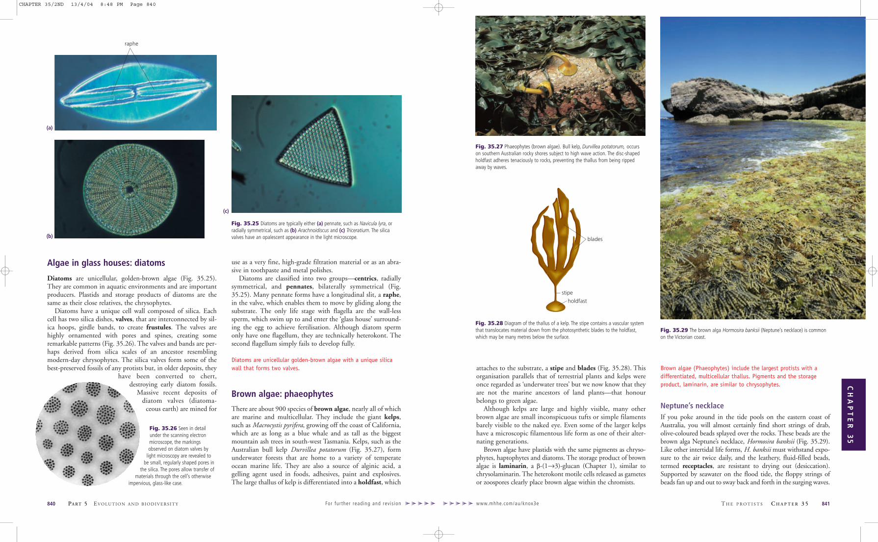
Chalk comes from dead algae: haptophytesHaptophytes are extremely abundant in oceans. Emiliana hux-leyi (named after T. H. Huxley) occurs in massive bloomsvisible in satellite photographs. A global correlation betweenthese satellite photographs and surface truthing (water samplestaken from oceanographic vessels at the same time) indicatesthat E. huxleyi may have the largest biomass of any singlespecies on earth. Several haptophytes are toxic to fish and shell-fish, and blooms of these algae can result in total decimation ofmarine life over great areas.
Haptophytes are thought to be close relatives of the chrys-ophytes because they have similar plastids and mitochondria.Their flagella are, however, quite different. The two flagella ofhaptophytes are both smooth and lack hairs (isokont), whichmeans that haptophytes are not true heterokonts (Fig. 35.23).Nevertheless, molecular trees indicate a close relationshipbetween haptophytes and true heterokonts.
The name haptophytes refers to the curious haptonema, athread-like (filiform) extension situated between the two fla-gella (Fig. 35.23). The haptonema can move, either bendingor coiling, and can capture prey, drawing them down to a‘mouth’ on the posterior of the cell for ingestion. It resemblesa flagellum but has a different structure and is driven by ahydraulic system employing endomembranes.
A major group of haptophytes is the coccolithophorids(Fig. 35.24), which are covered with intricately sculptured cal-cite plates, coccoliths. Coccoliths form by crystallisation ofCaCO3 within the cell and are extruded onto the cell surfacein overlapping arrays. The function of these elaborate struc-tures is unknown. Enormous chalk deposits, such as the white
cliffs of Dover, were formed from coccoliths and other protistskeletons accumulated over millions of years.
Haptophytes are unicellular and have plastids similar tochrysophytes. Although classified with heterokonts, they have twoidentical, smooth flagella. Between the flagella is a haptonema forcapturing prey.
www.mhhe.com/au/knox3e T H E P R O T I S T S C H A P T E R 3 5 839
CH
AP
TE
R 3
5
Fig. 35.23 Haptophytes have two equal flagella and a unique flagellum-likeorganelle known as the haptonema. Despite the unusual flagella, they are relatedto other heterokonts in the chromist group.
flagellum
cell
coccoliths(scales)
haptonema
flagellum
pigment, either phycocyanin or phycoerythrin. The product ofphotosynthesis is stored outside the plastid as starch.
Cryptomonads have a second small nucleus (a nucleo-morph) associated with the plastid. The nucleomorph is proofthat the endosymbiont was not a prokaryote but a photosyn-thetic eukaryote. Cryptomonads have thus acquired thecapacity to photosynthesise second-hand by cannibalising aeukaryote (a red alga) that had already formed a permanentassociation with a prokaryote (Fig. 35.20). The much-reducedsecond nucleus associated with the cryptomonad plastid is theremnant of the eukaryotic endosymbiont’s nucleus (Fig. 35.13).Cryptomonads are thus important as a kind of ‘missing link’in the process of plastid acquisition by secondary endosym-biosis. They are considered by some to be the earliest diverginglineage of the chromists but their plastids are different in thatthey have phycobilins and store starch. The presence of bothphycobilins and chlorophyll c make cryptomonads similar toboth chromists and red algae, further implicating them as alikely early offshoot from the chromists.
Golden flagellates: chrysophytes
Chrysophytes are golden-brown flagellates of marine and fresh-water habitats. Cells are unicellular or colonial (Fig. 35.21) andhave heterokont flagellation (p. 000).
Plastids contain chlorophylls a and c plus fucoxanthin,an accessory pigment giving the golden colour. Numerous
heterotrophic forms have a colourless plastid or no plastidwhatsoever, and even coloured photosynthetic forms caningest food particles. The product of photosynthesis is storedin the vacuole as chrysolaminarin (β-(1→3)-glucan). Variouscell coverings, including spines and scales composed of silicaor a lorica (external vase-shaped shell) made of either celluloseor chitin, adorn the cells. Silicoflagellates contain spectacular,star-shaped silica skeletons (Fig. 35.22).
Chrysophytes (golden-brown flagellates) are heterokonts, cellswith one smooth and one hairy flagellum.
838 PA RT 5 EVO LU T I O N A N D B I O D I V E R S I T Y For fur ther read ing and rev is ion
Fig. 35.20 Cryptomonads are single-celled protists with two flagella inserted ina crypt. They contain secondary red algal endosymbionts as plastids.
Fig. 35.21 Synura is a colonial chrysophyte common in fresh water.
flagella
connectedposteriorextension cells
Fig. 35.22 Chrysophytes have various cell coverings. The beautiful star-shapedskeleton of a silicoflagellate is made from silica.
Fig. 35.24 Haptophytes. (a) The filmy scales of Chrysocromulina are only visibleby high-resolution electron microscopy. The calcium carbonate armour plating ofcoccolithophorids can vary in shape from (b) flat discs, as in Pontosphaera, to(c) the elaborate trumpet-shaped structures of Discosphaera tubifera.
(a)
(b)
(c)
CHAPTER 35/2ND 13/4/04 8:48 PM Page 838
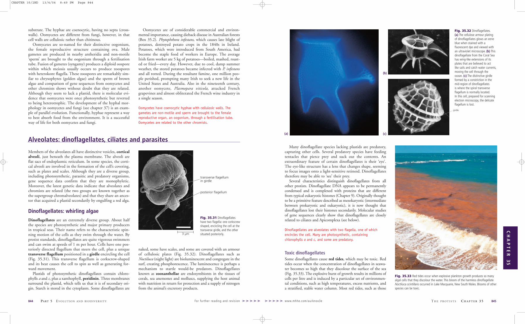
attaches to the substrate, a stipe and blades (Fig. 35.28). Thisorganisation parallels that of terrestrial plants and kelps wereonce regarded as ‘underwater trees’ but we now know that theyare not the marine ancestors of land plants—that honourbelongs to green algae.
Although kelps are large and highly visible, many otherbrown algae are small inconspicuous tufts or simple filamentsbarely visible to the naked eye. Even some of the larger kelpshave a microscopic filamentous life form as one of their alter-nating generations.
Brown algae have plastids with the same pigments as chryso-phytes, haptophytes and diatoms. The storage product of brownalgae is laminarin, a β-(1→3)-glucan (Chapter 1), similar tochrysolaminarin. The heterokont motile cells released as gametesor zoospores clearly place brown algae within the chromists.
Brown algae (Phaeophytes) include the largest protists with adifferentiated, multicellular thallus. Pigments and the storageproduct, laminarin, are similar to chrysophytes.
Neptune’s necklaceIf you poke around in the tide pools on the eastern coast ofAustralia, you will almost certainly find short strings of drab,olive-coloured beads splayed over the rocks. These beads are thebrown alga Neptune’s necklace, Hormosira banksii (Fig. 35.29).Like other intertidal life forms, H. banksii must withstand expo-sure to the air twice daily, and the leathery, fluid-filled beads,termed receptacles, are resistant to drying out (desiccation).Supported by seawater on the flood tide, the floppy strings ofbeads fan up and out to sway back and forth in the surging waves.
www.mhhe.com/au/knox3e T H E P R O T I S T S C H A P T E R 3 5 841
CH
AP
TE
R 3
5
Algae in glass houses: diatoms
Diatoms are unicellular, golden-brown algae (Fig. 35.25).They are common in aquatic environments and are importantproducers. Plastids and storage products of diatoms are thesame as their close relatives, the chrysophytes.
Diatoms have a unique cell wall composed of silica. Eachcell has two silica dishes, valves, that are interconnected by sil-ica hoops, girdle bands, to create frustules. The valves arehighly ornamented with pores and spines, creating someremarkable patterns (Fig. 35.26). The valves and bands are per-haps derived from silica scales of an ancestor resemblingmodern-day chrysophytes. The silica valves form some of thebest-preserved fossils of any protists but, in older deposits, they
have been converted to chert,destroying early diatom fossils.
Massive recent deposits ofdiatom valves (diatoma-
ceous earth) are mined for
use as a very fine, high-grade filtration material or as an abra-sive in toothpaste and metal polishes.
Diatoms are classified into two groups—centrics, radiallysymmetrical, and pennates, bilaterally symmetrical (Fig.35.25). Many pennate forms have a longitudinal slit, a raphe,in the valve, which enables them to move by gliding along thesubstrate. The only life stage with flagella are the wall-lesssperm, which swim up to and enter the ‘glass house’ surround-ing the egg to achieve fertilisation. Although diatom spermonly have one flagellum, they are technically heterokont. Thesecond flagellum simply fails to develop fully.
Diatoms are unicellular golden-brown algae with a unique silicawall that forms two valves.
Brown algae: phaeophytes
There are about 900 species of brown algae, nearly all of whichare marine and multicellular. They include the giant kelps,such as Macrocystis pyrifera, growing off the coast of California,which are as long as a blue whale and as tall as the biggestmountain ash trees in south-west Tasmania. Kelps, such as theAustralian bull kelp Durvillea potatorum (Fig. 35.27), formunderwater forests that are home to a variety of temperateocean marine life. They are also a source of alginic acid, agelling agent used in foods, adhesives, paint and explosives.The large thallus of kelp is differentiated into a holdfast, which
840 PA RT 5 EVO LU T I O N A N D B I O D I V E R S I T Y For fur ther read ing and rev is ion
Fig. 35.25 Diatoms are typically either (a) pennate, such as Navicula lyra, orradially symmetrical, such as (b) Arachnoidiscus and (c) Triceratium. The silicavalves have an opalescent appearance in the light microscope.
raphe
Fig. 35.26 Seen in detailunder the scanning electronmicroscope, the markingsobserved on diatom valves by
light microscopy are revealed tobe small, regularly shaped pores in
the silica. The pores allow transfer ofmaterials through the cell’s otherwise
impervious, glass-like case.
Fig. 35.28 Diagram of the thallus of a kelp. The stipe contains a vascular systemthat translocates material down from the photosynthetic blades to the holdfast,which may be many metres below the surface.
Fig. 35.29 The brown alga Hormosira banksii (Neptune’s necklace) is commonon the Victorian coast.
(a)
(c)
(b) blades
stipe
holdfast
Fig. 35.27 Phaeophytes (brown algae). Bull kelp, Durvillea potatorum, occurson southern Australian rocky shores subject to high wave action. The disc-shapedholdfast adheres tenaciously to rocks, preventing the thallus from being rippedaway by waves.
CHAPTER 35/2ND 13/4/04 8:48 PM Page 840

Hormosira banksii is dioecious, meaning it has male andfemale reproductive structures on separate thalli. Reproductivestructures are found within small warty growths, concepta-cles, which stud the surface of the receptacles (Fig. 35.30).Within the conceptacles on the male thallus are two types ofhairs: long, unbranched paraphyses and shorter, branchingantheridial hairs on which sperm-producing antheridia devel-op. Each antheridium undergoes meiosis and severalsubsequent rounds of mitosis to produce 64 sperm cells.Motile sperm are biflagellate heterokonts (having one smoothand one hairy flagellum) and bear an orange eyespot. At lowtide, an orange ooze of antheridia exudes from the concepta-cles on the male thallus. Sperm are released on the flood tide.
Eggs are produced by oogonia on a female thallus. Likeantheridia, oogonia develop in conceptacles. Four eggs (ova,
sing. ovum) are released from each oogonium. Ova have noflagella and drift motionless on the incoming tide. Sperm areattracted to a secretion produced by the ovum and clusteraround the ovum until one successfully fertilises it. The zygotesettles and, if it finds a suitable location, immediately developsinto a new, diploid, male or female thallus. The gametes arethe only haploid stage of the life cycle.
Water moulds and downy mildews:oomycetes
Water moulds and downy mildews, oomycetes, have a super-ficial resemblance to fungi (Chapter 37) since they producea network of filaments (hyphae) that penetrate their food
www.mhhe.com/au/knox3e T H E P R O T I S T S C H A P T E R 3 5 843
CH
AP
TE
R 3
5
842 PA RT 5 EVO LU T I O N A N D B I O D I V E R S I T Y For fur ther read ing and rev is ion
cross-sectionsof receptacles
receptacle
conceptacles
heterokont sperm (n)
conceptacle
plant
rock rock
ovum nucleus (n)
plant
antheridiaparaphyses
antheridialhairs
oogonium(4 ova)
oogonium
ovum (n)
paraphyses
Fig. 35.30 Reproductive structures of Hormosira banksii—conceptacles and receptacles.
In the 1920s there were a num-ber of reports of mysterious
deaths of jarrah trees, Eucalyptusmarginata, in Western Australianforests (Fig. B35.2a). Tree deathsappeared to follow bush tracksand logging sites and were at firstattributed to soil disturbance.When sand and gravel from thesecleared areas was transported toother regions, trees at these sitesalso died.
It was not until the late 1960sthat the cause of the forestdieback was identified as the oomycete, Phytophthora cinnamomi. Thispathogen attacks the roots of susceptible plants, causing problems inwater uptake and translocation. Infected trees show symptoms ofwater stress, with leaf yellowing and dieback of upper branches.Spread of the disease occurs underground by movement of flagellatedzoospores, which are able to swim through moist soil. Zoospores seeka host rootlet, attach themselves and produce hyphae that invade theplant’s root system (Fig. B35.2b). This mechanism of disease transferexplains how transport of contaminated soil or flushing of floodwaterspreads the disease.
Phytophthora cinnamomi is thought to originate from cinnamon trees inSumatra and was probably introduced to Australia by European colonists.Many endemic plants have no apparent resistance to dieback andsome highly susceptible Banksia species are threatened with extinction.The massive scale of the problem prevents the use of fungicide andoutbreaks of the disease must usually run their course before naturalantagonistic soil microbes bring the epidemic under control.
A U S T R A L I A N F O C U S
cystgerminatinghypha
Fig. B35.2(a) Dieback of jarrah treesin Western Australia caused by theoomycete Phytophthora cinnamomi.
BOX 35.2 Dieback disease
(a)
(b)
Fig. B35.2 (b) Cysts ofPhytophthora germinating on a plantrootlet. Phytophthora zoospores swimthrough soil water and encyst whenthey contact a plant root. The cyst thengerminates to produce hyphae thatpenetrate the root and invade thevascular system of the host, eventuallycausing dieback.
CHAPTER 35/2ND 13/4/04 8:48 PM Page 842
Many dinoflagellate species lacking plastids are predatory,capturing other cells. Several predatory species have feedingtentacles that pierce prey and suck out the contents. Anextraordinary feature of certain dinoflagellates is their ‘eye’.The eye-like structure has a lens that changes shape, seemingto focus images onto a light-sensitive retinoid. Dinoflagellatestherefore may be able to ‘see’ their prey.
Several characteristics distinguish dinoflagellates from allother protists. Dinoflagellate DNA appears to be permanentlycondensed and is complexed with proteins that are differentfrom typical eukaryotic histones (Chapter 9). Originally thoughtto be a primitive feature described as mesokaryotic (intermediatebetween prokaryotic and eukaryotic), it is now thought thatdinoflagellates lost their histones secondarily. Molecular studiesof gene sequences clearly show that dinoflagellates are closelyrelated to ciliates and Apicomplexa (see below).
Dinoflagellates are alveolates with two flagella, one of whichencircles the cell. Many are photosynthetic, containingchlorophylls a and c, and some are predatory.
Toxic dinoflagellatesSome dinoflagellates cause red tides, which may be toxic. Redtides occur when the concentration of dinoflagellates in seawa-ter becomes so high that they discolour the surface of the sea(Fig. 35.33). The explosive burst of growth results in millions ofcells per litre and is induced by a particular set of environmen-tal conditions, such as high temperatures, excess nutrients, anda stratified, stable water column. Most red tides, such as those
www.mhhe.com/au/knox3e T H E P R O T I S T S C H A P T E R 3 5 845
CH
AP
TE
R 3
5
substrate. The hyphae are coenocytic, having no septa (cross-walls). Oomycetes are different from fungi, however, in thatcell walls are cellulosic rather than chitinous.
Oomycetes are so-named for their distinctive oogonium,the female reproductive structure containing ova. Malegametes are produced in nearby antheridia and non-motile‘sperm’ are brought to the oogonium through a fertilisationtube. Fusion of gametes (syngamy) produces a diploid oosporewithin which meiosis usually occurs to produce zoosporeswith heterokont flagella. These zoospores are remarkably sim-ilar to chrysophytes (golden algae) and the sperm of brownalgae and comparison of gene sequences from oomycetes andother chromists shows without doubt that they are related.Although they seem to lack a plastid, there is molecular evi-dence that oomycetes were once photosynthetic but revertedto being heterotrophic. The development of the hyphal mor-phology in oomycetes and fungi (see chapter 37) is an exam-ple of parallel evolution. Functionally, hyphae represent a wayto best absorb food from the environment. It is a successfulway of life for both oomycetes and fungi.
Oomycetes are of considerable commercial and environ-mental importance, causing dieback disease in Australian forests(Box 35.2). Phytophthora infestans, which causes late blight ofpotatoes, destroyed potato crops in the 1840s in Ireland.Potatoes, which were introduced from South America, hadbecome the staple food of workers in Europe. The averageIrish farm worker ate 5 kg of potatoes—boiled, mashed, roast-ed or fried—every day. However, due to cool, damp summerweather, the stored potatoes became infected with P. infestansand all rotted. During the resultant famine, one million peo-ple perished, prompting many Irish to seek a new life in theUnited States and Australia. Also in the nineteenth century,another oomycete, Plasmopara viticola, attacked Frenchgrapevines and almost obliterated the French wine industry ina single season.
Oomycetes have coenocytic hyphae with cellulosic walls. Thegametes are non-motile and sperm are brought to the femalereproductive organ, an oogonium, through a fertilisation tube.Oomycetes are related to the other chromists.
844 PA RT 5 EVO LU T I O N A N D B I O D I V E R S I T Y For fur ther read ing and rev is ion
Members of the alveolates all have distinctive vesicles, corticalalveoli, just beneath the plasma membrane. The alveoli areflat sacs of endoplasmic reticulum. In some species, the corti-cal alveoli are involved in the formation of the cell’s covering,such as plates and scales. Although they are a diverse group,including photosynthetic, parasitic and predatory organisms,gene sequence data confirm that they are monophyletic.Moreover, the latest genetic data indicate that alveolates andchromists are related (the two groups are known together asthe supergroup chromalveolates) and that they share an ances-tor that acquired a plastid secondarily by engulfing a red alga.
Dinoflagellates: whirling algae
Dinoflagellates are an extremely diverse group. About halfthe species are photosynthetic and major primary producersin tropical seas. Their name refers to the characteristic spin-ning motion of the cells as they swim through the water. Byprotist standards, dinoflagellates are quite vigorous swimmersand can swim at speeds of 1 m per hour. Cells have one pos-teriorly directed flagellum that steers the cell, plus a uniquetransverse flagellum positioned in a girdle encircling the cell(Fig. 35.31). This transverse flagellum is corkscrew-shapedand its beat causes the cell to spin as well as generating for-ward movement.
Plastids of photosynthetic dinoflagellates contain chloro-phylls a and c, plus a xanthophyll, peridinin. Three membranessurround the plastid, which tells us that it is of secondary ori-gin. Starch is stored in the cytoplasm. Some dinoflagellates are
naked, some have scales, and some are covered with an armourof cellulosic plates (Fig. 35.32). Dinoflagellates such asNoctiluca (night light) are bioluminescent and congregate in thesurf, creating phosphorescence. The luminescence is perhaps amechanism to startle would-be predators. Dinoflagellatesknown as zooxanthellae are endosymbionts in the tissues ofcorals, sea anemones and molluscs, supplying the host animalwith nutrition in return for protection and a supply of nitrogenfrom the animal’s excretory products.
transverse flagellumin girdle
posterior flagellum
girdle
Fig. 35.31 Dinoflagellateshave two flagella: one corkscrewshaped, encircling the cell at thetransverse girdle, and the othersituated posteriorly.
Fig. 35.33 Red tides occur when explosive plankton growth produces so manyalgal cells that they discolour the water. This bloom of the harmless dinoflagellateNoctiluca scintillans occurred in Lake Macquarie, New South Wales. Blooms of otherspecies can be toxic.
(a)
(b)
(c)
Alveolates: dinoflagellates, ciliates and parasites
Fig. 35.32 Dinoflagellates.(a) The cellulose armour platingof dinoflagellates glows an eerieblue when stained with afluorescent dye and viewed withan ultraviolet microscope. (b) Thisdinoflagellate from the Coral Seahas wing-like extensions of itsplates that are believed to actlike sails and catch water currents,moving the cell through theocean. (c) The distinctive girdleformed by a constriction in themid-region of dinoflagellatesis where the spiral transverseflagellum is normally located.In this cell, prepared for scanningelectron microscopy, the delicateflagellum is lost.
3 µm
CHAPTER 35/2ND 13/4/04 8:49 PM Page 844

www.mhhe.com/au/knox3e T H E P R O T I S T S C H A P T E R 3 5 847
CH
AP
TE
R 3
5
caused by the bioluminescent dinoflagellate Noctiluca scintil-lans, appear to be harmless events. However, under exceptionalconditions, blooms of dinoflagellates can cause severe prob-lems. Sometimes the algae become so densely concentrated thatthey generate anoxic conditions, suffocating fish and inverte-brates in sheltered bays. Other dinoflagellates, such asGymnodinium mikimotoi, cause serious damage to fish inintensive aquaculture systems, either by the production ofmucus, which causes mechanical damage to fish gills, or by theproduction of haemolytic substances that destroy red bloodcells in gill tissues.
About 30 species of dinoflagellates produce potent toxinsthat move through food chains via fish or shellfish to humans.Dinoflagellate toxins are so potent that a pinhead-size quantity(about 500 mg), an amount easily accumulated in just one100-g serving of shellfish, could be fatal to humans. The toxinsinvolved rarely affect the nervous systems of fish or shellfish butthey evoke a variety of gastrointestinal and neurological symp-toms in humans. The resulting illnesses are known as paralyticshellfish poisoning (PSP), diarrhoetic shellfish poisoning(DSP) and ciguatera food poisoning.
Tasmania was the first Australian state to suffer problemswith toxic dinoflagellates contaminating the shellfish industry.In 1986, dense blooms of the chain-forming speciesGymnodinium catenatum, a species causing PSP, resulted in thetemporary closure of 15 Tasmanian shellfish farms. In 1988,the dinoflagellate Alexandrium catenella, which causes PSP,caused limited toxicity in wild mussels from Port Phillip Baybut fortunately no commercial shellfish farms were affected.Ciguatera poisoning caused by the coral reef dinoflagellateGambierdiscus toxicus poses an increasing danger in the GreatBarrier Reef region.
Small but deadly: apicomplexans
There are at least 5000 species of apicomplexans, most ofwhich are intracellular parasites of animals. Apicomplexa arenamed for their apical complex, a structure involved in thepenetration of host cells (Fig. 35.34). The apical complex is aconical arrangement of microtubules and secretory structures.The parasite attaches to the host at the apical complex andthen forces its way into the host.
Some apicomplexans alternate between a vertebrate host andan invertebrate host whereas others, such as Toxoplasma, movefrom vertebrate to vertebrate. Toxoplasma can leave one host asspores in the faeces and remain in the open environment untilthey can infect the second host. Humans can contract toxo-plasmosis by ingesting spores of Toxoplasma present on the furof cats carrying the infection. Toxoplasmosis is a commoninfection with flu-like symptoms but is usually only a problemduring pregnancy (when it can affect the developing foetus) orfor immunocompromised individuals (such as HIV sufferers)
who cannot combat infection and often die. Some apicom-plexa, such as Plasmodium, the causal agent of malaria (Box35.3), invade blood cells of vertebrates where they feed onhaemoglobin. Plasmodium species alternate between vertebrateand invertebrate hosts and have efficient ways of effectingcross-transfer between host species.
The protistology world was recently stunned by the dis-covery that apicomplexan parasites have a plastid similar tothose of plants and algae. The plastid (Fig. 35.34) of theseparasites is small and lacks chlorophyll but it contains a cir-cular DNA genome similar to that of all other plastids. Thisdiscovery tells us that these organisms were once photosyn-thetic but converted to a parasitic lifestyle. Why they kept theplastid remains a mystery but parasitologists are hopeful thatthe plastid might be the Achilles’ heel they have long searchedfor. Many processes in plastids can be blocked with herbicide-like drugs and antibacterials. Because humans lack a plastid,these types of drugs often have no side-effects on the patient.Therefore, targeting the plastid will kill the parasite but notthe human host
Apicomplexans are endoparasites of animals. They often parasitisetwo hosts and cause diseases such as malaria. An apical complexis used to penetrate host cells.
846 PA RT 5 EVO LU T I O N A N D B I O D I V E R S I T Y For fur ther read ing and rev is ion
nucleus
plastidGolgi apparatus
alveoli
apicalcomplex
Fig. 35.34 Apicomplexa have an apical complex to penetrate the host, alveolibeneath the plasma membrane and a non-photosynthetic plastid.
An estimated 500 million people suffer from malaria and eachyear the disease kills about two to three million people, mostly
infants in Africa and South America. Malaria is caused by apicomplexanprotists belonging to the genus Plasmodium. The Plasmodium life cycleinvolves two different hosts: a vertebrate and a blood-sucking insect.The parasite is transferred from one host to the other when insectssuck blood from vertebrates. Humans become infected withPlasmodium by the mosquito Anopheles.
As sporozoites (a stage that has the apical complex), Plasmodiumcells pass into the human bloodstream from the salivary glands of themosquito. The sporozoites quickly move to the liver and undergoasexual reproduction to produce numerous merozoites, which invadered blood cells. Merozoites transform into trophozoites, which eatthe contents of the red blood cells, then divide synchronously, andevery 48 or 72 hours (depending on the type of malaria) induce thelysis of red blood cells, causing the release of toxins and hence thecycles of fever and chills characteristic of malaria. After several rounds
of replication in red blood cells the parasite converts to the next cell
type, gametocytes. Gametocytes are ingested by mosquitoes while
the insect is sucking blood from a malaria sufferer. They pass into the
mosquito’s gut, where they develop into sperm and eggs, which fuse to
form a zygote. The zygote then undergoes meiosis to produce the
oocysts in the mosquito’s gut and eventually new haploid sporozoites,
which move into the mosquito’s salivary glands to complete the cycle.
The transfer from one host to the other can be risky and it is here
that malaria parasites excel. When a female mosquito takes her meal
of blood from a vertebrate, she infects the vertebrate with parasites.
The parasites multiply in the vertebrate and are then available to back-
infect the next generation of mosquitoes. By exploiting the relationship
between blood-sucking insects and vertebrates, malaria parasites
ensure their own reproduction and distribution. They also reduce the
defence strategies available to hosts since only a part of the life cycle
occurs in each host.
I N T E R N A T I O N A L F O C U S
BOX 35.3 Malaria
injectedsporozoitesmigrateto liver
sporozoites enterliver cells, multiply,emerge as merozoites
merozoites released
merozoitesreleased
merozoites enter red blood cells
stages in liver cells
stages in red bloodcells (repeats)
femalemosquitobites humanand ingestsgametocytes
gametocytesform
sporozoitesare releasedand migrateto salivaryglands
mosquitoinfectshuman byinjectingsalivacontainingsporozoites
ingestedgametocytesdevelopinto spermor eggs
femalegamete
malegamete
diploidAsexualcycle
haploid
Sexualcycle
fertilisation
Fig. B35.1 Life cycle of Plasmodium, the apicomplexan that causes malaria.
CHAPTER 35/2ND 13/4/04 8:49 PM Page 846

times after hot pursuit. Some predatory ciliates eat other ciliates.Cilia are often arranged in clusters, cirri, that beat synchro-
nously and work as paddles or feet for the cell. Sometimes ciliaare arranged evenly over the surface and beat in rhythmic wavesto propel cells. Cilia are all embedded in a layer of protein fibresthat are cross-linked. Elements of this layer are contractile, likeminiature muscles. In certain species they allow the ciliate tochange its shape. No plastids are found in ciliates. If it is true thata single secondary plastid acquisition created the chromalveo-lates, then ciliates must have lost the plastid.
Most ciliates are predatory, bacteria being the favoured prey.Prey are driven into an invagination, the buccal cavity, throughwhich they are ingested. Undigested material is excreted throughthe cytoproct (Fig. 35.36).
Ciliates have two types of nuclei: a micronucleus and amacronucleus. Cells usually have one of each type but somecells have several macronuclei. The micronucleus is diploid, con-tains normal chromosomes with all the ciliate’s genes and dividesmitotically. The macronucleus develops from the micronucleusand contains multiple copies of genes on relatively short piecesof DNA, some of which are circular. Macronuclei divide simplyby pinching into two approximately equal halves. Because themacronucleus contains multiple copies of the ciliate’s genes(sometimes several thousand copies), it is not essential that the
genetic material be accurately and equally divided, as occurs inmitosis. Messenger RNAs are transcribed from the multiple genecopies in the macronucleus while the master genes in themicronucleus are not transcribed. The macronucleus seems to bethe working copy of the DNA blueprint, while the micronucle-us is retained as a master copy to be passed on during the sexualprocess. This is analogous to making several working copies of acomputer program for daily use, while keeping the original pro-gram safely stored away.
Ciliates reproduce asexually by binary fission (Chapter 7)along the short axis of the cell. Sexual reproduction is by conju-gation, when two individual cells of opposite mating typesbecome attached to one another (Fig. 35.37). The two cellsremain attached over a period of several hours and exchange hap-loid versions of their micronuclei produced by meiosis. Thehaploid micronuclei fuse to form diploid micronuclei. After con-jugation, the macronuclei degenerate and new macronuclei areproduced from the recombinant micronuclei. Conjugation inciliates is essentially a form of sex in which haploid nuclei fromtwo individuals are brought together without ever needing toform gamete cells.
Ciliates are predatory unicells with numerous cilia and two typesof nuclei. They reproduce sexually by conjugation.
www.mhhe.com/au/knox3e T H E P R O T I S T S C H A P T E R 3 5 849
CH
AP
TE
R 3
5
Ciliates: eukaryotes with two differentnucleiCiliates are unicellular alveolates, ranging from 10 µm to 3 mmin length, with numerous cilia on the surface (Fig. 35.35).Watching ciliates under the microscope one can’t help but marvelat the complex behaviours exhibited by these unicellular crea-tures. They rove about their environment in a busy way exploringevery object they meet and testing it with their anterior ciliabefore ‘deciding’ how to react. If the object is large they simplyproceed around it. If the ciliate finds the object offensive, it willreverse away rapidly. When a predatory ciliate encounters a preyitem, edible bacteria and protists are rapidly swallowed, some-
848 PA RT 5 EVO LU T I O N A N D B I O D I V E R S I T Y For fur ther read ing and rev is ion
Fig 35.37 Sexual reproduction in the ciliate Paramecium. Two mature cells ofopposite mating strains fuse in a process called conjugation.
Two Parameciumindividuals of differentmating types comeinto contact.
The diploidmicronucleus in eachdivides by meiosis toproduce four haploidmicronuclei.
Three of the haploidmicronucleidegenerate. Theremainingmicronucleus in eachdivides by mitosis.
Mates exchangemicronuclei.
In each individual, thenew micronucleusfuses with themicronucleus alreadypresent, forming adiploid micronucleus.
The macronucleusdisintegrates, and thediploid micronucleusdivides by mitosis toproduce two identicaldiploid micronucleiwithin each individual.
One of thesemicronuclei is theprecursor of themicronucleus for thatcell, and the othereventually gives rise tothe macronucleus.
macronucleusmicronucleus (2n)
haploidmicronucleus (n)
dipliodmicronucleus (2n)
Fig. 35.35 Scanning electron micrograph of a ciliate from the Pacific Ocean. Thecilia are distributed uniformly across the surface.
cilia
Fig. 35.36 A ciliate is a single cell that moves, responds to stimuli and capturesprey. Food particles are wafted into the buccal cavity by the cilia and ingested throughthe cytostome into food vacuoles. Undigested material is ejected via the cytoproct.
This group includes flagellated unicells thatare photosynthetic or heterotrophic, somebeing parasitic, others free-living. They allhave an anterior depression (gullet) fromwhich the flagella emerge. Some of the het-erotrophic forms ingest food particles throughthis anterior gullet.
Euglenoid flagellates
Euglenoids (Fig. 35.38) are flagellates of bothmarine and freshwater habitats. About one-thirdof the species are photosynthetic but the otherslack chloroplasts and are heterotrophic. Evensome of the chloroplast-containing forms areoccasionally heterotrophic. When kept in dark-ness, their chloroplasts shrivel and they revert toheterotrophy, engulfing prey through the gullet.
The euglenoid chloroplast is similar to thatof green algae and land plants in that it containschlorophylls a and b and β-carotene. However,the euglenoid chloroplast is bounded by threemembranes, and the organisation of genes onthe chloroplast chromosome is unique. Unlikegreen algae, euglenoids do not store any starch
flagellum
stigma
secondflagellum
gullet
basal body
contractilevacuole
pelliclestrip
nucleus
chloroplast
paramylongranule
Fig. 35.38 (a) Diagram of a euglenoid. These flagellates can possess chloroplasts but are oftenheterotrophic. The helical proteinaceous strip forms a spiral pellicle, giving the cell its shape. Severalparamylon granules are distributed throughout the cell. (b) This species of Euglena is photosynthetic butcan also live heterotrophically.
(a) (b)
cytoproct
posteriorcontractilevacuole
buccal cavitycortex
cytostome
macronucleus
micronucleus
foodvacuole
cilia
anteriorcontractilevacuole
Euglenoids and kinetoplasts
CHAPTER 35/2ND 13/4/04 8:49 PM Page 848

occurs through bites from vampire bats.Another trypanosome causes Chagas’disease (Fig. 35.40), which infects about10–12 million people in South andCentral America. Again the disease istransmitted by blood-sucking insects,but in the case of Chagas’ disease the par-asite is conveyed by bites of the so-calledassassin bug (Fig. 35.40a). CharlesDarwin is thought to have contractedChagas’ disease while deliberately allow-ing assassin bugs to suck his blood inSouth America.
Leishmaniasis is an infection ofmacrophage cells (white blood cells thatnormally ingest foreign particles in thebloodstream) caused by the kinetoplastparasite Leishmania. Disease transmissionis by sandflies and the parasite occurs inSouth and Central America, Africa, theMiddle East, the Mediterranean andAsia. Relatively benign forms cause skin lesions (Fig. 35.41), butvisceral leishmaniasis attacks macrophages of the liver, spleen andbone marrow, often resulting in fatal anaemia.
A remarkable feature of trypanosomes, such as Leishmania,is their ability to survive in the host’s bloodstream and avoidelimination by the immune system. Trypanosomes do this byconstantly changing the molecules on their surface. Thus, nosooner does the host mount an immune response (Chapter 24)
to the invader, than the trypanosomesslip into another ‘jacket’ that theimmune system cannot yet ‘see’. Theparasite has up to 1000 different ver-sions of surface molecules that itproduces by sequentially rearranging thegenes that code for surface glycopro-teins. In this way, the parasite can stayone step ahead of the host’s immune sys-tem. Morphologically, kinetoplasts aresimilar to euglenoids and studies of genesequences confirm that these groups arerelated. Euglenoids differ in that theyare free-living, can have chloroplasts andnever have kinetoplast DNA. Although
kinetoplastids seem to lack a plastid, there is molecular evi-dence that they once were photosynthetic, suggesting areversion to heterotrophy.
Euglenoids and kinetoplasts are related flagellated cells.Euglenoids are free-living, some of which have chloroplasts andsome of which engulf prey through an anterior gullet. Kinetoplastsare parasitic with a unique mitochondrion.
www.mhhe.com/au/knox3e T H E P R O T I S T S C H A P T E R 3 5 851
CH
AP
TE
R 3
5
in the chloroplast. It is not exactly clear how photosyntheticeuglenoids came by their chloroplasts. Most biologists thinkeuglenoids stole their chloroplasts from green algae by second-ary endosymbiosis. Products of photosynthesis are stored asparamylon, a β-(1→3)-glucan, which forms solid granules inthe cytoplasm. At the anterior end of the cell is a small redorgan, the stigma, which is involved in the detection of light.Euglenoids usually swim with one long flagellum (a secondshort flagellum does not usually emerge from the gullet) andmany species perform a sinuous gyration or crawling motionknown as metaboly. Euglenoids are technically naked, havingno cell wall or ornamentation outside the plasma membrane,but many species have an elaborate proteinaceous pellicle,comprising overlocking helical strips that inter-slide as the cellmoves. Reproduction is principally by asexual division.
Flagellate parasites: kinetoplasts
Kinetoplasts are flagellate parasites known as trypanosomesand leishmanias and include disease-causing organisms of majormedical and veterinary significance. Species of Phytomonas infectplants and are a major problem in coconut palms, oil palms, cof-fee trees and various fruit crops in Latin America.
Kinetoplasts are unicellular with two flagella. The two fla-gella are inserted into an apical depression. The namekinetoplast refers to the large mass of DNA present in the sin-gle mitochondrion at the base of the flagella. A kinetoplast iscomposed of thousands of tiny DNA circles, linked togetherlike a chain, often forming an elongated rod-shaped structurein the mitochondrion (Fig. 35.39). The kinetoplast DNA alsocontains normal circular mitochondrial chromosomes.
Trypanosomes cause African sleeping sickness and nagana.These parasitic flagellates are free-swimming in the blood ofhumans and other vertebrates. Infection is usually transmittedby the blood-sucking tsetse fly. Occasional cross-infection
850 PA RT 5 EVO LU T I O N A N D B I O D I V E R S I T Y For fur ther read ing and rev is ion
Fig. 35.39 Kinetoplasts are parasitic flagellates. The name kinetoplast refers to aspecialised mitochondrion containing thousands of DNA minicircles, which arevisible here as a tangled mass of threads forming an elongate body. The kinetoplastlies at the base of the flagellum.
mitochondrial cristae
chain of DNA minicircles
kinetoplastinner and outer membranesof mitochondrion Golgi apparatus
flagellum
adhesionzone of cellwhere flagellumattaches
attached flagellum
mitochondrion
nucleus
basal body offlagellum
kinetoplast
Fig. 35.41 In Costa Rica, the small scars producedby cutaneous leishmaniasis, caused by the kinetoplastparasite Leishmania, are known as the ‘seal of theforest’. More severe forms of leishmaniasis oftenresult in death.
The cercozoa and forams are a new group recently recog-nised on the basis of genetic data. They are diverse but manymembers form amoeboid cells. Cercozoa and forams oftenform pseudopodia that are reticulated (one pseudopod con-nects to others). One group has secondary green plastids, butmost are heterotrophic.
Amoebae with second-hand chloroplasts:chlorarachniophytes
Chlorarachniophytes are a small group within the Cercozoansthat includes only a handful of species restricted to marine envi-ronments. They are important, however, as they are the onlymembers known to possess plastids. The best-known genus isChlorarachnion (Fig. 35.42), which exists principally as a plas-modium—a mass of amoeboid cells. Individual cells are linkedby a network of cytoplasmic strands called reticulopodia. Theplasmodial network (which resembles a spider web, hence thename Chlor, green, plus arachnion, spider-like) captures small
Fig. 35.42 Chlorarachnion has secondary chloroplasts obtained fromengulfing a green algae. Cells produce reticulopodia that link to other cells toform a plasmodium capable of catching prey cells.
reticulopodia
cell
Fig. 35.40 (a) Chagas’ disease, caused by Trypanasoma cruzi, is transmittedby bites of the so-called assassin bug. (b) Diagram of a trypanosome cell.(c) Numerous kinetoplastid parasites showing the nuclei and kinetoplasts.
kinetoplasts
nucleus
(a)
(b)
(c)
Cercozoa and forams
CHAPTER 35/2ND 13/4/04 8:49 PM Page 850
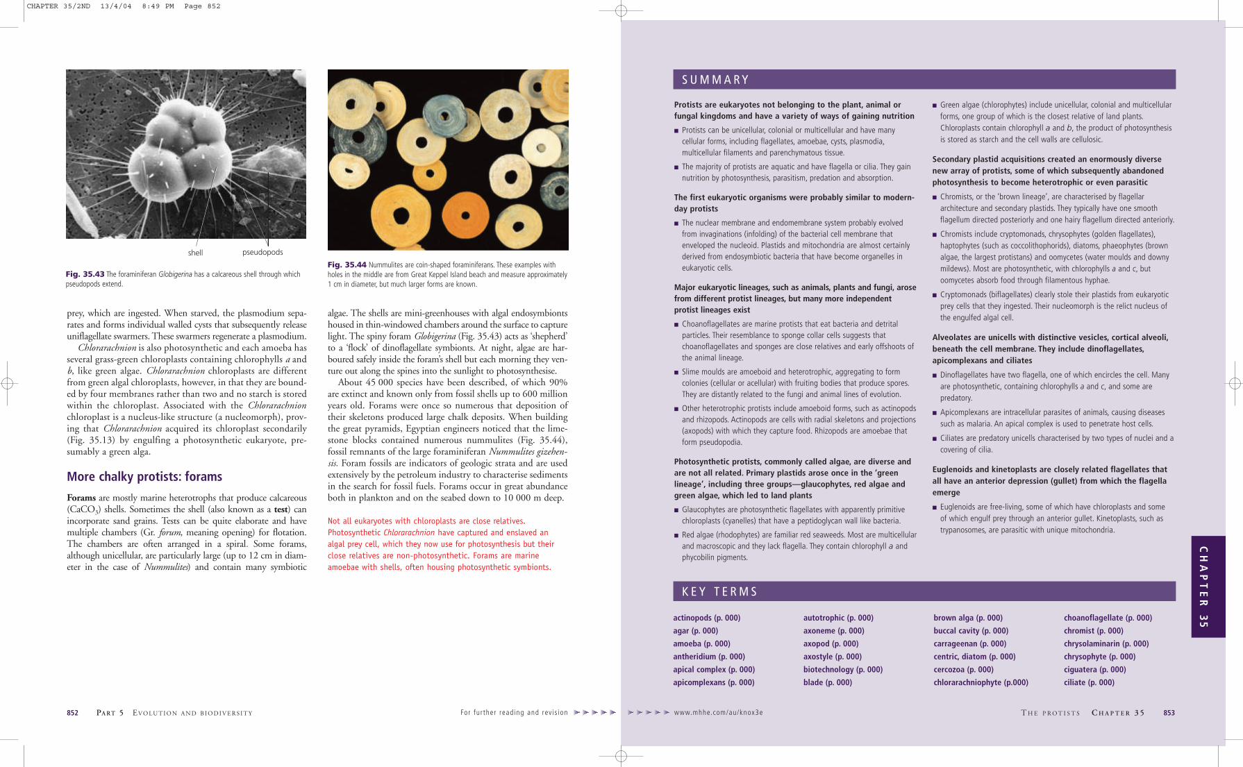
Protists are eukaryotes not belonging to the plant, animal orfungal kingdoms and have a variety of ways of gaining nutrition
� Protists can be unicellular, colonial or multicellular and have manycellular forms, including flagellates, amoebae, cysts, plasmodia,multicellular filaments and parenchymatous tissue.
� The majority of protists are aquatic and have flagella or cilia. They gainnutrition by photosynthesis, parasitism, predation and absorption.
The first eukaryotic organisms were probably similar to modern-day protists
� The nuclear membrane and endomembrane system probably evolvedfrom invaginations (infolding) of the bacterial cell membrane thatenveloped the nucleoid. Plastids and mitochondria are almost certainlyderived from endosymbiotic bacteria that have become organelles ineukaryotic cells.
Major eukaryotic lineages, such as animals, plants and fungi, arosefrom different protist lineages, but many more independentprotist lineages exist
� Choanoflagellates are marine protists that eat bacteria and detritalparticles. Their resemblance to sponge collar cells suggests thatchoanoflagellates and sponges are close relatives and early offshoots ofthe animal lineage.
� Slime moulds are amoeboid and heterotrophic, aggregating to formcolonies (cellular or acellular) with fruiting bodies that produce spores.They are distantly related to the fungi and animal lines of evolution.
� Other heterotrophic protists include amoeboid forms, such as actinopodsand rhizopods. Actinopods are cells with radial skeletons and projections(axopods) with which they capture food. Rhizopods are amoebae thatform pseudopodia.
Photosynthetic protists, commonly called algae, are diverse andare not all related. Primary plastids arose once in the ‘greenlineage’, including three groups—glaucophytes, red algae andgreen algae, which led to land plants
� Glaucophytes are photosynthetic flagellates with apparently primitivechloroplasts (cyanelles) that have a peptidoglycan wall like bacteria.
� Red algae (rhodophytes) are familiar red seaweeds. Most are multicellularand macroscopic and they lack flagella. They contain chlorophyll a andphycobilin pigments.
� Green algae (chlorophytes) include unicellular, colonial and multicellularforms, one group of which is the closest relative of land plants.Chloroplasts contain chlorophyll a and b, the product of photosynthesisis stored as starch and the cell walls are cellulosic.
Secondary plastid acquisitions created an enormously diversenew array of protists, some of which subsequently abandonedphotosynthesis to become heterotrophic or even parasitic
� Chromists, or the ‘brown lineage’, are characterised by flagellararchitecture and secondary plastids. They typically have one smoothflagellum directed posteriorly and one hairy flagellum directed anteriorly.
� Chromists include cryptomonads, chrysophytes (golden flagellates),haptophytes (such as coccolithophorids), diatoms, phaeophytes (brownalgae, the largest protistans) and oomycetes (water moulds and downymildews). Most are photosynthetic, with chlorophylls a and c, butoomycetes absorb food through filamentous hyphae.
� Cryptomonads (biflagellates) clearly stole their plastids from eukaryoticprey cells that they ingested. Their nucleomorph is the relict nucleus ofthe engulfed algal cell.
Alveolates are unicells with distinctive vesicles, cortical alveoli,beneath the cell membrane. They include dinoflagellates,apicomplexans and ciliates
� Dinoflagellates have two flagella, one of which encircles the cell. Manyare photosynthetic, containing chlorophylls a and c, and some arepredatory.
� Apicomplexans are intracellular parasites of animals, causing diseasessuch as malaria. An apical complex is used to penetrate host cells.
� Ciliates are predatory unicells characterised by two types of nuclei and acovering of cilia.
Euglenoids and kinetoplasts are closely related flagellates thatall have an anterior depression (gullet) from which the flagellaemerge
� Euglenoids are free-living, some of which have chloroplasts and someof which engulf prey through an anterior gullet. Kinetoplasts, such astrypanosomes, are parasitic with unique mitochondria.
www.mhhe.com/au/knox3e T H E P R O T I S T S C H A P T E R 3 5 853
CH
AP
TE
R 3
5
S U M M A R Y
actinopods (p. 000)
agar (p. 000)
amoeba (p. 000)
antheridium (p. 000)
apical complex (p. 000)
apicomplexans (p. 000)
autotrophic (p. 000)
axoneme (p. 000)
axopod (p. 000)
axostyle (p. 000)
biotechnology (p. 000)
blade (p. 000)
brown alga (p. 000)
buccal cavity (p. 000)
carrageenan (p. 000)
centric, diatom (p. 000)
cercozoa (p. 000)
chlorarachniophyte (p.000)
choanoflagellate (p. 000)
chromist (p. 000)
chrysolaminarin (p. 000)
chrysophyte (p. 000)
ciguatera (p. 000)
ciliate (p. 000)
K E Y T E R M S
prey, which are ingested. When starved, the plasmodium sepa-rates and forms individual walled cysts that subsequently releaseuniflagellate swarmers. These swarmers regenerate a plasmodium.
Chlorarachnion is also photosynthetic and each amoeba hasseveral grass-green chloroplasts containing chlorophylls a andb, like green algae. Chlorarachnion chloroplasts are differentfrom green algal chloroplasts, however, in that they are bound-ed by four membranes rather than two and no starch is storedwithin the chloroplast. Associated with the Chlorarachnionchloroplast is a nucleus-like structure (a nucleomorph), prov-ing that Chlorarachnion acquired its chloroplast secondarily(Fig. 35.13) by engulfing a photosynthetic eukaryote, pre-sumably a green alga.
More chalky protists: forams
Forams are mostly marine heterotrophs that produce calcareous(CaCO3) shells. Sometimes the shell (also known as a test) canincorporate sand grains. Tests can be quite elaborate and havemultiple chambers (Gr. forum, meaning opening) for flotation.The chambers are often arranged in a spiral. Some forams,although unicellular, are particularly large (up to 12 cm in diam-eter in the case of Nummulites) and contain many symbiotic
algae. The shells are mini-greenhouses with algal endosymbiontshoused in thin-windowed chambers around the surface to capturelight. The spiny foram Globigerina (Fig. 35.43) acts as ‘shepherd’to a ‘flock’ of dinoflagellate symbionts. At night, algae are har-boured safely inside the foram’s shell but each morning they ven-ture out along the spines into the sunlight to photosynthesise.
About 45 000 species have been described, of which 90%are extinct and known only from fossil shells up to 600 millionyears old. Forams were once so numerous that deposition oftheir skeletons produced large chalk deposits. When buildingthe great pyramids, Egyptian engineers noticed that the lime-stone blocks contained numerous nummulites (Fig. 35.44),fossil remnants of the large foraminiferan Nummulites gizehen-sis. Foram fossils are indicators of geologic strata and are usedextensively by the petroleum industry to characterise sedimentsin the search for fossil fuels. Forams occur in great abundanceboth in plankton and on the seabed down to 10 000 m deep.
Not all eukaryotes with chloroplasts are close relatives.Photosynthetic Chlorarachnion have captured and enslaved analgal prey cell, which they now use for photosynthesis but theirclose relatives are non-photosynthetic. Forams are marineamoebae with shells, often housing photosynthetic symbionts.
852 PA RT 5 EVO LU T I O N A N D B I O D I V E R S I T Y For fur ther read ing and rev is ion
Fig. 35.43 The foraminiferan Globigerina has a calcareous shell through whichpseudopods extend.
shell pseudopods
Fig. 35.44 Nummulites are coin-shaped foraminiferans. These examples withholes in the middle are from Great Keppel Island beach and measure approximately1 cm in diameter, but much larger forms are known.
CHAPTER 35/2ND 13/4/04 8:49 PM Page 852

www.mhhe.com/au/knox3e T H E P R O T I S T S C H A P T E R 3 5 855
CH
AP
TE
R 3
5
854 PA RT 5 EVO LU T I O N A N D B I O D I V E R S I T Y For fur ther read ing and rev is ion
coccolith (p. 000)
coccolithophorid (p. 000)
coenocytic (p. 000)
conceptacle (p. 000)
conjugation (p. 000)
cortical alveoli (p. 000)
cryptomonad (p. 000)
cyanelle (p. 000)
cytoproct (p. 000)
diarrhoetic shellfish poisoning(DSP) (p. 000)
diatom (p. 000)
dinoflagellate (p. 000)
diplomonad (p. 000)
endosymbiosis, primary andsecondary (p. 000)
euglenoid (p. 000)
foram (p. 000)
frustule (p. 000)
fucoxanthin (p. 000)
gametocyte (p. 000)
girdle (p. 000)
glaucophyte (p. 000)
green alga (p. 000)
gregarines (p. 000)
gullet (p. 000)
haptonema (p. 000)
haptophyte (p. 000)
heterokont (p. 000)
heterotrophic (p. 000)
holdfast (p. 000)
kelp (p. 000)
kinetoplast (p. 000)
laminarin (p. 000)
leishmania (p. 000)
Leishmaniasis (p. 000)
lorica (p. 000)
macronucleus (p. 000)
malaria (p. 000)
merozoite (p. 000)
metaboly (p. 000)
micronucleus (p. 000)
myxamoeba (p. 000)
nucleomorph (p. 000)
oogonium (p. 000)
oomycete (p. 000)
ovum (p. 000)
parabasalid (p. 000)
paralytic shellfish poisoning(PSP) (p. 000)
paramylon (p. 000)
pellicle (p. 000)
pennate, diatom (p. 000)
peptidoglycan (p. 000)
peridinin (p. 000)
phaetophyte (p. 000)
phragmoplast (p. 000)
phycobilin (p. 000)
phycocyanin (p. 000)
phycoerythrin (p. 000)
phycoplast (p. 000)
pit plug (p. 000)
plasmodium (p. 000)
polyphyletic (p. 000)
protist (p. 000)
pseudoplasmodium (p. 000)
raphe (p. 000)
receptacle (p. 000)
red alga (p. 000)
red tide (p. 000)
reticulopodium (p. 000)
rhizopod (p. 000)
rhodophyte (p. 000)
silicoflagellate (p. 000)
slime mould (p. 000)
sorocarp (p. 000)
sporozoite (p. 000)
stigma, of Euglena (p. 000)
stipe (p. 000)
stonewort (p. 000)
test (p. 000)
thallus (p. 000)
transverse flagellum (p. 000)
trophozoite (p. 000)
trypanosome (p. 000)
valve (p. 000)
zooxanthella (p. 000)
K E Y T E R M S
S E L F - A S S E S S M E N T Q U E S T I O N S
1. Choanoflagellate protists are morphologically similar to:
(a) red blood cells (b) sponge cells (c) sperm cells (d) macrophages
2. Chloroplasts of green algae contain:
(a) chlorophylls a and b (b) chlorophylls a and c(c) chlorophyll a and phycobilins (d) chlorophylls a, b and c
3. Parabasalid mitochondria are unique in that they:
(a) lack DNA(b) reduce protons to produce H2 gas(c) do not put electrons onto O2(d) all of the above
4. Flagellates with one smooth flagellum directed posteriorly and onehairy flagellum directed anteriorly are known as:
(a) trypanosomes (b) dinoflagellates(c) heterokonts (d) euglenoids
5. The storage product in euglenoids is:
(a) paramylon (b) starch(c) chrysolaminarin (d) glycogen
6. The flattened sacs of membrane, known as alveoli, beneath the plasmamembrane, are characteristic of which protists?
(a) ciliates (b) dinoflagellates(c) apicomplexans (d) all of the above
7. A pseudoplasmodium is:
(a) an aggregation of individual cells(b) a large multinucleate cell(c) a false form of malaria(d) a reproductive structure of red algae
8. Fucoxanthin is an accessory pigment found in:
(a) protostelids (b) red algae(c) green algae (d) haptophytes
9. The parasite that caused a health scare in Sydney water reservoirs in1998 was:
(a) an amoeba(b) the diplomonad Giardia(c) a pseudoplasmodium(d) a trypanosome
10. Which type of alga has plastids from a secondary endosymbiosis?
(a) cryptomonads (b) chlorophytes(c) rhodophytes (d) charophytes
SUGGESTED FURTHER READING
Canter-Lund, H. and Lund, J. W. G. (1995). Freshwater Algae. Bristol: Biopress.
Encyclopedia of Life Sciences. (2003). www.els.net (requires subscription).
Entwisle, T. J., Sonneman, J. A., Lewis, S. H. (1997). Freshwater Algae in Australia. Sydney: Sainty & Associates.
Margulis, L., Corliss, J. O., Melkonian, M., Chapman, D. J. (1989). Handbook of Protoctista. Boston: Jones & Bartlett.
O’Kelly, C. J. and Littlejohn, T. (1999). Protist Image Database: www.megasun.bch.umontreal.ca/protists/protists
Patterson, D. J. (1996). Free-living Freshwater Protozoa. New York: John Wiley & Sons.
Triemer, R. E. and Farmer, M. A. (1999). The Euglenid Project. www.lifesci.rutgers.edu/~triemer/index
ANWERS TOSELF-ASSESSMENT QUESTIONS
1.(b),2.(a),3.(b),4.(c),5.(a),6.(d),7.(a),8.(d),9.(b),10.(a)
R E V I E W Q U E S T I O N S
1. What major technological advances have allowed biologists todiscover great diversity and previously unrecognised evolutionaryrelationships in the protists? Why is the kingdom Protista consideredto be an artificial taxon? (p. 000)
2. Make a table comparing the main distinguishing features of red, greenand brown algae. What useful substances are obtained from red, greenand brown algae? (p. 000)
3. (a) Why are brown algae called heterokonts and what are their closest relatives? (p. 000)
(b) What organisms are green algae most closely related to? (p. 000)
4. What features would enable you to identify a protist as (a) a eugle-noid or (b) a dinoflagellate? Describe how each of these protistsmoves and how they gain their nutrition. (p. 000)
5. What are red tides? Why have they become a problem in Australia?(p. 000)
6. What is unusual about the genetic material in ciliates? How do ciliatesreproduce sexually? (p. 000)
7. What type of protist is a trypanosome? How do these parasites avoidbeing eliminated by the immune system when they are in the blood-stream of their host? (p. 000)
8. What groups of protists belong to the alveolates and what characteris-tics do they have in common? (p. 000)
9. What is the apical complex? Which protists possess one? What does itdo? (p. 000)
10. What is the difference between cellular slime moulds and plasmodialslime moulds? (p. 000)
E X T E N S I O N Q U E S T I O N S
1. Which protist causes jarrah dieback? What features of this protistexplain the extensive spread of the disease through Australian forests?How would you go about controlling the spread of this disease in alogging area or National Park?
2. It has been determined that not all protists that have a simple cellularstructure, such as those apparently lacking mitochondria, are earlyprimitive lineages among the eukaryotes. Discuss why this is the case.
3. Describe the life cycle of the protist that causes malaria and suggestwhy parasitising two different hosts may have been a selective advan-tage.
4. What are the possible explanations for the apparent increase in redtide problems in the world over the last few decades?
5. What do we mean by primary endosymbiosis and secondaryendosymbiosis? What is the impact of these processes on eukaryoticdiversity? (Hint: Add up the number of species in groups withsecondary plastids.)
To a c c e s s M a x M a r k a n d Po w e r We b g o t o t h e O n l i n e L e a r n i n g C e n t r e
w w w . m h h e . c o m / a u / k n o x 3 e
CHAPTER 35/2ND 13/4/04 8:49 PM Page 854