Embed Size (px)
Citation preview
N E V A D A T U B E R C U L O S I S P R O G R A M M A N U A L Diagnosis of Tuberculosis Disease 3 . 1
R e v i s e d A p r i l 2 0 1 8
Chapter 3 Diagnosis of Tuberculosis Disease
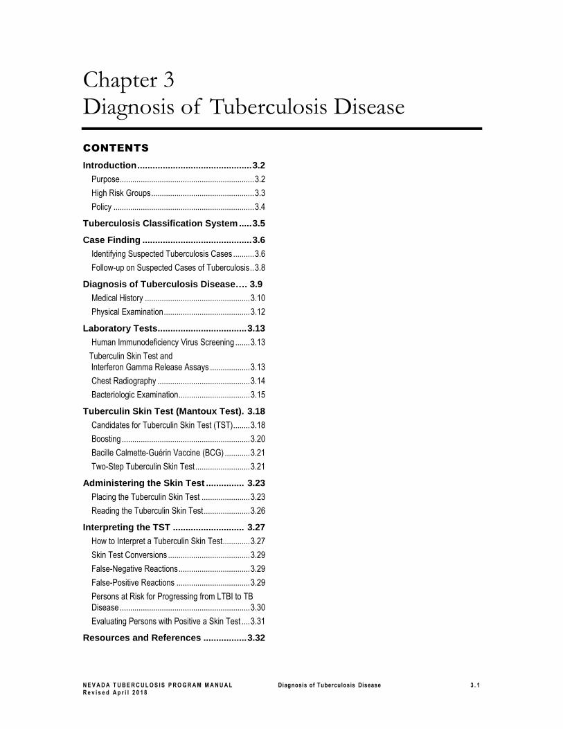
CONTENTS
Introduction ............................................. 3.2
Purpose................................................................ 3.2
High Risk Groups ................................................. 3.3
Policy ................................................................... 3.4
Tuberculosis Classification System ..... 3.5
Case Finding ........................................... 3.6
Identifying Suspected Tuberculosis Cases .......... 3.6
Follow-up on Suspected Cases of Tuberculosis .. 3.8
Diagnosis of Tuberculosis Disease…. 3.9
Medical History .................................................. 3.10
Physical Examination ......................................... 3.12
Laboratory Tests................................... 3.13
Human Immunodeficiency Virus Screening ....... 3.13
Tuberculin Skin Test and
Interferon Gamma Release Assays ................... 3.13
Chest Radiography ............................................ 3.14
Bacteriologic Examination .................................. 3.15
Tuberculin Skin Test (Mantoux Test). 3.18
Candidates for Tuberculin Skin Test (TST) ........ 3.18
Boosting ............................................................. 3.20
Bacille Calmette-Guérin Vaccine (BCG) ............ 3.21
Two-Step Tuberculin Skin Test .......................... 3.21
Administering the Skin Test ............... 3.23
Placing the Tuberculin Skin Test ....................... 3.23
Reading the Tuberculin Skin Test ...................... 3.26
Interpreting the TST ............................ 3.27
How to Interpret a Tuberculin Skin Test ............. 3.27
Skin Test Conversions ....................................... 3.29
False-Negative Reactions .................................. 3.29
False-Positive Reactions ................................... 3.29
Persons at Risk for Progressing from LTBI to TB
Disease .............................................................. 3.30
Evaluating Persons with Positive a Skin Test .... 3.31
Resources and References ................. 3.32
N E V A D A T U B E R C U L O S I S P R O G R A M M A N U A L Diagnosis of Tuberculosis Disease 3 . 2
R e v i s e d A p r i l 2 0 1 8
Introduction
Purpose
Use this section to understand and follow national and Nevada guidelines for
▪ Classifying patients with tuberculosis (TB) disease and latent TB infection (LTBI)
▪ Detecting suspected cases of TB
▪ Understanding when to report suspected or confirmed cases of TB and
▪ Diagnosing TB disease.
It is important to understand when a person should be evaluated further for TB disease.
Not recognizing TB symptoms promptly may lead to delays in initiating appropriate
treatment thus extending the possible infectious time, transmitting more TB disease, and
multiplying the number of contacts needing to be evaluated.
In the 2005 guideline, “Controlling Tuberculosis in the United States: Recommendations
from the American Thoracic Society, Centers for Disease Control and Prevention, and
the Infectious Diseases Society of America,” one of the recommended strategies to
achieve the goal of reduction of TB morbidity and mortality is early and accurate
detection, diagnosis, and reporting of TB cases, leading to initiation and completion of
treatment.1
Improvement in the detection of TB cases is essential to progress toward the elimination
of TB in the United States.2 Case detection includes the processes that lead to the;
presentation, evaluation, receipt of diagnosis, and reporting of persons with active TB.3
Detecting and reporting suspected cases of TB are key steps in stopping transmission of
Mycobacterium tuberculosis because it leads to prompt initiation of effective multiple-
drug treatment, which rapidly reduces infectiousness.4
TB is commonly diagnosed when a person seeks medical attention for symptoms
caused by the disease or a concomitant medical condition. Thus, healthcare providers,
particularly those providing primary healthcare to populations at high risk, are key
contributors to TB case detection.5 The majority of pulmonary TB cases continue to be
diagnosed at an advanced stage. Earlier diagnosis would result in less individual
morbidity and death, greater success in treatment, less transmission to contacts, and
fewer outbreaks of TB.6
A diagnosis of TB disease is usually based on positive cultures for M. tuberculosis.
However, in the absence of a positive culture, TB may also be diagnosed on the basis of
clinical signs and symptoms. Positive cultures for M. tuberculosis confirm the diagnosis
of Tuberculosis and provide an organism for susceptibility testing as well as genotyping.
Contacts are mentioned within this section, but the contact investigation
evaluation and follow-up are covered in more depth in Chapter 8, Contact
Investigation. For information on treatment, refer to Treatment of
Tuberculosis Disease, Chapter 4.
N E V A D A T U B E R C U L O S I S P R O G R A M M A N U A L Diagnosis of Tuberculosis Disease 3 . 3
R e v i s e d A p r i l 2 0 1 8
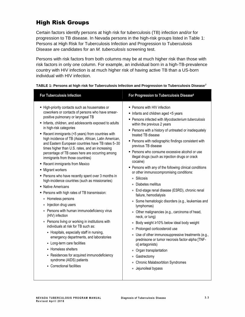
High Risk Groups
Certain factors identify persons at high risk for tuberculosis (TB) infection and/or for
progression to TB disease. In Nevada persons in the high-risk groups listed in Table 1:
Persons at High Risk for Tuberculosis Infection and Progression to Tuberculosis
Disease are candidates for an M. tuberculosis screening test.
Persons with risk factors from both columns may be at much higher risk than those with
risk factors in only one column. For example, an individual born in a high-TB-prevalence
country with HIV infection is at much higher risk of having active TB than a US-born
individual with HIV infection.
TABLE 1: Persons at high risk for Tuberculosis Infection and Progression to Tuberculosis Disease7
For Tuberculosis Infection For Progression to Tuberculosis Disease8
▪ High-priority contacts such as housemates or
coworkers or contacts of persons who have smear-
positive pulmonary or laryngeal TB
▪ Infants, children, and adolescents exposed to adults
in high-risk categories
▪ Recent immigrants (<5 years) from countries with
high incidence of TB (Asian, African, Latin American,
and Eastern European countries have TB rates 5–30
times higher than U.S. rates, and an increasing
percentage of TB cases here are occurring among
immigrants from those countries)
▪ Recent immigrants from Mexico
▪ Migrant workers
▪ Persons who have recently spent over 3 months in
high-incidence countries (such as missionaries)
▪ Native Americans
▪ Persons with high rates of TB transmission:
• Homeless persons
• Injection drug users
• Persons with human immunodeficiency virus
(HIV) infection
• Persons living or working in institutions with
individuals at risk for TB such as:
▪ Hospitals, especially staff in nursing,
emergency departments, and laboratories
▪ Long-term care facilities
▪ Homeless shelters
▪ Residences for acquired immunodeficiency
syndrome (AIDS) patients
▪ Correctional facilities
▪ Persons with HIV infection
▪ Infants and children aged <5 years
▪ Persons infected with Mycobacterium tuberculosis
within the previous 2 years
▪ Persons with a history of untreated or inadequately
treated TB disease
▪ Persons with radiographic findings consistent with
previous TB disease
▪ Persons who consume excessive alcohol or use
illegal drugs (such as injection drugs or crack
cocaine)
▪ Persons with any of the following clinical conditions
or other immunocompromising conditions:
• Silicosis
• Diabetes mellitus
• End-stage renal disease (ESRD), chronic renal
failure, hemodialysis
• Some hematologic disorders (e.g., leukemias and
lymphomas)
• Other malignancies (e.g., carcinoma of head,
neck, or lung)
• Body weight ≥10% below ideal body weight
• Prolonged corticosteroid use
• Use of other immunosuppressive treatments (e.g.,
prednisone or tumor necrosis factor-alpha [TNF-
α] antagonists)
• Organ transplantation
• Gastrectomy
• Chronic Malabsorbtion Syndromes
• Jejunoileal bypass
N E V A D A T U B E R C U L O S I S P R O G R A M M A N U A L Diagnosis of Tuberculosis Disease 3 . 4
R e v i s e d A p r i l 2 0 1 8
Policy
In Nevada:
▪ Persons who show or report signs and symptoms of TB should be evaluated for TB
disease as described in the “Diagnosis of Tuberculosis Disease” topic in this section
and reported as suspected cases of TB as described in the “Reporting Tuberculosis”
topic in the Surveillance section.
▪ Contacts should be evaluated as described in the Contact Investigation section,
Chapter 8.
For roles and responsibilities, refer to the “Roles, Responsibilities, and
Contact Information” topic in the Introduction, Chapter 1.
N E V A D A T U B E R C U L O S I S P R O G R A M M A N U A L Diagnosis of Tuberculosis Disease 3 . 5
R e v i s e d A p r i l 2 0 1 8
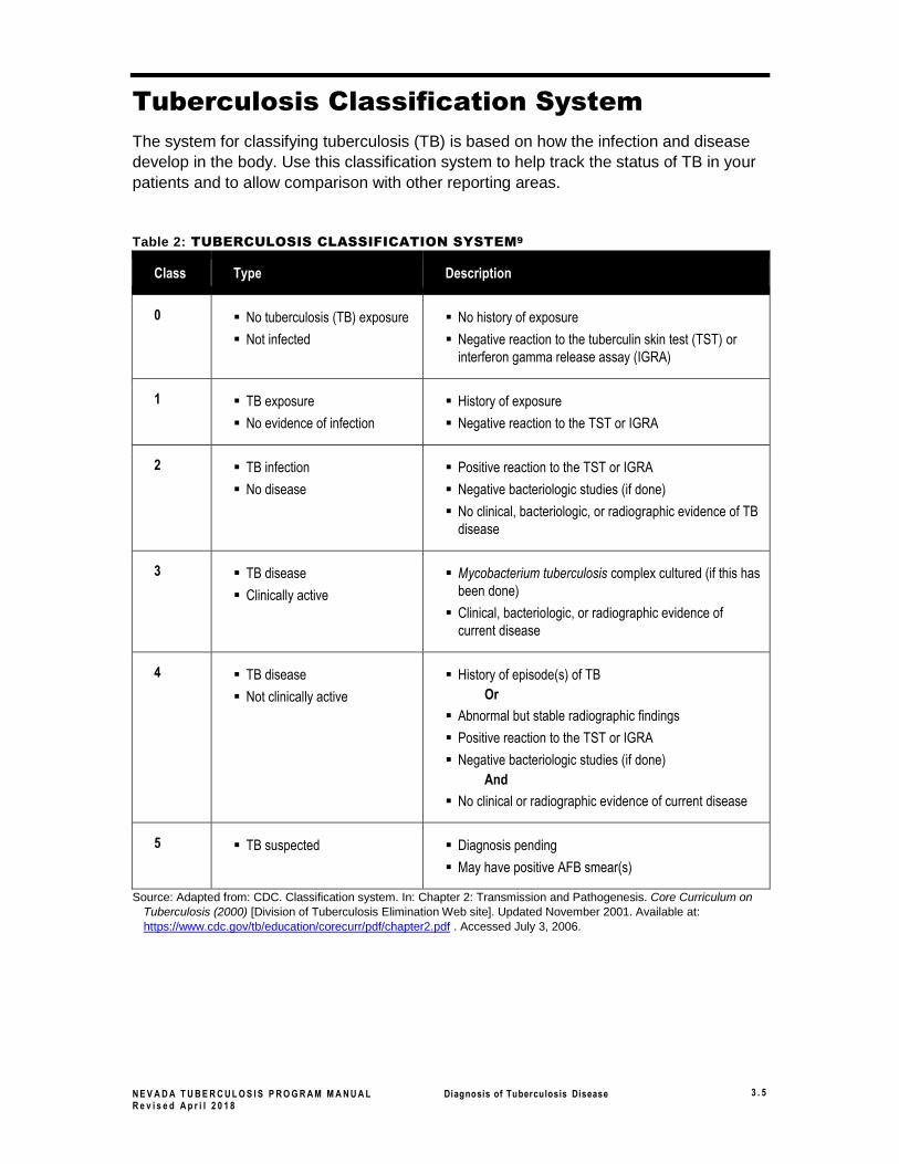
Tuberculosis Classification System
The system for classifying tuberculosis (TB) is based on how the infection and disease
develop in the body. Use this classification system to help track the status of TB in your
patients and to allow comparison with other reporting areas.
Table 2: TUBERCULOSIS CLASSIFICATION SYSTEM9
Class Type Description
0 ▪ No tuberculosis (TB) exposure
▪ Not infected
▪ No history of exposure
▪ Negative reaction to the tuberculin skin test (TST) or
interferon gamma release assay (IGRA)
1 ▪ TB exposure
▪ No evidence of infection
▪ History of exposure
▪ Negative reaction to the TST or IGRA
2 ▪ TB infection
▪ No disease
▪ Positive reaction to the TST or IGRA
▪ Negative bacteriologic studies (if done)
▪ No clinical, bacteriologic, or radiographic evidence of TB
disease
3 ▪ TB disease
▪ Clinically active
▪ Mycobacterium tuberculosis complex cultured (if this has
been done)
▪ Clinical, bacteriologic, or radiographic evidence of
current disease
4 ▪ TB disease
▪ Not clinically active
▪ History of episode(s) of TB
Or
▪ Abnormal but stable radiographic findings
▪ Positive reaction to the TST or IGRA
▪ Negative bacteriologic studies (if done)
And
▪ No clinical or radiographic evidence of current disease
5 ▪ TB suspected ▪ Diagnosis pending
▪ May have positive AFB smear(s)
Source: Adapted from: CDC. Classification system. In: Chapter 2: Transmission and Pathogenesis. Core Curriculum on
Tuberculosis (2000) [Division of Tuberculosis Elimination Web site]. Updated November 2001. Available at:
https://www.cdc.gov/tb/education/corecurr/pdf/chapter2.pdf . Accessed July 3, 2006.
N E V A D A T U B E R C U L O S I S P R O G R A M M A N U A L Diagnosis of Tuberculosis Disease 3 . 6
R e v i s e d A p r i l 2 0 1 8
Case Finding
Identifying Suspected Tuberculosis Cases
The majority of tuberculosis (TB) cases are detected during the medical evaluation of
symptomatic illnesses. Persons experiencing symptoms ultimately attributable to TB
usually seek care not at a public health TB clinic, but rather from other medical
practitioners in other healthcare settings.10 Professionals in the primary healthcare
sector, including hospital and emergency department clinicians, should be trained to
recognize patients with symptoms consistent with TB.11
Be alert for cases of TB among persons who have not sought medical care during
contact evaluations of patients with pulmonary TB and of other persons newly diagnosed
as infected with Latent Mycobacterium tuberculosis Infection (LTBI). Perform screening
for TB during evaluation of immigrants and refugees with Class B1, B2 or B3 TB
notification status, during evaluations of persons involved in TB outbreaks, and
occasionally in working with populations with a known high incidence of TB. Also, screen
for TB disease when the risk for TB in the population is high and when the
consequences of an undiagnosed case of TB are severe, such as in jails, prisons, and
other facilities with congregate settings and or high-risk populations.12
Suspect pulmonary TB and initiate a diagnostic investigation when the historic features,
signs, symptoms, and radiographic findings occur in adults. See these listed in Table 3,
When to Suspect Pulmonary Tuberculosis in Adults, below. The clinical presentation
of TB varies considerably as a result of the extent of the disease and the patient’s
response. TB should be suspected in any patient who has a persistent cough for more
than two to three weeks or other compatible signs and symptoms.13
Note that these symptoms should suggest a diagnosis of TB, but are not required. TB
should still be considered a diagnosis in asymptomatic patients who have risk factors for
TB and chest radiographs compatible with TB.
All persons who have a chronic cough for more than two to three weeks14
should be evaluated and be asked to use a mask or tissue to cover their
mouth. Hemoptysis (coughing up blood) is a serious symptom, and
patients who cough up blood should be evaluated as soon as possible. Be
sure to have these patients wear a mask or use tissues to cover their
cough.
N E V A D A T U B E R C U L O S I S P R O G R A M M A N U A L Diagnosis of Tuberculosis Disease 3 . 7
R e v i s e d A p r i l 2 0 1 8
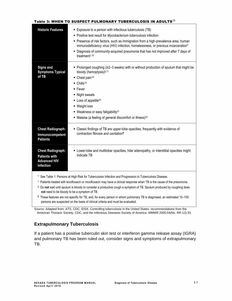
Table 3: WHEN TO SUSPECT PULMONARY TUBERCULOSIS IN ADULTS15
Historic Features ▪ Exposure to a person with infectious tuberculosis (TB)
▪ Positive test result for Mycobacterium tuberculosis infection
▪ Presence of risk factors, such as immigration from a high-prevalence area, human
immunodeficiency virus (HIV) infection, homelessness, or previous incarceration*
▪ Diagnosis of community-acquired pneumonia that has not improved after 7 days of
treatment†,16
Signs and
Symptoms Typical
of TB
▪ Prolonged coughing (≥2–3 weeks) with or without production of sputum that might be
bloody (hemoptysis)§,17
▪ Chest pain18
▪ Chills19
▪ Fever
▪ Night sweats
▪ Loss of appetite20
▪ Weight loss
▪ Weakness or easy fatigability21
▪ Malaise (a feeling of general discomfort or illness)22
Chest Radiograph:
Immunocompetent
Patients
▪ Classic findings of TB are upper-lobe opacities, frequently with evidence of
contraction fibrosis and cavitation¶
Chest Radiograph:
Patients with
Advanced HIV
Infection
▪ Lower-lobe and multilobar opacities, hilar adenopathy, or interstitial opacities might
indicate TB
* See Table 1: Persons at High Risk for Tuberculosis Infection and Progression to Tuberculosis Disease.
† Patients treated with levofloxacin or moxifloxacin may have a clinical response when TB is the cause of the pneumonia.
§ Do not wait until sputum is bloody to consider a productive cough a symptom of TB. Sputum produced by coughing does
not need to be bloody to be a symptom of TB.
¶ These features are not specific for TB, and, for every person in whom pulmonary TB is diagnosed, an estimated 10–100
persons are suspected on the basis of clinical criteria and must be evaluated.
Source: Adapted from: ATS, CDC, IDSA. Controlling tuberculosis in the United States: recommendations from the
American Thoracic Society, CDC, and the Infectious Diseases Society of America. MMWR 2005;54(No. RR-12):33.
Extrapulmonary Tuberculosis
If a patient has a positive tuberculin skin test or interferon gamma release assay (IGRA)
and pulmonary TB has been ruled out, consider signs and symptoms of extrapulmonary
TB.
N E V A D A T U B E R C U L O S I S P R O G R A M M A N U A L Diagnosis of Tuberculosis Disease 3 . 8
R e v i s e d A p r i l 2 0 1 8
Follow-up on Suspected Cases of Tuberculosis
When a person with signs and symptoms consistent with TB is identified, perform the
following:
Refer to Table 4: Guidelines for the Evaluation of Pulmonary
Tuberculosis in Adults in Five Clinical Scenarios in the “Diagnosis of
Tuberculosis Disease” topic in this section. This table presents guidelines
for the initial steps of TB case detection in five clinical scenarios
encountered by providers of primary healthcare, including those serving in
medical emergency departments.23
To formally report a suspected case of TB, see the “Reporting
Tuberculosis” topic in the Surveillance section.
The patient should be masked and immediately excluded from the
workplace or placed in airborne infection isolation (AII) until confirmed
noninfectious. For more information, see the “Isolation” topic in the
Infection Control section of this manual.
Laboratories are required to report positive smears or positives cultures,
and primary healthcare providers are required to report suspected or
confirmed cases of TB to the health department within 24 hours, as
specified in the “Reporting Tuberculosis” topic in the Surveillance section.
Prompt reporting allows the health department to organize treatment and
case management services and to initiate a contact investigation as
quickly as possible.24
Within 48 hours of suspect identification, administer a tuberculin skin test
(TST) or perform an interferon gamma release assay (IGRA) and/or obtain
a chest radiograph, if not already done. Evaluate the patient for TB disease
as specified in the “Diagnosis of Tuberculosis Disease” topic in this
section.

N E V A D A T U B E R C U L O S I S P R O G R A M M A N U A L Diagnosis of Tuberculosis Disease 3 . 9
R e v i s e d A p r i l 2 0 1 8
Diagnosis of Tuberculosis Disease
Consideration of tuberculosis (TB) disease as a possible diagnosis is the first step that
must be taken before further evaluation, diagnosis, and management can occur. The
diagnosis of TB disease is often overlooked because of the failure to consider it among
possible diagnoses. While a definitive diagnosis may involve the addition of laboratory
and radiographic findings, a high degree of suspicion can be based on epidemiology,
medical history, and physical examination. In considering TB disease, it is also important
to consider factors that may affect the typical presentation of TB, such as the patient’s
age, nutritional status, and coexisting diseases.
An individual who is suspected of having TB disease requires a complete medical
evaluation, including the following:
▪ Medical history, including exposure, symptoms, previous treatment for TB, and risk
factors
▪ Human Immunodeficiency Virus (HIV) screening
▪ Physical examination
▪ Tuberculin skin test or interferon gamma release assay
▪ Chest radiography
▪ Bacteriologic examination
When a suspected case of pulmonary TB is identified, refer to Table 4 for guidelines for
the initial steps of TB case detection in five clinical scenarios encountered by providers
of primary healthcare, including those serving in medical emergency departments.25
N E V A D A T U B E R C U L O S I S P R O G R A M M A N U A L Diagnosis of Tuberculosis Disease 3 . 1 0
R e v i s e d A p r i l 2 0 1 8
Table 4: GUIDELINES FOR THE EVALUATION OF PULMONARY TUBERCULOSIS IN
ADULTS IN FIVE CLINICAL SCENARIOS26
Patient and Setting
Recommended Evaluation
Any patient with a cough of ≥2–3 weeks duration Chest radiograph: If suggestive of tuberculosis (TB)*,
collect 3 sputum specimens for acid-fast bacilli (AFB)
smear microscopy, culture, and nucleic acid
amplification (NAA), if available27
Any patient at high risk for TB with an unexplained
illness, including respiratory symptoms of ≥2–3 weeks
duration†
Chest radiograph: If suggestive of TB, collect 3 sputum
specimens for AFB smear microscopy, culture, and
NAA, if available
Any patient with human immunodeficiency virus (HIV)
infection and unexplained cough or fever
Chest radiograph, and collect 3 sputum specimens for
AFB smear microscopy, culture, and NAA, if available
Any patient at high risk for TB with a diagnosis of
community-acquired pneumonia who has not improved
after 7 days of treatment†
Chest radiograph, and collect 3 sputum specimens for
AFB smear microscopy, culture, and NAA, if available
Any patient at high risk for TB with incidental findings
on chest radiograph suggestive of TB, even if
symptoms are minimal or absent†§
Review of previous chest radiographs, if available,
collect 3 sputum specimens for AFB smear
microscopy, culture, and NAA, if available
* Opacities with or without cavitation in the upper lobes or the superior segments of the lower lobes.28
† See Table 1: Persons at High Risk for Tuberculosis Infection and Progression to Tuberculosis Disease.
§ Chest radiograph performed for any reason, including targeted testing for latent TB infection and screening for TB disease.
Source: Adapted from: ATS, CDC, IDSA. Controlling tuberculosis in the United States: recommendations from the
American Thoracic Society, CDC, and the Infectious Diseases Society of America. MMWR 2005;54(No. RR-12):33.
Medical History
The clinician should interview patients to document their medical histories. A written
record of a patient’s medical history should include the following:
1. Exposure to infectious TB
2. Symptoms of TB disease (as listed in Table 3: When to Suspect Pulmonary
Tuberculosis in Adults, page 3.7, Table 4: Guidelines for the Evaluation of
Pulmonary Tuberculosis in Adults in Five Clinical Scenarios, page 3.10, and
Table 5: Symptoms of Tuberculosis Disease, page 3.11)
3. Previous TB infection or disease
4. Risk factors (as listed in Table 1: Persons at High Risk for Tuberculosis Infection
and Progression to Tuberculosis Disease, page 3.3)
5. Recent medical encounters (e.g., going to the emergency department for
pneumonia)
6. Previous antibiotic therapy
N E V A D A T U B E R C U L O S I S P R O G R A M M A N U A L Diagnosis of Tuberculosis Disease 3 . 1 1
R e v i s e d A p r i l 2 0 1 8
1. Exposure to Infectious TB: Ask patients if they have spent time with someone with infectious TB.
Question patients about whether they know of any contact in the recent or distant past with persons diagnosed with pulmonary or laryngeal TB. It is important to note that patients often refer to latent TB infection (LTBI) as TB disease. Be aware that most persons become infected with Mycobacterium tuberculosis without knowing they were exposed. Clinicians should also consider demographic factors that may increase a patient’s risk for exposure to TB disease and drug-resistant TB, such as country of origin, age, ethnic or racial group, occupation, and residence in congregate settings (such as a jail, homeless shelter, or refugee camp).
2. Symptoms of TB Disease: Ask patients about their symptoms.
Although TB disease does not always produce symptoms, most patients with TB disease have one or more symptoms that led them to seek medical care. When symptoms are present, they usually have developed gradually and been present for weeks or even months. Occasionally TB is discovered during a medical examination for an unrelated condition, such as ruling out a cancer diagnosis, or on a pre-op chest radiograph.
The symptoms in Table 5 below may be caused by other diseases, but they should prompt the clinician to suspect TB disease. For historic features and chest radiograph results that should raise suspicion of pulmonary TB disease, refer to Table 3: When to Suspect Pulmonary Tuberculosis in Adults, page 3.7.
Table 5: SYMPTOMS OF TUBERCULOSIS DISEASE29
Pulmonary General: Pulmonary and Extrapulmonary
Extrapulmonary
▪ Coughing
▪ Coughing up sputum or blood
▪ Pain in the chest when breathing or coughing
▪ Chills30
▪ Fever
▪ Night sweats
▪ Loss of appetite31
▪ Weight loss
▪ Weakness or easy fatigability32
▪ Malaise (a feeling of general
discomfort or illness)33
The symptoms depend on part of body affected by tuberculosis (TB) disease:
▪ TB of the spine may cause pain in the back.
▪ TB of the kidney may cause blood in the urine.
▪ Meningeal TB may cause headaches or psychiatric symptoms.
▪ Lymphatic TB may cause swollen and tender lymph nodes, often at the base of the neck.
Source: Adapted from: ATS, CDC, IDSA. Controlling tuberculosis in the United States: recommendations from the American Thoracic Society, CDC, and the Infectious Diseases Society of America. MMWR 2005;54(No. RR-12):33
N E V A D A T U B E R C U L O S I S P R O G R A M M A N U A L Diagnosis of Tuberculosis Disease 3 . 1 2
R e v i s e d A p r i l 2 0 1 8
3. Previous Latent TB Infection or TB Disease: Ask patients whether they have ever been diagnosed with or treated for TB infection or disease
Patients who have had LTBI or TB disease before should be asked when they were diagnosed and what treatment they received. If documentation of treatment is not available ask how many pills were taken per day (to determine what treatment regimen was used and whether they received injections) as well as the duration of the regimen. Ask if they experienced any adverse reactions to the medications, if they completed the regimen and if they didn’t complete the reason for discontinuing treatment.
If the regimen prescribed was inadequate or if the patient did not follow the recommended treatment, TB may recur, and it may be resistant to one or more of the standard four drugs used.
Patients known to have a positive skin test reaction may have TB infection. If they were infected within the past two years, they are at high risk for TB disease if certain immunosuppressive conditions exist or if immunosuppressive therapies are being taken. (See Table 1: Persons at High Risk for Tuberculosis Infection and Progression to Tuberculosis Disease.)34 For persons previously skin tested, an increase in induration of 10 mm within a two-year period is classified as a conversion to positive.
4. Risk Factors for Developing TB Disease: Determine whether the patient has any conditions or behaviors that are risk factors for developing TB disease.
For a list of behaviors and conditions that increase the risk that TB infection will progress to disease, see Table 1: Persons at High Risk for Tuberculosis Infection and Progression to Tuberculosis Disease.
5. Recent Medical Encounters: Determine what medical services patients have received for this condition.
Has the patient been diagnosed with pneumonia or another bacterial infection in the recent past?
6. Previous Antibiotic Therapy: Newly diagnosed TB patients might have fluoroquinolone resistance as the result of the wide use of fluoroquinolones for bacterial infections35.
Moxifloxacin is in the family of fluoroquinolones and is sometimes used to treat TB. If the patient has developed a resistance to fluoroquinolones, moxifloxacin will not be an effective medication to use to treat their TB.
Physical Examination
A physical examination is an essential part of the evaluation of any patient. It cannot be used to confirm or rule out TB, but it can provide valuable information about the patient’s overall condition; other factors, such as human immunodeficiency virus (HIV) infection, which may affect how TB is manifested; and the presence of extrapulmonary TB.36
N E V A D A T U B E R C U L O S I S P R O G R A M M A N U A L Diagnosis of Tuberculosis Disease 3 . 1 3
R e v i s e d A p r i l 2 0 1 8
Laboratory Tests
Human Immunodeficiency Virus Screening
Voluntary counseling and testing for human immunodeficiency virus (HIV) is
recommended for all patients with TB. HIV counseling and testing has also been
recommended for contacts of persons with TB.37
The Centers for Disease Control and Prevention (CDC) recommends the following:
▪ Routine HIV screening for all patients ages 13–64 seeking health care for any
reason, without regard to any patient’s known risks for HIV infection
▪ Annual HIV screening of patients known to be at high risk38
Tuberculin Skin Test and Interferon Gamma Release Assays
Use the Mantoux TST or an interferon gamma release assay (IGRA) to test for M.
tuberculosis infection. Note that for patients with a previous documented positive TST
reaction, a TST is not necessary. However, an IGRA can be done if there is suspicion
that the TST result was a false positive.
Blood Assay for Mycobacterium tuberculosis (BAMT) is a general term referring to recently developed in vitro diagnostic tests that assess for the presence of infection with M. tuberculosis. The term commonly used to discuss these tests is IGRAs (Interferon-Gamma Release Assays) which describes the mode of action these tests utilize. The IGRAs currently approved by the Food and Drug Administration (FDA) and available on the market are QuantiFERON®-TB Gold In-Tube (GIT), QuantiFERON®-TB Gold Plus In-Tube (GPIT), and the T-SPOT®.TB test, all of which can be used in all circumstances where the TST is used. Additional cytokine-based immunoassays may be developed and may also become useful in the diagnosis of M. tuberculosis infection. Future FDA-licensed products, in combination with Centers for Disease Control and Prevention (CDC)-issued recommendations, may provide additional diagnostic alternatives.39
The advantages of IGRA tests, compared with the TST, are that results can be obtained
after a single patient visit, and that, because it is a blood test performed in a qualified
laboratory, the variability associated with skin test placement and reading can be
eliminated.40 In addition, the Blood Assay for Mycobacterium tuberculosis (BAMT) are
not affected by past Bacille of Calmette-Guérin (BCG) vaccination and may eliminate the
unnecessary treatment of patients with BCG-related false-positive results.41 However,
the IGRA tests have practical limitations that include the need to draw blood and ensure
its receipt in a qualified laboratory in time for testing. Refer to www.quantiferon.com for
available test sites. Refer to the Qiagen web-site http://quantiferoncellestis.com/us for
additional information regarding QuantiFERON®-TB Gold In-Tube (IT) and visit
http://www.oxfordimmunotec.com/ for information regarding the T-SPOT®.TB test which
is the most recent test to have been approved by the FDA.
N E V A D A T U B E R C U L O S I S P R O G R A M M A N U A L Diagnosis of Tuberculosis Disease 3 . 1 4
R e v i s e d A p r i l 2 0 1 8
For both the TST and IGRA, additional tests, such as chest radiography and
bacteriologic examination, are required to confirm TB disease.42
Persons with a positive QFT-GIT result or a positive TST result, regardless of symptoms
and signs, must be evaluated for TB disease before LTBI is diagnosed. At minimum, a
chest radiograph is required to assess for abnormalities consistent with TB disease.43
A negative TST does not rule out TB disease44—as many as 20% of patients with TB
disease have a negative TST reaction.45 A negative TST result or a negative QFT-G
result should not be used alone to exclude M. tuberculosis infection in persons with
symptoms or signs suggestive of TB disease. Medical evaluation of such persons should
include a history and physical examination, chest radiograph, bacteriologic studies,
serology for human immunodeficiency virus (HIV), and, when indicated, other tests or
studies.46
For more information on the Mantoux TST, see the Diagnosis of Latent
Tuberculosis Infection section, Chapter 5. For more information on IGRAs and
the QuantiFERON®-TB Gold/Gold Plus In Tube (QFT-GIT/GPIT) Test, see the
CDC’s “Updated Guidelines for Interferon Gamma Release Assays to
Detecting Mycobacterium tuberculosis Infection, United States, 2010” (MMWR
2010:59(RR-5) at https://www.cdc.gov/mmwr/PDF/rr/rr5905.pdf
Chest Radiography
A posterior-anterior radiograph of the chest is the standard view used for the detection
and description of chest abnormalities in adults. In some instances, other views (e.g.,
lateral, lordotic) or additional studies (e.g., computed tomography [CT] scans) may be
necessary.
Children younger than 5 years of age should receive posterior-anterior and
lateral radiographs.47
Certain abnormalities on chest radiographs are suggestive, but are not diagnostic, of TB.
In pulmonary TB, radiographic abnormalities are often seen in the apical and posterior
segments of the upper lobe or in the superior segments of the lower lobe. However,
lesions may appear anywhere in the lungs and may differ in size, shape, density, and
presence or absence of cavitation, especially in HIV-infected and other
immunosuppressed persons.
In HIV-infected persons, pulmonary TB may present atypically on the chest radiograph.
For example, TB may cause opacities without cavities in any lung zone, or it may cause
mediastinal or hilar lymphadenopathy with or without accompanying opacities and/or
cavities. In HIV-infected persons, almost any abnormality on a chest radiograph may
N E V A D A T U B E R C U L O S I S P R O G R A M M A N U A L Diagnosis of Tuberculosis Disease 3 . 1 5
R e v i s e d A p r i l 2 0 1 8
indicate TB. In fact, the radiograph of an HIV-infected person with TB disease may even
appear entirely normal.48
For more information on chest radiography, see the Francis J. Curry National
Tuberculosis Center’s Radiographic Manifestations of Tuberculosis: A Primer
for Clinicians (2011) at
http://www.currytbcenter.ucsf.edu/products/radiographic-manifestations-
tuberculosis-primer-clinicians-second-edition .
http://www.currytbcenter.ucsf.edu/products/radiographic-
manifestations-tuberculosis-primer-clinicians-second-edition
Bacteriologic Examination
Refer to Table 6 below to determine the types of specimens needed to assist in the
diagnosis of TB.
Table 6: SPECIMENS FOR DIAGNOSING TUBERCULOSIS DISEASE
Suspected Diagnosis Specimen Needed
Pulmonary or laryngeal tuberculosis (TB)
Sputum (phlegm from deep in the lungs) samples for smear AND culture
examination.
A diagnosis of pulmonary TB cannot be established from sputum smear alone.
When Acid Fast Bacilli (AFB) is seen on smear, other procedures may be
necessary for identification, including nucleic acid amplification (NAA),
bronchoscopy, and gastric aspiration in children.
Extrapulmonary TB Depending on the anatomical site, other clinical specimens are necessary,
such as:
▪ Urine
▪ Cerebrospinal fluid
▪ Pleural fluid
▪ Pus or other aspirated fluid
▪ Biopsy specimens
▪ Blood (heparinized)Ensure both AFB smear AND culture is requested. DO
NOT put tissue specimens in formalin, as no culture can be obtained.

N E V A D A T U B E R C U L O S I S P R O G R A M M A N U A L Diagnosis of Tuberculosis Disease 3 . 1 6
R e v i s e d A p r i l 2 0 1 8
Refer to Table 7 below for information on the bacteriology tests used to diagnose TB.
Table 7: BACTERIOLOGY TESTS USED IN DIAGNOSING TUBERCULOSIS DISEASE49
Test Description Laboratory Turnaround Times
Acid-Fast Bacilli (AFB) Smear
▪ Provides the physician with a preliminary
confirmation of the diagnosis. It usually is the
first bacteriologic evidence of the presence of
mycobacteria in a clinical specimen.
▪ If positive, the laboratory gives a
semiquantitative estimate of the number of
bacilli being excreted (which is of vital clinical
and epidemiologic importance in assessing
the patient’s infectiousness).
▪ On-site test: results available within
24 hours from specimen collection.
▪ Off-site test: within 24 hours from
laboratory receipt of specimen (time
from specimen collection to
laboratory receipt should be 24
hours or less).50 Specimens are to
be refrigerated while being stored
and during transport to the lab.
Nucleic Acid Amplification (NAA) Assay51
▪ A test done on sputum specimens for the
direct and rapid identification of the
Mycobacterium tuberculosis complex.
▪ Allows for the amplification of specific target
sequences of nucleic acids that will be
detected by a nucleic acid probe.
▪ Does not replace the need for routine AFB
smear and culture.52
▪ Within 48 hours from positive smear
result and specimen arrival at the
laboratory performing NAA53,54
Culture ▪ Usually necessary for species identification of
all clinical specimens suspected of containing
mycobacteria.
▪ Is required for drug susceptibility testing and
genotyping.
▪ Mycobacterial growth detection:
usually within 14 days from
specimen collection
▪ Identification of mycobacteria:
usually within 21 days from culture
positive 55,56
Drug Susceptibility Testing
▪ For first-line drugs: Performed on initial
isolates of all patients to identify an effective
antituberculosis regimen.
▪ For both first-line and second-line drugs:
Repeated on interim isolates when a patient
remains culture-positive after 2 months of
treatment.57,58
▪ First-line drugs: may be available
within 30 days from specimen
collection
▪ Second-line drugs: within 4 weeks
from date of request or specimen
receipt at reference laboratory. The
provider must specify drugs to be
tested.
Sources: ATS, CDC, IDSA. Controlling tuberculosis in the United States: recommendations from the American Thoracic
Society, CDC, and the Infectious Diseases Society of America. MMWR 2005;54(No. RR-12):19; and Tenover, R., et al.
The resurgence of tuberculosis: is your laboratory ready? Journal of Clinical Microbiology 1993:767–770.
N E V A D A T U B E R C U L O S I S P R O G R A M M A N U A L Diagnosis of Tuberculosis Disease 3 . 1 7
R e v i s e d A p r i l 2 0 1 8
Laboratories are required to report positive smears or positives cultures, and primary
healthcare providers are required to report suspected or confirmed cases of TB to the
health department, as specified in the “Reporting Tuberculosis” topic in the Surveillance
section. Prompt reporting allows the health department to organize treatment and case
management services and to initiate a contact investigation as quickly as possible.59
For additional information on use of NAA Testing of sputum or other specimens,
see ATS, CDC, IDSA, “Updated Guidelines for the Use of Nucleic Acid
Amplification Tests in the Diagnosis of Tuberculosis” (MMWR 2009;58[01];
7-10). Available
at:https://www.cdc.gov/mmwr/preview/mmwrhtml/mm5801a3.htm
For information on reporting, see the “Reporting Tuberculosis” topic in the
Surveillance section.
For a list of all the laboratory services available and information on specimen
collection and shipment, see Chapter 9, Laboratory.
For laboratory services available in Nevada, contact The Nevada State Public
Health Laboratory at (775) 682-6218.

N E V A D A T U B E R C U L O S I S P R O G R A M M A N U A L Diagnosis of Tuberculosis Disease 3 . 1 8
R e v i s e d A p r i l 2 0 1 8
Tuberculin Skin Testing (Mantoux Test)
The Mantoux method of tuberculin skin testing has been used since the 1930’s as the
standard diagnostic test for detecting infection with Mycobacterium tuberculosis. It does
not distinguish between latent or active TB infection.
In general, it takes 2 to 10 weeks after a person becomes infected to develop a delayed-
type immune response to tuberculin that can be measured with the Mantoux tuberculin
skin test (TST).
During the test, tuberculin is injected into the skin. The immune system of most persons
infected with tuberculosis (TB) will recognize the tuberculin purified protein derivative
(PPD), causing a measurable reaction in the skin. The size of the measured induration
(a hard, dense, raised formation) and the patient's individual risk factors are used to
determine whether TB infection is diagnosed.
Candidates for Tuberculin Skin Testing (Mantoux TST)
The Mantoux TST can be administered to all persons, including pregnant women and
persons who have previously been vaccinated with Bacille Calmette-Guérin (BCG). It is
not possible to determine a true positive reaction from a false positive reaction in a BCG
vaccinated person.
A Mantoux TST may be administered to eligible clients when: their risk assessment
and/or symptoms indicate the possibility of M. tuberculosis infection, they will be in a
congregate setting, i.e., group home, jail or prison, or they are employees that are at risk
for exposure to tuberculosis i.e. healthcare workers, laboratorians and for persons
whose employers require it as pre-employment protocol.
TST of individuals and groups should be undertaken only if the diagnostic evaluation and
a course of preventative therapy can be completed. Routine testing of low risk
individuals is not recommended.
The Mantoux TST should not be administered until a minimum of four weeks after
vaccination with live-virus vaccines.
If the person being tested is a contact to an active case, follow the procedures outlined
in the Contact Investigation section, chapter 8.
Documented Prior Positive Tuberculin Skin Test
Persons who have tested positive in the past and can provide documentation of their
status do not need to have another TST. Instead, administer a TB symptom assessment
questionnaire to identify any symptoms of TB disease. Persons who are symptomatic
require a chest radiograph to determine the presence of active TB disease.
Evidence of severe scarring at an old TST site denotes a prior positive reaction and a
repeat TST may not be indicated.

N E V A D A T U B E R C U L O S I S P R O G R A M M A N U A L Diagnosis of Tuberculosis Disease 3 . 1 9
R e v i s e d A p r i l 2 0 1 8
Patients should be advised to retain documentation of positive TST results to avoid
having repeat TST’s performed unnecessarily.
Pregnancy
The risk of unrecognized tuberculosis in an expectant woman and the close post partum
contact between a mother (with active TB disease) and an infant can put the infant in
grave danger of becoming infected with tuberculosis and complications, such as TB
meningitis. Therefore, the prescribing physician should consider if the potential benefits
outweigh the possible risks for performing the TST on a pregnant woman
Tuberculin skin testing is considered safe and reliable throughout pregnancy by the
Advisory Council for The Elimination of Tuberculosis. It is recommended that pregnant
women at high risk for TB infection or disease be tested when they have any of the
following conditions:
▪ Symptoms suggestive of TB disease
▪ HIV infection
▪ Behavioral risk factors for HIV
▪ Medical conditions other than HIV infection that increase the risk for TB disease
▪ Close contact with a person who has pulmonary or laryngeal TB disease
▪ Immigration from an area of the world where incidence of TB is high
Things That Can Affect Mantoux Test Are:
Live-Virus Vaccines
The Mantoux TST can be administered in conjunction with all vaccines. However, the
measles, mumps, rubella (MMR) vaccine, varicella, and live attenuated influenza
vaccines—may transiently suppress the response to PPD. Therefore, if a vaccine
containing live virus has already been given, the TST should be deferred until (or
repeated) at least four weeks after the vaccine was administered.
When giving the TST and the MMR, one of the following three sequences should be
used:
▪ Apply the TST at same visit as the MMR
▪ Delay the TST at least four weeks if the MMR is given first
▪ Apply the TST first and wait to give the MMR until the TST has been measured
Anergy Testing
Anergy testing is a diagnostic procedure used to obtain information about the
competence of the cellular immune system. Conditions that cause an impaired cellular
immune system include HIV infection, severe or febrile illness, measles or other viral
infections, Hodgkin’s disease, sarcoidosis, live virus vaccination, and corticosteroid or
N E V A D A T U B E R C U L O S I S P R O G R A M M A N U A L Diagnosis of Tuberculosis Disease 3 . 2 0
R e v i s e d A p r i l 2 0 1 8
immunosuppressive therapy. Persons with conditions such as these may have
suppressed reactions to a TST even if infected with TB. However, there are no simple
skin testing protocols that can reliably identify persons as either anergic or nonanergic
that have been proven to be feasible for application in public health TB screening
programs.
Anergy testing is not routinely recommended in conjunction with TST for HIV-infected
persons in the U.S.
Do not rule out TB infection or disease based on results of anergy testing
Factors limiting the usefulness of anergy skin testing include the following:
▪ Problems with standardization and reproducibility
▪ Low risk for TB associated with a diagnosis of anergy
▪ Lack of apparent benefit of treatment for LTBI in groups of anergic HIV-infected
persons
▪ Overwhelming disease can cause false negative TST results, as well as anergy
tests.
Boosting
In some individuals infected with M. tuberculosis, the ability to react to the TST may
gradually diminish over the years. If skin tested at this point, these individuals may have
a false negative reaction. However, that skin test stimulates (the booster phenomenon)
the person’s ability to react to tuberculin tests placed after that, causing a positive
reaction to subsequent tests.1 This reaction may be misinterpreted as a new infection to
a recent exposure (conversion).
For this reason, the 2-step TST is performed which establishes an accurate baseline on
persons who will be routinely tested (i.e. group home residents, healthcare workers). If
the reaction to the first test is positive, consider the individual infected. If the reaction to
the first test is negative, a second test should be given 1 to 3 weeks later.
The booster phenomenon may occur at any age, but is more common in persons over
the age of fifty-five. Two step testing reduces the likelihood of interpreting a boosted
reaction as a new infection.
Boosted reactions may occur in persons infected with nontuberculous mycobacteria or in
persons who have had a prior BCG vaccination.2
1 Centers for Disease Control and Prevention (2000) Core Curriculum on Tuberculosis: What the Clinician Should Know; 4th edition; pp. 32-33 2 http://www.cdc.gov/tb/pubs/corecurr/Chapter4/Chapter_4_Skin_Testing.htm

N E V A D A T U B E R C U L O S I S P R O G R A M M A N U A L Diagnosis of Tuberculosis Disease 3 . 2 1
R e v i s e d A p r i l 2 0 1 8
Bacille Calmette-Guérin Vaccine
BCG vaccines are live vaccines derived from a strain of Mycobacterium bovis. They are
not recommended as a TB control strategy in the United States because their
effectiveness in preventing infectious forms of TB has never been demonstrated, except
under rare circumstances. They are however, used commonly in other countries that
have high incidence of TB infections and disease. A history of BCG vaccination is not a
contraindication for performing a tuberculin skin test, nor does it influence the indications
for a TST.
Administer and measure the TST in BCG-vaccinated persons in the same manner as in
those with no previous BCG vaccination. Diagnosis and treatment of LTBI should be
considered for BCG-vaccinated persons with a TST reaction of equal to or greater than
10 mm induration, especially if they are:
▪ continually exposed to populations with a high prevalence of TB (e.g., some
healthcare workers, employees and volunteers at homeless shelters, and workers at
drug treatment centers)
▪ born or have lived in a country with a high prevalence of TB
▪ exposed to someone with infectious TB, particularly if that person has transmitted TB
to others.
Evaluate these patients for symptoms of TB. If a patient has symptoms of TB disease,
obtain chest radiography and (if the patient is coughing) collect sputum specimens.
The Two-Step Tuberculin Skin Test
Two-step testing should be used for the initial skin testing of adults who will be retested
periodically, such as healthcare workers.
Two-step testing is used to reduce the likelihood that a boosted reaction will be misinterpreted as a recent infection.
Testing is recommended for staff and volunteers who meet the following criteria:
1) May be exposed to persons with TB on the job (e.g., staff of correctional facilities,
healthcare, and congregate living facilities). Testing the residents of some long-
term care facilities is also recommended.
2) Would pose a risk to large numbers of susceptible persons if they developed
infectious TB (e.g., staff of AIDS hospices).
Such persons should receive a two-step tuberculin skin test upon initial employment
and annually thereafter. This testing is done for two reasons:
• To detect TB infection or disease in staff so that they may be given treatment
• To determine whether TB is being transmitted in the facility (indicated by skin test
conversions among staff)

N E V A D A T U B E R C U L O S I S P R O G R A M M A N U A L Diagnosis of Tuberculosis Disease 3 . 2 2
R e v i s e d A p r i l 2 0 1 8
Table 8: FOUR APPOINTMENT SCHEDULE FOR TWO-STEP TESTING
Appointments Tasks
First appointment Apply the first tuberculin skin test (TST).
Second appointment
48 to 72 hours after applying the
first TST
Measure the reaction.
▪ If the reaction is negative, schedule a third appointment.
▪ If the reaction is positive, do not repeat the TST. Obtain a chest
radiograph.
Third appointment
1 to 3 weeks after measurement
of the first TST
Apply the second TST.
▪ If the reaction is negative and the patient returns over a week after the first
TST was applied, apply the second TST.
▪ Use the same dose and strength of tuberculin. Inject the tuberculin on the
other forearm, or at least 5 cm from the original test site.
Fourth appointment
48 to 72 hours after applying the
second TST
Measure the reaction.
▪ If the reaction is negative, classify the individual as uninfected.
▪ If the reaction is positive, obtain a chest radiograph.
The number of visits required may be reduced to 3 by using the following protocol: Table 9: THREE APPOINTMENT SCHEDULE FOR TWO-STEP TESTING
Appointments Tasks
First appointment Apply the first tuberculin skin test (TST).
Second appointment
7 days after applying the first
TST
Measure the reaction.
▪ If the reaction is negative, apply the second TST.
▪ Use the same dose and strength of tuberculin. Inject the tuberculin on the
other forearm, or at least 5 cm from the original test site.
▪ If the reaction is positive, do not repeat the TST. Obtain a chest
radiograph.
Third appointment
48 to 72 days after applying the
second TST (day 9 or 10)
Measure the reaction.
▪ If the reaction is negative, classify the individual as uninfected.
▪ If the reaction is positive, obtain a chest radiograph.
Sensitivity of this method The majority of significant TST reactions will remain “positive” 7 days after application. Those that have diminished or disappeared by day 7 will be boosted back to positive by the 2nd TST. Reducing the number of visits from 4 to 3 will not reduce the sensitivity of the two-step TST. Francis J. Curry National Tuberculosis Center http://www.nationaltbcenter.edu Updated March 2004

N E V A D A T U B E R C U L O S I S P R O G R A M M A N U A L Diagnosis of Tuberculosis Disease 3 . 2 3
R e v i s e d A p r i l 2 0 1 8
A positive reaction to the second test probably represents a boosted reaction (past
infection or prior BCG vaccination). Based on this second test result, the person should
be classified as previously infected and once active disease is ruled out, the person
should be offered LTBI treatment (unless previously treated).
In persons who have tested negative for both steps of the two step TST method, a
positive reaction to any subsequent test is likely to represent new infection with M.
tuberculosis (skin test conversion).
Administration of the Tuberculin Skin Test
CDC guidelines are used in the administration and evaluation of the Mantoux Tuberculin Skin Test (TST). CDC recommended Mantoux Skin Test Training can be found at: https://npin.cdc.gov/pages/mantoux-tuberculin-skin-test-training-materials-kit
The TST is to be placed by a healthcare worker who has received appropriate training
and is following written protocols.
Tuberculin Purified Protein Derivative
Read the PPD vial label carefully before administering a TST, including the tuberculin unit strength. The packaging of tetanus toxoid-containing vaccines (TTCVs) is similar to Tubersol® and Aplisol®, and all are refrigerated.
In order to maintain the potency of the tuberculin reagent, store and transport vials between 35-46o F or 2-8o C and avoid exposure to light.
PPD tuberculin vials must be dated upon opening and discarded 30 days
after.
Placing the Tuberculin Skin Test
1. Discuss why the skin test is given, what is involved in the procedure, and when the
patient should return for the test to be read. If a patient cannot return 48 to 72 hours
after the test is administered for the test to be read, reschedule the administration.
Encourage the patient to ask questions and talk about any anxieties (s)he may have.
If the patient’s written consent is required, obtain it, per health department
requirements.
2. Wipe the top of the vial with a new alcohol swab before drawing up the tuberculin
solution. Place the vial on a flat surface and insert a disposable tuberculin safety
needle and syringe (needle and syringe are one unit) through the neoprene stopper.
Invert the vial, the tip of the needle should be below the fluid level in the vial. Pull
back on the plunger and draw out slightly more than the one tenth of a milliliter
needed for the test. Remove the needle from the vial. Draw back slightly on the
plunger to create an air space in the syringe. Tap the syringe slightly to release any

N E V A D A T U B E R C U L O S I S P R O G R A M M A N U A L Diagnosis of Tuberculosis Disease 3 . 2 4
R e v i s e d A p r i l 2 0 1 8
air bubbles then push forward to expel the air and the small amount of excess fluid,
leaving exactly one tenth of a millimeter of tuberculin solution in the syringe.
3. The Mantoux Tuberculin Skin Test is an intradermal injection and should be placed
on the palm-side-up surface of the forearm, about two to four inches below the
elbow. The area selected should be free of any barriers to placing and reading the
skin test, such as muscle margins, heavy hair, veins, sores, tattoos, or scars.
4. After choosing the injection site, clean the area with an alcohol swab or alternative
skin cleanser by circling from the center of the site outward. Allow the site to dry
completely before the injection.
5. Stretch taut the selected area of skin between the thumb and forefinger. Inject 0.1
ml (one tenth of a milliliter) PPD tuberculin containing 5 tuberculin units (TU)
intradermally. Do this by inserting the needle, bevel facing up slowly at a 5-15-
degree angle (the angle is important because this layer of skin is very thin). The
needle bevel is advanced through the epidermis, the superficial layer of skin,
approximately 3 mm so that the entire bevel is covered and lies just under the skin
(the injection will produce inadequate results if the needle angle is too deep or too
shallow). Slowly inject the tuberculin solution. It is not unusual for a drop of blood to
appear at the site of the injection.
6. The injection should produce a discrete, pale elevation of the skin (a wheal) 6 to 10
mm in diameter. Note: If a 6- to10-mm wheal is not produced, repeat the test on the
opposite arm or the same arm, 2 inches from the original site. Do not: blot, dab,
press, massage or place a bandage over the wheal, as some of the tuberculin
reagent may be inadvertently drawn out of the injection site. Allow the entire 0.1 ml
injection to be absorbed naturally. The wheal should be at least 6 mm in diameter, if
it is not the test must be repeated.
7. Appropriately dispose of syringe and other supplies used and wash hands
thoroughly.
8. Record the date and time of TST administration, location of injection site, dose,
name of person who administered the test, name and manufacturer of tuberculin
product used, lot number, expiration date, and reason for testing.
9. Remind patient of importance of the 48-72-hour appointment to have the test result
read (A patient who does not return within 72 hours will need another skin test).
Explain that mild itching, swelling, irritation or erythema (reddening of the skin) may
occur and that these are normal reactions that do not require any treatment. These
reactions usually go away within a week. Explain that caring for the injection site
includes keeping the site clean and dry, avoid scratching, and do not apply creams,
lotions or adhesive bandages. Normal washing is appropriate but avoid vigorous
wiping or scrubbing.
N E V A D A T U B E R C U L O S I S P R O G R A M M A N U A L Diagnosis of Tuberculosis Disease 3 . 2 5
R e v i s e d A p r i l 2 0 1 8
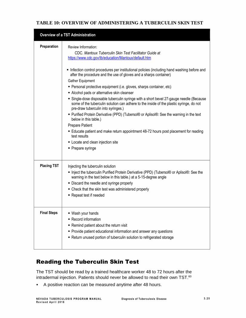
TABLE 10: OVERVIEW OF ADMINISTERING A TUBERCULIN SKIN TEST
Reading the Tuberculin Skin Test
The TST should be read by a trained healthcare worker 48 to 72 hours after the
intradermal injection. Patients should never be allowed to read their own TST.60
▪ A positive reaction can be measured anytime after 48 hours.
Overview of a TST Administration
Preparation
Review Information:
CDC. Mantoux Tuberculin Skin Test Facilitator Guide at https://www.cdc.gov/tb/education/Mantoux/default.htm
▪ Infection control procedures per institutional policies (including hand washing before and after the procedure and the use of gloves and a sharps container)
Gather Equipment
▪ Personal protective equipment (i.e. gloves, sharps container, etc)
▪ Alcohol pads or alternative skin cleanser
▪ Single-dose disposable tuberculin syringe with a short bevel 27-gauge needle (Because some of the tuberculin solution can adhere to the inside of the plastic syringe, do not pre-draw tuberculin into syringes.)
▪ Purified Protein Derivative (PPD) (Tubersol® or Aplisol®: See the warning in the text below in this table.)
Prepare Patient
▪ Educate patient and make return appointment 48-72 hours post placement for reading test results
▪ Locate and clean injection site
▪ Prepare syringe
Placing TST Injecting the tuberculin solution
▪ Inject the tuberculin Purified Protein Derivative (PPD) (Tubersol® or Aplisol®: See the warning in the text below in this table.) at a 5-15-degree angle
▪ Discard the needle and syringe properly
▪ Check that the skin test was administered properly
▪ Repeat test if needed
Final Steps ▪ Wash your hands
▪ Record information
▪ Remind patient about the return visit
▪ Provide patient educational information and answer any questions
▪ Return unused portion of tuberculin solution to refrigerated storage
N E V A D A T U B E R C U L O S I S P R O G R A M M A N U A L Diagnosis of Tuberculosis Disease 3 . 2 6
R e v i s e d A p r i l 2 0 1 8
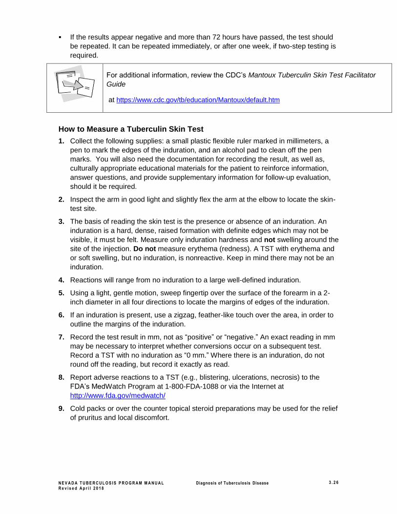
▪ If the results appear negative and more than 72 hours have passed, the test should
be repeated. It can be repeated immediately, or after one week, if two-step testing is
required.
For additional information, review the CDC’s Mantoux Tuberculin Skin Test Facilitator
Guide
at https://www.cdc.gov/tb/education/Mantoux/default.htm
How to Measure a Tuberculin Skin Test
1. Collect the following supplies: a small plastic flexible ruler marked in millimeters, a
pen to mark the edges of the induration, and an alcohol pad to clean off the pen
marks. You will also need the documentation for recording the result, as well as,
culturally appropriate educational materials for the patient to reinforce information,
answer questions, and provide supplementary information for follow-up evaluation,
should it be required.
2. Inspect the arm in good light and slightly flex the arm at the elbow to locate the skin-
test site.
3. The basis of reading the skin test is the presence or absence of an induration. An
induration is a hard, dense, raised formation with definite edges which may not be
visible, it must be felt. Measure only induration hardness and not swelling around the
site of the injection. Do not measure erythema (redness). A TST with erythema and
or soft swelling, but no induration, is nonreactive. Keep in mind there may not be an
induration.
4. Reactions will range from no induration to a large well-defined induration.
5. Using a light, gentle motion, sweep fingertip over the surface of the forearm in a 2-
inch diameter in all four directions to locate the margins of edges of the induration.
6. If an induration is present, use a zigzag, feather-like touch over the area, in order to
outline the margins of the induration.
7. Record the test result in mm, not as “positive” or “negative.” An exact reading in mm
may be necessary to interpret whether conversions occur on a subsequent test.
Record a TST with no induration as “0 mm.” Where there is an induration, do not
round off the reading, but record it exactly as read.
8. Report adverse reactions to a TST (e.g., blistering, ulcerations, necrosis) to the
FDA’s MedWatch Program at 1-800-FDA-1088 or via the Internet at
http://www.fda.gov/medwatch/
9. Cold packs or over the counter topical steroid preparations may be used for the relief
of pruritus and local discomfort.
N E V A D A T U B E R C U L O S I S P R O G R A M M A N U A L Diagnosis of Tuberculosis Disease 3 . 2 7
R e v i s e d A p r i l 2 0 1 8
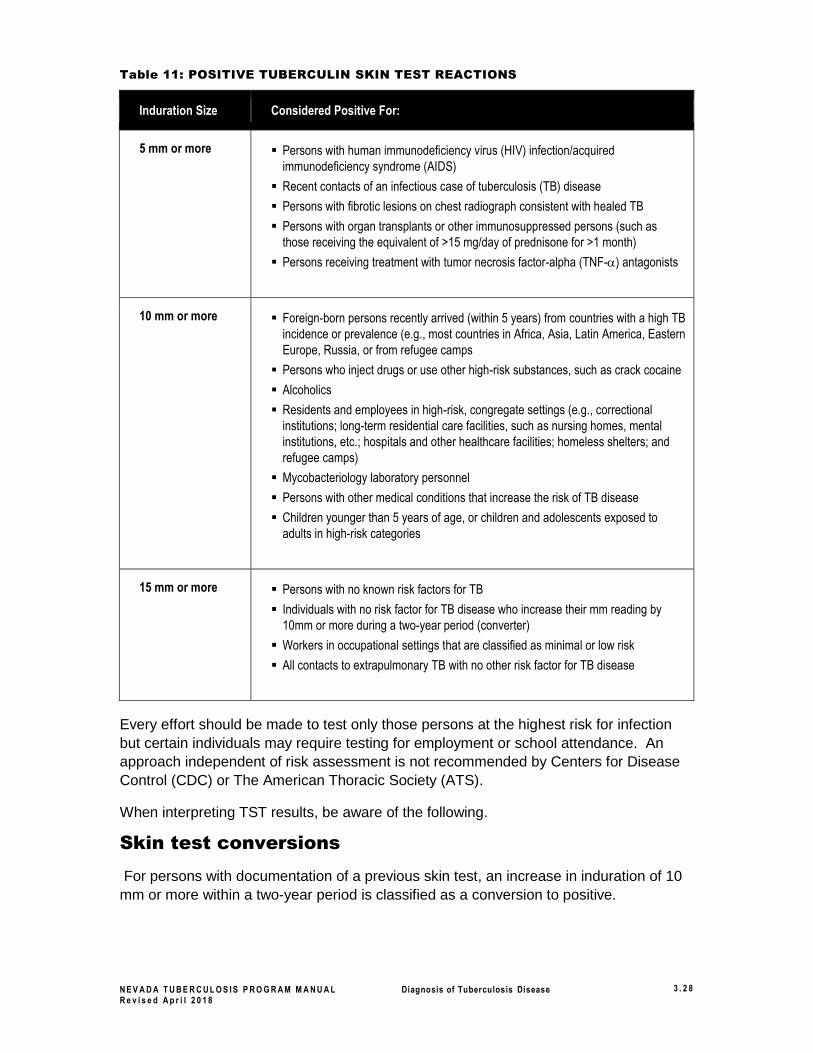
Interpretation of the Tuberculin Skin Test
TST's should be interpreted by a trained healthcare worker. Use Table 11 below to
interpret TST's.
Call your regional TB Control Program or the Nevada DPBH Tuberculosis
Program, (contact information is located in the Introduction, Chapter 1)
regarding TST reactions when interpretation and/or medical follow-up are
unclear.
Before interpreting a TST, information can be reviewed in the CDC’s Mantoux
Tuberculin
Skin Test Facilitator Guide at
https://www.cdc.gov/tb/education/Mantoux/default.htm.
How to Interpret a Tuberculin Skin Test
Based on the sensitivity and specificity of the Purified Protein Derivative (PPD), the
patients’ immune response capabilities, and the prevalence of TB in different groups,
three cut-points have been recommended for defining a positive tuberculin reaction:
▪ Greater than or equal to 5 mm
▪ Greater than or equal to 10 mm
▪ Greater than or equal to 15 mm of induration
Use Table 11: Positive Tuberculin Skin Test Reactions below, to determine what size
induration cut-point to use to interpret the TST measurement and ultimately decide
whether the test is positive or negative.
N E V A D A T U B E R C U L O S I S P R O G R A M M A N U A L Diagnosis of Tuberculosis Disease 3 . 2 8
R e v i s e d A p r i l 2 0 1 8
Table 11: POSITIVE TUBERCULIN SKIN TEST REACTIONS
Induration Size Considered Positive For:
5 mm or more ▪ Persons with human immunodeficiency virus (HIV) infection/acquired
immunodeficiency syndrome (AIDS)
▪ Recent contacts of an infectious case of tuberculosis (TB) disease
▪ Persons with fibrotic lesions on chest radiograph consistent with healed TB
▪ Persons with organ transplants or other immunosuppressed persons (such as
those receiving the equivalent of >15 mg/day of prednisone for >1 month)
▪ Persons receiving treatment with tumor necrosis factor-alpha (TNF-) antagonists
10 mm or more ▪ Foreign-born persons recently arrived (within 5 years) from countries with a high TB
incidence or prevalence (e.g., most countries in Africa, Asia, Latin America, Eastern
Europe, Russia, or from refugee camps
▪ Persons who inject drugs or use other high-risk substances, such as crack cocaine
▪ Alcoholics
▪ Residents and employees in high-risk, congregate settings (e.g., correctional
institutions; long-term residential care facilities, such as nursing homes, mental
institutions, etc.; hospitals and other healthcare facilities; homeless shelters; and
refugee camps)
▪ Mycobacteriology laboratory personnel
▪ Persons with other medical conditions that increase the risk of TB disease
▪ Children younger than 5 years of age, or children and adolescents exposed to
adults in high-risk categories
15 mm or more ▪ Persons with no known risk factors for TB
▪ Individuals with no risk factor for TB disease who increase their mm reading by
10mm or more during a two-year period (converter)
▪ Workers in occupational settings that are classified as minimal or low risk
▪ All contacts to extrapulmonary TB with no other risk factor for TB disease
Every effort should be made to test only those persons at the highest risk for infection
but certain individuals may require testing for employment or school attendance. An
approach independent of risk assessment is not recommended by Centers for Disease
Control (CDC) or The American Thoracic Society (ATS).
When interpreting TST results, be aware of the following.
Skin test conversions
For persons with documentation of a previous skin test, an increase in induration of 10
mm or more within a two-year period is classified as a conversion to positive.

N E V A D A T U B E R C U L O S I S P R O G R A M M A N U A L Diagnosis of Tuberculosis Disease 3 . 2 9
R e v i s e d A p r i l 2 0 1 8
False-Negative Reactions
False-negative reactions may be due to the following:
▪ Anergy
See “Anergy Testing” under “Candidates for Mantoux Tuberculin Skin Testing”
in this section.
▪ Recent TB infection (within the past 10 weeks)
▪ Very young age (less than 6 months of age, because the immune system is not fully
developed)
▪ Overwhelming TB disease
▪ Vaccination with live viruses (e.g., measles, mumps, rubella, varicella, smallpox, oral
polio, or yellow fever and live attenuated influenza vaccines).
TB skin testing should be done either on the same day as vaccination with live
virus or at least four weeks after vaccination.
▪ Some viral infections (measles, mumps, chickenpox, or HIV)
▪ Corticosteroids or other immunosuppressive agents given for two or more weeks
False-Positive Reactions
False-positive reactions may be due to the following:
▪ Nontuberculous mycobacteria (NTM) or mycobacterium other than tuberculosis
(MOTT)
▪ BCG vaccination
See “Bacille Calmette-Guérin Vaccine” under “Candidates for Mantoux
Tuberculin Skin Testing” in this section.

N E V A D A T U B E R C U L O S I S P R O G R A M M A N U A L Diagnosis of Tuberculosis Disease 3 . 3 0
R e v i s e d A p r i l 2 0 1 8
Persons at Risk for Progressing from Latent TB Infection (LTBI) to TB Disease 3
Every effort should be made to test only those persons at risk, interpret tuberculin skin
test (TST) reactions accurately, ensure appropriate treatment, and completion of the
recommended treatment regimen.
Generally, persons at risk for developing TB disease fall into two categories: those who
have been recently infected and those with clinical conditions that increase the risk of
progression from LTBI to TB disease.
Suspect recent infection in the following:
• Close contacts of a person with infectious TB
• Persons who have immigrated from areas of the world with high rates of TB
• Children < 5 years of age who have a positive TST result
• Recent converters (those with an increase of 10 mm or more in size of TST
reaction within a 2-year period)
• Groups with high rates of M. tuberculosis transmission, such as homeless
persons, injection drug users, and persons with HIV infection
• Persons who work or reside with people who are at high risk for TB in
facilities or institutions such as hospitals, homeless shelters, correctional
facilities, nursing homes, and residential homes for those with HIV
Clinical conditions that have been reported to increase the risk of progression
from LTBI to TB disease:
• HIV infection
• Radiographic evidence of prior TB
• Low body weight (> 10% below ideal)
• Silicosis
• Diabetes mellitus
• Chronic malabsorbtion syndrome
• Chronic renal failure or being on hemodialysis
• Gastrectomy or Jejunoileal bypass
• Leukemia, lymphomas, Hodgkin’s disease
• Solid organ transplant
• Cancer of the head or neck
• Prolonged use of immunosuppressive agents (e.g., prednisone, TNF-α
antagonists)
3 https://www.cdc.gov/tb/publications/factsheets/testing/skintestresults.htm
N E V A D A T U B E R C U L O S I S P R O G R A M M A N U A L Diagnosis of Tuberculosis Disease 3 . 3 1
R e v i s e d A p r i l 2 0 1 8
Evaluating persons with positive skin tests See section 3.9, Diagnosis of Tuberculosis Disease, of this Chapter, for more details
Sections:
Medical history ................................................... 3.10
Physical examination ......................................... 3.13
Human immunodeficiency virus screening ........ 3.13
About the Tuberculin skin test and
interferon gamma release assays ...................... 3.13
Chest radiography .............................................. 3.14
Bacteriologic examination .................................. 3.15
N E V A D A T U B E R C U L O S I S P R O G R A M M A N U A L Diagnosis of Tuberculosis Disease 3 . 3 2
R e v i s e d A p r i l 2 0 1 8
Resources and References
Resources
▪ ATS, CDC, IDSA. “Diagnostic Standards and Classification of Tuberculosis in Adults
and Children” (Am J Respir Crit Care Med 2000;161[4 Pt 1]). Available at:
https://www.cdc.gov/tb/publications/PDF/1376.pdf
▪ CDC. Self-Study Modules on Tuberculosis (Division of Tuberculosis Elimination Web
site; 1999). Available at: https://www.cdc.gov/tb/education/ssmodules/default.htm .
▪ CDC. Core Curriculum on Tuberculosis (2000) (Division of Tuberculosis Elimination Web
site; updated November 2001). Available at:
https://www.cdc.gov/tb/education/corecurr/index.htm.
▪ Tenover, R., et al. “The Resurgence of Tuberculosis: Is Your Laboratory Ready?” (Journal of
Clinical Microbiology 1993:767–770).
References
1 ATS, CDC, IDSA. Controlling tuberculosis in the United States: recommendations from the American Thoracic Society,
CDC, and the Infectious Diseases Society of America. MMWR 2005;54(No. RR-12):15. 2 ATS, CDC, IDSA. Controlling tuberculosis in the United States: recommendations from the American Thoracic Society,
CDC, and the Infectious Diseases Society of America. MMWR 2005;54(No. RR-12):32. 3 ATS, CDC, IDSA. Controlling tuberculosis in the United States: recommendations from the American Thoracic Society,
CDC, and the Infectious Diseases Society of America. MMWR 2005;54(No. RR-12):32. 4 ATS, CDC, IDSA. Controlling tuberculosis in the United States: recommendations from the American Thoracic Society,
CDC, and the Infectious Diseases Society of America. MMWR 2005;54(No. RR-12):15. 5 ATS, CDC, IDSA. Controlling tuberculosis in the United States: recommendations from the American Thoracic Society,
CDC, and the Infectious Diseases Society of America. MMWR 2005;54(No. RR-12):15–16. 6 ATS, CDC, IDSA. Controlling tuberculosis in the United States: recommendations from the American Thoracic Society,
CDC, and the Infectious Diseases Society of America. MMWR 2005;54(No. RR-12):32. 7 CDC. Guidelines for preventing the transmission of Mycobacterium tuberculosis in health-care settings, 2005. MMWR
2005;54(No. RR-17):4–5; CDC. Targeted tuberculin testing and treatment of latent tuberculosis infection. MMWR
2000;49(No. RR-6):7–9, 22. 8 CDC. Targeted tuberculin testing and treatment of latent tuberculosis infection. MMWR 2000;49(No. RR-6):8–9. 9 CDC. Classification system. In: Chapter 2: transmission and pathogenesis. Core Curriculum on Tuberculosis (2000)
[Division of Tuberculosis Elimination Web site]. Updated November 2001. Available at:
http://www.cdc.gov/tb/pubs/corecurr/default.htm . Accessed July 3, 2006. 10 ATS, CDC, IDSA. Controlling tuberculosis in the United States: recommendations from the American Thoracic Society,
CDC, and the Infectious Diseases Society of America. MMWR 2005;54(No. RR-12):32. 11 ATS, CDC, IDSA. Controlling tuberculosis in the United States: recommendations from the American Thoracic Society,
CDC, and the Infectious Diseases Society of America. MMWR 2005;54(No. RR-12):32. 12 ATS, CDC, IDSA. Controlling tuberculosis in the United States: recommendations from the American Thoracic Society,
CDC, and the Infectious Diseases Society of America. MMWR 2005;54(No. RR-12):34. 13 ATS, CDC, IDSA. Controlling tuberculosis in the United States: recommendations from the American Thoracic Society,
CDC, and the Infectious Diseases Society of America. MMWR 2005;54(No. RR-12):33. 14 ATS, CDC, IDSA. Controlling tuberculosis in the United States: recommendations from the American Thoracic Society,
CDC, and the Infectious Diseases Society of America. MMWR 2005;54(No. RR-12):33. 15 ATS, CDC, IDSA. Controlling tuberculosis in the United States: recommendations from the American Thoracic Society,
CDC, and the Infectious Diseases Society of America., MMWR 2005;54(No. RR-12):33; CDC. Medical evaluation. In:
Chapter 5: diagnosis of TB. Core Curriculum on Tuberculosis (2000) [Division of Tuberculosis Elimination Web site].
Updated November 2001. Available at: http://www.cdc.gov/tb/pubs/corecurr/default.htm . Accessed July 3, 2006; CDC.
Guidelines for preventing the transmission of Mycobacterium tuberculosis in health-care settings, 2005. MMWR
2005;54(No. RR-17):11; ATS, CDC, IDSA. Diagnostic standards and classification of tuberculosis in adults and children.
Am J Respir Crit Care Med. 2000; 161:1378; CDC. Module 3: diagnosis of tuberculosis infection and disease. Self-
N E V A D A T U B E R C U L O S I S P R O G R A M M A N U A L Diagnosis of Tuberculosis Disease 3 . 3 3
R e v i s e d A p r i l 2 0 1 8
Study Modules on Tuberculosis [Division of Tuberculosis Elimination Web site]. 1999:3. Available at:
http://www.cdc.gov/tb/pubs/ssmodules/default.htm . Accessed July 3, 2006. 16 ATS, CDC, IDSA. Controlling tuberculosis in the United States: recommendations from the American Thoracic Society,
CDC, and the Infectious Diseases Society of America. MMWR 2005;54(No. RR-12):33. 17 ATS, CDC, IDSA. Controlling tuberculosis in the United States: recommendations from the American Thoracic Society,
CDC, and the Infectious Diseases Society of America. MMWR 2005;54(No. RR-12):33. 18 CDC. Medical evaluation. In: Chapter 5: diagnosis of TB. Core Curriculum on Tuberculosis (2000) [Division of
Tuberculosis Elimination Web site]. Updated November 2001. Available at:
http://www.cdc.gov/tb/pubs/corecurr/default.htm . Accessed July 3, 2006; and CDC. Guidelines for preventing the
transmission of Mycobacterium tuberculosis in health-care settings, 2005. MMWR 2005;54(No. RR-17):11. 19 CDC. Medical evaluation. In: Chapter 5: diagnosis of TB. Core Curriculum on Tuberculosis (2000) [Division of
Tuberculosis Elimination Web site]. Updated November 2001. Available at:
http://www.cdc.gov/tb/pubs/corecurr/default.htm . Accessed July 3, 2006; and CDC. Guidelines for preventing the
transmission of Mycobacterium tuberculosis in health-care settings, 2005. MMWR 2005;54(No. RR-17):11. 20 ATS, CDC, IDSA. Diagnostic standards and classification of tuberculosis in adults and children. Am J Respir Crit Care
Med. 2000; 161:1378; CDC. Guidelines for preventing the transmission of Mycobacterium tuberculosis in health-care
settings, 2005. MMWR 2005;54(No. RR-17):11; and CDC. Medical evaluation. In: Chapter 5: diagnosis of TB. Core
Curriculum on Tuberculosis (2000) [Division of Tuberculosis Elimination Web site]. Updated November 2001. Available
at: http://www.cdc.gov/tb/pubs/corecurr/default.htm . Accessed July 3, 2006. 21 CDC. Guidelines for preventing the transmission of Mycobacterium tuberculosis in health-care settings, 2005. MMWR
2005;54(No. RR-17):11; and ATS, CDC, IDSA. Diagnostic standards and classification of tuberculosis in adults and
children. Am J Respir Crit Care Med. 2000; 161:1378. 22 CDC. Module 3: diagnosis of tuberculosis infection and disease. Self-Study Modules on Tuberculosis [Division of
Tuberculosis Elimination Web site]. 1999:3. Available at: http://www.cdc.gov/tb/pubs/ssmodules/default.htm . Accessed
July 3, 2006; and CDC. Guidelines for preventing the transmission of Mycobacterium tuberculosis in health-care
settings, 2005. MMWR 2005;54(No. RR-17):11. 23 ATS, CDC, IDSA. Controlling tuberculosis in the United States: recommendations from the American Thoracic Society,
CDC, and the Infectious Diseases Society of America. MMWR 2005;54(No. RR-12):33. 24 CDC. Diagnostic microbiology. In: Chapter 5: diagnosis of TB. Core Curriculum on Tuberculosis (2000) [Division of
Tuberculosis Elimination Web site]. Updated November 2001. Available at:
http://www.cdc.gov/tb/pubs/corecurr/default.htm . Accessed July 3, 2006. 25 ATS, CDC, IDSA. Controlling tuberculosis in the United States: recommendations from the American Thoracic Society,
CDC, and the Infectious Diseases Society of America. MMWR 2005;54(No. RR-12):33. 26 ATS, CDC, IDSA. Controlling tuberculosis in the United States: recommendations from the American Thoracic Society,
CDC, and the Infectious Diseases Society of America. MMWR 2005;54(No. RR-12):33. 27 Washington State Public Laboratory Tuberculosis Unit. Internal untitled report on the review, analysis, and
recommendations on the Gen-Probe Amplified Mycobacterium Tuberculosis Direct Test (MTD). January 2004. The
report includes the following references: (1) Gen-Probe Incorporated. Amplified Mycobacterium Tuberculosis Direct Test
Package Insert. Gen-Probe Incorporated, San Diego, CA, 2001; (2) ATS, CDC, IDSA. Diagnostic Standards and
Classification of tuberculosis in Adults and Children. American Thoracic Society, 1999; (3) Piersimoni, C. and Scarparo,
C. Relevance of commercial amplification methods for direct detection of Mycobacterium Tuberculosis Complex in
clinical samples. Journal of Clin. Micro., December 2003: 5355-5365; (4) Centers for Disease Control and Prevention.
Update: Nucleic acid amplification tests for tuberculosis. MMWR, 2000; 49:593-594; (5) Schluger, N.W. Changing
approaches to the diagnosis of tuberculosis. Am. J. of Resp. Crit. Care Med., 2001; 164:2020; (6) Catanzaro et al. The
role of Clinical suspicion in evaluation as a new diagnostic test for active tuberculosis. JAMA, Feb. 2, 2000; Vol. 283 No.
5 P.639. 28 Daley CL, Gotway MB, Jasmer RM. Radiographic manifestations of tuberculosis: a primer for clinicians. San Francisco,
CA: Francis J. Curry National Tuberculosis Center; 2003:1–30. 29 CDC. “The medical history.” In: Module 3: diagnosis of TB infection and disease Self-Study Modules on Tuberculosis
[Division of Tuberculosis Elimination Web site]. 1999:12. Available at:
http://www.cdc.gov/tb/pubs/ssmodules/default.htm . Accessed July 3, 2006. 30 CDC. Medical evaluation. In: Chapter 5: diagnosis of TB. Core Curriculum on Tuberculosis (2000). Updated November
2001; and CDC. Guidelines for preventing the transmission of Mycobacterium tuberculosis in health-care settings, 2005.
MMWR 2005;54(No. RR-17):11. 31 ATS, CDC, IDSA. Diagnostic standards and classification of tuberculosis in adults and children. Am J Respir Crit Care
Med 2000; 161:1378; CDC. Guidelines for preventing the transmission of Mycobacterium tuberculosis in health-care
settings, 2005. MMWR 2005;54(No. RR-17):11; and CDC. “Medical evaluation.” In: Chapter 5: diagnosis of TB. Core
Curriculum on Tuberculosis (2000). Updated November 2001. 32 CDC. Guidelines for preventing the transmission of Mycobacterium tuberculosis in health-care settings, 2005. MMWR
2005;54(No. RR-17):11; and ATS, CDC, IDSA. Diagnostic standards and classification of tuberculosis in adults and
children. Am J Respir Crit Care Med 2000; 161:1378. 33 CDC. Module 3: diagnosis of TB tuberculosis infection and disease. Self-Study Modules on Tuberculosis [Division of
Tuberculosis Elimination Web site]. 1999:3. Available at: http://www.cdc.gov/tb/pubs/ssmodules/default.htm . Accessed
N E V A D A T U B E R C U L O S I S P R O G R A M M A N U A L Diagnosis of Tuberculosis Disease 3 . 3 4
R e v i s e d A p r i l 2 0 1 8
July 3, 2006; and CDC. Guidelines for preventing the transmission of Mycobacterium tuberculosis in health-care
settings, 2005. MMWR 2005;54(No. RR-17):11. 34 CDC. The medical history. In: Module 3: diagnosis of TB infection and disease. Self-Study Modules on Tuberculosis
[Division of Tuberculosis Elimination Web site]. 1999:12. Available at:
http://www.cdc.gov/tb/pubs/ssmodules/default.htm . Accessed July 3, 2006. 35 Newly Diagnosed TB Patients at Risk of Fluoroquinolone Resistance, Peggy Peck, Accessed July 1, 2009
http://www.medscape.com/viewarticle/456061 36 CDC. Medical evaluation. In: Chapter 5: diagnosis of TB. Core Curriculum on Tuberculosis (2000) [Division of
Tuberculosis Elimination Web site]. Updated November 2001. Available at:
http://www.cdc.gov/tb/pubs/corecurr/default.htm . Accessed July 3, 2006; and Colorado Department of Public Health
and Environment. Tuberculosis Manual [Colorado Department of Public Health and Environment Web site]. (2004):3-1.
Available at: http://www.cdphe.state.co.us/dc/TB/tbman.html . Accessed November 1, 2006. 37 ATS, CDC, IDSA. Controlling tuberculosis in the United States: recommendations from the American Thoracic Society,
CDC, and the Infectious Diseases Society of America. MMWR 2005;54(No. RR-12):51. 38 CDC. Revised Recommendations for HIV Testing of Adults, Adolescents, and Pregnant Women in Health-Care
Settings. MMWR 2006;55(No. RR-14):1–17. 39 CDC. Guidelines for preventing the transmission of Mycobacterium tuberculosis in health-care settings, 2005. MMWR
2005;54(No. RR-17):4. 40 CDC. Guidelines for using the QuantiFERON-TB Gold test for detecting Mycobacterium tuberculosis infection, United
States. MMWR 2005;54(No RR-15):52. 41 CDC. Guidelines for using the QuantiFERON-TB Gold test for detecting Mycobacterium tuberculosis infection, United
States. MMWR 2005;54(No RR-15):50. 42 CDC. Guidelines for using the QuantiFERON®-TB Gold test for detecting Mycobacterium tuberculosis infection, United
States. MMWR. 2005; 54 (No. RR-15):52. 43 CDC. Guidelines for using the QuantiFERON®-TB Gold test for detecting Mycobacterium tuberculosis infection, United
States. MMWR. 2005; 54 (No. RR-15):52. 44 ATS, CDC, IDSA. Treatment of tuberculosis. MMWR. 2003;52(No. RR-11):3. 45 CDC. Module 3: diagnosis of TB infection and disease. Self-Study Modules on Tuberculosis [Division of Tuberculosis
Elimination Web site]. 1999:13. Available at: http://www.cdc.gov/tb/pubs/ssmodules/default.htm . Accessed July 3,
2006. 46 CDC. Guidelines for using the QuantiFERON-TB Gold test for detecting Mycobacterium tuberculosis infection, United
States. MMWR. 2005;54(No. RR-15):52. 47 CDC. Targeted tuberculin testing and treatment of latent tuberculosis infection. MMWR 2000;49(No. RR-6):25. 48 CDC. Medical evaluation. In: Chapter 5: diagnosis of TB. Core Curriculum on Tuberculosis (2000) [Division of
Tuberculosis Elimination Web site]. Updated November 2001. Available at:
http://www.cdc.gov/tb/pubs/corecurr/default.htm . Accessed July 3, 2006. 49 ATS, CDC, IDSA. Controlling tuberculosis in the United States: recommendations from the American Thoracic Society,
CDC, and the Infectious Diseases Society of America. MMWR 2005;54(No. RR-12):19; and Tenover, R., et al. The
resurgence of tuberculosis: is your laboratory ready? Journal of Clinical Microbiology 1993:767–770. 50 ATS, CDC, IDSA. Controlling tuberculosis in the United States: recommendations from the American Thoracic Society,
CDC, and the Infectious Diseases Society of America. MMWR 2005;54(No. RR-12):19; and Tenover, R., et al. The
resurgence of tuberculosis: is your laboratory ready? Journal of Clinical Microbiology 1993:767–770. 51 ATS, CDC, IDSA. Controlling tuberculosis in the United States: recommendations from the American Thoracic Society,
CDC, and the Infectious Diseases Society of America. MMWR 2005;54(No. RR-12):19. 52 ATS, CDC, IDSA. Diagnostic standards and classification of tuberculosis in adults and children. Am J Respir Crit Care
Med 2000; 161:1384. 53 ATS, CDC, IDSA. Controlling tuberculosis in the United States: recommendations from the American Thoracic Society,
CDC, and the Infectious Diseases Society of America. MMWR 2005;54(No. RR-12):19; and Tenover, R., et al. The
resurgence of tuberculosis: is your laboratory ready? Journal of Clinical Microbiology 1993:767–770. 54 CDC. National plan for reliable tuberculosis laboratory services using a systems approach–recommendations from CDC
and the Association of Public Health Laboratories Task Force on Tuberculosis Laboratory Services. MMWR
2005;54(No. RR-6):3. 55 ATS, CDC, IDSA. Controlling tuberculosis in the United States: recommendations from the American Thoracic Society,
CDC, and the Infectious Diseases Society of America. MMWR 2005;54(No. RR-12):19; and Tenover, R., et al. The
resurgence of tuberculosis: is your laboratory ready? Journal of Clinical Microbiology 1993:767–770. 56 CDC. National plan for reliable tuberculosis laboratory services using a systems approach–recommendations from CDC
and the Association of Public Health Laboratories Task Force on Tuberculosis Laboratory Services. MMWR
2005;54(No. RR-6):2. 57 Tenover, R., et al. The resurgence of tuberculosis: is your laboratory ready? Journal of Clinical Microbiology 1993:769;
and ATS, CDC, IDSA. Treatment of tuberculosis. MMWR 2003;52(No. RR-11):38.

N E V A D A T U B E R C U L O S I S P R O G R A M M A N U A L Diagnosis of Tuberculosis Disease 3 . 3 5
R e v i s e d A p r i l 2 0 1 8
58 ATS, CDC, IDSA. Treatment of tuberculosis. MMWR 2003;52(No. RR-11):12. 59 CDC. Diagnostic microbiology. In: Chapter 5: diagnosis of TB. Core Curriculum on Tuberculosis (2000) [Division of
Tuberculosis Elimination Web site]. Updated November 2001. Available at:
http://www.cdc.gov/tb/pubs/corecurr/default.htm . Accessed July 3, 2006.