Embed Size (px)
Citation preview

Chapter 28: Complex Regional Pain Syndrome
Mark Walsh PT, DPT, MS, CHT
Test Prep for the CHT Exam, 3rd Edition 1
Chapter 26: Complex Regional Pain Syndrome Mark Walsh PT, DPT, MS, CHT
I. Introduction/Definition: no one accepted definition
A. CRPS is a syndrome characterized by severe pain (out of proportion), swelling, stiffness, discoloration and decreased function. It can involve an entire extremity or a single finger.[1]
B. As established by the International Association for the Study of Pain in 1994, the
current recommended nomenclature is Chronic Regional Pain Syndrome (CRPS) Type I and Type II. Type I develops after an initiating noxious event, and Type II develops after a nerve injury. Complex regional pain syndrome Type I corresponds to Reflex Sympathetic Dystrophy (RSD) in that it does not have an identifiable nerve lesion. Type II designates causalgia, which presents similarly but occurs with injury to the peripheral nerve or its branch. Within the CRPS categories pain is regarded as sympathetically independent pain (SIP) occurring in the initial onset of the syndrome. Sympathetically maintained pain (SMP) is defined as "a symptom of CRPS and not a clinical entity” occurring after a period of time of the onset of the syndrome[2]. CRPS has strict inclusion criteria that do not encompass the presence or absence of SMP[3]. The international Association for the Study of Pain diagnostic criteria for CRPS are listed in the box below[4]. Also, the pain can be described as sympathetically maintained (SMP) or sympathetically independent (SIP) based on the response to treatment to the sympathetic system. (Figure 1A-C)
IASP DIAGNOSTIC CRITERIA
1. Presence of an inciting noxious event or a cause of immobilization.
2. Continuing pain, allodynia, or hyperalgesia with which pain is disproportionate to any inciting event.
3. Evidence at some time of edema, changes in skin blood flow or abnormal sudomotor activity in the region of pain.
4. This diagnosis is excluded by the existence of condition that would otherwise account for the degree of pain and dysfunction.
American Society of Hand TherapistsTM2
Chapter 28: Complex Regional Pain SyndromeMark Walsh PT, DPT, MS, CHT
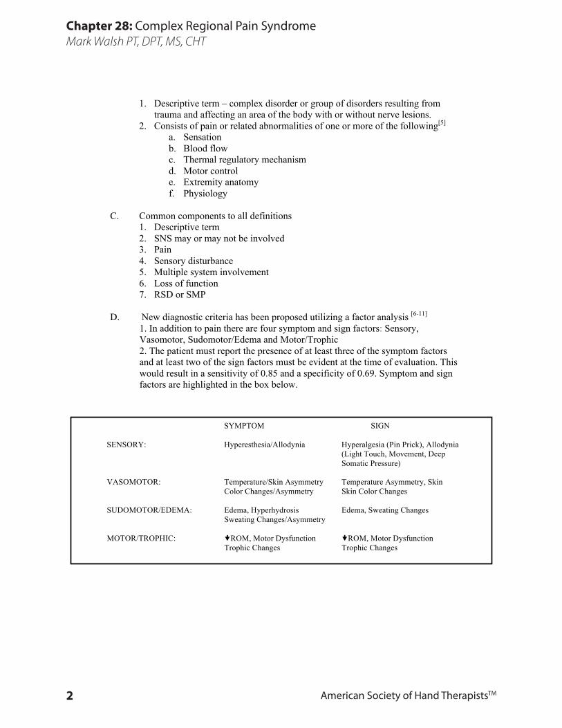
1. Descriptive term – complex disorder or group of disorders resulting from trauma and affecting an area of the body with or without nerve lesions.
2. Consists of pain or related abnormalities of one or more of the following[5] a. Sensation b. Blood flow c. Thermal regulatory mechanism d. Motor control e. Extremity anatomy f. Physiology
C. Common components to all definitions 1. Descriptive term 2. SNS may or may not be involved 3. Pain 4. Sensory disturbance 5. Multiple system involvement 6. Loss of function 7. RSD or SMP
D. New diagnostic criteria has been proposed utilizing a factor analysis [6-11] 1. In addition to pain there are four symptom and sign factors: Sensory, Vasomotor, Sudomotor/Edema and Motor/Trophic 2. The patient must report the presence of at least three of the symptom factors and at least two of the sign factors must be evident at the time of evaluation. This would result in a sensitivity of 0.85 and a specificity of 0.69. Symptom and sign factors are highlighted in the box below.
SYMPTOM SIGN
SENSORY: Hyperesthesia/Allodynia Hyperalgesia (Pin Prick), Allodynia (Light Touch, Movement, Deep Somatic Pressure) VASOMOTOR: Temperature/Skin Asymmetry Temperature Asymmetry, Skin Color Changes/Asymmetry Skin Color Changes SUDOMOTOR/EDEMA: Edema, Hyperhydrosis Edema, Sweating Changes
Sweating Changes/Asymmetry
MOTOR/TROPHIC: ROM, Motor Dysfunction ROM, Motor Dysfunction Trophic Changes Trophic Changes

Test Prep for the CHT Exam, 3rd Edition 3
Chapter 28: Complex Regional Pain SyndromeMark Walsh PT, DPT, MS, CHT
II. Proposed Mechanisms: no one accepted or proven mechanism[10]
A. Peripheral 1. Abnormal AV Shunting – SNS 2. Damage Peripheral Nerve 3. Local Demyelination – Sprouting Nociceptor Sensitivity 4. Vasomotor Tone - Nociceptor Activity 5. Sensory Afferent Impulses 6. Abnormal SNS Activity
B. Central
1. Internuncial Pool Abnormalities 2. Activity Substantia Gelatinosa (SG) 3. Abnormal Modulation WDR Neurons 4. Abnormal Modulation Afferents CNS
C. Summary
1. Above is only partial list of proposed central and/or peripheral mechanisms of pain mediation in RSD
2. Etiology remains in question 3. SNS involvement unanswered question – no known relationship to cause and
effect 4. Pain may or may not be related to the sympathetic nervous system 5. Syndrome as a whole is poorly understood
III. Primary Characteristics/Presentation
A. Pain 1. Persistent painful lesion disproportionate to the initial injury 2. Described as burning, sharp, stabbing, searing 3. Pain may be absent in 25% of cases 4. Allodynia: pain from non-painful stimulus 5. Hyperalgesia: increased pain from a known noxious stimulus 6. Hyperpathia: prolonged pain after removal of a known noxious stimulus 7. Pain may initially be in expected nerve distribution area of injury but
progresses to involve other areas 8. Pain that radiates proximally
B. Pain Terms Associated with Neuropathic Pain[12] 1. Allodynia Nonpainful Stimulus Provokes Pain 2. Hyperalgesia Increased Response to Painful Stimulus 3. Hyperpathia Increased Response to Painful Stimulus
Continues After It Is Withdrawn 4. Dysesthesia Unpleasant Abnormal Sensation
(Spontaneous or Evoked)

American Society of Hand TherapistsTM4
Chapter 28: Complex Regional Pain SyndromeMark Walsh PT, DPT, MS, CHT
5. Synchiria Stimulation Unaffected Limb Evokes Symptoms in Both Limbs
6. Dysynchiria Stimulation Unaffected Limb Evokes Symptoms in the Affected Limb
C. Factor 1: Sensory[4, 6, 8] 1. Reported Hypersensitivity to tactile stimulation anywhere from light to strong (light touch, movement, pressure etc.) 2. Hyperesthesia/Allodynia often a central area and peripheral area with decreasing sensitivity 3. Dysesthesia 4. Sensory disturbance may be intermittent and spontaneous
B. Factor 2: Vasomotor changes [4, 6, 8] (Figure 2) 1. Vasoconstriction: dusky, pallor, cool- common patient complaint 2. Vasodilatation: redness, shinny skin, warm to hot –especially in the
hypothenar eminence, dorsal MP’s and PIP’s 3. Varies in presentation; can see mixture of both even in same therapy visit 4. Vasomotor spasm 5. Prolonged capillary refill 6. Thermal Regulatory Control Dysfunction – cold intolerance
C. Factor 3: Sudomotor/Edema[4, 6, 8] (Figure 3) 1. Often expected following trauma 2. Progresses beyond initial injured tissue 3. Progresses from pitting to brawny edema 4. Chronic in nature – progresses to fibrosis and adhesions limiting motion 5. Hyper/hypohidrosis, Anhidrosis (dry)
D. Factor 4: Motor/Trophic[4, 6, 8] (Figure 4) 1. Stiffness/Limitation of Motion (Active or Passive)
a. Occurs partly due to chronic edema and to decreased motion secondary to pain b. Protective posturing promotes contracture c. Arthrofibrosis
2. Trophic changes a. Abnormal hair growth b. Loss of skin wrinkles; glossy shiny skin due to edema and subcutaneous
atrophy c. Penciling of tips due to atrophy of palmer pads d. Nail changes: ridges, brittle, conform to shape of atrophied pulp
3. Osteoporosis a. Demineralization of bone-commonly seen in metacarpals, phalanges, carpus b. Can be due to increased blood flow – washes out calcium

Test Prep for the CHT Exam, 3rd Edition 5
Chapter 28: Complex Regional Pain SyndromeMark Walsh PT, DPT, MS, CHT
Chapter 28 Figures
Figure 1. (A) Demonstrates a patient with CRPS in a vasodilated state. Note the shiny skin, the erythema about the entire aspect of the wrist and hand and MP extension and PIP flexion contractures. (B) A patient
with CRPS in the vasoconstrictive state. You’ll note that the hand is pale in color and edematous with limited AROM in comparison to the left non-involved side. (C) Isolated CRPS involving a single digit. Note the erythema of the entire distal portion of the middle finger edema and the loss of skin creases. This is
the result of incision and drainage for a felon.
A B C
Figure 2. A patient with CRPS in a mixed vasomotor state. Note comparing the left involved
to the right uninvolved side. The hand is edema-tous with aspects of vasodilatation (redness) over
the PIP MP joints and vasoconstriction (palor) over the dorsum of the metacarpals and phalanges.
Figure 3. An example of the Factor 3 Sudomotor/Edema in CRPS. Hyperhidrosis,
note the beads of perspiration over the palmer aspect of the hand.
(LEFT) Figure 4. Factor IV Motor/Trophic is a patient presenting with focal dystonia 4 years post onset of CRPS. Note the significant flexed posture of the elbow, wrist and digits.

American Society of Hand TherapistsTM6
Chapter 28: Complex Regional Pain SyndromeMark Walsh PT, DPT, MS, CHT
c. Can be due to immobilization 4. Palmer Fasciitis
a. Nodules; thickening of palmer fascia b. Progressive: contributes to flexion contractures
5. Motor Dysfunction a. Tremor b. Myoclonic Movements c. Dystonia e. Bradykinesia
IV. Clinical Types and Stages – No longer utilized[6, 13-15]
A. Types 1. Minor Causalgia
2. Minor Traumatic Dystrophy 3. Major Traumatic Dystrophy
4. Shoulder/Hand Syndrome 5. Major Causalgia
B. Stages: listed here for historical perspective as not used in current research 1. Stage I: average duration 1-3 months 2. Stage II: 3-9 months 3. Stage III: can last longer than 2 years
V. Incidence/Presentation
A. Females greater than males
B. Upper extremity greater than lower extremity
C. After trauma: major or trivial
D. Identifiable etiologic component less than 50%
E. Children and adolescents rare but severe when present
VI. Medical Management[16]
A. Primarily a clinical diagnosis: pain, swelling, stiffness, discoloration
B. Common Diagnostic Tests: Diagnostic testing should initially be geared toward ruling out other conditions. Some tests are used to aid in the diagnosis of CRPS, although they are not always positive.
1. X-Ray- see osteoporosis 2. Thermography
a. Reveals skin temperature changes b. RSD varies in presentation in regards to temperature c. Not specific; many feel has very little prognostic value
3. Triple phase bone scan a. Usefulness remains in question in terms of specificity and sensitivity

Test Prep for the CHT Exam, 3rd Edition 7
Chapter 28: Complex Regional Pain SyndromeMark Walsh PT, DPT, MS, CHT
b. Increased uptake especially in phase III around the carpus, metacarpals and PIP joints (supposed positive findings)
4. Cold stress test a. Looks for abnormal response to cold stress indicative of abnormal
sympathetic function b. Looks at neurovascular response by quantification of thermoregulatory
capacity by measuring digital temperature and blood flow c. May be suggestive of RSD but is not specific
VII. Medical Treatment[16]
Prevention and early recognition/treatment the most effective in resolving disorder.[17] Treatment must use team approach and include treating physician, anesthesia or pain management, therapy, and psychosocial treatment when appropriate. A. Elimination of inciting problem
1. Nerve compression 2. Nerve laceration or neuroma 3. Tight cast 4. Malunion 5. Appropriate pain management
B. Pharmacologic: drugs can be aimed at reducing sympathetic effects either by reducing sympathetic vasoconstrictive action of the peripheral vessels, blockade of the sympathetic nervous system or by vasodilatation of arterioles 1. Corticosteroids or NSAID’s 2. Antidepressant agents- decrease pain, decrease depressive symptoms, cause
sedation to assist with sleeping patterns 3. Anticonvulsants- Neurontin, Topamax, Lamictal, Lyrica 4. Topical analgesics- lidocaine transdermal patches 5. Opiates- rarely effective in Neuropathic Pain
C. Recommend cessation of smoking: smoking stimulates sympathetic nervous system
D. Neurologic blocks: lack of successful relief not necessarily indicative that RSD incorrect diagnosis 1. Somatic nerve blocks
a. Used more commonly for minor types of RSD (minor causalgia)
b. Uses a local anesthetic agent which produces interruption of abnormal sympathetic reflex because somatic nerves do carry sympathetic nerve fibers; prevents increased sympathetic activity from reaching involved area.
c. Median, ulnar, radial nerve block at wrist or at digital level

American Society of Hand TherapistsTM8
Chapter 28: Complex Regional Pain SyndromeMark Walsh PT, DPT, MS, CHT
d. Can be done in clinic initially with lidocaine to see response; follow with Marcaine (longer acting)
e. Can be given more proximally: axillary 2. Periodic perineurial infusion
a. Insert small catheter through which local anesthesia can be administered
b. Often used following a surgical procedure with patients with history of RSD
3. Stellate ganglion blocks a. Aimed at interrupting the abnormal sympathetic reflex b. Blockades all sympathetic efferent impulses into extremity c. Does not produce anesthesia or paralysis as it does not affect
peripheral nerves d. “Successful block” shows warming, drying, more normal color
and often improved motion and less pain e. Should see Horner’s sign: drooping eyelid, conjunctiva vessels
constriction of the pupil and drying/ warming of ipsilateral side of face
f. More effective in early stages g. May require several blocks to see effect h. Historically, has been used to confirm diagnosis and for
treatment 4. Continuous Stellate blocks 5. Sympathectomy: usually removal of first four thoracic sympathetic ganglia;
rare 6. Neuromodulation: is often beneficial if there is persistent pain, chronic in
nature, not relieved by conservative measures a. Includes use of spinal cord stimulation, peripheral nerve
stimulation and intrathecal analgesia to decrease painful symptoms allowing therapy to focus on improvement of motion and function.
VIII. Therapist Evaluation[18]
A. Total System Review; assess or evaluate- be careful to avoid/delay/modify
assessments that increase symptoms 1. Skin: trophic changes, hyper/hypohidrosis 2. Vascular: vasoconstriction, vasodilatation or mixed 3. Neurological
a. Threshold tests – Semmes Weinstein, vibration b. Innervation density: two point discrimination
4. Skeletal: osteoporosis 5. Connective tissue: periarticular thickening, soft tissue fibrosis, palmer nodules 6. Edema: describe nature i.e. brawny, pitting
a. Quantify: Circumference or Volumetrics

Test Prep for the CHT Exam, 3rd Edition 9
Chapter 28: Complex Regional Pain SyndromeMark Walsh PT, DPT, MS, CHT
7. Motion: active and passive- avoid passive testing if patient is painful or extremity is swollen and tender
8. Posturing: dystonia, co-contraction
B. Predictive Value of Symptoms[19, 20] 1. Visual analog scale greater than 3 cm 2. McGill with greater than three words 3. A temperature difference of >0.4°C 4. Volume difference >6. 5%
5. Range of motion limitation >15% could be used as diagnostic indicator of the presence of CRPS
C. Components of Pain/Pain Assessment
1. Descriptive: “fire”, “burning”, “intense stabbing”; nocturnal, hypersensitive, pain with motion[21]
2. Spontaneous pain can be present in 75% of the patients. 3. Pain can be amplified in 93% of the patients by dependency, striking, or
active range of motion of the effected extremity, greater at night, and with environmental temperature changes.
4. Sympathetic dysfunction occurs early in CRPS and normalizes with time. Early identification of vasomotor changes in conjunction with pain may lead to early diagnosis[22]
5. Clinical evaluation of pain (Figure 5) a. Spatial: body diagram indicating areas and type of pain b. Intensity: use of a verbal or visual analog scale c. Qualitative: McGill questionnaire d. Temporal: relationship of pain over time
i. Constant versus intermittent ii. Worse in evening iii. Length of exacerbation (hyperpathia) iv. Progression of pain: history of pain – past to present; has nature of
pain changed since onset 6. Sensory Evaluation[21] a. Sensory evaluation is undertaken for the purpose of identifying allodynia, hyperalgesia, sensory loss, in order to establish a baseline for desensitization or identifying a nerve lesion.
IX. Therapy Intervention: Practical Approach to Pain and the 4 Sign/Symptom Factors[10, 18] (Figure 6)
A. Efficacy of Therapy Intervention
1. There are limited randomized controlled trials examining the effectiveness of therapy intervention, adding to this confusion are the confounding variables that most of the literature also includes a medical intervention. This fact makes it difficult to specifically identify effective therapeutic interventions.

American Society of Hand TherapistsTM10
Chapter 28: Complex Regional Pain SyndromeMark Walsh PT, DPT, MS, CHT
Chapter 28 Figures
PAIN ASSESSMENTHAND & ORTHOPEDIC PHYSICAL THERAPY ASSOCIATES, P.C.
/ / ❏ Male ❏ FemaleName Date Date of Birth mm/dd/yyyy Gender
Therapist Therapist Signature
PAIN INTENSITY: VeAS (Verbal Analogue Scale) Scale: 0/10 = Pain 10/10 = Worst Pain Ever
Score: Now _________________________ Best _________________________ Worst _________________________
VeAS (Verbal Analogue Scale) Place a mark on the line describing your present pain level.
NO PAIN SEVERE PAIN
Pain Qualifiers Code❏ Aching 1❏ Burning 2❏ Cramping 3❏ Heaviness/Fatigue 4❏ Numbness 5❏ Sharp/Stabbing 6
Location: Using the body diagram below, outline your pain/symptoms and qualify using code numbers.
Pain Qualifiers Code❏ Stiffness 7❏ Swelling 8❏ Throbbing 9❏ Tingling/Pins & Needles 10❏ Weakness 11❏ Other 12
__________________________
Joint Pain0 = No Pain1 = Mild Pain with Deep Palpation2 = Severe Pain with Deep Palpation3 = Severe Pain with Mild Palpation4 = Hyperesthesia
Figure 5. Sample pain diagram utilized in the evaluation of the patient with CRPS.

Test Prep for the CHT Exam, 3rd Edition 11
Chapter 28: Complex Regional Pain SyndromeMark Walsh PT, DPT, MS, CHT
2. Multiple case reports successful treatment of CRPS with therapy intervention however these lack successful outcome measures other than the pain analog scale and motion measurements.[23-25] 3. Prospective randomized clinical study of CRPS has been reported in a series of four articles. PT and OT has resulted in a significant and more rapid improvement in impairment level sum scores, resulted in more rapid decrease in pain, increased range of motion, and was more cost effective.[25-28] 4. A recent systemic review article has demonstrated the effectiveness of physical therapy when utilizing Graded Motor Imagery (GMI)[29, 30] 5. There is no support in the literature to suggest an appropriate dosage or duration for treatment. It is only through ongoing evaluative process that these parameters can be determined.
B. Prevention/Early Detection 1. Often therapists detect early onset; communicate with physician immediately
to begin early intervention 2. Provide appropriate post-op/post-injury management; don’t ignore pain or
other abnormal symptoms 3. Patient education to include symptom management, joint protection, work
simplification/vocational rehabilitation, assistive devices to increase independence, support groups.
C. Pain: a meaningful reduction in pain of 50% is necessary for the patient with CRPS to report a successful treatment[31] 1. TENS- little evidence to support the use of TENS in the treatment of CRPS.
a. Conventional TENS (transcutaneous electrical nerve stimulation) i. Frequency 50 – 100 cps, width 40-75 uSec, amplitude 10-30 mAmp, sensory only ii. Perceptible paresthesia iii. Pain modulation based on gait control theory of pain stimulation of larger fibers iv. Increases circulation
b. Motor Level Stimulation i. Frequency 2-4 pps, width 100-200 µSec, amplitude 30-60 mAmp ii. Pain modulation and vasodilatation iii. Strong rhythmic contraction – increases arterial flow iv. Background paresthesia
c. Brief Intense TENS i. Frequency 100-250 cps, width 150-250 µSec, amp 30-80 mAmp; noxious ii. Pain modulation and vasoconstriction: 10-15 minute duration
TYPE RATE (Hz) WIDTH (uSec) AMP
(mAmp)
CLINICAL
RESPONSE
DESIRED
EFFECT
CONVENTIONAL 50-100 40-75 10-30 Paresthesia Pain Modulation
HIGH INTENSITY PULSE TRAIN 70-100 modulated @ 2 Hz 100-200 30-60 Rhythmic
Contraction
Pain Modulation
Vasodilatation
BRIEF INTENSE 100-250 100-200 30-80 Titanic Non-
rhythmic
Contraction
Pain Modulation
Vasoconstriction

American Society of Hand TherapistsTM12
Chapter 28: Complex Regional Pain SyndromeMark Walsh PT, DPT, MS, CHT
d. Electrode placement guidelines (Figure 7) i. Proximal to pain and hyperesthesia
ii. Anatomical site iii. Peripheral cutaneous nerve iv. Motor trigger acupuncture point v. Posterior primary ramus vi. Dermatomal distribution vii. Contralateral versus ipsilateral – TENS has been reported to be
effective for pain modulation and creation of vasodilatation/vasoconstriction when used on contralateral side
2. Pain related fear of physical activity or re-injury identified in CRPS patients.[32, 33]
a. Fear Avoidance Model[33-35] b. Treatment model of graded exposure in vivo has been recommended. c. Consists of educating the patient of the fear avoidance model to increase willingness to move (exposure) and graded exposure to those movements/situations that provoke pain related fear. Resulted in decreased pain related fear, pain intensity, disability and physiological signs and symptoms related to the autonomic and vasomotor disturbances in CPRS. d. Quality of Life- In acute and chronic CRPS (greater than three years) there is a marked decrease in the use of the involved extremity demonstrating sided neglect. The decrease in limb activity tended to correlate with disability and handicap.
3. Graded Motor Imagery (GMI) - has been demonstrated to be effective in the treatment of CRPS and utilizes the mirror neurons located in the somatosensory cortex.[30, 36] a. Laterality: The ability of the individual to identify correctly right and left extremities. This is often lost in CRPS and is related to the sided neglect previously alluded to.[37-39]
i. This is corrected utilizing flashcards for the patient to identify left and right. ii. Recovery of laterality is required before moving on to the next step.
b. Graded Motor Imagery: The use of imagery to simulate movement without actually moving the affected extremity.
i. Utilizing the same flashcards or other photos of movement the patient imagines producing that movement without actually performing it. When the patient is able to perform these imagined movements without discomfort in the affected extremity they then progress onto mirror visual therapy. c. Mirror Visual Feedback: this is the utilization of a mirror visualizing the unaffected extremity performing various activities. (Figure 8A and B)

Test Prep for the CHT Exam, 3rd Edition 13
Chapter 28: Complex Regional Pain SyndromeMark Walsh PT, DPT, MS, CHT
Chapter 28 Figures
Figure 6. Treatment algorithm developed to assist in decision-making for potential interventions in patients with CRPS.
Figure 7. Potential electrode placement sites for use of TENS. These same sites could be used on the non-involved side to obtain contralateral effects.

American Society of Hand TherapistsTM14
Chapter 28: Complex Regional Pain SyndromeMark Walsh PT, DPT, MS, CHT
d. Graded motor imagery is also performed as a home program in addition to clinic activities.
D. Factor 1: Sensory Hyperesthesia/Allodynia (Figure 9) [40-44]
1. Desensitization: Specifically outlined for the patient including duration and frequency as well as stimulus to be utilized.
a. Progress from the peripheral area of hypersensitivity towards the central region. b. Utilize (choose) the one stimulus of minimal threshold that just elicits the hypersensitivity. c. After the patient accommodates to this stimulus progress to the next hierarchy of stimuli that evokes the hypersensitivity d. Duration of 20 to 45 min. for a total time of 1 1/2 hours per day. e. Vibration capitalize utilizes a 100 Hz to 200 Hz frequency of a 6 cm² radiating area for the same dosage and duration. This has been shown to be effective in reducing pain by 50%. f. Desensitization Failure: occurs as the result of early abandonment of the program, inadequate time spent on each individual modality and inappropriate stimulus choice.
E. Factor 2: Vasomotor:
1. Reduce nicotine, caffeine 2. Increase general aerobic activities to assist in increasing cardiac output;
increase peripheral outflow to affected extremity 3. Vasoconstriction: pale, cool
a. Patient education in regards to vasomotor state b. Massage c. Modalities: Fluidotherapy, moist heat, paraffin, remote or contralateral
heating d. Contrast baths: not supported in literature in terms of effectiveness may
cause unstable vasomotor state e. Whirlpool – although increases tissue temperatures it also increases edema
due to dependent position[45, 46] f. TENS[47-49] g. Ultrasound[50, 51]- Over peripheral nerve in affected area
ii. Over Stellate ganglion 4. Vasodilatation: red, swollen, warm
a. Superficial cryotherapy including cold packs b. Cold emersion baths 18º C for 10-15 minute period c. TENS[52, 53] d. Patient education – avoid alcohol e. Biofeedback[54]
F. Factor 3: Sudomotor/Edema[10]
1. Assessment: volumetrics (preferred), circumferential (gross assessment)

Test Prep for the CHT Exam, 3rd Edition 15
Chapter 28: Complex Regional Pain SyndromeMark Walsh PT, DPT, MS, CHT
Chapter 28 Figures
Figure 8. These two photographs, (A) and (B), demonstrate the use of mirror therapy as part of the GMI program. Note that the patient is attempting to perform active
extension (A) and fisting (B) while observing the motion in the mirror.

American Society of Hand TherapistsTM16
Chapter 28: Complex Regional Pain SyndromeMark Walsh PT, DPT, MS, CHT
2. Elevation above heart level: decreases arterial hydrostatic pressure and assists in lymphatic and venous drainage ultimately decreasing interstitial volume
3. Judicious use of sling: avoid constant use - promotes stiffness and decreased functional use; monitor shoulder motion and maintain range
4. Retrograde massage and MEM (manual edema massage) 5. Active ROM (within pain limits) can elevate extremity while doing exercise
to decrease edema further. Active range of motion should also be functionally directed, specific and be performed with low repetitions throughout the day.
6. Compression wraps: Coban, ace, elastic sleeve, bulky dressing, gloves 7. Pneumatic Compression Pump or Sequential Intermittent Compression 8. Contrast baths- be cautious as baths can worsen abnormal vascular changes
and pain; never use extreme temperature a. 3 min warm, 1 min. cool; 2 warm, 1 cool; 2 warm, 1 cool, 2 min warm
9. Neuromuscular electrical stimulation – active muscle contraction to aid lymphatic system a. FES b. Interferential current
G. Factor 4: Motor/Trophic-ROM Dysfunction
1. Movement Disorders (Dystonia): focal weakness, spasms, tremor, difficulty initiating movement (bradykinesia) increased tone, co-contraction[55-64] a. Movement disorders have been in the literature and now recognized as one of four factors required for diagnosis. b. Mirror Visual Feedback (See GMI in Pain Treatment Section IX.C.3.) c. External Set in the presence of Bradykinesia[65] d. Physiologic motion of the entire body such as: large amplitude multi-joint movements such as reaching, PNF patterns, stationary bike and walking are examples.[66, 67] e. Biofeedback (Figure 10)
a) Facilitate neuromuscular re-education b) Inhibit or facilitate contraction c) Use with functional activities
2. Often affected extremity has been immobilized before onset of CRPS 3. Active exercise: surrounding unaffected joints as well as affected 4. Specific Blocking Exercises to isolate tendon excursion and IP motion and abduction/adduction of digits.[15] 5. Functional activities that are success oriented. 6. Modalities: moist heat, Fluidotherapy, contrast baths 7. Use of orthosis[68]
a. Preventive: web space contracture, safe resting hand position orthosis. b. Dynamic/static progressive: low load, prolonged stress well within
tolerance to correct joint contractures c. Incorporate function: wrist support so fingers can be used for ADL d. Serial casting: e.g. PIP joint contracture
8. CPM: there is no evidence to support its specific use for CRPS. a. Use in pain free range

Test Prep for the CHT Exam, 3rd Edition 17
Chapter 28: Complex Regional Pain SyndromeMark Walsh PT, DPT, MS, CHT
Chapter 28 Figures
Figure 9. The process of desensitization utilizing vibration. Note the cap is left on in order to maximize the irradiating surface
of the vibrator.
Figure 10. The use of biofeedback in the case of thenar motor bradykinesia in conjunction with functional activities to encourage
muscle activation.

American Society of Hand TherapistsTM18
Chapter 28: Complex Regional Pain SyndromeMark Walsh PT, DPT, MS, CHT
b. Periodically day/night as a supplement to functional activities/AROM c. Provides pain relief d. Can pause machine at end range as tolerated e. May be difficult to don/doff; often not reimbursed
9. PROM a. Can be used in later stages when pain is controlled! b. Contraindicated if hand placement of therapist is over hypersensitive area c. Do not be overly aggressive or flare may result
10. Ultrasound with gentle stretch a. Used as a preconditioning technique for other activities b. Temporarily increases tissue extensibility
11. Neuromuscular electrical stimulation: use in conjunction with activity, e.g. cone stacking
F. Stress Loading[69] a. Compressive loading followed by distraction b. Home program: initially, 3-5 minutes of compression alternating with distraction for 2-3 week period of time until a total of 10 minutes of compression and distraction is achieved c. Distraction: start with 1-5 lbs and increase as tolerated. Encourage distraction whenever patient is ambulating or standing by carrying an object in the hand such as a milk jug filled with the appropriate amount of water.

Test Prep for the CHT Exam, 3rd Edition 19
Chapter 28: Complex Regional Pain SyndromeMark Walsh PT, DPT, MS, CHT
References1. Merskey, H. and N. Bogduk, Classification of Chronic Pain: Descriptions of Chronic Pain Syndromes
and Definitions of Pain Terms. Second Edition ed, ed. H. Merskey and N. Bogduk. 1994: International Association for the Study of Pain.
2. Baron, R., H. Blumberg, and W. Janig, Reflex Sympathetic Dystrophy: A Reappraisal, in Progress in Pain Research and Management, W. Janig and M. Stanton-Hicks, Editors. 1996, IASP Press: Seattle. p. 25-48.
3. Campbell, J., Complex Regional Pain Syndrome and the Sympathetic Nervous System, in Pain 1996- An Updated Review - Refresher Course Syllabus, J. Campbell, Editor. 1996, IASP Press: Seattle. p. 89.
4. Mersky, H. and N. Bogduk, Classification of Chronic Pain: Descriptions of Chronic Pain Syndromes and Definition of Pain Terms. 2nd ed. 1994, Seattle: IASP Press. P.2 line 3.
5. Boas, R.A., Complex Regional Pain Syndromes: Symptoms, Signs and Differential Diagnosis, in Reflex Sympathetic Dystrophy: A Reappraisal, Progess in Pain Research and Management, W. Janig and M. Stanton-Hicks, Editors. 1994, IASP Press Seattle.
6. Bruehl, S., et al., External Validation of IASP Diagnostic Criteria for Complex Regional Pain Syndrome and Proposed Research Diagnostic Criteria. Pain, 1999. 81: p. 147-154.
7. Harden, R.N., et al., Complex Regional Pain Syndrome: Are the IASP Diagnostic Criteria Valid and Sufficiently Comprehensive? Pain, 1999. 83: p. 211-219.
8. Harden, R.N., et al., Proposed New Diagnostic Criteria for Complex Regional Pain Syndrome. Pain Medicine, 2007. 8(4): p. 326-331.
9. Veldman, P.H.J.M. and R.J.A. Goris, Multiple Reflex Sympathetic Dystrophy. Which Patients are at Risk for Developing a Recurrence of Reflex Sympathetic Dystrophy in the Same or Another Limb. Pain, 1996. 64: p. 463-466.
10. Wilson, P.R., M. Stanton-Hicks, and R.N. Harden, CRPS: Current Diagnosis and Therapy, in Progress in Pain Research and Management, P.R. Wilson, M. Stanton-Hicks, and R.N. Harden, Editors. 2005, IASP Press: Seattle. p. 45-58.
11. Wilson, P.R., P.A. Low, and M.D. Bedder, Diagnostic Algorithm for Complex Regional Pain Syn-dromes, in Reflex Sympathetic Dystrophy: A reapraisal, Progress in Pain Research and Management, W. Janig and M. Stanton-Hicks, Editors. 1996, IASP Press: Seattle. p. 93-105.
12. Acerra, N.E. and G.L. Moseley, Dysynchiria: Watching the Mirror Image of the Unaffected Limb Elic-its Pain on the Affected Side. Neurology, 2005. 65: p. 751-753.
13. Bonica, J., Causalgia and other Reflex Sympathetic Dystrophies, in The management of pain, J. Bonica, Editor. 1990, Lea & Febinger: Philadelphia.
14. Janig, W., Pathobiology of reflex sympathetic dystrophy: some general considerations., in Reflex sym-pathetic dystrophy, M. Stanton-Hicks, W. Janig, and R. Boas, Editors. 1990, Kluwer: Boston. p. 42-54.
15. Lankford, L., Reflex sympathetic dystrophy, in Rehabilitation of the hand, J. Hunter, et al., Editors. 1984, CV Mosby: St. Louis. p. 509-532.
16. Koman, L.A., et al., Comples Regional Pain Syndrome Types I and II, in Rehabilitation of the Hand and Upper Extremity, T. Skirven, et al., Editors. 2011, Elsevier Mosby: Philadelphia. p. 1470-1478.
17. Lankford, L., Reflex sympathetic dystrophy, in Operative hand surgery, D. Green, Editor. 1982, Chur-chill Livingstone: New York. p. 539-565.
18. Walsh, M., Therapist Management of Complex Regional Pain Syndrome, in Rehabilitation of the Hand and Upper Extremity, T. Skirven, et al., Editors. 2011, Elsevier Mosby: Philadelphia. p. 1479-1494.
19. Danidoff, G., et al., Pain Management in Reflex Sympathetic Dystrophy Syndrome. Pain, 1988. 32:27-34.
20. Perez, R.S.G.M., et al., Predictive Value of Symptom Level Measurements for Complex Regional Pain Syndrome Type I. European Journal of Pain, 2005. 9: p. 49-56.

American Society of Hand TherapistsTM20
Chapter 28: Complex Regional Pain SyndromeMark Walsh PT, DPT, MS, CHT
References21. Birklein, F., et al., Neurological Findings in Complex Regional Pain Syndromes - Analysis of 145 Cases.
ACTA Neurologica Scandinavica, 2000. 101: p. 262-269.22. Gradl, G. and M. Schürmann, Sympathetic Dysfunction as a Temporary Phenomenon in Acute Post-
traumatic CRPS I. Clin Auton Res, 2005. 15: p. 29-34.23. Husslage, P., Physiotherapy and its Regimen in the Treatment of Reflex Sympathetic Dystrophy. Pain
Clinic, 1995. 8(1): p. 77-79.24. Mak, P.H.K., M.G. Irwin, and S.L. Tsui, Functional Improvement after Physiotherapy with a Contin-
uous Infusion of Local Anaesthetics in Patients with Complex Regional Pain Syndrome. ACTA Anaes-thesiologica Scandinavica, 2003. 47: p. 94-97.
25. Oerlemans, H.M., et al., Do Physical Therapy and Occupational Therapy Reduce the Impairment Percentage in Reflex Sympathetic Dystrophy? American Journal of Physical Medicine & Rehabilitation, 1999. 78(6)(November/December): p. 533-539.
26. Oerlemans, H.M., et al., Pain and Reduced Mobility in Complex Regional Pain Syndrome I: Outcome of a Prospective Randomised Controlled Clinical Trial of Adjuvant Physical Therapy versus Occupa-tional Therapy. Pain, 1997. 83: p. 77-83.
27. Oerlemans, H.M., et al., Adjuvant Physical Therapy versus Occupational Therapy in Patients with Re-flex Sympathetic Dystrophy/Complex Regional Pain Syndrome Type 1. Arch Phys Med Rehabil, 2000. 81(January): p. 49-56.
28. Severens, J.L., et al., Cost-Effectiveness Analysis of Adjuvant Physical or Occupational Therapy for Patients with Reflex Sympathetic Dystrophy. Arch Phys Med Rehabil, 1999. 80(September): p. 1038-1043.
29. McCabe, C.S., et al., A Controlled Pilot Study of the Utility of Mirror Visual Feedback in the Treat-ment of Complex Regional Pain Syndrome (Type 1). British Society of Rheumatology, 2003. 42: p. 97-101.
30. Tichelaar, V., et al., Mirror Box Therapy Added to Cognitive Behavioural Therapy in Three Chronic Complex Regional Pain Syndrome Type I Patients: A Pilot Study. International Journal of Rehabilita-tion Research, 2007. 30(2): p. 181-188.
31. Forouzanfar, T., et al., What is a Meaningful Pain Reduction in Patients with Complex Regional Pain Syndrome Type 1? The Clinical Journal of Pain, 2003. 19(5)(September/October 2003): p. 281-285.
32. Crombez, G., et al., Pain-Related Fear is More Disabling than Pain Itself: Evidence on the Role of Pain-Related Fear in Chronic Back Pain Disability. Pain, 1999. 80: p. 329-339.
33. de Jong, J.R., et al., Reduction of Pain-Related Fear in Complex Regional Pain Syndrome Type I: The application of Graded Exposure in Vivo. Pain, 2005. 116: p. 264-275.
34. Boersma, K., et al., Lowering Fear-Avoidance and Enhancing Function through Exposure in Vivo. A Multiple Baseline Study across Six Patients with Back Pain. Pain, 2004. 108: p. 8-16.
35. Vlaeyen, J.W.S. and S.J. Linton, Fear-Avoidance and Its Consequences in Chronic Musculoskeletal Pain: A State of the Art. Pain, 2000. 85: p. 317-332.
36. McCabe, C.S., et al., A Controlled Pilot Study of the Utility of Mirror Visual Feedback in the Treat-ment of Complex Regional Pain Syndrome (Type 1). Rheumatology, 2003. 42: p. 97-101.
37. Moseley, G.L., Graded Motor Imagery is Effective for Long-Standing Complex Regional Pain Syn-drome: A Randomised Controlled Trial. Pain, 2004. 108: p. 192-198.
38. Moseley, G.L., Is Successful Rehabilitation of Complex Regional Pain Syndrome Due to Sustained Attention to the Affected Limb? A Randomised Clinical Trial. Pain, 2005. 114: p. 54-61.
39. Moseley, G.L., Graded Motor Imagery for Pathologic Pain - A Randomized Controlled Trial. Neurolo-gy, 2006. 67(December): p. 2129-2134.

Test Prep for the CHT Exam, 3rd Edition 21
Chapter 28: Complex Regional Pain SyndromeMark Walsh PT, DPT, MS, CHT
References40. Bin, G., G. Cruccu, and Hagbath, K., Analgesic Effect of Vibrations and Cooling on Pain Induced by
Intraneural Electrical Stimulation. Pain, 1984. 18:239-248.41. Gay, A., et al., Proprioceptive Feedback Enhancement induced by Vibratory Stimulation in Complex
Regional Pain Syndrome Type I: An Open Comparative Pilot Study in 11 Patients. Joint Bone Spine, 2007. 74: p. 461-466.
42. Lundeberg, T., Long term results of vibratory stimulation as a pain relief measure for chronic pain. Pain, 1984. 20:13-23.
43. Lundeberg, T., R. Nordemar, and D. Ottoson, Pain alleviation by vibratory stimulation. Pain, 1984. 20:25-44.
44. Acerra, N.E., T. Souvlis, and G.L. Moseley, Stroke, Complex Regional Pain Syndrome and Phantom Limb Pain: Can Commonalities Direct Future Management? Journal of Rehabilitation Medicine, 2007. 39(2): p. 109-114.
45. Walsh, M.T., Relationship of hand edema to upper extremity position and water temperature during whirlpool treatments in normals. 1983, Temple University: Philadelphia.
46. Schultz, K., The effect of active exercise during whirlpool on the hand,. 1982, San Jose State University: San Jose, California.
47. Kaada, B. and O. Eielson, In search of mediators of skin vasodilation induced by transcutaneous nerve stimulation, serotonin implicated. Gen Pharm, 1983. 14:635-641.
48. Kaada, B. and I. Lygren, Lower plasma levels of some gastrointestinal peptides in Raynaud’s disease influence of transcutaneous nerve stimulation. Gen Pharm, 1985. 16:153-156.
49. Leandri, M., O. Brunitti, and C. Parodi, Telethermographic findings after transcutaneous electrical nerve stimulation. Physical Therapy, 1986. 66:210-213.
50. Goodman, C., Treatment of Hand Shoulder Syndrome: Combined Ultrasonic Application to Stellate Ganglion and Physical Medicine. NY State Journal of Medicine, 1971: p. 559-562.
51. Portwood, M., J. Lieberman, and R. Taylor, Ultrasound treatment of reflex sympathetic dystrophy. Archives of Physical Medicine and Rehabilitation, 1987. 68:116-118.
52. Ernst, M. and M. Lee, Sympathetic Vasomotor Changes Induced by Manual and Electrical Acupunc-ture of the Hoken Point Visualized by Thermography. Pain, 1985. 21:25-35.
53. Wang, R. and D. Jette, Changes in sympathetic tone associated with different forms of transcutaneous electrical nerve stimulation in healthy subjects. Physical Therapy, 1984. 64:478-482.
54. Headley, B., Historical perspective of causalgia management of sympathetically maintained. Physical Therapy, 1987. 67:1370-1374.
55. Cardoso, F. and J. Jankovic, Peripherally Induced Tremor and Parkinsonism. Arch Neurol, 1991(52): p. 263-270.
56. Deuschl, g., H. Blumberg, and C.H. Lucking, Tremor in Reflex Sympathetic Dystrophy. Arch Neruol, 1991(48): p. 1247-1252.
57. Jankovic, J., Post-traumatic Movement Disorders: Central and Peripheral Mechanisms. Neurol, 1994. 44: p. 2006-2014.
58. Jankovic, J. and C. van der Laan, Dystonia and Tremor Induced by Peripheral Trauma: Predisposing Factors. Journal Neurol Neurosurg Psychiat, 1988(51): p. 1512-1519.
59. Juottonen, K., et al., Altered Central Sensorimotor Processing in Patients with Complex Regional Pain Syndrome. Pain, 2002. 98: p. 315-323.
60. Ribbers, G.M., et al., Reflex Sympathetic Dystrophy of the Left Hand and Motor Impairments of the Unaffected Right Hand: Impaired Central Motor Processing? Arch Phys Med Rehabil, 2002. 83(Janu-ary): p. 81-85.

American Society of Hand TherapistsTM22
Chapter 28: Complex Regional Pain SyndromeMark Walsh PT, DPT, MS, CHT
References61. Rommel, O., et al., Hemisensory Impairment in Patients with Complex Regional Pain Syndrome. Pain,
1999. 80: p. 95-101.62. Schwenkreis, P., et al., Bilateral Motor Cortex Disinhibition in Complex Regional Pain Syndrome
(CRPS) Type I of the Hand. Neurology, 2003. 61(August): p. 515-519.63. Van Der Laan, L., H.J. Terlaak, and A. Gabreels-Festen, Complex Regional Pain Syndrome Type I
(RSD): Pathology of Skeletal Muscle and Peripheral Nerve. Neurology, 1998. 51: p. 20-25.64. Verdugo, R.J. and J.L. Ochoa, Abnormal Movements in Complex Regional Pain Syndrome: Assessment
of Their Nature. Muscle & Nerve, 2000(February): p. 198-205.65. Majsak, M.J., et al., The Reaching Movements of Patients with Parkinson’s Disease Under Self-Deter-
mined Maximal Speed and Visually Cued Conditions. Brain, 1998. 121: p. 755-766.66. Byl, N.N. and M. Melnick, The Neural Consequences of Repetition: Clinical Implications of a Learn-
ing Hypothesis. Journal of Hand Therapy, 1997(10): p. 160-174.67. Kaas, J.H., M.M. Merzenich, and H.P. and Kllackery, The Reorganization of Somato Sensory Cortex
Following Peripheral Nerve Damage In Adult And Developing Mammals. Ann Rev Neurosci, 1983. 6: p. 325-356.
68. Mullins, P., Reflex sympathetic dystrophy, in Concepts in hand rehabilitation, B. Stanley and S. Tribuzi, Editors. 1992, FA Davis: Philadelphia.
69. Watson, H. and L. Carlson, Treatment of reflex sympathetic dystrophy of the hand with an active “stress loading” program. Journal Hand Surgery, 1987. 12A:779-785.
Test Prep for the CHT Exam, 3rd Edition 23
Chapter 28: Complex Regional Pain SyndromeMark Walsh PT, DPT, MS, CHT
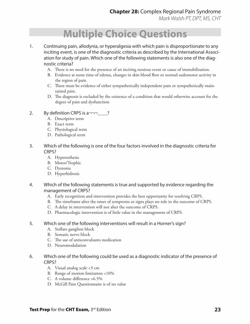
Multiple Choice Questions1. Continuing pain, allodynia, or hyperalgesia with which pain is disproportionate to any
inciting event, is one of the diagnostic criteria as described by the International Associ-ation for study of pain. Which one of the following statements is also one of the diag-nostic criteria?
A. There is no need for the presence of an inciting noxious event or cause of immobilization.B. Evidence at some time of edema, changes in skin blood flow or normal sudomotor activity in
the region of pain.C. There must be evidence of either sympathetically independent pain or sympathetically main-
tained pain.D. The diagnosis is excluded by the existence of a condition that would otherwise account for the
degree of pain and dysfunction.
2. By definition CRPS is a¬¬¬____?A. Descriptive termB. Exact termC. Physiological termD. Pathological term
3. Which of the following is one of the four factors involved in the diagnostic criteria for CRPS?
A. Hyperesthesia B. Motor/TrophicC. DystoniaD. Hyperhidrosis
4. Which of the following statements is true and supported by evidence regarding the management of CRPS?
A. Early recognition and intervention provides the best opportunity for resolving CRPS.B. The timeframe after the onset of symptoms or signs plays no role in the outcome of CRPS.C. A delay in intervention will not alter the outcome of CRPS.D. Pharmacologic intervention is of little value in the management of CRPS.
5. Which one of the following interventions will result in a Horner’s sign?A. Stellate ganglion blockB. Somatic nerve blockC. The use of anticonvulsants medicationD. Neuromodulation
6. Which one of the following could be used as a diagnostic indicator of the presence of CRPS?
A. Visual analog scale <3 cmB. Range of motion limitation <10%C. A volume difference >6.5%D. McGill Pain Questionnaire is of no value

American Society of Hand TherapistsTM24
Chapter 28: Complex Regional Pain SyndromeMark Walsh PT, DPT, MS, CHT
Multiple Choice Questions7. Which one of the following is an important component to consider in evaluation of
pain?A. Qualitative such as the use of the McGill Questionnaire.B. Vasomotor status.C. Hypersensitivity.D. Range of motion limitation.
8. Which one of the following statements is supported by the evidence for the efficacy of therapy intervention in CRPS?
A. There is no evidence that supports the use of therapy as an intervention for CRPS.B. There is strong evidence that supports the use of TENS in the intervention of CRPS.C. The use of Graded Motor Imagery has been demonstrated to be an effective therapy interven-
tion for CRPSD. Patriotism intervention is all that is necessary for therapy intervention for CRPS.
9. Which of the following value is a meaningful reduction in pain that results in the pa-tient reporting successful treatment of CRPS?
A. 75% or greater.B. 50% or greater.C. 25%.D. There is no meaningful value.
10. Which one of the following is a component of Graded Motor Imagery?A. LateralityB. Functional restorationC. DesensitizationD. Stress loading
11. Which of the following statement is true regarding Graded Motor Imagery?A. Mirror visual feedback is not utilized In Graded Motor Imagery.B. The fear avoidance model is a component Of Graded Motor Imagery.C. Graded Motor Imagery is only utilized in the clinic.D. Establishing laterality is required before moving onto the next step.
12. Which one of the following would be an appropriate intervention for Factor 2 Vasomo-tor?
A. Institute or increase general aerobic activities and capacity.B. The use of cryotherapy in the vasoconstrictive state.C. The use of heating modalities in the vasodilated state.D. Encourage upper extremity elevation.

Test Prep for the CHT Exam, 3rd Edition 25
Chapter 28: Complex Regional Pain SyndromeMark Walsh PT, DPT, MS, CHT
Multiple Choice Questions13. Choose the correct statement below regarding CRPS.
A. There is no movement disorders associated with CRPS.B. Dystonia and bradykinesia have been demonstrated to be present in CRPS.C. The use of “Internal Set” is appropriate in the presence of bradykinesia.D. Trophic changes are included in the Sudomotor/Edema factor of CRPS.
14. The intervention of stress loading requires which of the following components.A. Compression, Distraction and Home ProgramB. Compression only and Home ProgramC. Distraction only and Home ProgramD. Either Compression or Distraction and Home Program
Multiple Choice Question Answer KeyChapter 28
1-D, 2-A, 3-B, 4-A, 5-A, 6-C, 7-A, 8-C, 9-B, 10-A, 11-D, 12-A, 13-B, 14-A