Embed Size (px)
DESCRIPTION
Chapter 24 The Digestive System. Structure Gross Anatomy Histology Function Mechanical Chemical Development Disorders. Overview of GI tract Functions. Mouth---bite, chew, swallow Pharynx and esophagus----transport Stomach----mechanical disruption; absorption of water & alcohol - PowerPoint PPT Presentation
Citation preview
Tortora & Grabowski 9/e 2000 JWS 24-1
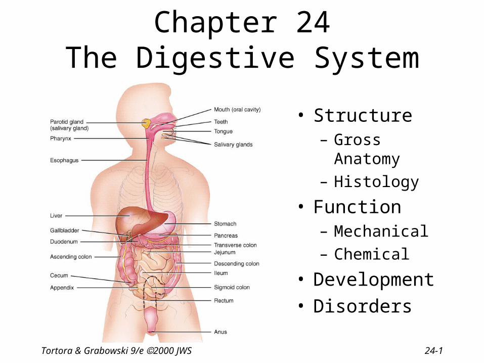
Chapter 24The Digestive System
• Structure – Gross Anatomy
– Histology
• Function– Mechanical
– Chemical
• Development• Disorders

Tortora & Grabowski 9/e 2000 JWS 24-2
Overview of GI tract Functions
• Mouth---bite, chew, swallow
• Pharynx and esophagus----transport
• Stomach----mechanical disruption; absorption of water & alcohol
• Small intestine--chemical & mechanical digestion & absorption
• Large intestine----absorb electrolytes & vitamins (B and K)
• Rectum and anus---defecation

Tortora & Grabowski 9/e 2000 JWS 24-3
Layers of the GI Tract
1. Mucosal layer
2. Submucosal layer
3. Muscularis layer
4. Serosa layer

Tortora & Grabowski 9/e 2000 JWS 24-4
Mucosa
• Epithelium– stratified squamous(in mouth,esophagus & anus) = tough
– simple columnar in the rest• secretes enzymes and absorbs nutrients
• specialized cells (goblet) secrete mucous onto cell surfaces
• enteroendocrine cells---secrete hormones controlling organ function
• Lamina propria– thin layer of loose connective tissue
– contains BV and lymphatic tissue
• Muscularis mucosae---thin layer of smooth muscle
– causes folds to form in mucosal layer
– increases local movements increasing absorption with exposure to “new” nutrients

Tortora & Grabowski 9/e 2000 JWS 24-5
Submucosa
• Loose connective tissue– containing BV, glands and lymphatic tissue
• Meissner’s plexus---– parasympathetic
– innervation• vasoconstriction
• local movement by muscularis mucosa smooth muscle
Tortora & Grabowski 9/e 2000 JWS 24-6
Muscularis
• Skeletal muscle = voluntary control– in mouth, pharynx , upper esophagus and anus– control over swallowing and defecation
• Smooth muscle = involuntary control– inner circular fibers & outer longitudinal fibers– mixes, crushes & propels food along by peristalsis
• Auerbach’s plexus (myenteric)--– both parasympathetic & sympathetic innervation of
circular and longitudinal smooth muscle layers

Tortora & Grabowski 9/e 2000 JWS 24-7
Serosa
• An example of a serous membrane
• Covers all organs and walls of cavities not open to the outside of the body
• Secretes slippery fluid
• Consists of connective tissue covered with simple squamous epithelium

Tortora & Grabowski 9/e 2000 JWS 24-8
Peritoneum
• Peritoneum– visceral layer covers
organs
– parietal layer lines the walls of body cavity
• Peritoneal cavity– potential space containing
a bit of serous fluid

Tortora & Grabowski 9/e 2000 JWS 24-9
Parts of the Peritoneum
• Mesentery• Mesocolon• Lesser omentum• Greater omentum• Peritonitis =
inflammation– trauma
– rupture of GI tract
– appendicitis
– perforated ulcer

Tortora & Grabowski 9/e 2000 JWS 24-10
Greater Omentum, Mesentery & Mesocolon

Tortora & Grabowski 9/e 2000 JWS 24-11
Lesser Omentum

Tortora & Grabowski 9/e 2000 JWS 24-12
Peritonitis
• Acute inflammation of the peritoneum
• Cause– contamination by infectious microbes during
surgery or from rupture of abdominal organs

Tortora & Grabowski 9/e 2000 JWS 24-13
Mouth
• Lips and cheeks-----contains buccinator muscle that keeps food between upper & lower teeth
• Vestibule---area between cheeks and teeth• Oral cavity proper---the roof = hard, soft palate and uvula
– floor = the tongue

Tortora & Grabowski 9/e 2000 JWS 24-14
Pharyngeal Arches
• Two skeletal muscles• Palatoglossal muscle
– extends from palate to tongue
– forms the first arch
– posterior limit of the mouth
• Palatopharyngeal muscle– extends from palate to
pharyngeal wall
– forms the second arch
– behind the palatine tonsil

Tortora & Grabowski 9/e 2000 JWS 24-15
Salivary Glands
• Parotid below your ear and over the masseter• Submandibular is under lower edge of mandible• Sublingual is deep to the tongue in floor of mouth• All have ducts that empty into the oral cavity

Tortora & Grabowski 9/e 2000 JWS 24-16
Composition and Functions of Saliva
• Wet food for easier swallowing
• Dissolves food for tasting
• Bicarbonate ions buffer acidic foods– bulemia---vomiting hurts the enamel on your teeth
• Chemical digestion of starch begins with enzyme (salivary amylase)
• Enzyme (lysozyme) ---helps destroy bacteria
• Protects mouth from infection with its rinsing action---1 to 1 and 1/2qts/day

Tortora & Grabowski 9/e 2000 JWS 24-17
Salivary Gland Cellular Structure
• Cells in acini (clusters)• Serous cells secrete a watery fluid• Mucous cells (pale staining) secrete a slimy, mucus
secretion

Tortora & Grabowski 9/e 2000 JWS 24-18
Salivation
• Increase salivation– sight, smell, sounds, memory of food, tongue
stimulation---rock in mouth– cerebral cortex signals the salivatory nuclei in
brainstem---(CN 7 & 9)– parasympathetic nn. (CN 7 & 9)
• Stop salivation – dry mouth when you are afraid– sympathetic nerves

Tortora & Grabowski 9/e 2000 JWS 24-19
Mumps
• Myxovirus that attacks the parotid gland
• Symptoms– inflammation and enlargement of the parotid– fever, malaise & sour throat (especially
swallowing sour foods)– swelling on one or both sides
• Sterility rarely possible in males with testicular involvement (only one side involved)
• Vaccine available since 1967
Tortora & Grabowski 9/e 2000 JWS 24-20
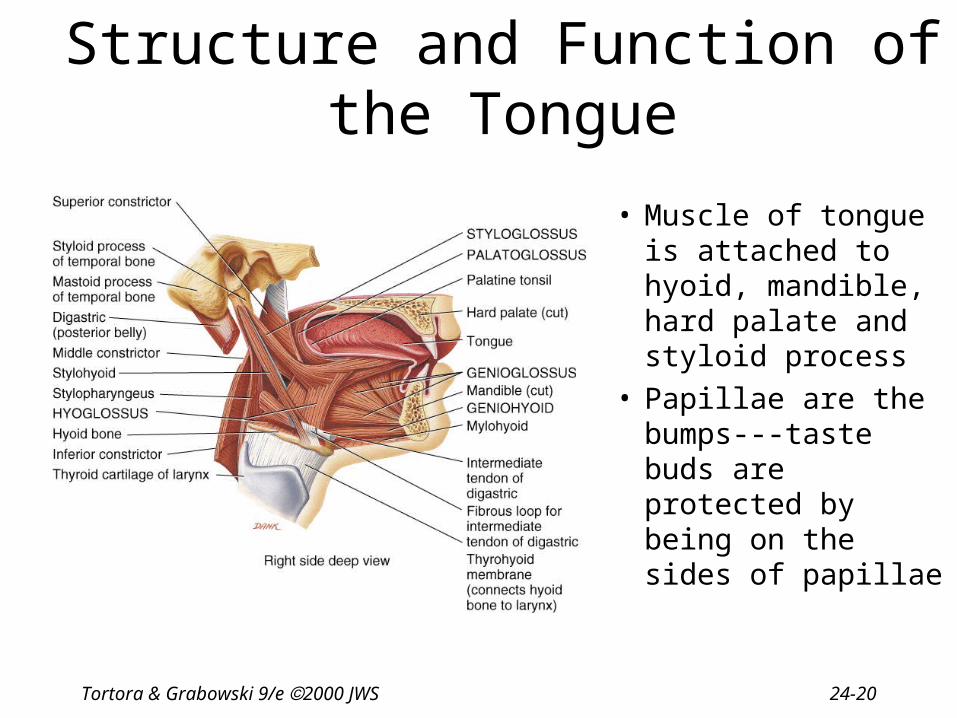
Structure and Function of the Tongue
• Muscle of tongue is attached to hyoid, mandible, hard palate and styloid process
• Papillae are the bumps---taste buds are protected by being on the sides of papillae

Tortora & Grabowski 9/e 2000 JWS 24-21
Tooth Structure
• Crown• Neck• Roots• Pulp cavity

Tortora & Grabowski 9/e 2000 JWS 24-22
Composition of Teeth
• Enamel– hardest substance in body
– calcium phosphate or carbonate
• Dentin– calcified connective tissue
• Cementum– bone-like
– periodontal ligament penetrates it
What is the gingiva?

Tortora & Grabowski 9/e 2000 JWS 24-23
Dentition
• Primary or baby teeth– 20 teeth that start erupting at 6 months– 1 new pair of teeth per month
• Permanent teeth– 32 teeth that erupt between 6 and 12 years of age– differing structures indicate function
• incisors for biting
• canines or cuspids for tearing
• premolars & molars for crushing and grinding food

Tortora & Grabowski 9/e 2000 JWS 24-24
Primary and Secondary Dentition
Tortora & Grabowski 9/e 2000 JWS 24-25
Digestion in the Mouth• Mechanical digestion (mastication or chewing)
• breaks into pieces• mixes with saliva so it forms a bolus
• Chemical digestion– amylase
• begins starch digestion at pH of 6.5 or 7.0 found in mouth• when bolus & enzyme hit the pH 2.5 gastric juices hydrolysis
ceases
– lingual lipase• secreted by glands in tongue• begins breakdown of triglycerides into fatty acids and glycerol

Tortora & Grabowski 9/e 2000 JWS 24-26
Pharynx• Funnel-shaped tube extending from internal
nares to the esophagus (posteriorly) and larynx (anteriorly)
• Skeletal muscle lined by mucous membrane• Deglutition or swallowing is facilitated by saliva
and mucus– starts when bolus is pushed into the oropharynx– sensory nerves send signals to deglutition center in
brainstem– soft palate is lifted to close nasopharynx– larynx is lifted as epiglottis is bent to cover glottis

Tortora & Grabowski 9/e 2000 JWS 24-27
Esophagus
• Collapsed muscular tube
• In front of vertebrae
• Posterior to trachea
• Posterior to the heart
• Pierces the diaphragm at hiatus– hiatal hernia or
diaphragmatic hernia

Tortora & Grabowski 9/e 2000 JWS 24-28
Histology of the Esophagus
• Mucosa = stratified squamous• Submucosa = large mucous glands• Muscularis = upper 1/3 is skeletal, middle is mixed,
lower 1/3 is smooth– upper & lower esophageal sphincters are prominent
circular muscle
• Adventitia = connective tissue blending with surrounding connective tissue--no peritoneum

Tortora & Grabowski 9/e 2000 JWS 24-29
Physiology of the Esophagus - Swallowing
• Voluntary phase---tongue pushes food to back of oral cavity• Involuntary phase----pharyngeal stage
– breathing stops & airways are closed– soft palate & uvula are lifted to close off nasopharynx– vocal cords close– epiglottis is bent over airway as larynx is lifted

Tortora & Grabowski 9/e 2000 JWS 24-30
Swallowing
• Upper sphincter relaxes when larynx is lifted
• Peristalsis pushes food down– circular fibers behind bolus
– longitudinal fibers in front of bolus shorten the distance of travel
• Travel time is 4-8 seconds for solids and 1 sec for liquids
• Lower sphincter relaxes as food approaches
Tortora & Grabowski 9/e 2000 JWS 24-31
Gastroesophageal Reflex Disease• If lower sphincter fails to open
– distension of esophagus feels like chest pain or heart attack
• If lower esophageal sphincter fails to close– stomach acids enter esophagus & cause heartburn (GERD)
– for a weak sphincter---don't eat a large meal and lay down in front of TV
– smoking and alcohol make the sphincter relax worsening the situation
• Control the symptoms by avoiding– coffee, chocolate, tomatoes, fatty foods, onions & mint
– take Tagamet HB or Pepcid AC 60 minutes before eating
– neutralize existing stomach acids with Tums

Tortora & Grabowski 9/e 2000 JWS 24-32
Anatomy of Stomach• Which side is it on? • Size when empty?
– large sausage– stretches due to rugae
• Parts of stomach– cardia– fundus---air in x-ray– body– pylorus---starts to narrow as approaches pyloric
sphincter
• Empties as small squirts of chyme leave the stomach through the pyloric valve

Tortora & Grabowski 9/e 2000 JWS 24-33
Pylorospasm and Pyloric Stenosis
• Abnormalities of the pyloric sphincter in infants
• Pylorospasm– muscle fibers of sphincter fail to relax trapping
food in the stomach – vomiting occurs to relieve pressure
• Pyloric stenosis– narrowing of sphincter indicated by projectile
vomiting– must be corrected surgically

Tortora & Grabowski 9/e 2000 JWS 24-34
Histology of the Stomach
Tortora & Grabowski 9/e 2000 JWS 24-35
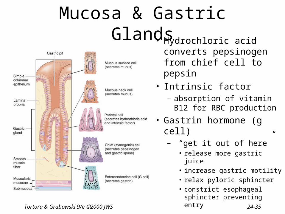
Mucosa & Gastric Glands• Hydrochloric acid
converts pepsinogen from chief cell to pepsin
• Intrinsic factor– absorption of vitamin B12
for RBC production
• Gastrin hormone (g cell)– “get it out of here”
• release more gastric juice
• increase gastric motility
• relax pyloric sphincter
• constrict esophageal sphincter preventing entry

Tortora & Grabowski 9/e 2000 JWS 24-36
Submucosa

Tortora & Grabowski 9/e 2000 JWS 24-37
Muscularis
• Three layers of smooth muscle--outer longitudinal, circular & inner oblique
• Permits greater churning & mixing of food with gastric juice

Tortora & Grabowski 9/e 2000 JWS 24-38
Serosa
• Simple squamous epithelium over a bit of connective tissue
• Also known as visceral peritoneum

Tortora & Grabowski 9/e 2000 JWS 24-39
Physiology--Mechanical Digestion
• Gentle mixing waves – every 15 to 25 seconds– mixes bolus with 2 quarts/day of gastric juice to
turn it into chyme (a thin liquid)
• More vigorous waves – travel from body of stomach to pyloric region
• Intense waves near the pylorus – open it and squirt out 1-2 teaspoons full with
each wave

Tortora & Grabowski 9/e 2000 JWS 24-40
Physiology--Chemical Digestion
• Protein digestion begins– HCl denatures (unfolds) protein molecules– HCl transforms pepsinogen into pepsin that breaks
peptides bonds between certain amino acids
• Fat digestion continues– gastric lipase splits the triglycerides in milk fat
• most effective at pH 5 to 6 (infant stomach)
• HCl kills microbes in food• Mucous cells protect stomach walls from being
digested with 1-3mm thick layer of mucous

Tortora & Grabowski 9/e 2000 JWS 24-41
Regulation of Gastric Secretion and Motility
• Cephalic phase
• Gastric phase
• Intestinal phase

Tortora & Grabowski 9/e 2000 JWS 24-42
Cephalic Phase = “Stomach Getting Ready”
• Cerebral cortex =sight, smell, taste & thought – stimulate parasympathetic nervous system
• Vagus nerve– increases stomach muscle and glandular
activity

Tortora & Grabowski 9/e 2000 JWS 24-43
Gastric Phase = “Stomach Working”
• Nervous control keeps stomach active– stretch receptors & chemoreceptors provide information– vigorous peristalsis and glandular secretions continue– chyme is released into the duodenum
• Endocrine influences over stomach activity– distention and presence of caffeine or protein cause G
cells secretion of gastrin into bloodstream– gastrin hormone increases stomach glandular secretion– gastrin hormone increases stomach churning and
sphincter relaxation

Tortora & Grabowski 9/e 2000 JWS 24-44
Intestinal Phase = “Stomach Emptying”
• Stretch receptors in duodenum slow stomach activity & increase intestinal activity
• Distension, fatty acids or sugar signals medulla – sympathetic nerves slow stomach activity
• Hormonal influences – secretin hormone decreases stomach secretions
– cholecystokinin(CCK) decreases stomach emptying
– gastric inhibitory peptide(GIP) decreases stomach secretions, motility & emptying

Tortora & Grabowski 9/e 2000 JWS 24-45
Absorption of Nutrients by the Stomach
• Water especially if it is cold• Electrolytes• Some drugs (especially aspirin) & alcohol• Fat content in the stomach slows the passage of alcohol to the
intestine where absorption is more rapid • Gastric mucosal cells contain alcohol dehydrogenase that converts
some alcohol to acetaldehyde-----more of this enzyme found in males than females
• Females have less total body fluid that same size male so end up with higher blood alcohol levels with same intake of alcohol
Tortora & Grabowski 9/e 2000 JWS 24-46
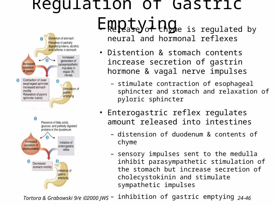
Regulation of Gastric Emptying• Release of chyme is regulated by neural and
hormonal reflexes
• Distention & stomach contents increase secretion of gastrin hormone & vagal nerve impulses
– stimulate contraction of esophageal sphincter and stomach and relaxation of pyloric sphincter
• Enterogastric reflex regulates amount released into intestines
– distension of duodenum & contents of chyme
– sensory impulses sent to the medulla inhibit parasympathetic stimulation of the stomach but increase secretion of cholecystokinin and stimulate sympathetic impulses
– inhibition of gastric emptying

Tortora & Grabowski 9/e 2000 JWS 24-47
Vomiting (emesis)
• Forceful expulsion of contents of stomach & duodenum through the mouth
• Cause– irritation or distension of stomach
– unpleasant sights, general anesthesia, dizziness & certain drugs
• Sensory input from medulla cause stomach contraction & complete sphincter relaxation
• Contents of stomach squeezed between abdominal muscles and diaphragm and forced through open mouth
• Serious because loss of acidic gastric juice can lead to alkalosis
Tortora & Grabowski 9/e 2000 JWS 24-48
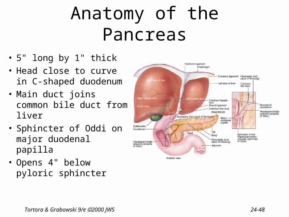
Anatomy of the Pancreas
• 5" long by 1" thick• Head close to curve in C-
shaped duodenum• Main duct joins common
bile duct from liver • Sphincter of Oddi on
major duodenal papilla• Opens 4" below pyloric
sphincter

Tortora & Grabowski 9/e 2000 JWS 24-49
Histology of the Pancreas
• Acini- dark clusters – 99% of gland
– produce pancreatic juice
• Islets of Langerhans– 1% of gland
– pale staining cells
– produce hormones

Tortora & Grabowski 9/e 2000 JWS 24-50
Composition and Functions of Pancreatic Juice
• 1 & 1/2 Quarts/day at pH of 7.1 to 8.2• Contains water, enzymes & sodium bicarbonate• Digestive enzymes
– pancreatic amylase, pancreatic lipase, proteases– trypsinogen---activated by enterokinase (a brush border enzyme)– chymotrypsinogen----activated by trypsin– procarboxypeptidase---activated by trypsin– proelastase---activated by trypsin– trypsin inhibitor---combines with any trypsin produced inside
pancreas
– ribonuclease----to digest nucleic acids
– deoxyribonuclease

Tortora & Grabowski 9/e 2000 JWS 24-51
Pancreatitis
• Pancreatitis---inflammation of the pancreas occurring with the mumps
• Acute pancreatitis---associated with heavy alcohol intake or biliary tract obstruction– result is patient secretes trypsin in the pancreas
& starts to digest himself
Tortora & Grabowski 9/e 2000 JWS 24-52
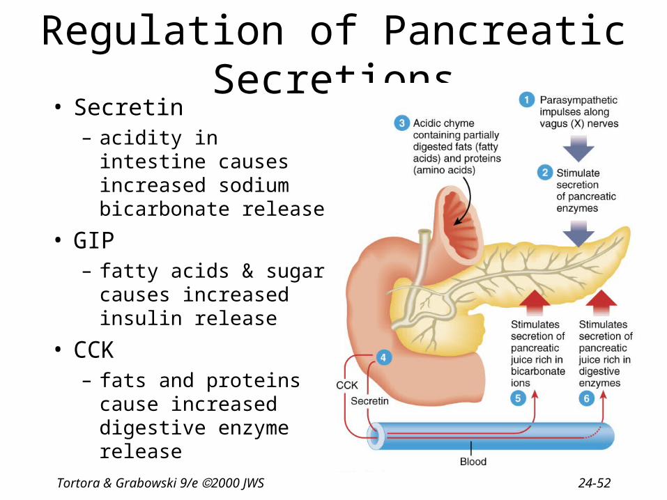
Regulation of Pancreatic Secretions• Secretin
– acidity in intestine causes increased sodium bicarbonate release
• GIP– fatty acids & sugar
causes increased insulin release
• CCK– fats and proteins cause
increased digestive enzyme release

Tortora & Grabowski 9/e 2000 JWS 24-53
Anatomy of the Liver and Gallbladder
• Liver– weighs 3 lbs.
– below diaphragm
– right lobe larger
– gallbladder on right lobe
– size causes right kidney to be lower than left
• Gallbladder– fundus, body & neck

Tortora & Grabowski 9/e 2000 JWS 24-54
Histology of the Liver
• Hepatocytes arranged in lobules • Sinusoids in between hepatocytes
are blood-filled spaces • Kupffer cells phagocytize microbes
& foreign matter

Tortora & Grabowski 9/e 2000 JWS 24-55
Histology of the Gallbladder
• Simple columnar epithelium• No submucosa• Three layers of smooth muscle• Serosa or visceral peritoneum

Tortora & Grabowski 9/e 2000 JWS 24-56
Flow of Fluids Within the Liver

Tortora & Grabowski 9/e 2000 JWS 24-57
Pathway of Bile Secretion
• Bile capillaries• Hepatic ducts connect to form common hepatic duct• Cystic duct from gallbladder & common hepatic duct
join to form common bile duct• Common bile duct & pancreatic duct empty into
duodenum

Tortora & Grabowski 9/e 2000 JWS 24-58
Blood Supply to the Liver• Hepatic portal vein
– nutrient rich blood from stomach, spleen & intestines
• Hepatic artery from branch off the aorta

Tortora & Grabowski 9/e 2000 JWS 24-59
Bile Production
• One quart of bile/day is secreted by the liver– yellow-green in color & pH 7.6 to 8.6
• Components– water & cholesterol– bile salts = Na & K salts of bile acids – bile pigments (bilirubin) from hemoglobin molecule
• globin = a reuseable protein
• heme = broken down into iron and bilirubin

Tortora & Grabowski 9/e 2000 JWS 24-60
Regulation of Bile Secretion

Tortora & Grabowski 9/e 2000 JWS 24-61
Liver Functions--Carbohydrate Metabolism
• Turn proteins into glucose
• Turn triglycerides into glucose
• Turn excess glucose into glycogen & store in the liver
• Turn glycogen back into glucose as needed

Tortora & Grabowski 9/e 2000 JWS 24-62
Liver Functions --Lipid Metabolism
• Synthesize cholesterol
• Synthesize lipoproteins----HDL and LDL(used to transport fatty acids in bloodstream)
• Stores some fat
• Breaks down some fatty acids

Tortora & Grabowski 9/e 2000 JWS 24-63
Liver Functions--Protein Metabolism
• Deamination = removes NH2 (amine group) from amino acids so can use what is left as energy source
• Converts resulting toxic ammonia (NH3) into urea for excretion by the kidney
• Synthesizes plasma proteins utilized in the clotting mechanism and immune system
• Convert one amino acid into another

Tortora & Grabowski 9/e 2000 JWS 24-64
Other Liver Functions
• Detoxifies the blood by removing or altering drugs & hormones(thyroid & estrogen)
• Removes the waste product--bilirubin• Releases bile salts help digestion by emulsification• Stores fat soluble vitamins-----A, B12, D, E, K• Stores iron and copper• Phagocytizes worn out blood cells & bacteria• Activates vitamin D (the skin can also do this with 1 hr
of sunlight a week)

Tortora & Grabowski 9/e 2000 JWS 24-65
Summary of Digestive Hormones
• Gastrin– stomach, gastric & ileocecal sphincters
• Gastric inhibitory peptide--GIP– stomach & pancreas
• Secretin– pancreas, liver & stomach
• Cholecystokinin--CCK– pancreas, gallbladder, sphincter of Oddi, &
stomach
Tortora & Grabowski 9/e 2000 JWS 24-66
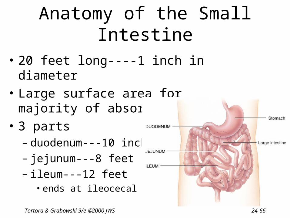
Anatomy of the Small Intestine
• 20 feet long----1 inch in diameter
• Large surface area for majority of absorption
• 3 parts– duodenum---10 inches– jejunum---8 feet– ileum---12 feet
• ends at ileocecal valve

Tortora & Grabowski 9/e 2000 JWS 24-67
Histology of Small Intestine
Tortora & Grabowski 9/e 2000 JWS 24-68
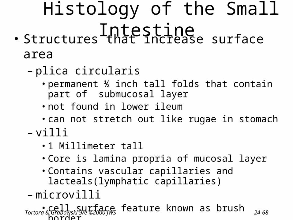
Histology of the Small Intestine• Structures that increase surface area
– plica circularis• permanent ½ inch tall folds that contain part of
submucosal layer• not found in lower ileum• can not stretch out like rugae in stomach
– villi• 1 Millimeter tall• Core is lamina propria of mucosal layer• Contains vascular capillaries and lacteals(lymphatic
capillaries)
– microvilli• cell surface feature known as brush border

Tortora & Grabowski 9/e 2000 JWS 24-69
Functions of Microvilli
• Absorption and digestion
• Digestive enzymes found at cell surface on microvilli
• Digestion occurs at cell surfaces
• Significant cell division within intestinal glands produces new cells that move up
• Once out of the way---rupturing and releasing their digestive enzymes & proteins

Tortora & Grabowski 9/e 2000 JWS 24-70
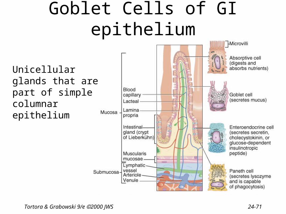
Cells of Intestinal Glands
• Absorptive cell• Goblet cell• Enteroendocrine
– secretin
– cholecystokinin
– gastric inhibitory peptide
• Paneth cells– secretes lysozyme
Tortora & Grabowski 9/e 2000 JWS 24-71
Goblet Cells of GI epithelium
Unicellular glands that are part of simple columnar epithelium

Tortora & Grabowski 9/e 2000 JWS 24-72
Roles of Intestinal Juice & Brush-Border Enzymes
• Submucosal layer has duodenal glands – secretes alkaline mucus
• Mucosal layer contains intestinal glands = Crypts of Lieberkuhn(deep to surface)– secretes intestinal juice
• 1-2 qt./day------ at pH 7.6
– brush border enzymes
– paneth cells secrete lysozyme kills bacteria

Tortora & Grabowski 9/e 2000 JWS 24-73
Mechanical Digestion in the Small Intestine
• Weak peristalsis in comparison to the stomach---chyme remains for 3 to 5 hours
• Segmentation---local mixing of chyme with intestinal juices---sloshing back & forth

Tortora & Grabowski 9/e 2000 JWS 24-74
Chemical Digestion in Small Intestine
• Chart page 853--groups enzymes by region where they are found
• Need to trace breakdown of nutrients– carbohydrates– proteins– lipids

Tortora & Grabowski 9/e 2000 JWS 24-75
Digestion of Carbohydrates
• Mouth---salivary amylase
• Esophagus & stomach---nothing happens
• Duodenum----pancreatic amylase
• Brush border enzymes (maltase, sucrase & lactose) act on disaccharides– produces monosaccharides--fructose, glucose &
galactose– lactose intolerance (no enzyme; bacteria
ferment sugar)--gas & diarrhea
Tortora & Grabowski 9/e 2000 JWS 24-76
Lactose Intolerance
• Mucosal cells of small intestine fail to produce lactase– essential for digestion of lactose sugar in milk– undigested lactose retains fluid in the feces– bacterial fermentation produces gases
• Symptoms– diarrhea, gas, bloating & abdominal cramps
• Dietary supplements are helpful

Tortora & Grabowski 9/e 2000 JWS 24-77
Digestion of Proteins
• Stomach– HCl denatures or unfolds proteins– pepsin turns proteins into peptides
• Pancreas– digestive enzymes---split peptide bonds between
different amino acids– brush border enzymes-----aminopeptidase or
dipeptidase------split off amino acid at amino end of molecule or split dipeptide

Tortora & Grabowski 9/e 2000 JWS 24-78
Digestion of Lipids
• Mouth----lingual lipase
• Small intestine– emulsification by bile– pancreatic lipase---splits into fatty acids &
monoglyceride– no enzymes in brush border

Tortora & Grabowski 9/e 2000 JWS 24-79
Digestion of Nucleic Acids
• Pancreatic juice contains 2 nucleases– ribonuclease which digests RNA– deoxyribonuclease which digests DNA
• Nucleotides produced are further digested by brush border enzymes (nucleosidease and phosphatase)– pentose, phosphate & nitrogenous bases
• Absorbed by active transport

Tortora & Grabowski 9/e 2000 JWS 24-80
Regulation of Secretion & Motility
• Enteric reflexes that respond to presence of chyme– increase intestinal motility– VIP (vasoactive intestinal polypeptide) stimulates the
production of intestinal juice– segmentation depends on distention which sends
impulses to the enteric plexus & CNS• distention produces more vigorous peristalsis• 10 cm per second
• Sympathetic impulses decrease motility

Tortora & Grabowski 9/e 2000 JWS 24-81
Absorption in Small Intestine

Tortora & Grabowski 9/e 2000 JWS 24-82
Where will the absorbed nutrients go?

Tortora & Grabowski 9/e 2000 JWS 24-83
Absorption of Monosaccharides
• Absorption into epithelial cell– glucose & galactose----sodium symporter(active transport)
– fructose-----facilitated diffusion
• Movement out of epithelial cell into bloodstream– by facilitated diffusion

Tortora & Grabowski 9/e 2000 JWS 24-84
Absorption of Amino Acids & Dipeptides
• Absorption into epithelial cell– active transport with Na+ or H+ ions (symporters)
• Movement out of epithelial cell into blood– diffusion

Tortora & Grabowski 9/e 2000 JWS 24-85
Absorption of Lipids • Small fatty acids enter cells & then blood by simple diffusion
• Larger lipids exist only within micelles (bile salts coating)
• Lipids enter cells by simple diffusion leaving bile salts behind in gut
• Bile salts reabsorbed into blood & reformed into bile in the liver
• Fat-soluble vitamins are enter cells since were within micelles
Tortora & Grabowski 9/e 2000 JWS 24-86
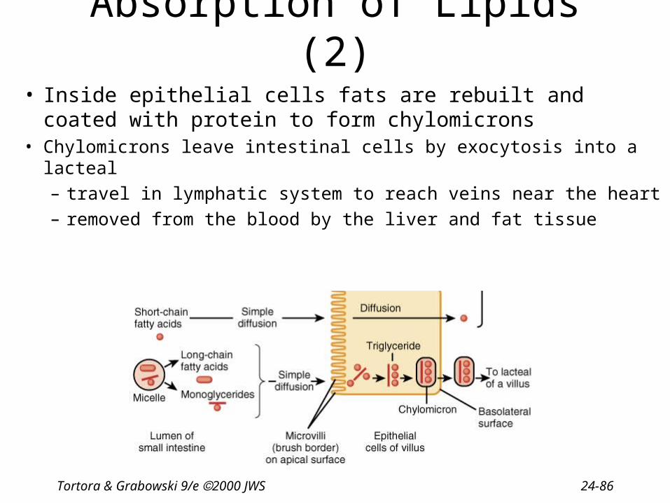
Absorption of Lipids (2)
• Inside epithelial cells fats are rebuilt and coated with protein to form chylomicrons
• Chylomicrons leave intestinal cells by exocytosis into a lacteal
– travel in lymphatic system to reach veins near the heart
– removed from the blood by the liver and fat tissue
Tortora & Grabowski 9/e 2000 JWS 24-87
Absorption of Electrolytes
• Sources of electrolytes– GI secretions & ingested foods and liquids
• Enter epithelial cells by diffusion & secondary active transport– sodium & potassium move = Na+/K+ pumps (active transport)
– chloride, iodide and nitrate = passively follow
– iron, magnesium & phosphate ions = active transport
• Intestinal Ca+ absorption requires vitamin D & parathyroid hormone

Tortora & Grabowski 9/e 2000 JWS 24-88
Absorption of Vitamins
• Fat-soluble vitamins – travel in micelles & are absorbed by simple
diffusion
• Water-soluble vitamins– absorbed by diffusion
• B12 combines with intrinsic factor before it is transported into the cells– receptor mediated endocytosis

Tortora & Grabowski 9/e 2000 JWS 24-89
Absorption of Water
• 9 liters of fluid dumped into GI tract each day
• Small intestine reabsorbs 8 liters
• Large intestine reabsorbs 90% of that last liter
• Absorption is by osmosis through cell walls into vascular capillaries inside villi

Tortora & Grabowski 9/e 2000 JWS 24-90
Anatomy of Large Intestine
• 5 feet long by 2½ inches in diameter• Ascending & descending colon are retroperitoneal• Cecum & appendix
• Rectum = last 8 inches of GI tract anterior to the sacrum & coccyx• Anal canal = last 1 inch of GI tract
– internal sphincter----smooth muscle & involuntary – external sphincter----skeletal muscle & voluntary control

Tortora & Grabowski 9/e 2000 JWS 24-91
Appendicitis• Inflammation of the appendix due to blockage
of the lumen by chyme, foreign body, carcinoma, stenosis, or kinking
• Symptoms– high fever, elevated WBC count, neutrophil count
above 75%– referred pain, anorexia, nausea and vomiting– pain localizes in right lower quadrant
• Infection may progress to gangrene and perforation within 24 to 36 hours
Tortora & Grabowski 9/e 2000 JWS 24-92
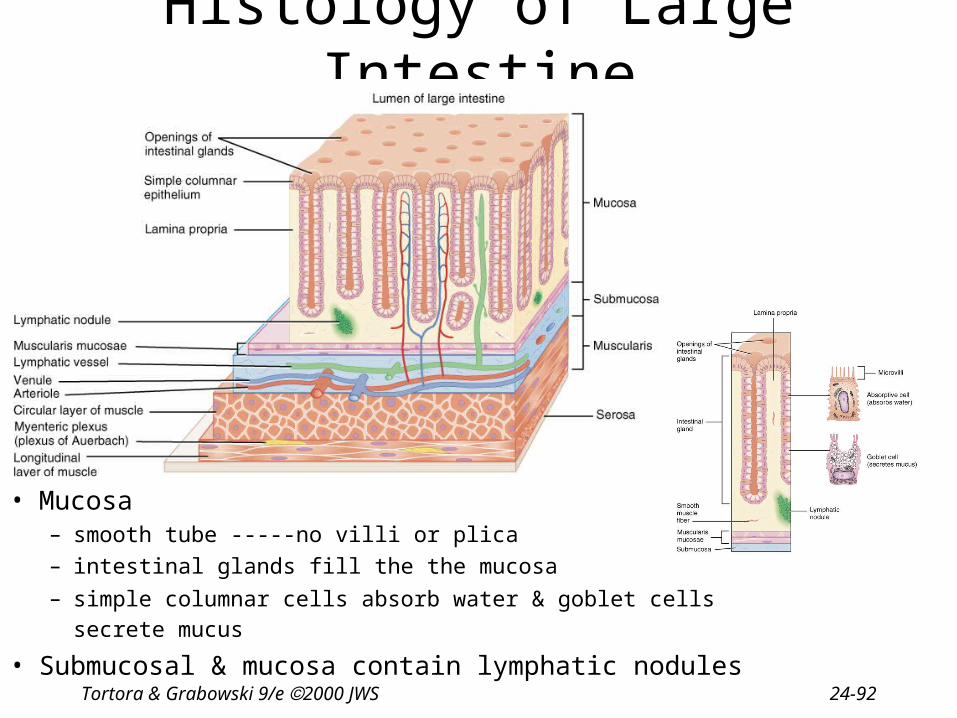
Histology of Large Intestine
• Mucosa– smooth tube -----no villi or plica
– intestinal glands fill the the mucosa
– simple columnar cells absorb water & goblet cells secrete mucus
• Submucosal & mucosa contain lymphatic nodules

Tortora & Grabowski 9/e 2000 JWS 24-93
Histology of Large Intestine
• Muscular layer– internal circular layer is normal
– outer longitudinal muscle• taeniae coli = shorter bands
• haustra (pouches) formed
• epiploic appendages
• Serosa = visceral peritoneum
• Appendix
– contains large amounts of lymphatic tissue
Tortora & Grabowski 9/e 2000 JWS 24-94
Mechanical Digestion in Large Intestine
• Smooth muscle = mechanical digestion• Peristaltic waves (3 to 12 contractions/minute)
– haustral churning----relaxed pouches are filled from below by muscular contractions (elevator)
– gastroilial reflex = when stomach is full, gastrin hormone relaxes ileocecal sphincter so small intestine will empty and make room
– gastrocolic reflex = when stomach fills, a strong peristaltic wave moves contents of transverse colon into rectum

Tortora & Grabowski 9/e 2000 JWS 24-95
Chemical Digestion in Large Intestine
• No enzymes are secreted only mucous
• Bacteria ferment– undigested carbohydrates into carbon dioxide
& methane gas– undigested proteins into simpler substances
(indoles)----odor– turn bilirubin into simpler substances that
produce color
• Bacteria produce vitamin K and B in colon

Tortora & Grabowski 9/e 2000 JWS 24-96
Absorption & Feces Formation in the Large Intestine
• Some electrolytes---Na+ and Cl-
• After 3 to 10 hours, 90% of H2O has been removed from chyme
• Feces are semisolid by time reaches transverse colon
• Feces = dead epithelial cells, undigested food such as cellulose, bacteria (live & dead)

Tortora & Grabowski 9/e 2000 JWS 24-97
Defecation
• Gastrocolic reflex moves feces into rectum
• Stretch receptors signal sacral spinal cord
• Parasympathetic nerves contract muscles of rectum & relax internal anal sphincter
• External sphincter is voluntarily controlled

Tortora & Grabowski 9/e 2000 JWS 24-98
Defecation Problems
• Diarrhea = chyme passes too quickly through intestine – H20 not reabsorbed
• Constipation--decreased intestinal motility– too much water is reabsorbed– remedy = fiber, exercise and water

Tortora & Grabowski 9/e 2000 JWS 24-99
Dietary Fiber
• Insoluble fiber– woody parts of plants (wheat bran, vegie skins)– speeds up transit time & reduces colon cancer
• Soluble fiber– gel-like consistency = beans, oats, citrus white
parts, apples– lowers blood cholesterol by preventing
reabsorption of bile salts so liver has to use cholesterol to make more

Tortora & Grabowski 9/e 2000 JWS 24-100
Development of the Digestive System
• Endoderm forms primitive gut with help from the splanchnic mesoderm --- resulting tube is made up of epithelial, glandular, muscle & connective tissue
• Differentiates into foregut, midgut & hindgut• Endoderm grows into the mesoderm to form salivary glands, liver,
gallbladder & pancreas

Tortora & Grabowski 9/e 2000 JWS 24-101
Development of the Digestive System
• Stomodeum develops into oral cavity– oral membrane
ruptures
• Proctodeum develops into anus– cloacal membrane
ruptures

Tortora & Grabowski 9/e 2000 JWS 24-102
Aging and the Digestive System• Changes that occur
– decreased secretory mechanisms
– decreased motility
– loss of strength & tone of muscular tissue
– changes in neurosensory feedback
– diminished response to pain & internal stimuli
• Symptoms– sores, loss of taste, peridontal disease, difficulty swallowing,
hernia, gastritis, ulcers, malabsorption, jaundice, cirrhosis, pancreatitis, hemorrhoids and constipation
• Cancer of the colon or rectum is common
Tortora & Grabowski 9/e 2000 JWS 24-103
Diseases of the GI Tract
• Dental caries and periodontal disease • Peptic Ulcers• Diverticulitis• Colorectal cancer• Hepatitis• Anorexia nervosa