Embed Size (px)
Citation preview

Atrial Fibrillation - Current Understanding and Management Strategies 169
IntroductionAtrial Fibrillation (AF) is an arrhythmia characterized by irregular, disorganized and chaotic electrical activity of the atrium. The ECG classically is represented by low amplitude, variable and irregular base-line undulations corresponding to atrial rate in the range of 400-600/min. The ventricular rate is irregularly irregular (Fig. 1). The mean ventricular rate is usually between 100 to 150/min and is markedly susceptible to acceleration by vagolysis and catecholamines (i.e. standing, exercise).
AF can present as:-
a. Paroxysmal AF- recurrent episodes which terminate spontaneously.b. Persistent AF – lasts for more than 48 hours and terminates after electric or pharmacologic
cardioversion.c. Permanent AF- fails to terminate despite intervention.
Incidence and PrevalanceAtrial fibrillation was observed in humans as a clinical entity as early as 19091. It is the most common sustained arrhythmia occurring in approximately 0.4% to 1% of the general population. The prevalence of AF increases with age, affecting up to 4% of the population over age 60. Atrial fibrillation accounts for 1/3rd of all patient discharges with arrhythmias as principal diagnosis. More than 50% of the patients with severe rheumatic mitral valve disease have atrial fibrillation. Table 1 enlists the various causes of atrial fibrillation.
Mechanism of AFTwo hypotheses have been proposed1:
a ‘Multiple wavelet’ theory advanced by Moe, in which AF is a result of reentry mechanism in the atrial muscle. Maintenance of fibrillation is favoured by a large mass of atrial tissue.
b. Ectopic focus theory which suggests that AF is due to one or more areas of ectopic automaticity that fires high frequency electric discharges to the neighbouring atria culminating into AF.
C h a p t e r 2 1
Atrial Fibrillation - Current Understanding and Management Strategies
Amit Vora1, Swapna Athawale2
1 Consultant Cardiologist, Nanavati & Lilavati Hospital, Mumbai2 Clinical Assistant, Glenmark Cardiac Centre, Mumbai

170 CME 2004
Table 1 : Etiology of Atrial Fibrillation
I. Cardiac Causes 1. Valvular heart disease i. Rheumatic mitral valve disease ii. Mitral valve prolapse- mitral regurgitation 2. Hypertensive heart disease 3. Ischemic heart disease 4. Pericarditis 5. Cardiac tumors; atrial myxoma 6. Sick sinus syndrome, WPW syndrome 7. Cardiomyopathy - Hypertrophic - Idiopathic dilated 8. Post coronary bypass surgery
II. Non-cardiac causes- 1. Pulmonary i. Chronic obstructive lung disease ii. Pneumonia iii. Pulmonary embolism 2. Hyperthyroidism 3. Toxic : alcohol (‘holiday heart’ syndrome) 4. Miscellaneous- Carbon monoxide poisoning, pheochromocytoma, hypoxia, hypokalemia, sepsis, physiologic
stress, caffeine.
III. “Lone” AF
Fig. 1 : 8 12- lead showing atrial fibrillation
* “AF begets AF”AF induces changes in atrial refractoriness that may promote its perpetuation. Brief episodes of AF may shorten atrial refractoriness for several minutes, leading to a heightened tendency for the reinduction of AF after conversion to sinus rhythm2. The atria get larger during periods of sustained AF and with restoration of sinus rhythm, the AF is reversible if it has not been present for long. The longer the duration of AF, the more difficult it may be to achieve successful conversion and maintenance of sinus rhythm2.
Clinical PresentationAtrial fibrillation can be an incidental finding in an asymptomatic individual. The patients can present

Atrial Fibrillation - Current Understanding and Management Strategies 171
with variable symptoms like palpitations, fatigue, dyspnoea, angina, reduced sense of well-being, syncope, diaphoresis, dizziness or congestive heart failure. Sometimes, patients can present with embolic stroke resulting from left atrial clot. In some cases, a cognitive disability has been reported presumably due to silent cerebral ischemia.
Complications
1. Thromboembolism and stroke Sustained or chronic AF presents a considerable risk for thromboembolism; presumably
following clot formation in LA, more commonly in LA appendage.
Predictors of thromboembolic risk in AF:- • Rheumatic heart disease (RHD)- 18-20% stroke/year • Hypertension • Prior stroke or transient ischemic attack (TIA) • Diabetes • Congestive heart failure • Age > 65 years The risk of stroke in high risk patients is 5-8%/ year. The echocardiographic risk factors for
stroke in patients with AF are presence of LV systolic dysfunction and increased LA size. The importance of non-rheumatic AF as a risk factor for ischemic stroke appears to depend largely on the presence of spontaneous LA contrast on echocardiography3.
2. Congestive Heart Failure (CHF) Patients with organic heart disease such as obstructive hypertrophic cardiomyopathy or chronic
stable CHF may decompensate rapidly with the onset of AF due to loss of atrioventricular (AV) synchrony, the uncontrolled ventricular rate and the loss of atrial ‘kick’ i.e. loss of effective atrial contraction with its contribution to cardiac output. Patients with chronic, long standing AF develop rate related cardiomyopathy due to rapid ventricular response, thereby leading to CHF.
Physical Examination and Investigations
1. Clinical signs • Irregularly irregular heart rate • Apex pulse deficit (heart rate by palpation less than that by auscultation) • Variable first heart sound • Jugular venous pulse may show absent ‘a’ waves or fibrillatory waves may be appreciated
and pulse amplitude is variable.
2. Electrocardiogram (ECG) P waves are absent. Atrial activity is chaotic and fibrillatory (f) waves are present. The baseline
of the ECG is often undulating and may occasionally have coarse, irregular activity that can resemble atiral flutter but is not as stereotype from wave to wave as flutter tends to be. The atrial rate is generally in the range of 400-600 /min with a venticular conduction being variable and generally in the range of 100-150 /min in the absence of drug therapy.
Intraventicular conduction of the impulses from fibrillating atria may at times, be associated with phasic aberrant ventricular conduction. Its importance lies in the fact that aberrantly conducted beats may be mistaken for venticular ectopic beats and this may influence the physician to withhold digitalis when it is needed or to give antiarrhythmic drugs where they are not needed and may even be contraindicated. Aberration is particularly likely to develop when a lengthening

172 CME 2004
of venticular cycle is immediately followed by a short cycle. The beat ending the short cycle shows aberrant conduction. This short-long-short cycle sequence promoting aberation is called as Ashman’s phenomenon5.
3. 2D Echocardiography Role of echo in AF: • identify structural heart disease • identify LVH , LV systolic function • quantify LA enlargement • detect ‘smoke’ i.e. spontaneous LA contrast/ clot in LA Most clots are in the left atrial appendage (LAA), but poorly visualised by transthoracic echo.
Transesophageal echocardiography (TEE) has a 92% sensitivity and 98% specificity for detecting LAA clot
Management of AFA flow chart as elaborated in figure 2 seems to be the most practical approach to the management of atrial fibrillation (Fig. 2).
Patient with AF
Rheumatic valvular heart disease +ve → ← Rheumatic valvular heart disease –ve
Anticoagulation
Surgery required Surgery not required
Valve ± MAZE surgery
Pre-discharge electricCardioversion
Sinus RhythmAsymptomatic
AFpersists
Amiodarone±
Electric cardioversion
Sinus rhythm AF persists
Rate controlmeasures
Lone AF
Antiplatelets
Risk factors for thromboembolism
Warfarin
Age < 65 yrs.HOCM, DCM
Age > 65 yrs.
Rhythmcontrol
Ratecontrol
Fig. 2 : Therapeutic approach to atrial fibrillation
Atrial Fibrillation - Current Understanding and Management Strategies 173
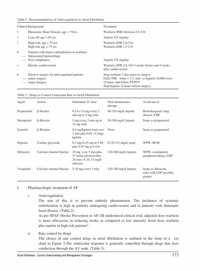
I. Pharmacologic treatment of AF
i. Anticoagulation The aim of this is to prevent embolic phenomenon. The incidence of systemic
embolisation is high in patients undergoing cardioversion and in patients with rheumatic heart disease. (Table 2).
As per SPAF (Stroke Prevention in AF) III randomized clinical trial, adjusted dose warfarin is more efficacious in reducing stroke as compared to low intensity fixed–dose warfarin plus aspirin in high risk patients6.
ii. Rate control by drugs The choice of rate control drugs in atrial fibrillation is outlined in the form of a flow
chart in Figure 3.The ventricular response is generally controlled through drugs that slow conduction through the AV node. (Table 3).
Table 2 : Recommendations of Anticoagulation in Atrial Fibrillation
Clinical Background Treatment
• Rheumatic Heart Disease, age < 75yrs. Warfarin (INR) between 2.0-3.0)
• Lone AF age < 65 yrs Arpirin 325 mg/day
• High risk, age < 75 yrs Warfarin (INR 2.0-3.0)High risk age > 75 yrs Warfarin (INR 1.5-2.5)
• Patients with major contradictions to warfarin:— Intracranial haemorrhage— Poor compliance Aspirin 325 mg/day
• Electric cardioversion Warfarin (INR 2.0-3.0) 4 weeks before and 4 weeks after cardioversion
• Elective surgery for anticoagulated patients Stop warfarin 7 days prior to surgery Daily INR - when < 1.5, start sc heparin 10,000 every 12 hours and follow PT/PTTStop heparin 12 hours before surgery.
— minor surgery— major Surgery
Table 3 : Drugs to Control Ventricular Rate in Atrial Fibrillation
Agent Action Immediate IV dose Oral maintenance therapy
Avoid use in
Propranolol β-blocker 0.5 to 1.0 mg every 5 min up to 5 mg total
80-320 mg/d; hepatic Bronchospastic lung disease, CHF
Metoprolol β-Blocker 5 mg every 5 min up to 15 mg total
50-200 mg/d; hepatic Same as propranolol
Esmolol β-Blocker 0.5 mg/kg/min load over 1 min plus 0.05 –0.3mg/kg/min
None Same as propranolol
Digoxin Cardiac glycoside 0.5 mg+0.25 mg in 5-6h plus 0.25 mg in 4-6 h
0.125-0.5 mg/d; renal WPW, HCM
Diltiazem Calcium channel blocker 20 mg over 5 min plus 2nd bolus allowed after 20 min +5,10, 15 mg/h infusion
120-360 mg/d; hepatic WPW, constipation peripheral edema, CHF
Verapamil Calcium channel blocker 5-10 mg every 5 min 120-240 mg/d; hepatic Same as diltiazem, risks with CHF possibly greater

174 CME 2004
iii. Rhythm control drugs The drugs to suppress AF are (Table 4) : • Class 1 agents
IA: quinidine, procainamide, disopyramide
IC: flecainide, propafenone
• Class III agents
Amiodarone, sotalol
Ibutilide, dofetilide
The antiarrhythmic therapy proves highly efficacious for some patients, at least initially (< 50% of all patients). It is non-invasive but, this approach is not curative and is associated with high recurrence rate, adverse effects of the drugs with potentially lethal proarrhythmias and a high long term cost.
Rate vs. Rhythm control for AF With the advent of new pharmacologic and non-pharmacologic therapy for restoration of
sinus rhythm in atrial fibrillation, the role of the traditional rate control approach for AF was questioned.
Rationale for rhythm control: Rhythm control would help to achieve an appropriate physiologic rate with regularization of the rhythm. This also restores the atrial contribution to cardiac output. Hopefully, this prevents left atrial dilation, prevents left ventricular dysfunction and reduces the incidence of thromboembolism/ stroke and thus may probably obviate the need for anticoagulation.
In patients with RHD, primarily the valvular anomaly should be corrected following which the
Table 4 : Drugs for Rhythm Control in Atrial Fibrillation
Drug Oral Dose Useful In Avoid In
Class IA
Quinidine gluconate 300-600 mg every 6-8 hr; sustained release preparations: 324-972 mg every 8-12 hrs.
Chronic renal failure CHF, liver failure
Procainamide 0.5-1.5g every 12 hr Renal failure, CHF, joint disease.
Disopyramide 200-400 mg every 12 hr Older men at risk for urinary retention, CHF, glaucoma, renal failure
Class IC
Flecainide 75-150 mg every12 hr Failure of Class IA drugs CHF , CAD
Propafenone 150-300 mg every 8 hr Failure of Class IA drugs CHF
Class III
Sotalol 80-240 mg every12 hr Failure of IA or IC drug; may be used with mild-moderate LV dysfunction
Where beta blockade is contraindicated
Amiodarone 1200 mg OD for 5 days followed by 400 mg OD for 1 month then 200-400 mg OD. Many alternative dosing regimens
Severe LV dysfunction, failure of other drugs. CHF, renal failure
Pulmonary disease.

Atrial Fibrillation - Current Understanding and Management Strategies 175
aim should be to maintain the patient in normal sinus rhythm using rhythm control drugs. As per ‘CRRAFT’ (Control of rate versus rhythm in rheumatic atrial fibrillation trial) study done at KEM hospital, Mumbai, it was shown that rhythm control approach was better than rate control for rheumatic AF7.
The other trials like AFFIRM and RACE did not show any change in mortality with use of rhythm control drugs as compared to rate control drugs8.
AFFIRM AFFIRM investigators conducted a randomized, multicenter comparison of rate versus rhythm
control treatment strategies in patients with atrial fibrillation and a high risk of stroke or death. The primary end point was overall mortality. A total of 4060 patients (mean age – 70 years) were enrolled in the study.
In this trial rate control regime appeared to be as good as rhythm control approach and in fact with a tendency to higher mortality in the latter group though not statistically significant. The implications of this study seem relevant for the elderly patient and cannot be extrapolated to the younger patient population. Also the means for achieving sinus rhythm were left to the discretion of the physician and therefore multiple rhythm control strategies were used and this by itself may have led to poor outcomes.
II. Non-pharmacologic treatment of AF Understanding the limitations of the drug therapy for AF, last decade has shown many novel
non-pharmacologic treatment modalities (Table 5).
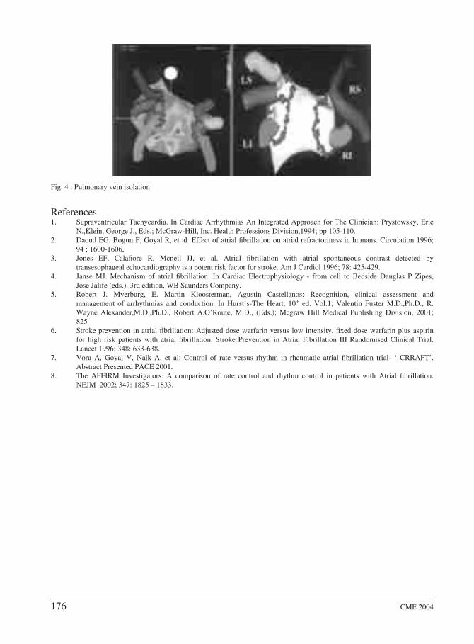
Pulmonary vein isolation (Figure 4) There is a suggestion that many patients with atrial fibrillation have the foci of the arrhythmia in
the pulmonary veins. Isolation of these foci followed by their radiofrequency catheter ablation is a new, effective curative procedure, which is still under investigation. Its success rate is about 50-70% with a recurrence rate of 20-30%, with complications like pulmonary vein stenosis, pericardial effusion occurring in about 10% of the patients.
ConclusionAtrial fibrillation continues to be a menacing problem, however, the recent advance in the non-drug therapy is promising to overcome it, hopefully in the near future. Till such time, the traditional approach of rate control with anticoagulation seems to be the mainstay of therapy for most patients.
Table 5 : Non-pharmacologic therapy
Type of AF Therapy
1. Paroxysmal AF Pacemaker therapy- overdrive atrial pacing or dual site right atrial or biatrial pacing.
2. Persistent AF Atrial defibrillation- internal cardioversion of the atria via percutaneously directed transvenous devices.
3. Permanent AF I) Radiofrequency ablation:
a) Palliative- Atrio-venticular node ablation with implantation of rate responsive pacemaker (VVIR pacing) - advisable in tachycardiomyopathy setting.
b) Curative-
i) Pulmonary vein isolation and ablation of the ectopic focus
ii) Catheter ‘maze’ procedure
II) Surgical ‘maze’
176 CME 2004
References1. Supraventricular Tachycardia. In Cardiac Arrhythmias An Integrated Approach for The Clinician; Prystowsky, Eric
N.,Klein, George J., Eds.; McGraw-Hill, Inc. Health Professions Division,1994; pp 105-110.2. Daoud EG, Bogun F, Goyal R, et al. Effect of atrial fibrillation on atrial refractoriness in humans. Circulation 1996;
94 : 1600-1606, 3. Jones EF, Calafiore R, Mcneil JJ, et al. Atrial fibrillation with atrial spontaneous contrast detected by
transesophageal echocardiography is a potent risk factor for stroke. Am J Cardiol 1996; 78: 425-429.4. Janse MJ. Mechanism of atrial fibrillation. In Cardiac Electrophysiology - from cell to Bedside Danglas P Zipes,
Jose Jalife (eds.). 3rd edition, WB Saunders Company.5. Robert J. Myerburg, E. Martin Kloosterman, Agustin Castellanos: Recognition, clinical assessment and
management of arrhythmias and conduction. In Hurst’s-The Heart, 10th ed. Vol.1; Valentin Fuster M.D.,Ph.D., R. Wayne Alexander,M.D.,Ph.D., Robert A.O’Route, M.D., (Eds.); Mcgraw Hill Medical Publishing Division, 2001; 825
6. Stroke prevention in atrial fibrillation: Adjusted dose warfarin versus low intensity, fixed dose warfarin plus aspirin for high risk patients with atrial fibrillation: Stroke Prevention in Atrial Fibrillation III Randomised Clinical Trial. Lancet 1996; 348: 633-638.
7. Vora A, Goyal V, Naik A, et al: Control of rate versus rhythm in rheumatic atrial fibrillation trial- ‘ CRRAFT’. Abstract Presented PACE 2001.
8. The AFFIRM Investigators. A comparison of rate control and rhythm control in patients with Atrial fibrillation. NEJM 2002; 347: 1825 – 1833.
Fig. 4 : Pulmonary vein isolation