Embed Size (px)
Citation preview

Chapter 20:
The Cardiovascular System:
The Heart

Video
■ Heart surgery in short -
https://youtu.be/MxP2cMgPx5s
■ Khan Academy The Heart:
https://youtu.be/7XaftdE_h60


Layers of the Heart Wall
1. Superior vena cava –
deoxygenated blood from the
anterior (head and chest).
2. Inferior vena cava –
deoxygenated blood from the
posterior (legs).
3. Right/left coronary artery –
oxygenated blood to the heart
muscle.
4. Cardiac veins –
deoxygenated blood from the
heart.

Pericardium and Heart Wall

Layers of the Heart Wall
1. Epicardium
(external layer)
2. Myocardium
❑ 95% of heart is
cardiac muscle
3. Endocardium (inner
layer)
❑ Smooth lining for
chambers of heart

Structure of the Heart
2 atria – receiving chambers
Auricles increase capacity
2 ventricles – pumping chambers

Internal Anatomy of the Heart

Right Atrium
❑ Receives blood from
■ Superior vena cava
■ Inferior vena cava
❑ Blood passes through
tricuspid valve (right
atrioventricular valve)
into right ventricle

Right Ventricle
■ Receives blood from
the right atrium.
■ Blood leaves through
pulmonary valve
(pulmonary semilunar
valve) into pulmonary
trunk and then right and
left pulmonary arteries
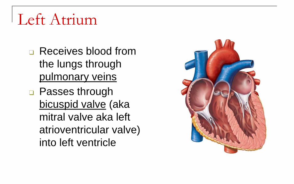
Left Atrium
❑ Receives blood from
the lungs through
pulmonary veins
❑ Passes through
bicuspid valve (aka
mitral valve aka left
atrioventricular valve)
into left ventricle

Left Ventricle
❑ Thickest chamber of the heart
❑ Blood passes through aortic valve (aortic semilunar valve) into ascending aorta
❑ Some blood flows into coronary arteries,remainder to body

Myocardial thickness
❑ Right ventricle pumps
blood to lungs
■ Shorter distance, lower
pressure, less
resistance
❑ Left ventricle pumps
blood to body
■ Longer distance,
higher pressure, more
resistance
Copyright 2009, John Wiley & Sons, Inc.
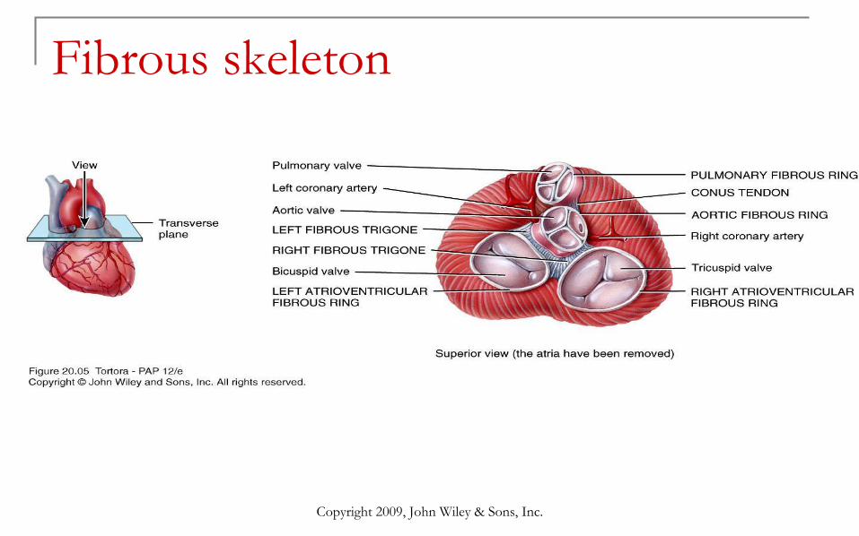
Fibrous skeleton

Copyright 2009, John Wiley & Sons, Inc.

Systemic and pulmonary
circulation - 2 circuits in series❑ Systemic circuit
■ Left side of heart
■ Receives blood from lungs
■ Ejects blood into aorta

Systemic and pulmonary
circulation - 2 circuits in series❑ Pulmonary circuit
■ Right side of heart
■ Receives blood from systemic circulation
■ Ejects blood into pulmonary trunk then to pulmonary arteries
■ Gas exchange in pulmonary capillaries
■ Pulmonary veins takes blood to left atrium

Autorhythmic Fibers
❑ Specialized cardiac
muscle fibers
❑ Repeatedly generate
action potentials that
trigger heart
contractions
❑ 2 important functions
1. Act as pacemaker
2. Form conduction
system

Conduction System
■ SA node acts as
natural pacemaker
❑ Faster than other
autorhythmic fibers
❑ Initiates 100 times per
second

Conduction System
■ Nerve impulses from
autonomic nervous
system (ANS) and
hormones modify
timing and strength
of each heartbeat

Heart Sounds
■ Auscultation
■ Sound of heartbeat
comes primarily from
blood turbulence
caused by closing of
heart valves
■ 4 heart sounds in each
cardiac cycle – only 2
loud enough to be
heard