Embed Size (px)
Citation preview

Chapter 20. Spine Medical Treatment Guidelines Subchapter A. Cervical Spine Injury
Editor’s Note: Form LWC-WC 1009. Disputed Claim for Medical Treatment has been moved to §2328 of this Part. §2001. Introduction
A. This document has been prepared by the Louisiana Workforce Commission, Office of Workers’ Compensation (OWCA) and should be interpreted within the context of guidelines for physicians/providers treating individuals qualifying under Louisiana’s Workers’ Compensation Act as injured workers with cervical spine injuries. These guidelines are enforceable under the Louisiana Workers Compensation Act. All medical care, services, and treatment owed by the employer to the employee in accordance with the Louisiana Workers’ Compensation Act shall mean care, services, and treatment in accordance with these guidelines. Medical care, services and treat Medical care, services, and treatment that varies from these guidelines shall also be due by the employer when it is demonstrated to the medical director of the office by a preponderance of the scientific medical evidence, that a variance from these guidelines is reasonably required to cure or relieve the injured worker from the effects of the injury or occupational disease given the circumstances. Therefore, these guidelines are not relevant as evidence of a provider’s legal standard of professional care. To properly utilize this document, the reader should not skip nor overlook any sections.
AUTHORITY NOTE: Promulgated in accordance with R.S. 23:1203.1. HISTORICAL NOTE: Promulgated by the Louisiana Workforce Commission, Office of Workers Compensation
Administration, LR 37:1631 (June 2011), amended by the Louisiana Workforce Commission, Office of Workers Compensation, LR 40:1119 (June 2014). §2003. General Guideline Principles
A. The principles summarized in this section are key to the intended implementation of all Office of Workers’ Compensation guidelines and critical to the reader’s application of the guidelines in this document.
1. Application of Guidelines. The OWCA provides procedures to implement medical treatment guidelines and to foster communication to resolve disputes among the provider, payer, and patient through the Office of Worker’s Compensation.
2. Education. Education of the patient and family, as well as the employer, insurer, policy makers and the community should be the primary emphasis in the treatment of workers’ compensation injuries. Currently, practitioners often think of education last, after medications, manual therapy, and surgery. Practitioners must develop and implement an effective strategy and skills to educate patients, employers, insurance systems, policy makers, and the community as a whole. An education-based paradigm should always start with inexpensive communication providing reassuring information to the patient. More in-depth education currently exists within a treatment regime employing functional restorative and innovative programs of prevention and rehabilitation. No treatment plan is complete without addressing issues of individual and/or group patient education as a means of facilitating self-management of symptoms and prevention.
3. Treatment Parameter Duration. Time frames for specific interventions commence once treatments have been initiated, not on the date of injury. Obviously, duration will be impacted by patient compliance, as well as availability of services. Clinical judgment may substantiate the need to accelerate or decelerate the time frames discussed in this document. Such deviation shall be in accordance with La. R.S. 23:1203.1.
4. Active Interventions. Emphasizing patient responsibility, such as therapeutic exercise and/or functional treatment, are generally emphasized over passive modalities, especially as treatment progresses. Generally, passive interventions are viewed as a means to facilitate progress in an active rehabilitation program with concomitant attainment of objective functional gains.
5. Active Therapeutic Exercise Program. Exercise program goals should incorporate patient strength, endurance, flexibility, coordination, and education. This includes functional application in vocational or community settings.
6. Positive Patient Response. Positive results are defined primarily as functional gains that can be objectively measured. Standard measurement tools, including outcome measures, should be used.
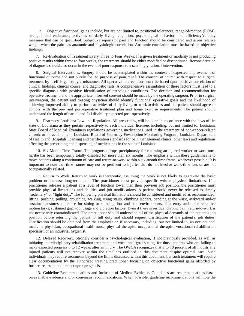
a. Objective functional gains include, but are not limited to, positional tolerances, range-of-motion (ROM), strength, and endurance, activities of daily living, cognition, psychological behavior, and efficiency/velocity measures that can be quantified. Subjective reports of pain and function should be considered and given relative weight when the pain has anatomic and physiologic correlation. Anatomic correlation must be based on objective findings.
7. Re-Evaluation of Treatment Every Three to Four Weeks. If a given treatment or modality is not producing positive results within three to four weeks, the treatment should be either modified or discontinued. Reconsideration of diagnosis should also occur in the event of poor response to a seemingly rational intervention.
8. Surgical Interventions. Surgery should be contemplated within the context of expected improvement of functional outcome and not purely for the purpose of pain relief. The concept of "cure” with respect to surgical treatment by itself is generally a misnomer. All operative interventions must be based upon positive correlation of clinical findings, clinical course, and diagnostic tests. A comprehensive assimilation of these factors must lead to a specific diagnosis with positive identification of pathologic conditions. The decision and recommendation for operative treatment, and the appropriate informed consent should be made by the operating surgeon. Prior to surgical intervention, the patient and treating physician should identify functional operative goals and the likelihood of achieving improved ability to perform activities of daily living or work activities and the patient should agree to comply with the pre- and post-operative treatment plan and home exercise requirements. The patient should understand the length of partial and full disability expected post-operatively.
9. Pharmacy-Louisiana Law and Regulation. All prescribing will be done in accordance with the laws of the state of Louisiana as they pertain respectively to each individual licensee, including, but not limited to: Louisiana State Board of Medical Examiners regulations governing medications used in the treatment of non-cancer-related chronic or intractable pain; Louisiana Board of Pharmacy Prescription Monitoring Program; Louisiana Department of Health and Hospitals licensing and certification standards for pain management clinics; other laws and regulations affecting the prescribing and dispensing of medications in the state of Louisiana.
10. Six Month Time Frame. The prognosis drops precipitously for returning an injured worker to work once he/she has been temporarily totally disabled for more than six months. The emphasis within these guidelines is to move patients along a continuum of care and return-to-work within a six-month time frame, whenever possible. It is important to note that time frames may not be pertinent to injuries that do not involve work-time loss or are not occupationally related.
11. Return to Work. Return to work is therapeutic, assuming the work is not likely to aggravate the basic problem or increase long-term pain. The practitioner must provide specific written physical limitations. If a practitioner releases a patient at a level of function lower than their previous job position, the practitioner must provide physical limitations and abilities and job modifications. A patient should never be released to simply “sedentary” or “light duty.” The following physical limitations should be considered and modified as recommended: lifting, pushing, pulling, crouching, walking, using stairs, climbing ladders, bending at the waist, awkward and/or sustained postures, tolerance for sitting or standing, hot and cold environments, data entry and other repetitive motion tasks, sustained grip, tool usage and vibration factors. Even if there is residual chronic pain, return-to-work is not necessarily contraindicated. The practitioner should understand all of the physical demands of the patient’s job position before returning the patient to full duty and should request clarification of the patient’s job duties. Clarification should be obtained from the employer or, if necessary, including, but not limited to, an occupational medicine physician, occupational health nurse, physical therapist, occupational therapist, vocational rehabilitation specialist, or an industrial hygienist.
12. Delayed Recovery. Strongly consider a psychological evaluation, if not previously provided, as well as initiating interdisciplinary rehabilitation treatment and vocational goal setting, for those patients who are failing to make expected progress 6 to 12 weeks after an injury. The OWCA recognizes that 3 to 10 percent of all industrially injured patients will not recover within the timelines outlined in this document despite optimal care. Such individuals may require treatments beyond the limits discussed within this document, but such treatment will require clear documentation by the authorized treating practitioner focusing on objective functional gains afforded by further treatment and impact upon prognosis.
13. Guideline Recommendations and Inclusion of Medical Evidence. Guidelines are recommendations based on available evidence and/or consensus recommendations. When possible, guideline recommendations will note the

level of evidence supporting the treatment recommendation. When interpreting medical evidence statements in the guideline, the following apply to the strength of recommendation.
Strong Level 1 Evidence We Recommend Moderate Level 2 and Level 3
Evidence We Suggest
Weak Level 4 Evidence Treatment is an Option Inconclusive Evidence is Either Insufficient of Conflicting
a. Consensus guidelines are generated by a professional organization that the guidelines are intended to
serve. A committee of specialists and experts are selected by the organization to create an unbiased, vetted recommendation for the treatment of specific issues within the realm of their expertise. All recommendations in the guideline are considered to represent reasonable care in appropriately selected cases, regardless of the level of evidence or consensus statement attached to it. Those procedures considered inappropriate, unreasonable, or unnecessary are designated in the guideline as “not recommended.”
B. The remainder of this document should be interpreted within the parameters of these guideline principles that may lead to more optimal medical and functional outcomes for injured workers.
AUTHORITY NOTE: Promulgated in accordance with R.S. 23:1203.1. HISTORICAL NOTE: Promulgated by the Louisiana Workforce Commission, Office of Workers Compensation
Administration, LR 37:1631 (June 2011), amended by the Louisiana Workforce Commission, Office of Workers Compensation, LR 40:1119 (June 2014).
§2005. Initial Diagnostic Procedures
A. The OWCA recommends the following diagnostic procedures be considered, at least initially, the responsibility of the workers’ compensation carrier to ensure that an accurate diagnosis and treatment plan can be established. Standard procedures, that should be utilized when initially diagnosing a work-related cervical spine complaint, are listed below.
1. History-taking and physical examination (Hx and PE). These are generally accepted, well-established and widely used procedures that establish the foundation/basis for and dictate subsequent stages of diagnostic and therapeutic procedures. When findings of clinical evaluations and those of other diagnostic procedures are not complementing each other, the objective clinical findings should have preference. The medical records should reasonably document the following.
a. History of Present Injury. A detailed history, taken in temporal proximity to the time of injury, should primarily guide evaluation and treatment. The history should include pertinent, positive and negative information regarding the following:
i. Mechanism of Injury. This includes details of symptom onset and progression. The mechanism of injury should include a detailed description of the incident and the position of the body before, during, and at the end of the incident. Inclusion of normal work body postures, frequency during the workday and lifting/push/pull requirements, should be included in the absence of a known specific incident;
ii. Location of pain, nature of symptoms, and alleviating/exacerbating factors (e.g. sleep positions). Of particular importance, is whether raising the arm over the head alleviates radicular-type symptoms. The history should include both the primary and secondary complaints (e.g., primary neck pain, secondary arm pain, headaches, and shoulder girdle complaints). The use of a patient completed pain drawing, Visual Analog Scale (VAS) is highly recommended, especially during the first two weeks following injury to assure that all work related symptoms are being addressed;
iii. presence and distribution of upper and/or lower extremity numbness, paresthesias, or weakness, especially if precipitated by coughing or sneezing;
iv. alteration of bowel, bladder, or sexual function; and for female patients, alteration in their menstrual cycle;
v. any treatment for current injury and result; and
vi. ability to perform job duties and activities of daily living.
b. Past history:

i. past medical history includes neoplasm, arthritis, and diabetes;
ii. review of systems includes symptoms of rheumatologic, neurologic, endocrine, neoplastic, infectious, and other systemic diseases;
iii. smoking history;
iv. vocational and recreational pursuits;
v. history of depression, anxiety, or other psychiatric illness.
vi. The examiner will screen for concurrent emotional disorders/conditions and, when possible, other known psychosocial predictors of poor outcome;
vii. prior occupational and non-occupational injuries to the same area including specific prior treatment, chronic or recurrent symptoms, and any functional limitations; specific history regarding prior motor vehicle accidents may be helpful.
c. Physical Examination should include accepted tests and exam techniques applicable to the area being examined, including:
i. general and visual inspection, including posture, stance and gait;
ii. palpation of spinous processes, facets, and muscles noting myofacial tightness, tenderness, and trigger points;
iii. cervical range-of-motion, quality of motion, and presence of muscle spasm. Motion evaluation of specific joints may be indicated. Range-of-motion should not be checked in acute trauma cases until fracture and instability have been ruled out on clinical examination, with or without radiographic evaluation;
iv. examination of thoracic spine;
v. motor and sensory examination of the upper muscle groups with specific nerve root focus, as well as sensation to light touch, pin prick, temperature, position and vibration. More than 2 cm difference in the circumferential measurements of the two upper extremities may indicate chronic muscle wasting; and
vi. Deep tendon reflexes. Asymmetry may indicate pathology. Inverted reflexes (e.g. arm flexion or triceps tap) may indicate nerve root or spinal cord pathology at the tested level. Pathologic reflexes include wrist, clonus, grasp reflex, and Hoffman’s sign.
d. Relationship to Work: This includes a statement of the probability that the illness or injury is work-related. If further information is necessary to determine work relatedness, the physician should clearly state what additional diagnostic studies or job information is required.
e. Spinal Cord Evaluation: In cases where the mechanism of injury, history, or clinical presentation suggests a possible severe injury, additional evaluation is indicated. A full neurological examination for possible spinal cord injury may include:
i. Sharp and light touch, deep pressure, temperature, and proprioceptive sensory function;
ii. strength testing;
iii. anal sphincter tone and/or perianal sensation;
iv. presence of pathological reflexes of the upper and lower extremities; or
v. evidence of an Incomplete Spinal Cord Injury Syndrome:
(a). Anterior Cord Syndrome is characterized by the loss of motor function and perception of pain and temperature below the level of the lesion with preservation of touch, vibration, and proprioception. This is typically seen after a significant compressive or flexion injury. Emergent CT or MRI is necessary to look for a possible reversible compressive lesion requiring immediate surgical intervention. The prognosis for recovery is the worst of the incomplete syndromes.
(b). Brown-Sequard Syndrome is characterized by ipsilateral motor weakness and proprioceptive disturbance with contralateral alteration in pain and temperature perception below the level of the lesion. This is

usually seen in cases of penetrating trauma or lateral mass fracture. Surgery is not specifically required, although debridement of the open wound may be.
(c). Central Cord Syndrome is characterized by sensory and motor disturbance of all limbs, often upper extremity more than lower, and loss of bowel and bladder function with preservation of perianal sensation. This is typically seen in elderly patients with a rigid spine following hyperextension injuries. Surgery is not usually required.
(d). Posterior Cord Syndrome, a rare condition, is characterized by loss of sensation below the level of the injury, but intact motor function.
vi. Spinal cord lesions should be classified according to the American Spine Injury Association (ASIA) impairment scale.
Asia Impairment Scale A=Complete: No motor or sensory function is preserved in the sacral segments S4-S5 B=Incomplete: Sensory but not motor function is preserved below the neurological level and includes the sacral segments S4-S5 C=Incomplete: Motor function is preserved below the neurological level, and more than half of key muscles below the neurological level have a muscle grade less than 3 D=Incomplete: Motor function is preserved below the neurological level, and at least half of key muscles below the neurological level have a grade of 3 or more E= Normal: motor and sensory function are normal
vii. A worksheet which details dermatomes and muscle testing required is available from ASIA.
f. Soft Tissue Injury Evaluation. Soft tissue injuries are traumatic injuries to the muscles, ligaments, tendons, and/or connective tissue. The most common mechanism is sudden hyperextension and/or hyperflexion of the neck. Acceleration/deceleration on the lateral plane may also result in one of these syndromes. A true isolated cervical strain is not associated with focal neurological symptoms. The signs and pathophysiology of these injuries are not well understood. Soft tissue injuries may include cervical strain, myofascial syndromes, somatic dysfunction, and fractures. The Quebec Classification is used to categorize soft tissue and more severe cervical injuries.
i. Grade Ineck complaints of pain, stiffness, or tenderness only, without physical signs. Lesion not serious enough to cause muscle spasm. Includes whiplash injury, minor cervical sprains, or strains.
ii. Grade IIneck complaints with musculoskeletal signs, such as limited range-of-motion. Includes muscle spasm related to soft tissue injury, whiplash, cervical sprain, and cervicalgia with headaches, sprained cervical facet joints and ligaments.
iii. Grade IIIneck complaints, such as limited range-of-motion, combined with neurologic signs. Includes whiplash, cervicobrachialgia, herniated disc, cervicalgia with headaches.
iv. Grade IVneck complaints with fracture or dislocation.
2. Radiographic imaging of the cervical spine is a generally accepted, well-established and widely used diagnostic procedure. Basic views are the anteroposterior (AP), lateral, right, and left obliques, swimmer’s, and odontoid. CT scans may be necessary to visualize C7 and odontoid in some patients. Lateral flexion and extension views are done to evaluate instability but may have a limited role in the acute setting. MRI or CT is indicated when spinal cord injury is suspected. The mechanism of injury and specific indications for the imaging should be listed on the request form to aid the radiologist and x-ray technician. Alert, non-intoxicated patients, who have isolated cervical complaints without palpable midline cervical tenderness, neurologic findings, or other acute or distracting injuries elsewhere in the body, may not require imaging. The following suggested indications are:
a. history of significant trauma, especially high impact motor vehicle accident, rollover, ejection, bicycle, or recreational vehicle collision or fall from height greater than one meter;
b. age over 65 years;
c. suspicion of fracture, dislocation, instability, or neurologic deficitQuebec Classification Grade III and IV;
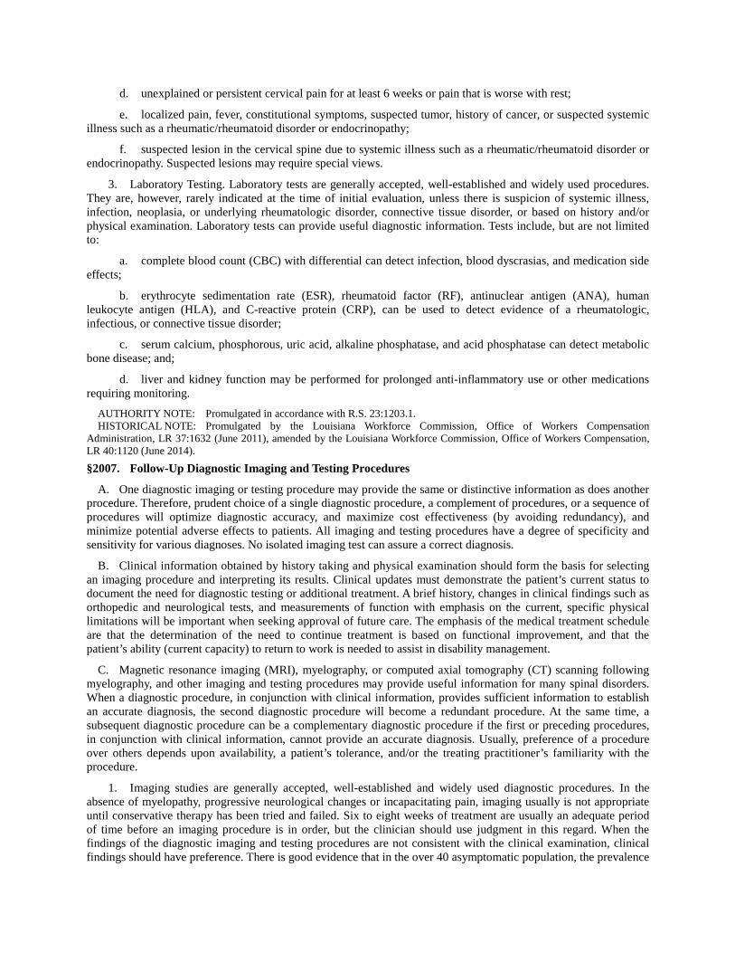
d. unexplained or persistent cervical pain for at least 6 weeks or pain that is worse with rest;
e. localized pain, fever, constitutional symptoms, suspected tumor, history of cancer, or suspected systemic illness such as a rheumatic/rheumatoid disorder or endocrinopathy;
f. suspected lesion in the cervical spine due to systemic illness such as a rheumatic/rheumatoid disorder or endocrinopathy. Suspected lesions may require special views.
3. Laboratory Testing. Laboratory tests are generally accepted, well-established and widely used procedures. They are, however, rarely indicated at the time of initial evaluation, unless there is suspicion of systemic illness, infection, neoplasia, or underlying rheumatologic disorder, connective tissue disorder, or based on history and/or physical examination. Laboratory tests can provide useful diagnostic information. Tests include, but are not limited to:
a. complete blood count (CBC) with differential can detect infection, blood dyscrasias, and medication side effects;
b. erythrocyte sedimentation rate (ESR), rheumatoid factor (RF), antinuclear antigen (ANA), human leukocyte antigen (HLA), and C-reactive protein (CRP), can be used to detect evidence of a rheumatologic, infectious, or connective tissue disorder;
c. serum calcium, phosphorous, uric acid, alkaline phosphatase, and acid phosphatase can detect metabolic bone disease; and;
d. liver and kidney function may be performed for prolonged anti-inflammatory use or other medications requiring monitoring.
AUTHORITY NOTE: Promulgated in accordance with R.S. 23:1203.1. HISTORICAL NOTE: Promulgated by the Louisiana Workforce Commission, Office of Workers Compensation
Administration, LR 37:1632 (June 2011), amended by the Louisiana Workforce Commission, Office of Workers Compensation, LR 40:1120 (June 2014).
§2007. Follow-Up Diagnostic Imaging and Testing Procedures
A. One diagnostic imaging or testing procedure may provide the same or distinctive information as does another procedure. Therefore, prudent choice of a single diagnostic procedure, a complement of procedures, or a sequence of procedures will optimize diagnostic accuracy, and maximize cost effectiveness (by avoiding redundancy), and minimize potential adverse effects to patients. All imaging and testing procedures have a degree of specificity and sensitivity for various diagnoses. No isolated imaging test can assure a correct diagnosis.
B. Clinical information obtained by history taking and physical examination should form the basis for selecting an imaging procedure and interpreting its results. Clinical updates must demonstrate the patient’s current status to document the need for diagnostic testing or additional treatment. A brief history, changes in clinical findings such as orthopedic and neurological tests, and measurements of function with emphasis on the current, specific physical limitations will be important when seeking approval of future care. The emphasis of the medical treatment schedule are that the determination of the need to continue treatment is based on functional improvement, and that the patient’s ability (current capacity) to return to work is needed to assist in disability management.
C. Magnetic resonance imaging (MRI), myelography, or computed axial tomography (CT) scanning following myelography, and other imaging and testing procedures may provide useful information for many spinal disorders. When a diagnostic procedure, in conjunction with clinical information, provides sufficient information to establish an accurate diagnosis, the second diagnostic procedure will become a redundant procedure. At the same time, a subsequent diagnostic procedure can be a complementary diagnostic procedure if the first or preceding procedures, in conjunction with clinical information, cannot provide an accurate diagnosis. Usually, preference of a procedure over others depends upon availability, a patient’s tolerance, and/or the treating practitioner’s familiarity with the procedure.
1. Imaging studies are generally accepted, well-established and widely used diagnostic procedures. In the absence of myelopathy, progressive neurological changes or incapacitating pain, imaging usually is not appropriate until conservative therapy has been tried and failed. Six to eight weeks of treatment are usually an adequate period of time before an imaging procedure is in order, but the clinician should use judgment in this regard. When the findings of the diagnostic imaging and testing procedures are not consistent with the clinical examination, clinical findings should have preference. There is good evidence that in the over 40 asymptomatic population, the prevalence

of disc degeneration is greater than 50 percent. Disc degeneration, seen as loss of signal intensity on MRI, may be due to age-related biochemical changes rather than structural deterioration, and may not have pathological significance. Disc bulging and posterior disc protrusion, while not rare, is more commonly symptomatic in the cervical spine than in the lumbar spine due to the smaller cervical spinal canal. Mild reduction in the cross-sectional area of the spinal cord may be seen without myelopathy in patients older than 40, therefore, clinical correlation is required. The studies below are listed in frequency of use, not importance.
a. Magnetic Resonance Imaging (MRI) is the imaging study of choice for most abnormalities of the cervical spine. MRI is useful in suspected nerve root compression, in myelopathy to evaluate the spinal cord and/or masses, infections such as epidural abscesses or disc space infection, bone marrow involvement by metastatic disease, and/or suspected disc herniation or cord contusion following severe neck injury. MRI should be performed immediately if there is a question of infection or metastatic disease with cord compression. MRI is contraindicated in patients with certain implanted devices.
i. In general, the high field, conventional, MRI provides better resolution. A lower field scan may be indicated when a patient cannot fit into a high field scanner or is too claustrophobic despite sedation. Inadequate resolution on the first scan may require a second MRI using a different technique. All questions in this regard should be discussed with the MRI center and/or radiologist.
ii. Specialized MRI Scans
(a). MRI with Three-Dimensional Reconstruction. On rare occasions, MRI with three-dimensional reconstruction views may be used as a pre-surgical diagnostic procedure to obtain accurate information of characteristics, location, and spatial relationships among soft tissue and bony structures;
(b). Dynamic-Kinetic MRI of the Spine. Dynamic-kinetic MRI of the spine uses an MRI unit configured with a top-front open design which enables upright, weight-bearing patient positioning in a variety of postures not obtainable with the recumbent images derived from conventional, closed unit MRI systems. Imaging can be obtained in flexion, extension, and rotation of the spine, as well as in erect positioning. There is a theoretical advantage to imaging sequences obtained under more physiologic conditions than in the supine position. There is currently ongoing research to establish whether the theoretical advantages of positional and kinetic MRI result in improved sensitivity and specificity in detecting spine pathology. Currently it remains investigational, and is not recommended until the correlation with clinical syndromes is firmly established.
b. Computed axial tomography (CT) provides excellent visualization of bone and is used to further evaluate bony masses and suspected fractures not clearly identified on radiographic evaluation. It may sometimes be done as a complement to MRI scanning to better delineate bony osteophyte formation in the neural foramen. CT is usually utilized for suspected cervical spine fracture in a patient with negative plain films, or to further delineate a cervical fracture. CT scanning is also quite useful for congenital anomalies at the skull base and at the C1-2 levels. Plain CT scanning is poor for the C6-7 or C7-T1 levels because of shoulder artifact. Instrument-scatter reduction software provides better resolution when metallic artifact is of concern.
c. Myelography is the injection of radiopaque material into the spinal subarachnoid space, with x-rays then taken to define anatomy. It may be used as a diagnostic procedure to obtain accurate information of characteristics, location, and spatial relationships among soft tissue and bony structures. Myelography is an invasive procedure with complications including nausea, vomiting, headache, convulsion, arachnoiditis, CSF leakage, allergic reactions, bleeding, and infection. Therefore, myelography should only be considered when CT and MRI are unavailable, for morbidly obese patients or those who have undergone multiple operations, and when other tests prove non-diagnostic. The use of small needles and a less toxic, water-soluble, nonionic contrast is recommended.
d. CT myelogram provides more detailed information about relationships between neural elements and surrounding anatomy and is appropriate in patients with multiple prior operations or tumorous conditions.
e. Single Photon Emission Computerized Tomography (SPECT). A scanning technique which may be helpful to localize facet joint pathology and is useful in determining which patients are likely to have a response to facet injection. SPECT combines bone scans and CT scans in looking for facet joint pathology.
f. Bone scan (radioisotope bone scanning) is generally accepted, well-established and widely used. Bone scanning is more sensitive but less specific than MRI. 99MTechnetium diphosphonate uptake reflects osteoblastic activity and may be useful in diagnosing metastatic/primary bone tumors, stress fractures, osteomyelitis, and

inflammatory lesions, but cannot distinguish between these entities. In the cervical spine, the usual indication is to evaluate for neoplastic conditions. Other indications include occult fracture or infection.
g. Other radioisotope scanning indium and gallium scans are generally accepted, well-established, and widely used procedures, usually to help diagnose lesions seen on other diagnostic imaging studies. 67Gallium citrate scans are used to localize tumor, infection, and abscesses. 111Indium-labeled leukocyte scanning is utilized for localizing infection or inflammation and is usually not used for the cervical spine.
h. Dynamic [digital] fluoroscopy dynamic [digital] fluoroscopy of the cervical spine measures the motion of intervertebral segments using a videofluoroscopy unit to capture images as the subject performs cervical flexion and extension, storing the anatomic motion of the spine in a computer. Dynamic Fluoroscopy may be used in state-designated trauma centers to evaluate the cervical spine. Its superiority over MRI has not been established. If performed, full visualization of the cervical spine (C1 - T1), in accordance with §2005.A.2. (Initial Diagnostic Procedures-Imaging), should be accomplished prior to the procedure. In the post-acute setting in some rare cases, Dynamic [Digital] Fluoroscopy may be used but is primarily an investigational tool and therefore, requires prior authorization in the post-acute setting. No studies have yet demonstrated predictive value in terms of standard operative and non-operative therapeutic outcomes.
2. Other Tests. The following diagnostic procedures are listed in alphabetical order, not by importance.
a. Electrodiagnostic Testing
i. Electromyography (EMG), and Nerve Conduction Studies. (NCS). These are generally accepted, well-established and widely used diagnostic procedures. EMG and NCS, when performed and interpreted by a trained physician/electrophysiologist, may be useful for patients with suspected neural involvement whose symptoms are persistent or unresponsive to initial conservative treatments. They are used to differentiate peripheral neural deficits from radicular and spinal cord neural deficits and to rule out concomitant myopathy. However, F-Wave Latencies are not diagnostic for radiculopathy.
ii. In general, EMG and NCS are complementary to imaging procedures such as CT, MRI, and/or myelography or diagnostic injection procedures. Electrodiagnostic studies may provide useful, correlative neuropathophysiological information that would be otherwise unobtainable from the radiologic studies discussed above.
iii. Portable Automated Electrodiagnostic Device (also known as Surface EMG) this is not a substitute for conventional diagnostic testing in clinical decision-making and therefore, is not recommended.
iv. Somatosensory Evoked Potential (SSEP) is useful for the evaluation of myelopathy. It is not recommended to identify radiculopathy.
v. Current Perception Threshold Evaluation (CPT) may be useful as a screening tool, but its diagnostic efficacy in the evaluation of cervical spine pain has not been determined. Therefore, CPT is not recommended as a diagnostic tool
b. InjectionsDiagnostic
i. Description Diagnostic cervical injections are generally accepted well-established procedures. These injections may be useful for localizing the source of pain, and may have added therapeutic value when combined with injection of therapeutic medication(s). Each diagnostic injection has inherent risks, and risk versus benefit should always be evaluated when considering injection therapy.
ii. Since these procedures are invasive, less invasive or non-invasive procedures should be considered first. Selection of patients, choice of procedure, and localization of the level for injection should be determined by clinical information indicating strong suspicion for pathologic condition(s) and the source of pain symptoms. Because injections are invasive with an inherent risk, the number of diagnostic procedures should be limited in any individual patient to those most likely to be primary pain generators. Patients should not receive all of the diagnostic blocks listed merely in an attempt to identify 100 percent of the pain generators.
iii. The interpretation of the test results are primarily based on functional change, symptom report, and pain response (via a recognized pain scale before and at an appropriate time after the injection). The diagnostic significance of the test result should be evaluated in conjunction with clinical information and the results of other diagnostic procedures. Injections with local anesthetics of differing duration may be used to support a diagnosis. In

some cases, injections at multiple levels may be required to accurately diagnose cervical conditions. Refer to Injections – Therapeutic for information on specific injections.
(a). It is obligatory that sufficient data be accumulated by the examiner performing this procedure such that the diagnostic value of the procedure is evident to other reviewers. This entails, at a minimum, documentation of patient response immediately following the procedure with details of any symptoms with a response and the degree of response. Additionally, a log must be recorded as part of the medical records which documents response, if any, on an hourly basis for, at a minimum, the expected duration of local anesthetic phase of the procedure. Responses must be identified as to specific body part (e.g., neck, arm pain). The practitioner must identify the local anesthetic used and the expected duration of response for diagnostic purposes.
(b). Multiple injections provided at the same session without staging may seriously dilute the diagnostic value of these procedures. Practitioners must carefully weigh the diagnostic value of the procedure against the possible therapeutic value.
iv. Special Requirements for Diagnostic Injections. Since multi-planar fluoroscopy during procedures is required to document technique and needle placement, an experienced physician should perform the procedure. Permanent images are required to verify needle placement. The subspecialty disciplines of the physicians performing the injections may be varied, including, but not limited to: anesthesiology, radiology, surgery, or physiatry. The practitioner should have completed fellowship training in pain medicine with interventional training, or its equivalent. They must also be knowledgeable in radiation safety.
v. Complications. General complications of diagnostic injections may include transient neurapraxia, nerve injury, infection, headache, vasovagal effects, as well as epidural hematoma, permanent neurologic damage, dural perforation and CSF leakage, and spinal meningeal abscess. Severe complications are remote but can include spinal cord damage, quadriplegia, and/or death. Injections at a C2-C3 level frequently cause temporary neuritis with ataxia. vi. Contraindications
(a). Absolute contraindications to diagnostic injections include:
(i). bacterial infectionsystemic or localized to region of injection;
(ii). bleeding diatheses;
(iii). hematological conditions; and
(iv). possible pregnancy.
(b). Relative contraindications to diagnostic injections may include: allergy to contrast, poorly controlled diabetes mellitus, and hypertension.
(c). Drugs affecting coagulation may require restriction from use. Anti-platelet therapy and anti-coagulations should be addressed individually by a knowledgeable specialist. It is recommended to refer to the American Society of Regional Anesthesia for anticoagulation guidelines.
vii. Specific Diagnostic Injections. In general, relief should last for at least the duration of the local anesthetic used and should significantly relieve pain and result in functional improvement. Refer to “InjectionsTherapeutic” for information on specific therapeutic injections.
(a). Medial branch blocks are generally-accepted diagnostic injections, used to determine whether a patient is a candidate for radiofrequency medial branch neurotomy (also known as facet rhizotomy). The International Spine Intervention Society (ISIS) suggests controlled blocksusing either placebo or an anesthetic with a varying length of activity (i.e., bupivacaine longer than lidocaine). To be a positive diagnostic block, the patient should report a reduction of pain of 50 percent or greater from baseline for the length of time appropriate for the local anesthetic used. In almost all cases, this will mean a reduction of pain to 1 or 2 on the Visual Analog Scale (VAS) 10-point scale correlated with functional improvement. The patient should also identify activities of daily living (which may include measurements of range-of-motion) that are impeded by their pain and can be observed to document functional improvement in the clinical setting. Ideally, these activities should be assessed throughout the observation period for function. The observer should not be the physician who performed the procedure. It is suggested that this be recorded on a form similar to ISIS recommendations.
(i). A separate comparative block on a different date may be performed to confirm the level of involvement. A comparative block uses anesthetics with varying lengths of activity. Medial Branch blocks are probably not helpful to determine the likelihood of success for spinal fusion.
(ii). Frequency and maximum duration may be repeated once for comparative blocks. Limited to four levels / five medial branches.
(b). Atlanto-axial and atlanto-occipital injections are generally accepted for diagnosis and treatment but do not lend themselves to denervation techniques owing to variable neuroanatomy. Injection of this articulation is complicated by the proximity of the vertebral artery, which may be tortuous at the level of the C1 joint. Inadvertent injection of the vertebral artery may cause respiratory arrest, seizure, stroke, or permanent neurological sequelae. Only practitioners skilled in these injections should perform them:
(i). frequency and maximum duration: once per side.
(c). Transforaminal injections / Spinal selective nerve root blocks are generally accepted and useful in identifying spinal pathology. When performed for diagnosis, small amounts of local anesthetic should be used to determine the level of nerve root irritation. A positive diagnostic block should result in a positive diagnostic functional benefit and a 50 percent reduction in nerve-root generated pain appropriate for the anesthetic used as measured by accepted pain scales (such as a VAS):
(i). time to produce effect: less than 30 minutes for local anesthesia; corticosteroids up to 72 hours for most patients;
(ii). frequency and maximum duration: once per suspected level. limited to two levels.
(d). Zygapophyseal (Facet) Blocks. Facet blocks are generally accepted but should not be considered diagnostic blocks for the purposes of determining the need for a rhizotomy (radiofrequency medial branch neurotomy), nor should they be done with medial branch blocks. These blocks should not be considered a definitive diagnostic tool. They may be used diagnostically to direct functional rehabilitation programs. A positive diagnostic block should result in a positive diagnostic functional benefit and a 50 percent reduction in pain appropriate for the anesthetic used as measured by accepted pain scales (such as a VAS). They then may be repeated per the therapeutic guidelines when they are accompanied by a functional rehabilitation program. (Refer to Therapeutic Spinal Injections):
(i). time to produce effect: less than 30 minutes for local anesthesia; corticosteroids up to 72 hours for most patients;
(ii). frequency and maximum duration: once per suspected level, limited to two levels.
c. Personality/ Psychological/ Psychiatric/ Psychosocial Evaluation. These are generally accepted and well-established diagnostic procedures with selective use in the upper extremity population, but have more widespread use in subacute and chronic upper extremity populations. Diagnostic testing procedures may be useful for patients with symptoms of depression, delayed recovery, chronic pain, recurrent painful conditions, disability problems, and for preoperative evaluation. Psychological/psychosocial and measures have been shown to have predictive value for postoperative response, and therefore should be strongly considered for use pre-operatively when the surgeon has concerns about the relationship between symptoms and findings, or when the surgeon is aware of indications of psychological complication or risk factors for psychological complication (e.g. childhood psychological trauma). Psychological testing should provide differentiation between pre-existing conditions versus injury caused psychological conditions, including depression and posttraumatic stress disorder. Psychological testing should incorporate measures that have been shown, empirically, to identify comorbidities or risk factors that are linked to poor outcome or delayed recovery.
i. Formal psychological or psychosocial evaluation should be performed on patients not making expected progress within 6 to 12 weeks following injury and whose subjective symptoms do not correlate with objective signs and test results. In addition to the customary initial exam, the evaluation of the injured worker should specifically address the following areas:
(a). employment history;
(b). interpersonal relationships-both social and work;
(c). patient activities;
(d). current perception of the medical system;
(e). current perception/attitudes toward employer/job
(f). results of current treatment
(g). risk factors and psychological comorbidities that may influence outcome and that may require treatment
(h). childhood history, including history of childhood psychological trauma, abuse and family history of disability.
ii. Personality/ psychological/ psychosocial evaluations consist of two components, clinical interview and psychological testing. Results should help clinicians with a better understanding of the patient in a number of ways. Thus the evaluation result will determine the need for further psychosocial interventions; and in those cases, Diagnostic and Statistical Manual of Mental Disorders (DSM) diagnosis should be determined and documented. The evaluation should also include examination of both psychological comorbidities and psychological risk factors that are empirically associated with poor outcome and/or delayed recovery. An individual with a Ph.D., Psy.D, or psychiatric M.D./D.O. credentials should perform initial evaluations, which are generally completed within one to two hours. A professional fluent in the primary language of the patient is preferred. When such a provider is not available, services of a professional language interpreter should be provided.
iii. Frequency: one-time visit for the clinical interview. If psychometric testing is indicated as a part of the initial evaluation, time for such testing should not exceed an additional two hours of professional time.
d. Provocation Discography
i. Description. Discography is an accepted, but rarely indicated, invasive diagnostic procedure to identify or refute a discogenic source of pain for patients who are surgical candidates. Discography should only be performed by physicians who are experienced and have been proctored in the technique. Discograms have a significant false positive rate. It is essential that all indications, preconditions, special considerations, procedures, reporting requirements, and results, are carefully and specifically followed. Results should be interpreted judiciously. Fewer studies have been published on cervical and thoracic discography than on lumbar discography.
ii. Indications. Discography may be indicated when a patient has a history of functionally limiting, unremitting cervical pain of greater than four months duration, with or without arm pain, which has been unresponsive to all conservative interventions. A patient who does not desire operative therapeutic intervention is not a candidate for an invasive non-therapeutic intervention, such as provocation discography.
iii. Discography may prove useful for the evaluation of the pre-surgical spine, discogenic pain at levels above or below a prior spinal fusion, annular tear, or internal disc disruption.
iv. Discography may show disc degeneration and annular disruption in the absence of cervical pain. Discography may also elicit concordant pain in patients with mild and functionally inconsequential neck pain. Because patients with mild neck pain should not be considered for invasive treatment, discography should not be performed on these patients. The presence of an annular tear does not necessarily identify the tear as a pain generator.
v. Discography is not useful in previously operated discs. Discography may prove useful in evaluating the number of cervical spine levels that might require fusion. CT Discography provides further detailed information about morphological abnormalities of the disc and possible lateral disc herniations.
vi. Preconditions for provocation discography include all of the following:
(a). A patient with functionally limiting, unremitting neck and/or arm pain of greater than four months duration in whom conservative treatment has been unsuccessful and in whom the specific diagnosis of the pain generator has not been made apparent on the basis of other noninvasive imaging studies (e.g., MRI, CT, plain films, etc.). It is recommended that discography be reserved for use in patients with equivocal MRI findings, especially at levels adjacent to clearly pathological levels. Discography may be more sensitive than MRI or CT in detecting radial annular tears. However, radial tears must always be correlated with clinical presentation.
(b). Psychosocial evaluation has been completed. There is some evidence that false positives and complaints of long-term pain arising from the procedure itself occur more frequently in patients with somatoform

disorders. Therefore, discograms should not be performed on patients with non-anatomic symptoms consistent with somatoform disorders.
(c). Patients who are considered surgical candidates (e.g., symptoms are of sufficient magnitude and the patient has been informed of the possible surgical options that may be available based upon the results of discography). Discography should never be the sole indication for surgery.
(d). Informed consent regarding the risks and potential diagnostic benefits of discography has been obtained.
vii. Complications include, but are not limited to, discitis, nerve damage, retropharyngeal abscess, chemical meningitis, pain exacerbation, and anaphylaxis. Therefore, prior to consideration of discography, the patient should undergo other diagnostic modalities in an effort to define the etiology of the patient's complaint including psychological evaluation, myelography, CT and MRI.
viii. Contraindications include:
(a). active infection of any type or continuing antibiotic treatment for infection; and/or
(b). bleeding diathesis or pharmaceutical anticoagulation with warfarin, etc.; and/or
(c). significant spinal stenosis at the level being studied as visualized by MRI, myelography or CT scan; and/or
(d). presence of clinical myelopathy; and/or
(e). effacement of the cord, thecal sac or circumferential absence of epidural fat; and
(f). known allergic reactions.
ix. Special Considerations
(a). Discography should not be performed by the physician expected to perform the therapeutic procedure. The procedure should be carried out by an experienced individual who has received specialized training in the technique of provocation discography.
(b). Discography should be performed in a blinded format that avoids leading the patient with anticipated responses. The procedure should always include one or more disc levels thought to be normal or nonpainful in order to serve as an internal control. The patient should not know what level is being injected in order to avoidspurious results. Adjacent discs may be identified as pain generators in more than half of cases in which discogenic pain is identified at one level. Because surgery is likely to fail in multi-level discogenic pain, injection of as many levels as feasible can prevent many operative failures. Abnormal disc levels may be repeated to confirm concordance.
(c). Sterile technique must be utilized.
(d). Judicious use of light sedation during the procedure is acceptable, represents the most common practice nationally at the current time, and is recommended by most experts in the field. The patient must be awake and able to accurately report pain levels during the provocation portion of the procedure.
(e). CT or MRI should establish cervical spinal dimensions and ruled out spinal stenosis.
(f). Intradiscal injection of local anesthetic may be carried out after the provocation portion of the examination and the patient’s response.
(g). It is recommended that a post-discogram CT be considered as it frequently provides additional useful information about disc morphology or other pathology.
x. Reporting of Discography. In addition to a narrative report, the discography report should contain a standardized classification of disc morphology and the pain response. All results should be clearly separated in the report from the narrative portion. Asymptomatic annular tears are common and the concordant pain response is an essential finding for a positive discogram.
xi. When discography is performed to identify the source of a patient’s neck pain, both a concordant pain response and morphological abnormalities must be present at the pathological level prior to initiating any treatment
directed at that level. The patient must be awake during the provocation phase of the procedure; therefore, sedative medication must be carefully titrated.
xii. Caution should be used when interpreting results from discography. One study using asymptomatic volunteers reported pain in the majority of discs injected, but no subjects reported pain exceeding 6/10 on a pain scale in a normal disc.
xiii. Reporting disc morphology as visualized by the post-injection CT scan (when available) should follow the Modified Dallas Discogram Scale where:
(a). Grade 0 = Normal Nucleus.
(b). Grade 1 = Annular tear confined to inner one-third of annulus fibrosis.
(c). Grade 2 = Annular tear extending to the middle third of the annulus fibrosis.
(d). Grade 3 = Annular tear extending to the outer one-third of the annulus fibrosis.
(e). Grade 4 = A grade 3 tear plus dissection within the outer annulus to involve more than 30 degrees of the disc circumference.
(f). Grade 5 = Full thickness tear with extra-annular leakage of contrast, either focal or diffuse.
xiv. Reporting of pain response should be consistent with the operational criteria of the International Spine Intervention Society Guidelines (ISIS). The report must include the level of concordance for neck and arm pain separately using a 10-point VAS, or similar quantitative assessment. It should be noted that the change in the VAS score before and after provocation is more important than the number reported.
xv. The diagnosis of discogenic pain is less likely when there are more discs with dissimilar pain and fewer with no pain. At least two discs with no pain on stimulation and one disc with concordant pain registering at least 7 on a 10-point VAS or equivalent should be present to qualify for a diagnosis of discogenic pain. The VAS score prior to the discogram should be taken into account when interpreting the VAS score reported by the patient during the discogram.
(a). Time parameters for provocation discography are as follows:
(i). frequency: one time only;
(ii). maximum: repeat discography is rarely indicated.
xvi. Thermography is an accepted and established procedure, but has no use as a diagnostic test for cervical pain. It may be used to diagnose regional pain disorders and in these cases, refer to the Complex Regional Pain Syndrome/Reflex Sympathetic Dystrophy Medical Treatment Guidelines.
3. Special tests are generally well-accepted tests and are performed as part of a skilled assessment of the patients’ capacity to return-to-work, his/her strength capacities, and physical work demand classifications and tolerance. The procedures in this subsection are listed in alphabetical order, not by importance.
a. Computer-Enhanced Evaluations may include isotonic, isometric, isokinetic and/or isoinertial measurement of movement, range-of-motion, endurance, or strength. Values obtained can include degrees of motion, torque forces, pressures, or resistance. Indications include determining validity of effort, effectiveness of treatment and demonstrated motivation. These evaluations should not be used alone to determine return-to-work restrictions.
i. Frequency: One time for evaluation. Can monitor improvements in strength every three to four weeks up to a total of six evaluations.
b. Functional Capacity Evaluation (FCE): is a comprehensive or modified evaluation of the various aspects of function as they relate to the worker’s ability to return to work. Areas such as endurance, lifting (dynamic and static), postural tolerance, specific range-of-motion, coordination and strength, worker habits, employability, as well as psychosocial, cognitive, and sensory perceptual aspects of competitive employment may be evaluated. Components of this evaluation may include: musculoskeletal screen; cardiovascular profile/aerobic capacity; coordination; lift/carrying analysis; job-specific activity tolerance; maximum voluntary effort; pain assessment/psychological screening; non-material and material handling activities cognitive; visual; and sensory perceptual factors.

i. When an FCE is being used to determine return to a specific jobsite, the provider is responsible for fully understanding the job duties. A jobsite evaluation is frequently necessary. FCEs cannot be used in isolation to determine work restrictions. The authorized treating physician must interpret the FCE in light of the individual patient's presentation and medical and personal perceptions. FCEs should not be used as the sole criteria to diagnose malingering.
ii. Full FCEs are sometimes not necessary. If Partial FCEs are performed, it is recognized that all parts of the FCE that are not performed are considered normal. In many cases, a work tolerance screening will identify the ability to perform the necessary job tasks.
iii. Frequency: can be used initially to determine baseline status. Additional evaluations can be performed to monitor and assess progress and aid in determining the endpoint for treatment.
c. Job site evaluation is a comprehensive analysis of the physical, mental and sensory components of a specific job. These components may include, but are not limited to; postural tolerance (static and dynamic); aerobic requirements; range-of-motion; torque/force; lifting/carrying; cognitive demands; social interactions; visual perceptual; sensation; coordination; environmental requirements of a job; repetitiveness; and essential job functions.
i. Job descriptions provided by the employer are helpful but should not be used as a substitute for direct observation. A jobsite evaluation may include observation and instruction of how work is done, what material changes (desk, chair) should be made, and determination of readiness to return-to-work.
ii. Requests for a jobsite evaluation should describe the expected goals for the evaluation. Goals may include, but are not limited to the following.
(a). to determine if there are potential contributing factors to the person’s condition and/or for the physician to assess causality;
(b). to make recommendations for, and to assess the potential for ergonomic changes;
(c). to determine the essential demands of the job. To provide a detailed description of the physical and cognitive job requirements;
(d). to assist the patient in their return-to-work by educating them on how they may be able to do their job more safely and in a more bio-mechanically appropriate manner;
(e). to give detailed work/activity restrictions.
iii. Frequency: One time with additional visits as needed for follow-up per jobsite.
d. Vocational Assessment. The vocational assessment should provide valuable guidance in the determination of future rehabilitation program goals. It should clarify rehabilitation goals, which optimize both patient motivation and utilization of rehabilitation resources. If prognosis for return to former occupation is determined to be poor, except in the most extenuating circumstances, vocational assessment should be implemented within 3 to 12 months post-injury. Declaration of MMI should not be delayed solely due to lack of attainment of a vocational assessment:
i. frequency: one time with additional visits as needed for follow-up.
e. Work tolerance screening is a determination of an individual's tolerance for performing a specific job based on a job activity or task and may be used when a full Functional Capacity Evaluation is not indicated. It may include a test or procedure to specifically identify and quantify work-relevant cardiovascular, physical fitness and postural tolerance. It may also address ergonomic issues affecting the patient’s return-to-work potential. Full job description should include a physical assessment of the job requirements:
i. frequency: one time for initial screen. May monitor improvements in strength every 3 to 4 weeks up to a total of six visits.
AUTHORITY NOTE: Promulgated in accordance with R.S. 23:1203.1. HISTORICAL NOTE: Promulgated by the Louisiana Workforce Commission, Office of Workers Compensation
Administration, LR 37:1634 (June 2011), amended by the Louisiana Workforce Commission, Office of Workers Compensation, LR 40:1121 (June 2014).

§2009. Therapeutic ProceduresNon-Operative
A. Before initiation of any therapeutic procedure, the authorized treating provider, employer, and insurer must consider these important issues in the care of the injured worker.
B. First, patients undergoing therapeutic procedure(s) should be released or returned to modified or restricted duty during their rehabilitation at the earliest appropriate time. Refer to “Return-to-Work” in this section for detailed information.
C. Second, cessation and/or review of treatment modalities should be undertaken when no further significant subjective or objective improvement in the patient’s condition is noted. If patients are not responding within the recommended duration periods, alternative treatment interventions, further diagnostic studies or consultations should be pursued.
1. Reassessment of the patient’s status in terms of functional improvement should be documented after each treatment. If patients are not responding within the recommended time periods, alternative treatment interventions, further diagnostic studies or consultations should be pursued. Continued treatment should be monitored using objective measures such as:
a. return-to-work or maintaining work status;
b. fewer restrictions at work or performing activities of daily living;
c. decrease in usage of medications;
d. measurable functional gains, such as increased range of motion or documented increase in strength;
D. Third, providers should provide and document education to the patient. No treatment plan is complete without addressing issues of individual and/or group patient education as a means of facilitating self-management of symptoms
E. Lastly, formal psychological or psychosocial evaluation should be performed on patients not making expected progress within 6 to 12 weeks following injury and whose subjective symptoms do not correlate with objective signs and tests.
F. Home therapy is an important component of therapy and may include active and passive therapeutic procedures as well as other modalities to assist in alleviating pain, swelling, and abnormal muscle tone.
G. The following procedures are listed in alphabetical order.
1. Acupuncture is an accepted and widely used procedure for the relief of pain and inflammation, and there is some scientific evidence to support its use. The exact mode of action is only partially understood. Western medicine studies suggest that acupuncture stimulates the nervous system at the level of the brain, promotes deep relaxation, and affects the release of neurotransmitters. Acupuncture is commonly used as an alternative or in addition to traditional Western pharmaceuticals. While it is commonly used when pain medication is reduced or not tolerated, it may be used as an adjunct to physical rehabilitation and/or surgical intervention to hasten the return of functional activity. Acupuncture should be performed by licensed practitioners.
a. Acupuncture is the insertion and removal of filiform needles to stimulate acupoints (acupuncture points). Needles may be inserted, manipulated, and retained for a period of time. Acupuncture can be used to reduce pain, reduce inflammation, increase blood flow, increase range-of-motion, decrease the side effect of medication-induced nausea, promote relaxation in an anxious patient, and reduce muscle spasm. Indications include joint pain, joint stiffness, soft tissue pain and inflammation, paresthesia, post-surgical pain relief, muscle spasm, and scar tissue pain.
b. Acupuncture with Electrical Stimulation: is the use of electrical current (micro-amperage or milli-amperage) on the needles at the acupuncture site. It is used to increase effectiveness of the needles by continuous stimulation of the acupoint. Physiological effects (depending on location and settings) can include endorphin release for pain relief, reduction of inflammation, increased blood circulation, analgesia through interruption of pain stimulus, and muscle relaxation.
i. It is indicated to treat chronic pain conditions, radiating pain along a nerve pathway, muscle spasm, inflammation, scar tissue pain, and pain located in multiple sites.

c. Total Time Frames for Acupuncture and Acupuncture with Electrical Stimulation: Time frames are not meant to be applied to each of the above sections separately. The time frames are to be applied to all acupuncture treatments regardless of the type or combination of therapies being provided.
i. Time to Produce Effect: three to six treatments
ii. Frequency: one to three times per week.
iii. Optimum Duration: one to two months.
iv. Maximum Duration: 14 treatments.
v. Any of the above acupuncture treatments may extend longer if objective functional gains can be documented or when symptomatic benefits facilitate progression in the patient’s treatment program. Treatment beyond 14 treatments must be documented with respect to need and ability to facilitate positive symptomatic or functional gains. Such care should be re-evaluated and documented with each series of treatments.
d. Other Acupuncture Modalities. Acupuncture treatment is based on individual patient needs and therefore treatment may include a combination of procedures to enhance treatment effect. Other procedures may include the use of heat, soft tissue manipulation/massage, and exercise. Refer to active therapy (therapeutic exercise) and passive therapy sections (massage and superficial heat and cold therapy) for a description of these adjunctive acupuncture modalities and time frames.
2. Biofeedback is a form of behavioral medicine that helps patients learn self-awareness and self-regulation skills for the purpose of gaining greater control of their physiology, such as muscle activity, brain waves, and measures of autonomic nervous system activity. Electronic instrumentation is used to monitor the targeted physiology and then displayed or fed back to the patient visually, auditorially, or tactilely, with coaching by a biofeedback specialist. Biofeedback is provided by clinicians certified in biofeedback and/or who have documented specialized education, advanced training, or direct or supervised experience qualifying them to provide the specialized treatment needed (e.g., surface EMG, EEG, or other).
a. Treatment is individualized to the patient’s work-related diagnosis and needs. Home practice of skills is required for mastery and may be facilitated by the use of home training tapes. The ultimate goal of biofeedback treatment is to normalize the physiology to the pre-injury status to the extent possible, and involves transfer of learned skills to the workplace and daily life. Candidates for biofeedback therapy or training must be motivated to learn and practice biofeedback and self-regulation techniques.
b. Indications for biofeedback include individuals who are suffering from musculoskeletal injury in which muscle dysfunction or other physiological indicators of excessive or prolonged stress response affects and/or delays recovery. Other applications include training to improve self-management of emotional stress/pain responses such as anxiety, depression, anger, sleep disturbance, and other central and autonomic nervous system imbalances. Biofeedback is often used in conjunction with other treatment modalities:
i. time to produce effect: three to four sessions;
ii. frequency: one to two times per week;
iii. optimum duration: five to six sessions;
iv. maximum duration: 10 to 12 sessions. Treatment beyond 12 sessions must be documented with respect to need, expectation, and ability to facilitate positive functional gains.
3. InjectionsTherapeutic
a. Therapeutic Spinal Injections. Therapeutic spinal injections may be used after initial conservative treatments, such as physical and occupational therapy, medication, manual therapy, exercise, acupuncture, have been undertaken. Therapeutic injections should be used only after imaging studies and diagnostic injections have established pathology. Injections are invasive procedures that can cause catastrophic complications; thus clinical indications and contraindications should be closely adhered to. The purpose of spinal injections is to facilitate active therapy by providing short-term relief through reduction of pain and inflammation. All patients should continue appropriate exercise with functionally directed rehabilitation. Active treatment, which patients should have had prior to injections, will frequently require a repeat of the sessions previously ordered (Refer to Active Therapy). Injections, by themselves, are not likely to provide long-term relief. Rather, active rehabilitation with modified work

achieves long-term relief by increasing active ROM, strength, and stability. Subjective reports of pain response (via a recognized pain scale) and function should be considered and given relative weight when the pain has anatomic and physiologic correlation. Anatomic correlation must be based on objective findings.
i. Special Considerationsfor all injections (excluding trigger point and occipital nerve blocks) multi-planar fluoroscopy, during procedures is required to document technique and needle placement, and should be performed by a physician experienced in the procedure. Permanent images are required to verify needle placement. The subspecialty disciplines of the physicians performing injections may be varied, including, but not limited to: anesthesiology, radiology, surgery, neurology or physiatry. The practitioner should have completed fellowship training in pain medicine with interventional training, or its equivalent. They must also be knowledgeable in radiation safety.
ii. Complications. Appropriate medical disclosures with regard to potential complications should be provided to the patient as deemed appropriate by the treating physician.
iii. Contraindications. Absolute contraindications to therapeutic injections include: bacterial infection – systemic or localized to region of injection, bleeding diatheses, hematological conditions, and possible pregnancy.
(a). Relative contraindications to diagnostic injections may include: allergy to contrast, poorly controlled diabetes mellitus and hypertension. Drugs affecting coagulation may require restriction from use. Anti-platelet therapy and anti-coagulations should be addressed individually by a knowledgeable specialist. It is recommended to refer to American Society of Regional Anesthesia for anticoagulation guidelines.
b. Cervical Epidural Steroid Injection (ESI)
i. Description. Cervical ESIs are injections of corticosteroid into the epidural space. The purpose of ESI is to reduce pain and inflammation in the acute or subacute phases of injury, restoring range-of-motion, and thereby, facilitating progress in more active treatment programs.
ii. Needle Placement. Multi-planar fluoroscopic imaging is required for all epidural steroid injections. Contrast epidurograms allow one to verify the flow of medication into the epidural space. Permanent images are required to verify needle placement.
iii. Indications
(a). Cervical ESIs are useful in patients with symptoms of cervical radicular pain syndromes. They have less defined usefulness in non-radicular pain. There is some evidence that epidural steroid injections are effective for patients with radicular pain or radiculopathy (sensory or motor loss in a specific dermatome or myotome). In one study, 53 percent of patients had 50 percent or greater relief of pain at 6 months with only 20 percent having similar relief at 12 months.
(b). There is some evidence to suggest that epidural injections are not effective for cervical axial pain; however, it is an accepted intervention. Only patients who have: pain affected by activity and annular tears verified by appropriate imaging may have injections for axial pain.
(c). There is some evidence in studies of the lumbar spine that patients who smoke or who have pain unaffected by rest or activity are less likely to have a successful outcome from ESIs. This may also apply to the cervical spine although there are currently no studies to verify this finding. MRI or CT scans are required prior to thoracic and cervical ESIs, to assure that adequate epidural space is present.
iv. Time/Frequency/Duration
(a). Time to Produce Effect. Local anesthetic, less than 30 minutes; corticosteroid, 48 to 72 hours for 80 percent of patients and 72 hours to 2 weeks for 20 percent of patients.
(b). Frequency. One or more divided levels can be injected in one session. Whether injections are repeated depends upon the patient’s response to the previous injection. Subsequent injections may occur after one to two weeks if there is a positive patient response. Positive patient response results are defined primarily as functional gains that can be objectively measured. Objective functional gains include, but are not limited to, positional tolerances, range of motion (ROM), strength, endurance, activities of daily living, cognition, psychological behavior, and efficiency/velocity measures that can be quantified. Subjective reports of pain response (via a recognized pain
scale) and function should be considered and given relative weight when the pain has anatomic and physiologic correlation. Anatomic correlation must be based on objective findings.
(c). Injections can be repeated after a hiatus of six months if the patient has demonstrated functional gain and pain returns or worsens. If the first injection does not provide a diagnostic response with temporary and sustained pain relief (at least two to six weeks) substantiated by accepted pain scales (i.e., 50 percent pain reduction as measured by tools such as VAS), and improvement in function, similar injections should not be repeated.
(d). Optimal Duration. Usually one to three injection(s), over a period of six months depending upon each patient’s response and functional gain.
(e).Maximum Duration: Two sessions consisting of up to three injections each may be done in one year, as per the patient’s response to pain and function. Patients should be reassessed after each injection for a 50 percent improvement in pain (as measured by accepted pain scales) and evidence of functional improvement.
c. Zygapophyseal (Facet) Injection
i. Description. A generally accepted intra-articular or pericapsular injection of local anesthetic and corticosteroid. There is conflicting evidence to support long-term therapeutic effect using facet injections. There is no justification for a combined facet and medial branch block.
ii. Indications. Patients with pain suspected to be facet in origin based on exam findings and affecting activity; or patients who have refused a rhizotomy; or patients who have facet findings with a thoracic component. In these patients, facet injections may be occasionally useful in facilitating a functionally-directed rehabilitation program and to aid in identifying pain generators. Patients with recurrent pain should be evaluated with more definitive diagnostic injections, such as medial nerve branch injections, to determine the need for a rhizotomy. Because facet injections are not likely to produce long-term benefit by themselves and are not the most accurate diagnostic tool, they should not be performed at more than two levels.
iii. Timing/Frequency/Duration
(a). Time to Produce Effect: Up to 30 minutes for local anesthetic; corticosteroid up to 72 hours.
(b). Frequency: 1 injection per level with a diagnostic response. If the first injection does not provide a diagnostic response of temporary and sustained pain relief substantiated by accepted pain scales, (i.e., 50 percent pain reduction substantiated by tools such as VAS), and improvement in function, similar injections should not be repeated. At least four to six weeks of functional benefit should be obtained with each therapeutic injection.
(c). Optimum Duration: two to three injections for each applicable joint per year. Not to exceed two joint levels.
(d). Maximum Duration: four per level per year. Prior authorization must be obtained for injections beyond two levels.
(e). Facet injections may be repeated if they result in increased documented functional benefit for at least 4 to 6 weeks and at least a 50 percent initial improvement in pain scales as measured by accepted pain scales (such as VAS).
d. Intradiscal Steroid Therapy. Intradiscal steroid therapy consists of injection of a steroid preparation into the intervertebral disc under fluoroscopic guidance at the time of discography. There is good evidence that it is not effective in the treatment of suspected discogenic low back pain. There is no support for its use in the cervical spine and its use is not recommended.
e. Radio Frequency (RF) Medial Branch Neurotomy/ Facet Rhizotomy
i. Description. A procedure designed to denervate the facet joint by ablating the corresponding sensory medial branches. Continuous percutaneous radio-frequency is the method generally used.
ii. There is good evidence to support this procedure in the cervical spine but benefits beyond one year are not yet established. Radio-frequency medial branch neurotomy is the procedure of choice over alcohol, phenol, or cryoablation. Precise positioning of the probe under fluoroscopic guidance is required since the maximum effective diameter of the device is a 5 x 8 millimeter oval. Permanent images should be recorded to verify placement of the device.

iii. Indications. Those patients with proven, significant, facetogenic pain. This procedure is not recommended for patients with multiple pain generators or involvement of more than three medial branch nerves.
iv. Individuals should have met the following indications: pain of well-documented facet origin, unresponsive to active and/or passive therapy, manual therapy, and in which a psychosocial screening has been performed (e.g., pain diagram, thorough psychosocial history, screening questionnaire). It is generally recommended that this procedure not be performed until three months of active therapy and manual therapy have been completed. All patients should continue appropriate exercise with functionally directed rehabilitation. Active treatment, which patients will have had prior to the procedure, will frequently require a repeat of the sessions previously ordered (Refer to Active Therapy).
v. All patients should have a successful response to a diagnostic medial nerve branch block and a separate comparative block. ISIS suggests controlled blocks-using either placebo or anesthetics with varying lengths of activity (i.e., bupivacaine longer than lidocaine). To be a positive diagnostic block the patient should report a reduction of pain of 50 percent or greater from baseline for the length of time appropriate for the local anesthetic used. In almost all cases this will mean a reduction of pain to 1 or 2 on the VAS 10-point scale correlated with functional improvement. The patient should also identify activities of daily living (which may include measurements of range-of-motion) that are impeded by their pain and can be observed to document functional improvement in the clinical setting. Ideally, these activities should be assessed throughout the observation period for function. The observer should not be the physician who performed the procedure. It is suggested that this be recorded on a form similar to ISIS recommendations.
vi. A separate comparative block may be performed on a different date to confirm the level of involvement. A comparative block uses anesthetics with varying lengths of activity.
vii. Complications. Appropriate medical disclosures should be provided to the patient as deemed necessary by the treating physician.
viii. Post-Procedure Therapy. Active therapy. Implementation of a gentle reconditioning program within the first post-procedure week is recommended, barring complications. Instruction and participation in a long-term home-based program of ROM, cervical, scapular, and thoracic strengthening, postural or neuromuscular re-education, endurance, and stability exercises should be accomplished over a period of four to ten visits post-procedure.
ix. Requirements for repeat RF neurotomy (or additional level RF neurotomies). In some cases pain may recur [ISIS]. Successful rhizotomy usually provides from six to eighteen months of relief.
(a). Before a repeat RF neurotomy is done, a confirmatory medial branch injection should be performed if the patient’s pain pattern presents differently than in the initial evaluation. In occasional patients, additional levels of RF neurotomy may be necessary. The same indications and limitations apply.
f. Occipital Nerve Block
i. Description. Occipital nerve blocks are generally accepted injections used both diagnostically and therapeutically in the treatment of occipital neuralgia. The greater occipital nerve is the target.
ii. Indications. Diagnosis and treatment of occipital neuralgia/cephalgia. Peripheral block of the greater occipital nerve may be appropriate as initial treatment. It may be indicated in patients unresponsive to peripheral nerve block or those patients in need of additional diagnostic information.
iii. Complications. Bleeding, infection, neural injury. Post procedural ataxia is common and usually lasts 30 minutes post procedure. Because the occipital artery runs with the occipital nerve, inadvertent intravascular injection is a risk of this procedure and may lead to systemic toxicity and/or seizures.
(a). Time to Produce Effect: Approximately 30 minutes for local anesthetic; 48 to 72 hours for corticosteroid.
(b). Optimal Duration: one to three sessions for each nerve
(c). Maximum Duration: Continue up to three injections if progressive symptomatic and functional improvement can be documented.
g. Trigger Point Injections
i. Description. Trigger point injections are a generally accepted treatment. Trigger point treatment can consist of injection of local anesthetic with or without, corticosteroid into highly localized, extremely sensitive bands of skeletal muscle fibers that produce local and referred pain when activated. Medication is injected in a four-quadrant manner in the area of maximum tenderness. Injection efficacy can be enhanced if injections are immediately followed by myofascial therapeutic interventions, such as vapo-coolant spray and stretch, ischemic pressure massage (myotherapy), specific soft tissue mobilization and physical modalities. There is conflicting evidence regarding the benefit of trigger point injections. A truly blinded study comparing dry needle treatment of trigger points is not feasible. There is no evidence that injection of medications improves the results of trigger-point injections. Needling alone may account for some of the therapeutic response.
ii. There is no indication for conscious sedation for patients receiving trigger point injections. The patient must be alert to help identify the site of the injection.
iii. Indications. Trigger point injections may be used to relieve myofascial pain and facilitate active therapy and stretching of the affected areas. They are to be used as an adjunctive treatment in combination with other treatment modalities such as functional restoration programs. Trigger point injections should be utilized primarily for the purpose of facilitating functional progress. Patients should continue in an aggressive aerobic and stretching therapeutic exercise program as tolerated throughout the time period they are undergoing intensive myofascial interventions. Myofascial pain is often associated with other underlying structural problems and any abnormalities need to be ruled out prior to injection.
iv. Trigger point injections are indicated in those patients where well circumscribed trigger points have been consistently observed, demonstrating a local twitch response, characteristic radiation of pain pattern and local autonomic reaction, such as persistent hyperemia following palpation. Generally, these injections are not necessary unless consistently observed trigger points are not responding to specific, noninvasive, myofascial interventions within approximately a six-week time frame.
v. Complications. Potential but rare complications of trigger point injections include infection, pneumothorax, anaphylaxis, neurapraxia, and neuropathy. If corticosteroids are injected in addition to local anesthetic, there is a risk of local myopathy developing. Severe pain on injection suggests the possibility of an intraneural injection, and the needle should be immediately repositioned.
vi. Timing/Frequency/Duration
(a). Time to Produce Effect: Local anesthetic 30 minutes; no anesthesia 24 to 48 hours
(b). Frequency: Weekly, suggest no more than four injection sites per session per week to avoid significant post-injection soreness
(c). Optimal Duration: four Weeks
(d). Maximum Duration: eight weeks. Occasional patients may require two to four repetitions of trigger point injection series over a one to two year period.
h. Prolotherapy: also known as sclerotherapy consists of a series of injections of hypertonic dextrose, with or without glycerine and phenol, into the ligamentous structures of the neck. There is no evidence that Prolotherapy is effective in cervical pain. The injections are invasive, may be painful to the patient, and are not generally accepted or widely used. Therefore, the use of Prolotherapy for cervical pain is not recommended.
4. Epiduroscopy and Epidural Lysis of Adhesions: is not recommended in the cervical spine secondary to the potential for dural puncture, hematoma, and spinal cord injury.
5. Medications/Pharmacy. Medication used in the treatment of cervical injuries is appropriate for controlling acute and chronic pain and inflammation. Use of medications will vary widely due to the spectrum of injuries from simple strains to post-surgical healing. All drugs should be used according to patient needs. A thorough medication history, including use of alternative and over the counter medications, should be performed at the time of the initial visit and updated periodically. Treatment for pain control is initially accomplished with acetaminophen and/or NSAIDs. The patient should be educated regarding the interaction with prescription and over-the-counter medications as well as the contents of over-the-counter herbal products. The following are listed in alphabetical order:

a. Acetaminophen: is an effective analgesic with antipyretic but not anti-inflammatory activity. Acetaminophen is generally well tolerated, causes little or no gastrointestinal irritation, and is not associated with ulcer formation. Acetaminophen has been associated with liver toxicity in overdose situations or in chronic alcohol use. Patients may not realize that many over-the-counter preparations may contain acetaminophen. The total daily dose of acetaminophen is recommended not to exceed 2250 mg per 24-hour period from all sources, including narcotic-acetaminophen combination preparations. Higher doses may result in liver toxicity.
i. Optimum Duration: 7 to 10 days.
ii. Maximum Duration: Chronic use as indicated on a case-by-case basis. Use of this substance long-term for three days per week or greater may be associated with rebound pain upon cessation.
b. Muscle Relaxants: are appropriate for muscle spasm with pain. There is strong evidence that muscle relaxants are more effective than placebo for providing short-term pain relief in acute low back pain. Similar effects can be expected for cervical pain. When prescribing these agents, physicians must seriously consider side effects of drowsiness or dizziness, and the fact that benzodiazepines may be habit-forming.
i. Optimum Duration: one week.
ii. Maximum Duration: two weeks (or longer if used only at night)
c. Narcotics should be primarily reserved for the treatment of severe cervical pain. In mild-to-moderate cases of cervical pain, narcotic medication should be used cautiously on a case-by-case basis. Adverse effects include respiratory depression, the development of physical and psychological dependence, and impaired alertness.
d. Narcotic medications should be prescribed with strict time, quantity, and duration guidelines, and with definitive cessation parameters. Pain is subjective in nature and should be evaluated using a scale to rate effectiveness of the narcotic prescribed. Any use beyond the maximum should be documented and justified based on the diagnosis and/or invasive procedures.
i. Optimum Duration: three to seven days.
ii. Maximum Duration: two weeks. Use beyond two weeks is acceptable in appropriate cases. Refer to Chronic Pain Guidelines which gives a detailed discussion regarding medication use in chronic pain management.
e. Nonsteroidal Anti-Inflammatory Drugs (NSAIDs): are useful for pain and inflammation. In mild cases, they may be the only drugs required for analgesia. There are several classes of NSAIDs, and the response of the individual injured worker to a specific medication is unpredictable. For this reason, a range of NSAIDs may be tried in each case with the most effective preparation being continued. Patients should be closely monitored for adverse reactions. The US Food and Drug Administration advise that many NSAIDs may cause an increased risk of serious cardiovascular thrombotic events, myocardial infarction, and stroke, which can be fatal. Naproxen sodium does not appear to be associated with increased risk of vascular events. Administration of proton pump inhibitors, histamine 2 blockers, or prostaglandin analog misoprostol along with these NSAIDs may reduce the risk of duodenal and gastric ulceration but do not impact possible cardiovascular complications. Due to the cross-reactivity between aspirin and NSAIDs, NSAIDs should not be used in aspirin-sensitive patients, and should be used with caution in all asthma patients. NSAIDs are associated with abnormal renal function, including renal failure, as well as abnormal liver function. Certain NSAIDs may have interactions with various other medications. Individuals may have adverse events not listed above. Intervals for metabolic screening are dependent upon the patient's age, general health status and should be within parameters listed for each specific medication. Complete blood count (CBC), and liver and renal function should be monitored at least every six months in patients on chronic NSAIDs and initially when indicated.
i. Non-selective Nonsteroidal Anti-Inflammatory Drugs
(a). Includes NSAIDs and acetylsalicylic acid (aspirin). Serious GI toxicity, such as bleeding, perforation, and ulceration can occur at any time, with or without warning symptoms in patients treated with traditional NSAIDs. Physicians should inform patients about the signs and/or symptoms of serious gastrointestinal toxicity and what steps to take if they occur. Anaphylactoid reactions may occur in patients taking NSAIDs. NSAIDs may interfere with platelet function. Fluid retention and edema have been observed in some patients taking NSAIDs.
(i). Optimal Duration: one week.

(ii). Maximum Duration: one year Use of these substances long-term for (three days per week or greater) is associated with rebound pain upon cessation.
ii. Selective Cyclo-oxygenase-2 (COX-2) Inhibitors
(a). Selective cyclo-oxygenase-2 (COX-2) inhibitors are more recent NSAIDs and differ in adverse side effect profiles from the traditional NSAIDs. The major advantages of selective COX-2 inhibitors over traditional NSAIDs are that they have less gastrointestinal toxicity and no platelet effects. COX-2 inhibitors can worsen renal function in patients with renal insufficiency, thus renal function may need monitoring.
(b). COX-2 inhibitors should not be first-line for low risk patients who will be using an NSAID short term but are indicated in select patients for whom traditional NSAIDs are not tolerated. Serious upper GI adverse events can occur even in asymptomatic patients. Patients at high risk for GI bleed include those who use alcohol, smoke, are older than 65, take corticosteroids or anti-coagulants, or have a longer duration of therapy. Celecoxib is contraindicated in sulfonamide allergic patients.
(i). Optimal Duration: 7 to 10 days.
(ii). Maximum Duration: Chronic use is appropriate in individual cases. Use of these substances long-term (for three days per week or greater) is associated with rebound pain upon cessation.
f. Oral Steroids: have limited use but are accepted in cases requiring potent anti-inflammatory drug effect and should not be routinely recommended.
g. Intravenous Steroids: The risks of permanent neurological damage from acute spinal cord compression generally outweigh the risks of pharmacologic side effects of steroids in an emergent situation.
h. Psychotropic/Anti-anxiety/Hypnotic Agents: may be useful for treatment of mild and chronic pain, dysesthesias, sleep disorders, and depression. Antidepressant medications, such as tricyclics and Selective Serotonin Reuptake Inhibitors (SSRIs), are useful for affective disorder and chronic pain management. Tricyclic antidepressant agents, in low dose, are useful for chronic pain but have more frequent side effects.
i. Anti-anxiety medications should generally be limited to short-term use. Combinations of the above agents may be useful.
ii. As a general rule, providers (physicians or medical psychologist) should access the patient’s prior history of substance abuse or depression prior to prescribing any of these agents. Due to the habit-forming potential of the benzodiazepines and other drugs found in this class, they are not routinely recommended. Refer to the Chronic Pain Guidelines which give a detailed discussion regarding medication use in chronic pain management.
(a). Optimum Duration: one to six months.
(b). Maximum Duration: 6 to 12 months, with monitoring.
i. Tramadol: is useful in relief of pain and has been shown to provide pain relief equivalent to that of commonly prescribed NSAIDs. Although Tramadol may cause impaired alertness, it is generally well tolerated, does not cause gastrointestinal ulceration, or exacerbate hypertension or congestive heart failure. Tramadol should be used cautiously in patients who have a history of seizures or who are taking medication that may lower the seizure threshold, such as monoamine oxidase (MAO) inhibitors, SSRIs, and tricyclic antidepressants. This medication has physically addictive properties and withdrawal may follow abrupt discontinuation and is not recommended for those with prior opioid addiction.
i. Optimal Duration: three to seven days
ii. Maximum Duration: two weeks. Use beyond two weeks is acceptable in appropriate cases.
6. Occupational Rehabilitation Programs
a. Non-Interdisciplinary. These generally accepted programs are work-related, outcome-focused, individualized treatment programs. Objectives of the program include, but are not limited to, improvement of cardiopulmonary and neuro-musculoskeletal functions (strength, endurance, movement, flexibility, stability, and motor control functions), patient education, and symptom relief. The goal is for patients to gain full or optimal function and return to work. The service may include the time-limited use of passive modalities with progression to active treatment and/or simulated/real work.

i. Work Conditioning
(a). These programs are usually initiated once reconditioning has been completed but may be offered at any time throughout the recovery phase. It should be initiated when imminent return of a patient to modified or full duty is not an option, but the prognosis for returning the patient to work at completion of the program is at least fair to good.
(i). Length of Visit: one to two hours per day.
(ii). Frequency: two to five visits per week.
(iii). Optimum Duration: two to four weeks.
(iv). Maximum Duration: six weeks. Participation in a program beyond six weeks must be documented with respect to need and the ability to facilitate positive symptomatic or functional gains.
ii. Work Simulation
(a). Work simulation is a program where an individual completes specific work-related tasks for a particular job and return to work. Use of this program is appropriate when modified duty can only be partially accommodated in the work place, when modified duty in the work place is unavailable, or when the patient requires more structured supervision. The need for work place simulation should be based upon the results of a Functional Capacity Evaluation and/or Jobsite Analysis.
(i). Length of Visit: two to six hours per day.
(ii). Frequency: two to five visits per week.
(iii). Optimum Duration: two to four weeks.
(iv). Maximum Duration: six weeks. Participation in a program beyond six weeks must be documented with respect to need and the ability to facilitate positive symptomatic or functional gains.
b. Interdisciplinaryprograms are well-established treatment for patients with sub-acute and functionally impairing cervical spine pain. They are characterized by a variety of disciplines that participate in the assessment, planning, and/or implementation of an injured workers program with the goal for patients to gain full or optimal function and return to work. There should be close interaction and integration among the disciplines to ensure that all members of the team interact to achieve team goals. Programs should include cognitive-behavioral therapy as there is good evidence for its effectiveness in patients with chronic low back pain and it is probably effective in cervical spine pain. These programs are for patients with greater levels of disability, dysfunction, deconditioning and psychological involvement. For patients with chronic pain, refer to the Chronic Pain Disorder Medical Treatment Guidelines.
i. Work Hardening
(a). Work Hardening is an interdisciplinary program addressing a patient’s employability and return to work. It includes a progressive increase in the number of hours per day that a patient completes work simulation tasks until the patient can tolerate a full workday. This is accomplished by addressing the medical, psychological, behavioral, physical, functional, and vocational components of employability and return-to-work.
(b). This can include a highly structured program involving a team approach or can involve any of the components thereof. The interdisciplinary team should, at a minimum, be comprised of a qualified medical director who is board certified with documented training in occupational rehabilitation; team physicians having experience in occupational rehabilitation; occupational therapy; physical therapy; case manager; and psychologist. As appropriate, the team may also include: chiropractor, RN, vocational specialist or Certified Biofeedback Therapist.
(i). Length of Visit: up to eight hours/day.
(ii). Frequency: two to five visits per week.
(iii). Optimal Duration: two to four weeks.
(iv). Maximum Duration: six weeks. Participation in a program beyond six weeks must be documented with respect to need and the ability to facilitate positive symptomatic or functional gains.

ii. Spinal Cord Programs
(a). Spinal Cord Systems of Care provide coordinated, case-managed, and integrated service for people with spinal cord dysfunction, whether due to trauma or disease. The system includes an inpatient component in an organization licensed as a hospital and an outpatient component. Each component endorses the active participation and choice of the persons served throughout the entire program. The Spinal Cord System of Care also provides or formally links with key components of care that address the lifelong needs of the persons served.
(b). This can include a highly structured program involving a team approach or can involve any of the components thereof. The interdisciplinary team should, at a minimum, be comprised of a qualified medical director who is board certified and trained in rehabilitation, a case manager, occupational therapy, physical therapy, psychologist, rehabilitation RN and MD, and therapeutic recreation specialist. As appropriate, the team may also include: rehabilitation counselor, respiratory therapist, social worker, or speech-language pathologist.
(c). Timeframe durations for any spinal cord program should be determined based upon the extent of the patient’s injury and at the discretion of the rehabilitation physician in charge.
7. Orthotics. Primary principles and objectives of the application of cervical orthosis include, control of the position through the use of control forces; application of corrective forces to abnormal curvatures; aid in spinal stability when soft tissues or osteoligamentous structures cannot sufficiently perform their role as spinal stabilizers; and restrict spinal segment movement after acute trauma or surgical procedure. In cases of traumatic cervical injury, the most important objective is the protection of the spinal cord and nerve root.
a. Cervical Collars
i. Soft Collars are well-tolerated by most patients but may not significantly restrict motion in any plane and are associated with delayed recovery. There is no evidence that their use promotes recovery from cervical sprain. In acute strain/sprain type injuries, use of cervical collars may prolong disability, limit early mobilization, promote psychological dependence, and limit self-activity. There is some evidence that patients encouraged to continue usual activity have less neck stiffness and headache than patients placed in cervical collars following motor vehicle crashes.
ii. Rigid Collars, such as a Philadelphia Orthosis, are useful post-operative or in emergency situations. These collars restrict flexion and extension motion, and to a lesser degree, lateral bending and rotation. Duration of wear post-surgery is dependent upon the surgeon and degree of cervical healing but is generally not used beyond eight weeks.
b. Poster Appliances: such as the Miami brace, restrict flexion and extension motion to about the same degree as a Philadelphia collar, and to a greater degree, lateral bending and rotation. Not recommended in sprain or strain injuries.
c. Cervicothoracic Orthosis: such as Yale and sternal occipital mandibular immobilization (SOMI) type braces, restrict flexion and extension motion to a fuller degree than the Philadelphia collar and to a better degree lateral bending and rotation. Not recommended in sprain or strain type injuries.
d. Halo Devices: are used in the treatment of cervical fracture, dislocation, and instability at the discretion of the treating surgeon. Refer to Halo Devices in the Operative Treatment section.
e. Other Orthosis Devices and Equipment: Special orthosis or equipment may have a role in the rehabilitation of a cervical injury such as those injuries to a cervical nerve root resulting in upper extremity weakness or a spinal cord injury with some degree of paraparesis or tetraparesis. Use of such devices would be in a structured rehabilitation setting as part of a comprehensive rehabilitation program.
8. Patient education. No treatment plan is complete without addressing issues of individual and/or group patient education as a means of prolonging the beneficial effects of treatment, as well as, facilitating self-management of symptoms and injury prevention. The patient should be encouraged to take an active role in the establishment of functional outcome goals. They should be educated on their specific injury, assessment findings, and plan of treatment. Instruction on proper body mechanics and posture, positions to avoid, self-care for exacerbation of symptoms, and home exercise should also be addressed.
a. Time to Produce Effect: Varies with individual patient.
b. Frequency: Should occur at each visit.

9. Personality/psychological/psychiatric/ psychosocial intervention is a generally accepted, widely used, and well-established intervention. This group of therapeutic and diagnostic modalities includes, but is not limited to; individual counseling, group therapy, stress management, psychosocial crises intervention, hypnosis and meditation. Any evaluation or diagnostic workup should clarify and distinguish between pre-existing versus aggravated versus purely causative psychological conditions. Psychosocial intervention is recommended as an important component in the total management program that should be implemented as soon as the problem is identified. This can be used alone or in conjunction with other treatment modalities. Providers treating patients with chronic pain should refer to the Chronic Pain Disorder Medical Treatment Guidelines.
a. Time to Produce Effect: two to four weeks.
b. Frequency: one to three times weekly for the first four weeks (excluding hospitalization, if required), decreasing to one to two times per week for the second month. Thereafter, two to four times monthly.
c. Optimum Duration: six weeks to three months.
d. Maximum Duration: 3 to 12 months. Counseling is not intended to delay but to enhance functional recovery. For select patients, longer supervised treatment may be required, and if further counseling beyond three months is indicated, extensive documentation addressing which pertinent issues are preexisting versus aggravated versus causative, as well as projecting a realistic functional prognosis, should be provided by the authorized treating practitioner every four to six weeks during treatment.
10. Restriction of activities. There is some evidence to support the continuation of normal daily activities as the recommended treatment for acute and chronic cervical injuries without neurologic symptoms. Complete work cessation should be avoided, if possible, since it often further aggravates the pain presentation. Modified return-to-work is almost always more efficacious and rarely contraindicated in the vast majority of injured workers with cervical spine injuries.
11. Return-to-work: Early return-to-work should be a prime goal in treating occupational injuries given the poor return-to-work prognosis for an injured worker who has been out of work for more than six months. It is imperative that the patient be educated regarding the benefits of return-to-work, restrictions, and follow-up if problems arise. When attempting to return a patient to work after a specific injury, clear objective restrictions of activity level should be made. An accurate job description with detailed physical duty descriptions is often necessary to assist the physician in making return-to-work recommendations. This may require a jobsite evaluation.
a. Employers should be prepared to offer transitional work. This may consist of temporary work in a less demanding position, return to the regular job with restrictions, or gradual return to the regular job. Company policies which encourage return-to-work with positive communication are most likely to have decreased worker disability.
b. Return-to-work is defined as any work or duty that the patient is able to perform safely. It may not be the patient’s regular work. Due to the large spectrum of injuries of varying severity and varying physical demands in the work place, it is not possible to make specific return-to-work guidelines for each injury. Therefore, the OWCA recommends the following.
i. Establishment of a Return-To-Work Status: Ascertaining a return-to-work status is part of medical care, should be included in the treatment and rehabilitation plan, and addressed at every visit. A description of daily activity limitations is part of any treatment plan and should be the basis for restriction of work activities. In most non-surgical cases, the patient should be able to return to work in some capacity or in an alternate position consistent with medical treatment within several days unless there are extenuating circumstances. Injuries requiring more than two weeks off work should be thoroughly documented.
ii. Establishment of Activity Level Restrictions: Communication is essential between the patient, employer, and provider to determine appropriate restrictions and return-to-work dates. It is the responsibility of the physician to provide clear concise restrictions, and it is the employer’s responsibility to determine if temporary duties can be provided within the restrictions. For cervical spine injuries, the following should be addressed when describing the patient’s activity level:
(a). Total body position including upper trunk, especially rotation and flexion. To include duration and frequency.
(b). Upper extremity requirements including reaching above the shoulder, repetitive motions, pushing, pulling, and lifting or carrying requirements. Duration and frequency should be included.

(c). Sitting duration and frequency with regard to posture, work height(s), and movements of the head and neck.
(d). Visual field requirements in respect to limitations in head and neck movements and tolerance to looking upward and downward.
(e). Use of adaptive devices or equipment for proper office ergonomics or to enhance capacities can be included.
(f). The effect of any medications that may pose a safety risk to the patient, co-workers or the general public should be considered with regard to the workplace and home.
iii. Compliance with Activity Restrictions: In some cases, compliance with restriction of activity levels may require a complete jobsite evaluation, a functional capacity evaluation (FCE), or other special testing. Refer to the “Special Tests” section of this guideline.
12. TherapyActive. The following active therapies are widely used and accepted methods of care for a variety of work-related injuries. They are based on the philosophy that therapeutic exercise and/or activity are beneficial for restoring flexibility, strength, endurance, function, range-of-motion, and can alleviate discomfort. Active therapy requires an internal effort by the individual to complete a specific exercise or task. This form of therapy requires supervision from a therapist or medical provider such as verbal, visual, and/or tactile instruction(s). At times, the provider may help stabilize the patient or guide the movement pattern but the energy required to complete the task is predominately executed by the patient.
a. On occasion, specific diagnoses and post-surgical conditions may warrant durations of treatment beyond those listed as "maximum”. Factors such as exacerbation of symptoms, re-injury, interrupted continuity of care and co-morbidities may also extend durations of care. Specific goals with objectively measured functional improvement during treatment must be cited to justify extended durations of care. It is recommended that, if no functional gain is observed after the number of treatments under “time to produce effect” has been completed alternative treatment interventions, further diagnostic studies or further consultations should be pursued.
b. Patients should be instructed to continue active therapies at home as an extension of the treatment process in order to maintain improvement levels. Follow-up visits to reinforce and monitor progress and proper technique are recommended. Home exercise can include exercise with or without mechanical assistance or resistance and functional activities with assistive devices.
c. The following active therapies are listed in alphabetical order.
i. Activities of Daily Living (ADL) are well-established interventions which involve instruction, active-assisted training, and/or adaptation of activities or equipment to improve a person's capacity in normal daily activities such as self-care, work re-integration training, homemaking, and driving.
(a). Time to Produce Effect: four to five treatments.
(b). Frequency: three to five times per week.
(c). Optimum Duration: four to six weeks.
(d). Maximum Duration: six weeks.
ii. Aquatic Therapy is a well-accepted treatment which consists of the therapeutic use of aquatic immersion for therapeutic exercise to promote strengthening, core stabilization, endurance, range-of-motion, flexibility, body mechanics, and pain management. Aquatic therapy includes the implementation of active therapeutic procedures in a swimming or therapeutic pool. The water provides a buoyancy force that lessens the amount of force gravity applies to the body. The decreased gravity effect allows the patient to have a mechanical advantage and more likely have a successful trial of therapeutic exercise. The therapy may be indicated for individuals who:
(a). cannot tolerate active land-based or full-weight bearing therapeutic procedures;
(b). require increased support in the presence of proprioceptive deficit;
(c). are at risk of compression fracture due to decreased bone density;

(d). have symptoms that are exacerbated in a dry environment;
(e). would have a higher probability of meeting active therapeutic goals than in a dry environment;
(f). the pool should be large enough to allow full extremity range-of-motion and fully erect posture. Aquatic vests, belts, and other devices may be used to provide stability, balance, buoyancy, and resistance.
(i). Time to Produce Effect: four to five treatments
(ii). Frequency: three to five times per week.
(iii). Optimum Duration: four to six weeks.
(iv). Maximum Duration: eight weeks.
(v.). A self-directed program is recommended after the supervised aquatics program has been established, or, alternatively a transition to a self-directed dry environment exercise program.
iii. Functional activities are well-established interventions which involve the use of therapeutic activities to enhance mobility, body mechanics, employability, coordination, balance, and sensory motor integration.
(a). Time to Produce Effect: four to five treatments
(b). Frequency: three to five times per week
(c). Optimum Duration: four to six weeks
(d). Maximum Duration: six weeks
iv. Functional electrical stimulation is an accepted treatment in which the application of electrical current to elicit involuntary or assisted contractions of atrophied and/or impaired muscles. It may be indicated for muscle atrophy due to radiculopathy.
(a). Time to Produce Effect: two to six treatments.
(b). Frequency: three times per week.
(c). Optimum Duration: eight weeks.
(d). Maximum Duration: eight weeks. If beneficial, provide with home unit.
v. Neuromuscular re-education is a generally accepted treatment. It is the skilled application of exercise with manual, mechanical, or electrical facilitation to enhance strength, movement patterns, neuromuscular response, proprioception, kinesthetic sense, and coordination, education of movement, balance, and posture. Indications include the need to promote neuromuscular responses through carefully timed proprioceptive stimuli, to elicit and improve motor activity in patterns similar to normal neurologically developed sequences, and improve neuromotor response with independent control.
(a). Time to Produce Effect: two to six treatments.
(b). Frequency: three times per week.
(c). Optimum Duration: four to eight weeks.
(d). Maximum Duration: eight weeks.
vi. Spinal stabilization is a generally accepted treatment. The goal of this therapeutic program is to strengthen the spine in its neural and anatomic position. The stabilization is dynamic which allows whole body movements while maintaining a stabilized spine. It is the ability to move and function normally through postures and activities without creating undue vertebral stress.
(a). Time to Produce Effect: four to eight treatments.
(b). Frequency: three to five times per week.
(c). Optimum Duration: four to eight weeks.
(d). Maximum Duration: eight weeks.

vii. Therapeutic exercise is a generally well-accepted treatment. Therapeutic exercise with or without mechanical assistance or resistance, may include isoinertial, isotonic, isometric and isokinetic types of exercises. Indications include the need for cardiovascular fitness, reduced edema, improved muscle strength, improved connective tissue strength and integrity, increased bone density, promotion of circulation to enhance soft tissue healing, improvement of muscle recruitment, improved proprioception and coordination, increased range-of-motion and are used to promote normal movement patterns. Therapeutic exercise can also include complementary/alternative exercise movement therapy (with oversight of a physician or appropriate healthcare professional).
(a). Time to Produce Effect: two to six treatments.
(b). Frequency: three to five times per week.
(c). Optimum Duration: four to eight weeks.
(d). Maximum Duration: eight weeks.
13. TherapyPassive. Most of the following passive therapies and modalities are generally accepted methods of care for a variety of work-related injuries. Passive therapy includes those treatment modalities that do not require energy expenditure on the part of the patient. They are principally effective during the early phases of treatment and are directed at controlling symptoms such as pain, inflammation and swelling and to improve the rate of healing soft tissue injuries. They should be used adjunctively with active therapies such as postural stabilization and exercise programs to help control swelling, pain, and inflammation during the active rehabilitation process. Please refer to General Guideline Principles, Active Interventions. Passive therapies may be used intermittently as a therapist deems appropriate or regularly if there are specific goals with objectively measured functional improvements during treatment.
a. On occasion, specific diagnoses and post-surgical conditions may warrant durations of treatment beyond those listed as "maximum”. Factors such as exacerbation of symptoms, re-injury, interrupted continuity of care and co-morbidities may also extend durations of care. Specific goals with objectively measured functional improvement during treatment must be cited to justify extended durations of care. It is recommended that, if no functional gain is observed after the number of treatments under “time to produce effect” has been completed alternative treatment interventions, further diagnostic studies or further consultations should be pursued.
i. The following passive therapies are listed in alphabetical order:
(a). Electrical Stimulation (Unattended): is an accepted treatment. Once applied, unattended electrical stimulation requires minimal on-site supervision by the physical therapist, occupational therapist, or other provider. Indications include pain, inflammation, muscle spasm, atrophy, decreased circulation, and the need for osteogenic stimulation. A home unit should be purchased if treatment is effective and frequent use is recommended.
(i). Time to Produce Effect: two to four treatments.
(ii). Frequency: Varies, depending upon indication, between two to three times/day to 1 time/week. A home unit should be purchased if treatment is effective and frequent use is recommended.
(iii). Optimum Duration: four treatments for clinic use.
(iv). Maximum Duration: eight treatments for clinic use.
(b). Iontophoresis: is an accepted treatment which consists of the transfer of medication, including, but not limited to, steroidal anti-inflammatories and anesthetics, through the use of electrical stimulation. Indications include pain (Lidocaine), inflammation (hydrocortisone, salicylate), edema (mecholyl, hyaluronidase, salicylate), ischemia (magnesium, mecholyl, iodine), muscle spasm (magnesium, calcium), calcific deposits (acetate), scars, and keloids (sodium chloride, iodine, acetate). There is no proven benefit for this therapy in the cervical spine.
(i). Time to Produce Effect: one to four treatments.
(ii). Frequency: 3 times per week with at least 48 hours between treatments.
(iii). Optimum Duration: four to six weeks.
(iv). Maximum Duration: six weeks.
(c). Manipulation: is a generally accepted, well-established, and widely used therapeutic intervention for cervical pain. Manipulative treatment (not therapy) is defined as the therapeutic application of manually guided forces by an operator to improve physiologic function and/or support homeostasis that has been altered by the injury or occupational disease, and has associated clinical significance.
(i). High velocity, low amplitude (HVLA) technique, chiropractic manipulation, osteopathic manipulation, muscle energy techniques, counter strain, and non-force techniques are all types of manipulative treatment. This may be applied by osteopathic physicians (D.O.), chiropractors (D.C.), properly trained physical therapists (P.T.), properly trained occupational therapists (O.T.), or properly trained medical physicians. Under these different types of manipulation exist many subsets of different techniques that can be described as a) direct- a forceful engagement of a restrictive/pathologic barrier, b) indirect- a gentle/non-forceful dis-engagement of a restrictive/pathologic barrier, c) the patient actively assisting in the treatment and d) the patient relaxing, allowing the practitioner to move the body tissues. When the proper diagnosis is made and coupled with the appropriate technique, manipulation has no contraindications and can be applied to all tissues of the body. Pre-treatment assessment should be performed as part of each manipulative treatment visit to ensure that the correct diagnosis and correct treatment is employed.
(ii). High velocity, low amplitude (HVLA) manipulation is performed by taking a joint to its end range of motion and moving the articulation into the zone of accessory joint movement, well within the limits of anatomical integrity. There is good scientific evidence to suggest that HVLA manipulation can be effective for relieving pain and decreasing muscle spasm to improve function for patients with cervical pain. There is some evidence to show that manipulation of the cervical spine with exercise may be effective prophylactic treatment for cervicogenic headaches. Contraindications to HVLA manipulation include joint instability, fractures, severe osteoporosis, infection, metastatic cancer, active inflammatory arthritides, and signs of progressive neurologic deficits, myelopathy, vertebrobasilar insufficiency, or carotid artery disease. Relative contraindications include stenosis, spondylosis, and disc herniation.
b. Manipulation / Grade I - V
i. Time to produce effect for all types of manipulative treatment: one to six treatments.
ii. Frequency: Up to three times per week for the first four weeks as indicated by the severity of involvement and the desired effect, then up to two treatments per week for the next four weeks. For further treatments, twice per week or less to maintain function.
iii. Optimum Duration: 8 to 12 weeks.
iv. Maximum Duration: three months. Extended durations of care beyond what is considered “maximum” may be necessary in cases of re-injury, interrupted continuity of care, exacerbation of symptoms, and in those patients with comorbidities. Refer to the Chronic Pain Guidelines for care beyond three months.
c. Manipulation under General Anesthesia (MUA) refers to manual manipulation of the cervical spine in combination with the use of a general anesthetic or conscious sedation. It is intended to improve the success of manipulation when pain, muscle spasm, guarding, and fibrosis appear to be limiting its application in patients otherwise suitable for its use. There have been no high quality studies to justify MUAs benefits. Given the risks of general anesthetic and conscious sedation, it is not recommended.
d. Manipulation under Joint Anesthesia (MUJA) refers to manipulation of the cervical spine in combination with a fluoroscopically guided injection of anesthetic with or without corticosteroid agents into the facet joint at the level being manipulated. There are no controlled clinical trials to support its use. It is not recommended.
e. Massage. Manual or Mechanical. Massage is a generally well-accepted treatment consisting of manipulation of soft tissue with broad ranging relaxation and circulatory benefits. This may include stimulation of acupuncture points and acupuncture channels (acupressure), application of suction cups and techniques that include pressing, lifting, rubbing, pinching of soft tissues by or with the practitioner's hands. Indications include edema (peripheral or hard and non-pliable edema), muscle spasm, adhesions, the need to improve peripheral circulation and range-of-motion, or to increase muscle relaxation and flexibility prior to exercise.
i. As with all passive therapies, massage must be accompanied by exercise and patient education.
ii. MobilizationGrade I - V

(a). Time to Produce Effect: Immediate
(b). Frequency: one to two times per week
(c). Optimum Duration: six weeks
(d). Maximum Duration: two months
f. Mobilization (Joint) is a generally well-accepted treatment consisting of passive movement involving oscillatory motions to the vertebral segment(s). The passive mobility is performed in a graded manner (I, II, III, IV, or V), which depicts the speed and depth of joint motion during the maneuver. For further discussion on Level V joint mobilization please see section on HVLA manipulation [Section 12. c.]. It may include skilled manual joint tissue stretching. Indications include the need to improve joint play, segmental alignment, improve intracapsular arthrokinematics, or reduce pain associated with tissue impingement. Mobilization should be accompanied by active therapy. For Level V mobilization, contraindications include joint instability, fractures, severe osteoporosis, infection, metastatic cancer, active inflammatory arthritis, and signs of progressive neurologic deficits, myelopathy, vertebrobasilar insufficiency, or carotid artery disease. Relative contraindications include stenosis, spondylosis, and disc herniation.
i. Time to Produce Effect: six to nine treatments.
ii. Frequency: Up to three times per week.
iii. Optimum Duration: four to six weeks.
iv. Maximum Duration: six weeks.
g. Intramuscular Manual Therapy: Trigger Point Dry Needling. IMT involves using filament needles to treat "Trigger Points" within muscle. It may require multiple advances of a filament needle to achieve a local twitch response to release muscle tension and pain. Dry needling is an effective treatment for acute and chronic pain of neuropathic origin with very few side effects. Dry needling is a technique to treat the neuro-musculoskeletal system based on pain patterns, muscular dysfunction and other orthopedic signs and symptoms.
i. Time to produce effect: immediate
ii. Frequency: one to two times a week
iii. Optimum duration: 6 weeks
iv. Maximum duration: 2 months
h. Mobilization (Soft Tissue): is a generally well-accepted treatment. Mobilization of soft tissue is the skilled application of muscle energy, strain/counter strain, myofascial release, manual trigger point release, and other manual therapy techniques designed to improve or normalize movement patterns through the reduction of soft tissue pain and restrictions. These can be interactive with the patient participating or can be with the patient relaxing and letting the practitioner move the body tissues. Indications include muscle spasm around a joint, trigger points, adhesions, and neural compression. Mobilization should be accompanied by active therapy.
i. Time to Produce Effect: four to nine treatments.
ii. Frequency: Up to three times per week.
iii. Optimum Duration: four to six weeks.
iv. Maximum Duration: six weeks.
i Short-Wave Diathermy is an accepted treatment which involves the use of equipment that exposes soft tissue to a magnetic or electrical field. Indications include enhanced collagen extensibility before stretching, reduced muscle guarding, reduced inflammatory response, and enhanced reabsorption of hemorrhage/hematoma or edema.
i. Time to Produce Effect: two to four treatments
ii. Frequency: two to three times per week up to three weeks
iii. Optimum Duration: three to five weeks
iv. Maximum Duration: five weeks
j. Superficial Heat and Cold Therapy (Excluding Infrared Therapy is a generally accepted treatment. Superficial heat and cold are thermal agents applied in various manners that lower or raise the body tissue temperature for the reduction of pain, inflammation, and/or effusion resulting from injury or induced by exercise. It includes application of heat just above the surface of the skin at acupuncture points. Indications include acute pain, edema and hemorrhage, need to increase pain threshold, reduce muscle spasm, and promote stretching/flexibility. Cold and heat packs can be used at home as an extension of therapy in the clinic setting.
i. Time to Produce Effect: Immediate
ii. Frequency: two to five times per week
iii. Optimum Duration: three weeks as primary or intermittently as an adjunct to other therapeutic procedures up to two months
iv. Maximum Duration: two months
k. Traction-Manualis an accepted treatment and an integral part of manual manipulation or joint mobilization. Indications include decreased joint space, muscle spasm around joints, and the need for increased synovial nutrition and response. Manual traction is contraindicated in patients with tumor, infection, fracture, or fracture dislocation.
i. Time to Produce Effect: one to three sessions
ii. Frequency: two to three times per week
iii. Optimum Duration: 30 days
iv. Maximum Duration: one month
l. Traction. Mechanical is a generally accepted treatment and most commonly used for patients with radicular findings. It is sometimes used to treat symptoms from decreased joint space and muscle spasm around the joints. If successful it should be shifted to home traction. Traction modalities are contraindicated in patients with tumor, infections, fracture, or fracture dislocation. Non-oscillating inversion traction methods are contraindicated in patients with glaucoma or hypertension. A home cervical traction unit may be purchased if therapy proves effective.
i. Time to Produce Effect: 1 to 3 sessions up to 30 minutes. If response is negative after 3 treatments, discontinue this modality
ii. Frequency: two to three times per week. A home cervical traction unit may be purchased if therapy proves effective.
iii. Optimum Duration: four weeks.
iv. Maximum Duration: four weeks.
m. Transcutaneous electrical nerve stimulation (TENS) is a generally accepted treatment which should include at least one instructional session for proper application and use. Indications include muscle spasm, atrophy, and decreased circulation and pain control. Minimal TENS unit parameters should include pulse rate, pulse width and amplitude modulation. Consistent, measurable, functional improvement must be documented prior to the purchase of a home unit.
i. Time to Produce Effect: Immediate
ii. Frequency: Variable
iii. Optimum Duration: three sessions
iv. Maximum Duration: three sessions. Purchase or provide with home unit if effective.
n. Ultrasound (including phonophoresis) is an accepted treatment which uses sonic generators to deliver acoustic energy for therapeutic thermal and/or non-thermal soft tissue effects. Indications include scar tissue, adhesions, collagen fiber and muscle spasm, and the need to extend muscle tissue or accelerate the soft tissue healing. Ultrasound with electrical stimulation is concurrent delivery of electrical energy that involves dispersive electrode placement. Indications include muscle spasm, scar tissue, pain modulation, and muscle facilitation.

i. Phonophoresis is the transfer of medication through the use of sonic generators to the target tissue to control inflammation and pain. These topical medications include, but are not limited to, steroidal anti-inflammatory and anesthetics.
(a). Time to Produce Effect: 6 to 15 treatments
(b). Frequency: three times per week
(c). Optimum Duration: four to eight weeks
(d). Maximum Duration: eight weeks
14. Vocational Rehabilitation is a generally accepted intervention. Initiation of vocational rehabilitation requires adequate evaluation of patients for quantification highest functional level, motivation, and achievement of maximum medical improvement. Vocational rehabilitation may be as simple as returning to the original job or as complicated as being retrained for a new occupation.
a. It may also be beneficial for full vocational rehabilitation to be started before MMI if it is evident that the injured worker will be unable to return to his/her previous occupation. A positive goal and direction may aid the patient in decreasing stress and depression and promote optimum rehabilitation.
AUTHORITY NOTE: Promulgated in accordance with R.S. 23:1203.1. HISTORICAL NOTE: Promulgated by the Louisiana Workforce Commission, Office of Workers Compensation
Administration, LR 37:1640 (June 2011), amended by the Louisiana Workforce Commission, Office of Workers Compensation, LR 40:1126 (June 2014).
§2011. Therapeutic ProceduresOperative
A. All operative interventions should be based on a positive correlation with clinical findings, the natural history of the disease, the clinical course, and diagnostic tests. A comprehensive assimilation of these factors should have led to a specific diagnosis with positive identification of the pathologic condition(s). It is imperative for the clinician to rule out non-physiologic modifiers of pain presentation, or non-operative conditions mimicking radiculopathy or instability (peripheral compressive neuropathy, chronic soft tissue injuries, and psychological conditions), prior to consideration of elective surgical intervention. Early intervention may be required in acute incapacitating pain or in the presence of progressive neurological deficits. Patients who are not candidates for or refuse surgical treatment should be treated with non-operative therapy as indicated.
B. Operative treatment is indicated when the natural history of surgically treated lesions is better than the natural history for non-operatively treated lesions. All patients being considered for surgical intervention should first undergo a comprehensive neuromusculoskeletal examination to identify mechanical pain generators that may respond to non-surgical techniques, or may be refractory to surgical intervention.
1. Referral for surgical evaluation and treatment. Consultation should be made to an appropriate surgical specialist for surgical evaluation and treatment when operative treatment is considered.
a. The decision and recommendation for operative treatment, and the appropriate informed consent should be made by the operating surgeon.
b. Prior to surgical intervention, the patient and treating physician should identify functional operative goals and the likelihood of achieving improved ability to perform activities of daily living or work activities and the patient should agree to comply with the pre- and post-operative treatment plan and home exercise requirements. The patient should understand the length of partial and full disability expected post-operatively
C. In situations requiring the possible need for re-surgery, a second opinion may be necessary. Psychological evaluation is strongly encouraged when surgery is being performed for isolated axial pain to determine if the patient will likely benefit from the treatment.
D. Interdisciplinary interventions should be strongly considered post-operatively in patients not making functional progress within expected time frames (Refer to Interdisciplinary Programs).
E. Return to work activity restrictions should be specific according to the recommendations in Return to Work. Most cervical non-fusion surgical patients can return to a limited level of duty between three to six weeks. Full activity is generally achieved between six weeks to six months, depending on the procedure and healing of the individual.
F. Cervical Operative Procedures and Conditions
1. Acute fractures and dislocations: Decisions regarding the need for surgery in acute traumatic injury will depend on the specific injury type and possibility of long-term neurologic damage. Acute disc herniations may occur in the presence of traumatic injury.
a. Halo Immobilization
i. Description. Intervention that restricts flexion-extension motion. Halo vest will provide significant but not complete rotational control and is the most effective device for treating unstable injuries to the cervical spine.
ii. Complications. May include pin infection, pin loosening, and palsy of the sixth cranial nerve.
iii. Surgical Indications. Cervical fractures requiring the need for nearly complete restriction of rotational control, and to prevent graft dislodgment, spine mal-alignment, or pseudarthrosis. Decision for use of halo is at the discretion of the surgeon based upon the patients’ specific injury. Not indicated for unstable skull fractures or if skin overlying pin sites is traumatized.
iv. Operative Treatment. Placement of the pins and apparatus.
v. Post-Operative Therapy. Traction may be required for re-alignment and or fracture reduction (amount to be determined by surgeon), active and/or passive therapy, pin care.
b. Anterior or Posterior Decompression with Fusion
i. Descriptionto provide relief of pressure on the cervical spinal cord and nerve roots, and alignment and stabilization of the spine. May involve the use of bone grafts, sometimes combined with metal devices, to produce a rigid connection between two or more adjacent vertebrae.
ii. Complicationsappropriate medical disclosures should be provided to the patient as deemed necessary by the treating physician.
iii. Surgical Indicationswhen a significant or progressive neurological deficit exists in the presence of spinal canal compromise. Whether early decompression and reduction of neural structures enhances neurological recovery continues to be debated. Currently, a reasonable approach would be to treat non-progressive neurological deficits on a semi-urgent basis, when the patient's systemic condition is medically stable.
iv. Operative Treatmentboth anterior and posterior surgical decompression of the cervical spine are widely accepted. The approach is guided by location of the compressive pathology as well as the presence of other concomitant injuries. Posterior stabilization and fusion alone may be indicated for patients who have been realigned with traction and do not have significant canal compromise. The anterior approach is acceptable if there is disc and/or vertebral body anteriorly compromising the canal. The posterior approach may be indicated in radiculopathy in the absence of myelopathy and with evidence of pseudarthrosis on radiographs, or if the compression pathology is arising posteriorly.
(a). The number of levels involved in the fracture pattern determines the choice between the use of wire techniques versus spinal plates. In injuries treated with an anterior decompression procedure, anterior bone grafting alone does not provide immediate internal fixation and an anterior cervical plate is significantly beneficial. Patients who undergo surgery for significant fracture dislocations of the spine (three level injury) with canal compromise are best managed with anterior cervical decompression, fusion, and plating but in some cases posterior stabilization and fusion are also considered.
(b). Recombinant human bone morphogenetic protein (rhBMP-2) is a member of a family of cytokines capable of inducing bone formation. It is produced from genetically modified cell lines using molecular cloning techniques. Use of rhBMP-2 in the cervical spine may carry a risk of swelling and ectopic bone formation which can encroach on neurovascular structures and on the esophagus. BMP usage in the anterior cervical spine is generally not indicated.
v. Post-Operative Treatment. Cervical bracing may be appropriate (usually 6-12 weeks with fusion). Home programs with instruction in ADLs, sitting, posture, and a daily walking program should be an early part of the rehabilitation process. Referral to a formal rehabilitation program, with emphasis on cervical, scapular, and thoracic strengthening, and restoration of ROM, is appropriate once the fusion is solid and without complication. Active treatment, which patients should have had prior to surgery, will frequently require a repeat of the sessions

previously ordered. The goals of the therapy program should include instruction in a long-term home-based exercise program. (Refer to Active Therapy).
2. Disc herniation and other cervical conditions. Operative treatment is indicated only when the natural history of an operatively treatable problem is better than the natural history of the problem without operative treatment. All patients being considered for surgical intervention should undergo a comprehensive neuromuscular examination to identify pain generators that may respond to nonsurgical techniques or may be refractory to surgical intervention. Timely decision making for operative intervention is critical to avoid deconditioning, and increased disability of the cervical spine.
a. General Recommendations. There is some evidence to suggest that recovery from cervical radiculopathy in patients without clinical signs of spinal cord compression at one year is similar with one-level fusion, physical therapy, or rigid cervical collar use. For patients with whiplash injury (Quebec Classification Grade Levels I or II), there is no evidence of any beneficial effect of operative treatment. Refer to (Soft Tissue Injury Evaluation), for Discussion on Quebec Classification Levels.
b. If cervical fusion is being considered, it is recommended that the injured worker refrain from smoking for at least six weeks prior to surgery and during the time of healing. Because smokers have a higher risk of non-union and higher post-operative costs, it is recommended that insurers cover a smoking cessation program peri-operatively.
c. General indications for surgery. operative intervention should be considered and a consultation obtained when improvement of symptoms has plateaued and the residual symptoms of pain and functional disability are unacceptable at the end of six weeks of treatment, or at the end of longer duration of non-operative intervention for debilitated patients with complex problems. Choice of hardware instrumentation is based on anatomy, the patient’s pathology, and surgeon’s experience and preference.
i. Specific indications include:
(a). for patients with myelopathy immediate surgical evaluation and treatment is indicated;
(b). for patients with cervical radiculopathy:
(i). early intervention may be required for acute incapacitating pain or in the presence of progressive neurological deficits;
(ii). persistent or recurrent arm pain with functional limitations, unresponsive to conservative treatment after six weeks; or
(iii). progressive functional neurological deficit; or
(iv). static neurological deficit associated with significant radicular pain; and
(v). confirmatory imaging studies consistent with clinical findings;
(c). for patients with persistent non-radicular cervical pain: in the absence of a radiculopathy, it is recommended that a decisive commitment to surgical or nonsurgical interventions be made within four to five months following injury. The effectiveness of three-level cervical fusion for non-radicular pain has not been established. In patients with non-radicular cervical pain for whom fusion is being considered, required pre-operative indications include all of the following.
(i). In general, if the program of non-operative treatment fails, operative treatment is indicated when:
[a]. improvement of the symptoms has plateaued, and the residual symptoms of pain and functional disability are unacceptable at the end of 6 to 12 weeks of active treatment, or at the end of longer duration of non-operative programs for debilitated patients with complex problems; and/or
[b]. frequent recurrences of symptoms cause serious functional limitations even if a non-operative active treatment program provides satisfactory relief of symptoms, and restoration of function on each recurrence;
[c]. mere passage of time with poorly guided treatment is not considered an active treatment program;
(ii). all pain generators are adequately defined and treated; and

(iii). all physical medicine and manual therapy interventions are completed; and
(iv). x-ray, MRI, or CT/discography demonstrating disc pathology or spinal instability; and
(v). spine pathology limited to two levels; and
(vi). psychosocial evaluation for confounding issues addressed;
(vii). for any potential surgery, it is recommended that the injured worker refrain from smoking for at least six weeks prior to surgery and during the period of healing. Because smokers have a higher risk of non-union and higher post-operative costs, it is recommended that insurers cover a smoking cessation program peri-operatively.
ii. Surgical procedures include:
(a). Cervical Discectomy with or without Fusion
(i). Description. Procedure to relieve pressure on one or more nerve roots or spinal cord. It may be performed with or without the use of a microscope.
(ii). Complications. Appropriate medical disclosures should be provided to the patient as deemed necessary by the treating physician.
(iii). Surgical Indications. Radiculopathy from ruptured disc or spondylosis, spinal instability, or patients with non-radicular neck pain meeting fusion criteria. There is no evidence that discectomy with fusion versus discectomy without fusion has superior long-term results. Discectomy alone is generally considered in patients with pure radicular symptoms from their herniated disc and who have sufficiently large foramen that disc space collapse is unlikely to further compromise the nerve root. Failure rates increase with disease at more than two levels.
(iv). Operative Treatment. Cervical plating may be used to prevent graft dislodgment especially for multi-level disease.
[a]. Recombinant Human Bone Morphogenetic Protein (rhBMP-2) is a member of a family of cytokines capable of inducing bone formation. It is produced from genetically modified cell lines using molecular cloning techniques. Use of rhBMP-2 in the cervical spine may carry a risk of swelling and ectopic bone formation which can encroach on neurovascular structures and on the esophagus. BMP usage in the anterior cervical spine is generally not indicated.
(v). Post-Operative Therapy. Cervical bracing may be appropriate (usually 6 - 12 weeks with fusion). Home programs with instruction in ADLs, sitting, posture, and a daily walking program should be an early part of the rehabilitation process. Referral to a formal rehabilitation program, with emphasis on cervical, scapular, and thoracic strengthening and restoration of ROM is appropriate, once fusion is solid and without complication. Active treatment, which patients should have had prior to surgery, will frequently require a repeat of the sessions previously ordered. The goals of the therapy program should include instruction in a long-term home-based exercise program. (Refer to Active Therapy).
(b). Cervical Corpectomy
(i). Description. Removal of a portion or the entire vertebral body from the front of the spine. May also include removal of the adjacent discs. Usually involves fusion.
(ii). Complications. Appropriate medical disclosures should be provided to the patient as deemed necessary by the treating physician.
(iii). Surgical Indications. Single or two-level spinal stenosis, spondylolisthesis, or severe kyphosis, with cord compression.
(iv). Operative Treatment. Neural decompression, fusion with instrumentation, or halo vest placement to maintain cervical position. Hemicorpectomy may be done when only a portion of the vertebral body needs to be resected. Allografts may be used for single bone graft fusion; however, autografts are generally preferable for multi-level fusions unless a large strut graft is required.
(v). Post-Operative Therapydependent upon number of vertebral bodies involved, healing time may be longer than discectomy. Halo vest care is required. Home programs with instruction in ADLs, sitting, posture, and

a daily walking program should be an early part of the rehabilitation process. Referral to a formal rehabilitation program with emphasis on cervical, scapular, and thoracic strengthening is appropriate for most patients once the cervical spine is deemed stable and without complication. The goals of the therapy program should include instruction in a long-term home-based exercise program. (Refer to Active Therapy).
(c). Cervical Laminectomy with or without Foraminotomy or Fusion:
(i). Description. Surgical removal of the posterior portion of a vertebrae in order to gain access to the spinal cord or nerve roots with or without stabilization fusion. /instrumentation.
(ii). Complications. May include perineural fibrosis, kyphosis in fractures without fusion or with failed fusion, nerve injury, post surgical instability (with foraminotomies), CSF leak, infection, in-hospital mortality, non-union of fusion, donor site pain (autograft only).
(iii). Surgical Indications. Neural compression.
(iv). Operative Treatment. Laminotomy, partial discectomy, and nerve root decompression.
(v). Post-Operative Therapy. Cervical bracing may be appropriate (usually 6 to 12 weeks with fusion). Home programs with instruction in ADLs, sitting, posture, and a daily walking program should be an early part of the rehabilitation process. Referral to a formal rehabilitation program with emphasis on cervical, scapular, and thoracic strengthening and restoration of ROM is appropriate for most patients once the cervical spine is deemed stable and without complication. The goals of the therapy program should include instruction in a long-term home-based exercise program. (Refer to Active Therapy).
(d). Cervical Laminoplasty
(i). Description. Technique that increases anterior or posterior dimensions of the spinal canal while leaving posterior elements partially intact. It may be performed with or without the use of a microscope.
(ii). Complications. Loss of cervical motion, especially extension.
(iii). Surgical Indications. Multi-level disease: cervical spinal stenosis or spondylitic myelopathy. Not indicated in cervical kyphosis.
(iv). Operative Treatment. Posterior approach, with or without instrumentation.
(v). Post-Operative Therapy. May include 4 to 12 weeks of cervical bracing. Home programs with instruction in ADLs, sitting, posture, and daily walking program should be an early part of the rehabilitation process. Referral to a formal rehabilitation program with emphasis on cervical, scapular, and thoracic strengthening and restoration of ROM is appropriate once the cervical spine is stable and without complication. Active treatment which patients should have had prior to surgery will frequently require a repeat of the sessions previously ordered. The goals of the therapy program should include instruction in a long-term, home-based exercise program. (Refer to Active Therapy).
(e). Percutaneous Discectomy:
(i). Description. An invasive operative procedure to accomplish partial removal of the disc through a needle which allows aspiration of a portion of the disc trocar under imaging control.
(ii). Complications include, but are not limited to, injuries to the nerve or vessel, infection, and hematoma.
(iii). Surgical Indications. Percutaneous discectomy is indicated only in cases of suspected septic discitis in order to obtain diagnostic tissue. The procedure is not recommended for contained disc herniations or bulges with associated radiculopathy due to lack of evidence to support long-term improvement.
(iv). Operative Treatment: Partial Discectomy
3. Artificial Cervical Disc Replacement. This involves the insertion of an FDA approved prosthetic device into the cervical intervertebral space with the goal of maintaining physiologic motion at the treated cervical segment. The use of artificial discs in motion-preserving technology should be based on the surgeon’s skill and training.
4. Percutaneous radiofrequencey disc decompression of the cervical spine is an investigational procedure which introduces a 19 gauge cannula under local anesthesia and fluoroscopic guidance into the nucleus pulposus of
a contained herniated disc, using radiofrequency energy to dissolve and remove disc material. Pressure inside the disc is lowered as a result. There have been no randomized clinical trials of this procedure at this time. It is not recommended.
5. Epiduroscopy and Epidural Lysis of Adhesions. Refer to Therapeutic Injections.
6. Intraoperative neurophysiologic monitoring (IONM) is a battery of neurophysiologic tests used to assess the functional integrity of the spinal cord, nerve roots, and other peripheral nervous system structures (eg, brachial plexus) during spinal surgery. The underlying principle of IONM is to identify emerging insult to nervous system structures, pathways, and/or related vascular supply and to provide feedback regarding correlative changes in neural function before development of irreversible neural injury. IONM data provide an opportunity for intervention to prevent or minimize postoperative neurologic deficit. Current multimodality monitoring techniques permit intraoperative assessment of the functional integrity of afferent dorsal sensory spinal cord tracts, efferent ventral spinal cord motor tracts, and nerve roots. Combined use of these techniques is useful during complex spinal surgery because these monitoring modalities provide important complementary information to the surgery team. Intraoperative neurophysiologic monitoring should be used during spinal surgery when information regarding spinal cord and nerve root function is desired. The appropriate diagnostic modality for the proposed surgical intervention should be utilized at the discretion of the surgeon.
7. Non invasive electrical bone growth stimulators may be considered:
a. as an adjunct to becomespinal fusion surgery for those at high risk for pseudoarthrosis, including one or more of the following fusion failure risk factors:
i. one or more previous failed spinal fusion(s);
ii. grade ii or worse spondylolisthesis;
iii. fusion to be performed at more than one level;
iv. presence of other risk factors that may contribute to non-healing:
(a). current smoking;
(b). diabetes;
(c). renal disease;
(d). other metabolic diseases where bone healing is likely to be compromised (e.g.: significant osteoporosis);
(e). active alcoholism;
(f). common Morbid obesity BMI >40;
b. as treatment for individuals with failed spinal fusion. Failed spinal fusion is defined as percutaneous spinal procedures gain greater acceptance.a spinal fusion that has not healed at a minimum of 6 months after the original surgery, as evidenced by serial x-rays over a course of 3 months during the latter portion of the 6 month period;
c. no strict criteria for device removal are suggested in the literature. Implanted devices are generally removed only when the patient complains of discomfort, when there is device malfunction, or to allow for future ability to use MRI. Removal of batteries is not recommended unless there is a device malfunction or other complication.
AUTHORITY NOTE: Promulgated in accordance with R.S. 23:1203.1. HISTORICAL NOTE: Promulgated by the Louisiana Workforce Commission, Office of Workers Compensation
Administration, LR 37:1651 (June 2011), amended by the Louisiana Workforce Commission, Office of Workers Compensation, LR 40:1132 (June 2014).