Embed Size (px)
Citation preview
Chapter 16
Drosophila guttifera as a Model Systemfor Unraveling Color Pattern Formation
Shigeyuki Koshikawa, Yuichi Fukutomi, and Keiji Matsumoto
Abstract A polka-dotted fruit fly, Drosophila guttifera, has a unique pigmentation
pattern made of black melanin and serves as a good model system to study color
pattern formation. Because of its short generation time and the availability of
transgenics, it is suitable for dissecting the genetic mechanisms of color pattern
formation. While the ecology and life history of D. guttifera in the wild are not wellunderstood, it is known to be resistant to a mushroom toxin, and this physiological
trait is under molecular scrutiny. Pigmentation around crossveins and longitudinal
vein tips is common in closely related species of the quinaria group, in addition to
which D. guttifera has evolved species-specific pigmentation spots around the
campaniform sensilla. Regulatory evolution of the Wnt signaling ligand Wingless,
which locally induces pigmentation in the developing wing epithelium, has driven
the evolution of distinct aspects of wing and body pigmentation. A melanin
biosynthesis pathway gene, yellow, is also involved in the elaboration of these
traits, downstream of wingless. Unraveling the detailed mechanism of pigmentation
pattern formation of this species sheds light on the general principles of morpho-
logical evolution and foreshadows potential parallels with other systems, such as
the pigmented wings of butterflies.
Keywords Drosophila guttifera • Pigmentation • Color pattern • Evolution •
Development • Transgenic • Cis-regulatory element • Phylogeny • Ecology • Life
history • Taxonomy
S. Koshikawa (*)
The Hakubi Center for Advanced Research, Kyoto University, Sakyo-ku, Kyoto 606-8501,
Japan
Graduate School of Science, Kyoto University, Sakyo-ku, Kyoto 606-8501, Japan
e-mail: [email protected]
Y. Fukutomi
Graduate School of Science, Kyoto University, Sakyo-ku, Kyoto 606-8501, Japan
K. Matsumoto
Graduate School of Science, Kyoto University, Sakyo-ku, Kyoto 606-8501, Japan
Graduate School of Science, Osaka City University, Sumiyoshi-ku, Osaka 558-8585, Japan
© The Author(s) 2017
T. Sekimura, H.F. Nijhout (eds.), Diversity and Evolution of Butterfly WingPatterns, DOI 10.1007/978-981-10-4956-9_16
287
16.1 Introduction
Research on butterfly color patterns has greatly advanced in recent years. Knowl-
edge of the characteristics of the genome, mechanisms of pattern formation, and the
function and evolutionary mode of the pattern is rapidly growing. This was enabled
by utilization of multiple model species, including species of Bicyclus, Heliconius,Junonia, Vanessa, Papilio, and others, and by the best use of characteristics of
materials (Nijhout 1991; Carroll et al. 1994; Brakefield et al. 1996; Joron et al.
2011; Reed et al. 2011; The Heliconius Genome Consortium 2012; Martin et al.
2012; Kunte et al. 2014; Monteiro 2015; Nishikawa et al. 2015; Beldade and Peralta
2017).
In vertebrates, zebrafish (Danio rerio) has been a model of color pattern forma-
tion, and recently, domestic and wild cats and a four-striped mouse (Rhabdomyspumilio) were also used for research, making this an exciting time for color pattern
studies (Singh and Nüsslein-Volhard 2015; Kaelin et al. 2012; Mallarino
et al. 2016).
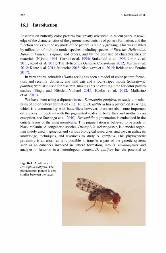
We have been using a dipteran insect, Drosophila guttifera, to study a mecha-
nism of color pattern formation (Fig. 16.1). D. guttifera has a pattern on its wings,
which is a commonality with butterflies; however, there are also some important
differences. In contrast with the pigmented scales of butterflies and moths (as an
exception, see Stavenga et al. 2010), Drosophila pigmentation is embedded in the
cuticle layers of the wing membrane. This pigmentation is believed to be made of
black melanin. A congeneric species, Drosophila melanogaster, is a model organ-
ism widely used in genetics and various biological researches, and we can utilize its
knowledge, techniques, and resources to study D. guttifera. This phylogenetic
proximity is an asset, as it is possible to transfer a part of the genetic system,
such as an enhancer involved in pattern formation, into D. melanogaster and
analyze its function in a heterologous context. D. guttifera has the potential to
Fig. 16.1 Adult male of
Drosophila guttifera. Thepigmentation pattern is very
similar between the sexes
288 S. Koshikawa et al.

approach the same problem of color pattern as in butterflies but from a different
angle. It also enables a good comparison, since its complex pigmentation patterns
evolved independently from the ones seen in butterflies.
In this chapter, we present an overview of the biology of D. guttifera. Then we
discuss differences in pattern formation between D. guttifera and butterflies and theadvantage and potential of D. guttifera to contribute to the general understanding ofanimal color pattern formation.
16.2 Phylogenetic Position of D. guttifera
Fruit flies (drosophilid flies) belong to family Drosophilidae, order Diptera, and
consist of 72 genera and more than 4000 described species (Yassin 2013). Among
them, genus Drosophila includes more than 1160 described species (Markow and
O’Grady 2006; Toda 2017). The best-studied species, D. melanogaster, also
belongs to this genus. It should be noted, however, that the genus Drosophila is
not monophyletic and potentially includes multiple genera within this clade, and
there is ongoing debate on the proper taxonomic treatment of this genus (O’Grady2010).
D. guttiferawas described by an English entomologist, Francis Walker, based on
a specimen collected in Florida (Walker 1849). This description consisted of 4 lines
in Latin and 21 lines in English with no illustration and was one of many descrip-
tions of a museum collection of the British Museum. In his taxonomic revision of
North American drosophilids, Sturtevant (1921) examined multiple specimens of
D. guttifera and redescribed the morphological features. Sturtevant (1942)
established “species groups” to classify species within the genus Drosophila.D. guttifera was assigned to a monospecific guttifera group. He also established
the quinaria group, which includes 11 species (D. quinaria, deflecta, palustris,subpalustris, occidentalis, suboccidentalis, munda, subquinaria, transversa, andpossibly phalerata and nigromaculata). Patterson (1943) revised drosophilids of
the Southwestern United States and Northern Mexico and redescribed many species
with beautiful illustrations. D. guttifera was redescribed with illustrations of a pupaand internal organs of reproduction and a color illustration of the whole body.
Patterson also described three new species in the quinaria group (D. suffusca,tenebrosa, and innubila). After that, many species were described in the quinariagroup, and currently it includes 31 species (Markow and O’Grady 2006, Toda
2017).
The close relationship between D. guttifera and the quinaria group is almost
certain at this time, based on molecular genetic evidence (Perlman et al. 2003;
Izumitani et al. 2016). Morphological similarity between D. guttifera and the
quinaria group was also noticed (Patterson and Stone 1952), and some authors
even placed D. guttifera in the quinaria group (Throckmorton 1962, 1975; Markow
and O’Grady 2006). Species-level relationships among D. guttifera and species of
the quinaria group are not completely resolved; however, the commonly supported
16 Drosophila guttifera as a Model System for Unraveling Color Pattern Formation 289
result is bifurcation into two clades, one including mostly North American species
and one including mostly Eurasian species (Perlman et al. 2003, Markow and
O’Grady 2006, Izumitani et al. 2016).
There are species with pigment patterns on the thorax, abdomen, and wings to
various degrees in the quinaria group (Patterson 1943; Werner and Jaenike 2017),
but D. guttifera has distinctive vertical stripes on the thorax and a polka dot patternon the abdomen and wings. Even when compared with the quinaria group species,
D. guttifera has the most prominently pigmented appearance.
16.3 Food Habits, Poison Resistance, and BehavioralEcology of D. guttifera
The life history and ecology of D. guttifera in the wild have not been well studied.
There are many species of the quinaria group that utilize mushrooms as a food
source. Sturtevant (1921) assumed D. guttifera is also a mushroom feeder based on
the facts that D. guttifera was found around mushrooms and that he could rear
D. guttifera, from eggs to adults, with mushrooms (he noted that both gill fungi and
pore fungi can be utilized, but he did not describe mushroom species). Bunyard and
Foote (1990a) studied what kind of dipteran insects emerged from mushrooms
collected in the state of Ohio and reported that D. guttifera emerged from two
mushroom species, Psilocybe polytrichophila and Collybia dryophila. They tested
oviposition site preference among commercial Agaricus bisporus, banana, tomato,
lettuce, and agar and found that Agaricus was the most preferred site (Bunyard and
Foote 1990b). They also confirmed that D. guttifera can grow from eggs to adults
with Agaricus. In laboratory conditions, however, we can keep strains of
D. guttifera with artificial food containing sugar/corn meal/yeast/agar (sugar
food) or molasses/corn meal/yeast/agar (molasses food) without adding
mushrooms.
Some fungus-feeding drosophilids are known to have high tolerance to a mush-
room toxin, alpha-amanitin, which is highly poisonous to most animals (Spicer and
Jaenike 1996). D. guttifera has the potential to be a model system to study this
phenomenon. Alpha-amanitin exerts its toxicity by binding to RNA polymerase II,
an enzyme essential for transcription. A mutant strain of D. melanogaster with highalpha-amanitin tolerance had an amino acid substitution in RNA polymerase II
(Chen et al. 1993). However, D. guttifera and other species with the tolerance do
not have the same substitution, indicating that other mechanisms are involved
(Stump et al. 2011). There are other strains of D. melanogaster with alpha-amanitin
tolerance but without RNA polymerase II mutation. The responsible locus was
mapped, and gene expression profiles were analyzed in these strains (Begum and
Whitley 2000; Mitchell et al. 2014, 2015).
There are some other studies of D. guttifera behavior. Oviposition site prefer-
ence of D. guttifera was affected by larval food condition, and this is known as a
290 S. Koshikawa et al.
classic example of olfactory conditioning of animals (Cushing 1941). The mating
behavior of D. guttifera was also studied (Grossfield 1977). The ecological signif-
icance and function of pigmentation patterns of D. guttifera is not well understood.Some drosophilids are known to use wing pigmentation in courtship displays
(Ringo and Hodosh 1978; Yeh et al. 2006; Fuyama 1979). Dombeck and Jaenike
(2004) analyzed fitness effects of abdominal spot number in D. falleni.
16.4 The Evolution of Wing Pigmentation Pattern
Dombeck and Jaenike (2004) analyzed and illustrated the evolutionary path of wing
and abdominal pigmentations of D. guttifera and seven species of the quinariagroup. We summarize here the evolution of wing pigmentation pattern of
D. guttifera and the quinaria group species based on molecular phylogenetics
(Fig. 16.2). As previously explained, the quinaria group is divided into two major
clades (Perlman et al. 2003; Markow and O’Grady 2006, Izumitani et al. 2016). We
defined the clade with mostly North American species as “clade A” and the clade
with mostly Eurasian species as “clade B.” Species in clade A have relatively
simple patterns; pigmentations are formed only around crossveins except in
D. innubila, which has no pigmentation. The evolution of patterns in clade B is
rather complicated. The relationships among basal species of clade B [D. guttifera,nigromaculata, and (deflecta + palustris + subpalustris)] have not been completely
resolved, because the topologies of the phylogenetic trees depend on the analytical
methods. These four species have pigmentations around crossveins and longitudi-
nal vein tips. In addition, D. guttifera has pigmentations around the campaniform
sensilla, which is unique to this species [at least unique among the clade of
(quinaria group + D. guttifera) and probably among the genus
Drosophila]. Among the rest of the species in clade B, D. quinaria has weak
pigmentations on the tips of longitudinal veins in addition to crossveins.
D. recens and many other species within this cluster have pigmentations around
crossveins.D. kuntzei, which has a similar pattern toD. quinaria, branches from the
most basal position of clade B according to Perlman et al. (2003), although the
statistical support for this topology was low. Due to the lack of a robust phylogeny,
it would be premature to propose a simple scenario stepwise pattern of gain and loss
within the quinaria group. It is plausible that the instances of longitudinal vein tip
pigmentation are the result of convergent evolution, perhaps via parallel mecha-
nisms, although we cannot exclude the possibility of a single gain of the longitu-
dinal vein tip pigmentation and a secondary loss in derived species of clade
B. Nevertheless, the other dot-like patterns of D. guttifera, which overlap in
position with innervated cupules known as campaniform sensilla (see below), are
unique to this species and are assumed to form a true evolutionary novelty.
16 Drosophila guttifera as a Model System for Unraveling Color Pattern Formation 291

16.5 Wing Pigmentation Pattern Formation in Drosophila
The initial study of the mechanism of wing pigmentation pattern formation was
done by True et al. (1999). They argued that patterns are formed through patterning
by gene expression and subsequent elaboration by precursor trafficking through
wing veins, based on experiments using Drosophila grimshawi (synonym of
D. falleni
D. innubila
D. phalerata+ other species
D. nigromaculata
D. kuntzei
D. quinaria
D. recens+ other species
D. deflectaD. palustrisD. subpalustris
D. guttifera
Clade A
Clade B
quinaria group
I. grimshawi
(Hawaiian Drosophila)
D. biarmipes
D. melanogaster melanogaster group
Fig. 16.2 Phylogenetic relationships of D. guttifera and species in the quinaria group. The
topology was drawn from a consensus between Perlman et al. (2003) and Izumitani et al.
(2016). See also Fig. 16.3 for interpretation of pigmentation
292 S. Koshikawa et al.

Idiomyia grimshawi), D. rajasekari (synonym of D. biarmipes), and mutants and
transgenics of D. melanogaster. Wittkopp et al. (2002) studied the function of
yellow and ebony genes in the body trunk and wings of D. melanogaster. Theyalso showed that the future spot position had more Yellow protein and less Ebony
protein. Yellow is known to enhance black melanin synthesis, and Ebony is an
enzyme that conjugates beta-alanine to dopamine and produces NBAD (N-beta-
alanyldopamine) resulting in repression of black melanin synthesis. Gompel et al.
(2005) analyzed the regulation of yellow gene expression in D. biarmipes and
showed that evolution of an enhancer (a sequence that enhances expression of a
nearby gene) was involved in the gain of pigmentation. In D. biarmipes and
D. guttifera, they showed that Yellow protein was localized in future black spots
and Ebony protein was localized in future transparent (no pigmentation) places. The
yellow expression in the anteriodistal part of the wing in D. biarmipes results fromregulation by at least two factors: posterior expression of engrailed repressing the
yellow expression and anteriodistal expression of Distal-less enhancing expressionsof yellow and other pigmentation genes (Gompel et al. 2005; Arnoult et al. 2013).
16.6 Features of Wing Pigmentation Pattern in D. guttifera
D. guttifera has prominent black polka dots on its wings, and these are believed to
be made with melanin (Fig. 16.3). Pigmentations are formed around crossveins,
longitudinal vein tips, and the campaniform sensilla. Weak pigmentations are also
formed in intervein regions. As mentioned previously, crossvein pigmentation is
widely observed in the quinaria group and also found in many species in other
species groups. The crossvein pigmentation in D. guttifera is constricted in the
center, forming an hourglass shape (or calabash shape), and this is unique to this
species. Longitudinal vein tip pigmentations are observed in a few species, but the
pigmentation area is largest in D. guttifera. Campaniform sensilla pigmentation is a
trait unique to D. guttifera, although some species, such as a Hawaiian species,
Idiomyia grimshawi (synonym of Drosophila grimshawi), have dappled spots all
over the wings. The campaniform sensilla are lined on the third longitudinal vein in
the same way as in other drosophilids, but in D. guttifera, one campaniform
sensillum is also found on the fifth longitudinal vein, which is unique to this species.
This campaniform sensillum is also surrounded by pigmentation (Sturtevant 1921;
Werner et al. 2010). The wing pigmentation of D. guttifera starts to form in the
pupal period, and it continues until one day old adult (Fukutomi et al. 2017).
16 Drosophila guttifera as a Model System for Unraveling Color Pattern Formation 293

16.7 Wingless Gene Induces Pigmentation PatternFormation in D. guttifera
Werner et al. (2010) analyzed the cis-regulatory region of the yellow gene and
identified vein spot CRE, which is an enhancer driving expression in all the polka
dots, and intervein shade CRE, which is an enhancer driving expression in the
intervein region. Vein spot CRE drove polka dots in D. guttifera but drove around
crossveins and longitudinal vein tips if introduced in D. melanogaster. This differ-ence means there is a difference in localization of a trans-regulatory factor that hasan input to vein spot CRE. Gene expression patterns were known for several genes
in D. melanogaster, and therefore they found candidate genes from genes showing
similar expression with the vein spot CRE pattern. Among the candidate genes,
wingless, a gene encoding a ligand of the Wnt signaling pathway, showed expres-
sion in the center of future spot positions (crossveins, longitudinal vein tips, and
campaniform sensilla) in D. guttifera. There was no wingless expression in the
campaniform sensilla in a closely related species, D. deflecta, which does not have
pigmentation around them. A spontaneous mutant line of D. guttifera, schwarzvier,has additional pigmentation on the fourth longitudinal vein. In this mutant line,
wingless was ectopically expressed on the fourth longitudinal vein. To obtain direct
Fig. 16.3 Top Wing pigmentation of D. guttifera. Bottom Interpretation of the pigmentation
pattern. Blue marks pigmentations around crossveins, purple marks longitudinal vein tips, yellow
marks campaniform sensilla, and red marks intervein shading
294 S. Koshikawa et al.

functional evidence, they tried to make ectopic expressions of wingless by con-
struction of the GAL4/UAS system in D. guttifera. Although they did not obtain
optimal GAL4 lines, they found that one of the UAS-wingless lines had ectopic
expression of wingless, probably caused by the enhancer trap mechanism. In this
line, wingless was expressed ectopically on the second, third, and fourth longitudi-
nal veins of pupal wings, and additional pigmentation was formed on these veins in
adult wings. With these evidences, they concluded that wingless is the upstream
trans-factor that induces pigmentation.
In Heliconius and Limenitis butterflies, the WntA gene, which also seems to
encode a ligand of Wnt signaling, is involved in specifying wing pattern shapes,
including in melanic elements (Martin et al. 2012; Gallant et al. 2014; Martin and
Reed 2014). In Junonia coenia and some other butterfly species, wingless is knownto be expressed in future pattern elements called basal (B), discal (DI and DII), and
marginal (EI) elements (Carroll et al. 1994; Martin and Reed 2010, 2014; Huber
et al. 2015) and was also identified at the center of eyespot patterns (Monteiro et al.
2006). The thoracic pattern of larval Bombyx mori is also regulated by Wnt1(homolog of wingless) (Yamaguchi et al. 2013). Evolutionary roles of secreted
ligand genes such as wingless are reviewed in chapter 4 of this book (Martin and
Courtier-Orgogozo 2017).
Werner et al. (2010) proposed a model of pigmentation pattern formation based
on the assumption that Wingless protein diffuses from the source and serves as a
long-range signal. There are a limited number of cells expressing wingless, and theyare located in centers of future pigmented spots. In their model, secreted Wingless
protein is diffused or transported to wider regions and transduces the signal. The
signal is probably mediated by an unknown transcription factor and activates
transcription of melanin synthesis-related genes, including yellow. Melanin should
be synthesized by products of these genes and wings are consequently pigmented.
This model should be validated by future research.
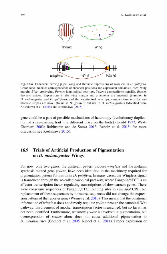
16.8 Cis-Regulatory Evolution of Wingless
The expression pattern of wingless evolved uniquely in D. guttifera. To examine
how this unique expression pattern evolved, the genomic region around winglesswas analyzed using a fluorescent reporter assay. As a result, three novel enhancer
activities (in longitudinal vein tips, campaniform sensilla, and thoracic stripes) were
found (Fig. 16.4). These novel enhancer activities are thought to have been
involved in the evolution of the novel pigmentation pattern (Koshikawa et al.
2015). This study provided unique insights into the evolution of novel traits,
illustrating how gains of novel enhancer activities at developmental regulatory
gene were associated with derived expression domains and the emergence of
novel traits (Rebeiz et al. 2011; Koshikawa et al. 2015; Rebeiz and Williams 2017).
We can generalize this concept as follows. In many organisms, gains of novel
expression domains by gains of enhancer activities for a developmental regulatory
16 Drosophila guttifera as a Model System for Unraveling Color Pattern Formation 295
gene could be a part of possible mechanisms of heterotopy (evolutionary duplica-
tion of a pre-existing trait in a different place on the body) (Gould 1977; West-
Eberhard 2003; Rubinstein and de Souza 2013; Rebeiz et al. 2015; for more
discussion see Koshikawa 2015).
16.9 Trials of Artificial Production of Pigmentationon D. melanogaster Wings
For now, only two genes, the upstream pattern inducer wingless and the melanin
synthesis-related gene yellow, have been identified in the machinery required for
pigmentation pattern formation in D. guttifera. In many cases, the Wingless signal
is transduced through the so-called canonical pathway, where Pangolin/dTCF is an
effector transcription factor regulating transcriptions of downstream genes. There
were consensus sequences of Pangolin/dTCF binding sites in vein spot CRE, butreplacement of these sequences by nonsense sequences did not change the expres-
sion pattern of the reporter gene (Werner et al. 2010). This means that the positional
information of wingless does not directly regulate yellow through the canonical Wnt
pathway. Involvement of another transcription factor is assumed, but so far it has
not been identified. Furthermore, we know yellow is involved in pigmentation, but
overexpression of yellow alone does not cause additional pigmentation in
D. melanogaster (Gompel et al. 2005; Riedel et al. 2011). Proper expression or
wingless Wnt6 Wnt10
Thorax Wing
Fig. 16.4 Enhancers driving pupal wing and thoracic expressions of wingless in D. guttifera.Color code indicates correspondence of enhancer positions and expression domains. Green: wingmargin. Blue: crossveins. Purple: longitudinal vein tips. Yellow: campaniform sensilla. Brown:thoracic stripes. Expressions in the wing margin and crossveins are ancestral (common in
D. melanogaster and D. guttifera), and the longitudinal vein tips, campaniform sensilla, and
thoracic stripes are novel (found in D. guttifera but not in D. melanogaster) (Modified from
Koshikawa et al. (2015) and Koshikawa (2015))
296 S. Koshikawa et al.
repression of melanin synthesis-related genes and/or proper supply of melanin
precursors, such as dopa and dopamine, could be required for artificial production
of pigmentation in D. melanogaster wings.
16.10 Diversity and Generality in Color Pattern Formation
We summarized above what was revealed by studies of D. guttifera, but will itapply to pattern formation in other organisms? Due to the experimental strengths of
this system, we can be optimistic that we will reach an integrated model for
pigmentation pattern formation inDrosophila. Butterflies show interesting parallels
with the Drosophila wing patterning genes, as Wnt genes and Distal-less are key
players in both lineages (Werner et al. 2010; Martin et al. 2012; Brakefield et al.
1996; Arnoult et al. 2013). If we expand the comparison to vertebrates, there are
large differences in genes involved in pattern formation and melanin synthesis
(Kopp 2009; Kronforst et al. 2012; Kaelin et al. 2012; Mallarino et al. 2016). Still
we assume we can find some common mechanisms, such as a way of measuring
distance in a tissue, and a hierarchical regulatory architecture. Comparing compre-
hensive datasets will be instrumental in answering this question of fundamental
interest for our understanding of the mechanisms that generate biodiversity on
Earth.
Acknowledgments We thank Toshiro Sekimura, Frederik H. Nijhout, and persons involved in
the meeting at Chubu University in 2016 for stimulating us to write this chapter. We also thank
Arnaud Martin and Takao K. Suzuki for reviewing this chapter, Masanori J. Toda for advice on
taxonomy, Elizabeth Nakajima for English editing, and Noriko Funayama for hosting our research.
A part of the writing was supported by KAKENHI (15K18586) and the Sumitomo Foundation.
References
Arnoult L, Su KF, Manoel D, Minervino C, Magri~na J, Gompel N, Prud’homme B (2013)
Emergence and diversification of fly pigmentation through evolution of a gene regulatory
module. Science 339(6126):1423–1426. doi:10.1126/science.1233749
Begun DJ, Whitley P (2000) Genetics of alpha-amanitin resistance in a natural population of
Drosophila melanogaster. Heredity 85(2):184–190. doi:10.1046/j.1365-2540.2000.00729.x
Beldade P, Peralta CM (2017) Developmental and evolutionary mechanisms shaping butterfly
eyespots. Curr Opin Insect Sci 19:22–29. doi:10.1016/j.cois.2016.10.006
Brakefield PM, Gates J, Keys D, Kesbeke F, Wijngaarden PJ, Monteiro A, French V, Carroll SB
(1996) Development, plasticity and evolution of butterfly eyespot patterns. Nature 384
(6606):236–242. doi:10.1038/384236a0
Bunyard B, Foote BA (1990a) Acalyptrate Diptera reared from higher fungi in northeastern Ohio.
Entomol News 101(2):117–121
Bunyard B, Foote BA (1990b) Biological notes on Drosophila guttifera (Diptera: Drosophilidae),
a consumer of mushrooms. Entomol News 101(3):161–163
16 Drosophila guttifera as a Model System for Unraveling Color Pattern Formation 297

Carroll SB, Gates J, Keys DN, Paddock SW, Panganiban GE, Selegue JE, Williams JA (1994)
Pattern formation and eyespot determination in butterfly wings. Science 265(5168):109–114.
doi:10.1126/science.7912449
Chen Y,Weeks J, Mortin MA, Greenleaf AL (1993) Mapping mutations in genes encoding the two
large subunits of Drosophila RNA polymerase II defines domains essential for basic transcrip-
tion functions and for proper expression of developmental genes. Mol Cell Biol 13
(7):4214–4222. doi:10.1128/MCB.13.7.4214
Cushing JE (1941) An experiment in olfactory conditioning inDrosophila guttifera. Proc Natl AcaSci U S A 27(11):496–499
Dombeck I, Jaenike J (2004) Ecological genetics of abdominal pigmentation inDrosophila falleni.Evolution 58(3):587–596. doi:10.1554/03-299
Fukutomi Y, Matsumoto K, Agata K, Funayama N, Koshikawa S (2017) Pupal development and
pigmentation process of a polka-dotted fruit fly, Drosophila guttifera (Insecta, Diptera). Dev
Genes Evol 227(3):171–180. doi:10.1007/s00427-017-0578-3
Fuyama Y (1979) A visual stimulus in the courtship of Drosophila suzukii. Experientia 35
(10):1327–1328
Gallant JR, Imhoff VE, Martin A, Savage WK, Chamberlain NL, Pote BL, Peterson C, Smith GE,
Evans B, Reed RD, Kronforst MR, Mullen SP (2014) Ancient homology underlies adaptive
mimetic diversity across butterflies. Nat Commun 5:4817. doi:10.1038/ncomms5817
Gompel N, Prud’homme B, Wittkopp PJ, Kassner VA, Carroll SB (2005) Chance caught on the
wing: cis-regulatory evolution and the origin of pigment patterns in Drosophila. Nature 433
(7025):481–487. doi:10.1038/nature03235
Gould SJ (1977) Ontogeny and phylogeny. Harvard University Press, Cambridge
Grossfield J (1977) Drosophila courtship: decapitated quinaria group females. J NY Entomol Soc
85(3):119–126
Huber B, Whibley A, Poul YL, Navarro N, Martin A, Baxter S, Shah A, Gilles B, Wirth T,
McMillan WO, Joron M (2015) Conservatism and novelty in the genetic architecture of
adaptation in Heliconius butterflies. Heredity 114(5):515–524. doi:10.1038/hdy.2015.22
Izumitani HF, Kusaka Y, Koshikawa S, TodaMJ, Katoh T (2016) Phylogeography of the subgenus
Drosophila (Diptera: Drosophilidae): evolutionary history of faunal divergence between the
old and the new worlds. PLoS One 11(7), e0160051. doi:10.1371/journal.pone.0160051
Joron M, Frezal L, Jones RT, Chamberlain NL, Lee SF, Haag CR, Whibley A, Becuwe M, Baxter
SW, Ferguson L, Wilkinson PA, Salazar C, Davidson C, Clark R, Quail MA, Beasley H,
Glithero R, Lloyd C, Sims S, Jones MC, Rogers J, Jiggins CD, ffrench-Constant RH (2011)
Chromosomal rearrangements maintain a polymorphic supergene controlling butterfly mim-
icry. Nature 477(7363):203–206. doi:10.1038/nature10341
Kaelin CB, Xu X, Hong LZ, David VA, McGowan KA, Schmidt-Küntzel A, Roelke ME, Pino J,
Pontius J, Cooper GM, Manuel H, Swanson WF, Marker L, Harper CK, van Dyk A, Yue B,
Mullikin JC, Warren WC, Eizirik E, Kos L, O’Brien SJ, Barsh GS, Menotti-Raymond M
(2012) Specifying and sustaining pigmentation patterns in domestic and wild cats. Science 337
(6101):1536–1541. doi:10.1126/science.1220893
Kopp A (2009) Metamodels and phylogenetic replication: a systematic approach to the evolution
of developmental pathways. Evolution 63(11):2771–2789. doi:10.1111/j.1558-5646.2009.
00761.x
Koshikawa S (2015) Enhancer modularity and the evolution of new traits. Fly (Austin) 9
(4):155–159. doi:10.1080/19336934.2016.1151129
Koshikawa S, Giorgianni MW, Vaccaro K, Kassner VA, Yoder JH, Werner T, Carroll SB (2015)
Gain of cis-regulatory activities underlies novel domains of wingless gene expression in
Drosophila. Proc Natl Acad Sci U S A 112(24):7524–7529. doi:10.1073/pnas.1509022112
Kronforst MR, Barsh GS, Kopp A, Mallet J, Monteiro A, Mullen SP, Protas M, Rosenblum EB,
Schneider CJ, Hoekstra HE (2012) Unraveling the thread of nature’s tapestry: the genetics ofdiversity and convergence in animal pigmentation. Pigment Cell Melanoma Res 25
(4):411–433. doi:10.1111/j.1755-148X.2012.01014.x
298 S. Koshikawa et al.
Kunte K, Zhang W, Tenger-Trolander A, Palmer DH, Martin A, Reed RD, Mullen SP, Kronforst
MR (2014) Doublesex is a mimicry supergene. Nature 507(7491):229–232. doi:10.1038/
nature13112
Mallarino R, Henegar C, Mirasierra M, Manceau M, Schradin C, Vallejo M, Beronja S, Barsh GS,
Hoekstra HE (2016) Developmental mechanisms of stripe patterns in rodents. Nature 539
(7630):518–523. doi:10.1038/nature20109
Martin A, Reed RD (2010)Wingless and aristaless2 define a developmental ground plan for moth
and butterfly wing pattern evolution. Mol Biol Evol 27(12):2864–2878. doi:10.1093/molbev/
msq173
Martin A, Papa R, Nadeau NJ, Hill RI, Counterman BA, Halder G, Jiggins CD, Kronforst MR,
Long AD, McMillan WO, Reed RD (2012) Diversification of complex butterfly wing patterns
by repeated regulatory evolution of a Wnt ligand. Proc Natl Aca Sci U S A 109
(31):12632–12637. doi:10.1073/pnas.1204800109
Martin A, Reed RD (2014) Wnt signaling underlies evolution and development of the butterfly
wing pattern symmetry systems. Dev Biol 395(2):367–378. doi:10.1016/j.ydbio.2014.08.031
Martin A, Courtier-Orgogozo V (2017) Morphological evolution repeatedly caused by mutations
in signaling ligand genes. In: Diversity and evolution of butterfly wing patterns: an integrative
approach. Springer, New York
Markow TA, O’Grady PM (2006) Drosophila: a guide to species identification and use. Academic
Press, New York
Mitchell CL, Saul MC, Lei L, Wei H, Werner T (2014) The mechanisms underlying α-amanitin
resistance in Drosophila melanogaster: a microarray analysis. PLoS One 9(4):e93489. doi:10.
1371/journal.pone.0093489
Mitchell CL, Yeager RD, Johnson ZJ, D’Annunzio SE, Vogel KR, Werner T (2015) Long-term
resistance of Drosophila melanogaster to the mushroom toxin alpha-amanitin. PLoS One 10
(5):e0127569. doi:10.1371/journal.pone.0127569
Monteiro A (2015) Origin, development, and evolution of butterfly eyespots. Annu Rev Entomol
60:253–271. doi:10.1146/annurev-ento-010814-020942
Monteiro A, Glaser G, Stockslager S, Glansdorp N, Ramos D (2006) Comparative insights into
questions of lepidopteran wing pattern homology. BMC Dev Biol 6:52. doi:10.1186/1471-
213X-6-52
Nijhout HF (1991) The development and evolution of butterfly wing patterns. Smithsonian
Institution Press, Washington, DC
Nishikawa H, Iijima T, Kajitani R, Yamaguchi J, Ando T, Suzuki Y, Sugano S, Fujiyama A,
Kosugi S, Hirakawa H, Tabata S, Ozaki K, Morimoto H, Ihara K, Obara M, Hori H, Itoh T,
Fujiwara H (2015) A genetic mechanism for female-limited Batesian mimicry in Papiliobutterfly. Nat Genet 47(4):405–409. doi:10.1038/ng.3241
O’Grady PM (2010) Whither Drosophila? Genetics 185(2):703–705. doi:10.1534/genetics.110.
118232
Patterson JT (1943) The Drosophilidae of the Southwest. University of Texas Publication
4313:7–216
Patterson JT, Stone WS (1952) Evolution in the genus Drosophila. Macmillan Company,
New York
Perlman SJ, Spicer GS, Shoemaker DD, Jaenike J (2003) Associations between mycophagous
Drosophila and their Howardula nematode parasites: a worldwide phylogenetic shuffle. Mol
Ecol 12(1):237–249. doi:10.1046/j.1365-294X.2003.01721.x
Rebeiz M, Jikomes N, Kassner VA, Carroll SB (2011) Evolutionary origin of a novel gene
expression pattern through co-option of the latent activities of existing regulatory sequences.
Proc Natl Acad Sci U S A 108(25):10036–10043. doi:10.1073/pnas.1105937108
16 Drosophila guttifera as a Model System for Unraveling Color Pattern Formation 299

Rebeiz M, Patel NH, Hinman VF (2015) Unraveling the tangled skein: the evolution of transcrip-
tional regulatory networks in development. Annu Rev Genomics Hum Genet 16:103–131.
doi:10.1146/annurev-genom-091212-153423
Rebeiz M, Williams TM (2017) Using Drosophila pigmentation traits to study the mechanisms of
cis-regulatory evolution. Curr Opin Insect Sci 19:1–7. doi:10.1016/j.cois.2016.10.002
Reed RD, Papa R, Martin A, Hines HM, Counterman BA, Pardo-Diaz C, Jiggins CD, Chamberlain
NL, Kronforst MR, Chen R, Halder G, Nijhout HF, McMillan WO (2011) optix drives the
repeated convergent evolution of butterfly wing pattern mimicry. Science 333
(6046):1137–1141. doi:10.1126/science.1208227
Riedel F, Vorkel D, Eaton S (2011) Megalin-dependent yellow endocytosis restricts melanization
in the Drosophila cuticle. Development 138(1):149–158. doi:10.1242/dev.056309
Ringo JM, Hodosh RJ (1978) A multivariate analysis of behavioral divergence among closely
related species of endemic Hawaiian Drosophila. Evolution 32(2):389–397. doi:10.1111/j.
1558-5646.1978.tb00654.x
Rubinstein M, de Souza FSJ (2013) Evolution of transcriptional enhancers and animal diversity.
Philos Trans R Soc B 368(1632):20130017–20130017. doi:10.1098/rstb.2013.0017
Singh AP, Nüsslein-Volhard C (2015) Zebrafish stripes as a model for vertebrate colour pattern
formation. Curr Biol 25(2):R81–R92. doi:10.1016/j.cub.2014.11.013
Spicer GS, Jaenike J (1996) Phylogenetic analysis of breeding site use and α-amanitin tolerance
within the Drosophila quinaria species group. Evolution 50(6):2328–2337. doi:10.2307/
2410701
Stavenga DG, Giraldo MA, Leertouwer HL (2010) Butterfly wing colors: glass scales ofGraphiumsarpedon cause polarized iridescence and enhance blue/green pigment coloration of the wing
membrane. J Exp Biol 213(10):1731–1739. doi:10.1242/jeb.041434
Stump AD, Jablonski SE, Bouton L, Wilder JA (2011) Distribution and mechanism of α-amanitin
tolerance in mycophagous Drosophila (Diptera: Drosophilidae). Environ Entomol 40
(6):1604–1612. doi:10.1603/EN11136
Sturtevant AH (1921) The North American species of Drosophila. Carnegie Institution of
Washington, Washington, DC
Sturtevant AH (1942) The classification of the genus Drosophila, with the description of nine newspecies. University of Texas Publication 4213:5–51
The Heliconius Genome Consortium (2012) Butterfly genome reveals promiscuous exchange of
mimicry adaptations among species. Nature 487(7405):94–98. doi:10.1038/nature11041
Throckmorton LH (1962) The problem of phylogeny in the genus Drosophila. University of TexasPublication 6205:207–343
Throckmorton LH (1975) The phylogeny, ecology, and geography of Drosophila. In: King RC
(ed) Handbook of genetics, vol 3. Plenum Press, New York, pp 421–469
Toda MJ (2017) DrosWLD-Species: taxonomic information database for world species of
Drosophilidae. Available at: http://bioinfo.lowtem.hokudai.ac.jp/db/. Accessed 25 Jan 2017
True JR, Edwards KA, Yamamoto D, Carroll SB (1999) Drosophila wing melanin patterns form
by vein-dependent elaboration of enzymatic prepatterns. Curr Biol 9(23):1382–1391. doi:10.
1016/S0960-9822(00)80083-4
Walker F (1849) List of specimens of dipterous insects of the collection of the British Museum.
Part 4. British Museum (N.H.), London, pp 689–1172
Werner T, Jaenike J (2017) Drosophilids of the Midwest and Northeast. River Campus Libraries,
University of Rochester, Rochester
Werner T, Koshikawa S, Williams TM, Carroll SB (2010) Generation of a novel wing colour
pattern by the Wingless morphogen. Nature 464(7292):1143–1148. doi:10.1038/nature08896
West-Eberhard MJ (2003) Developmental plasticity and evolution. Oxford University Press,
Oxford
300 S. Koshikawa et al.

Wittkopp PJ, True JR, Carroll SB (2002) Reciprocal functions of the Drosophila yellow and ebony
proteins in the development and evolution of pigment patterns. Development 129
(8):1849–1858
Yamaguchi J, Banno Y, Mita K, Yamamoto K, Ando T, Fujiwara H (2013) Periodic Wnt1expression in response to ecdysteroid generates twin-spot markings on caterpillars. Nat
Commun 4:1857. doi:10.1038/ncomms2778
Yassin A (2013) Phylogenetic classification of the Drosophilidae Rondani (Diptera): the role of
morphology in the postgenomic era. Syst Entomol 38(2):349–364. doi:10.1111/j.1365-3113.
2012.00665.x
Yeh SD, Liou SR, True JR (2006) Genetics of divergence in male wing pigmentation and courtship
behavior between Drosophila elegans and D. gunungcola. Heredity 96(5):383–395. doi:10.
1038/sj.hdy.6800814
Open Access This chapter is licensed under the terms of the Creative Commons Attribution 4.0
International License (http://creativecommons.org/licenses/by/4.0/), which permits use, sharing,
adaptation, distribution and reproduction in any medium or format, as long as you give appropriate
credit to the original author(s) and the source, provide a link to the Creative Commons license and
indicate if changes were made.
The images or other third party material in this chapter are included in the chapter’s CreativeCommons license, unless indicated otherwise in a credit line to the material. If material is not
included in the chapter’s Creative Commons license and your intended use is not permitted by
statutory regulation or exceeds the permitted use, you will need to obtain permission directly from
the copyright holder.
16 Drosophila guttifera as a Model System for Unraveling Color Pattern Formation 301