Embed Size (px)
Citation preview
Chapter 13: The Spinal Cord and Spinal Nerves
Copyright 2009, John Wiley & Sons, Inc.
Spinal Cord Anatomy
Protective and stabilizing structures: Vertebral column Meninges – continuous around spinal cord and
brain cerebrospinal fluid denticulate ligament
Copyright 2009, John Wiley & Sons, Inc.
Spinal Cord Anatomy
Meninges – 3 layers1. Dura mater
most superficial dense irregular connective tissue Ends at second sacral vertebra Cushion of fat and connective tissue in epidural space Subdural space filled with interstitial fluid
2. Arachnoid mater Spider web arrangement of delicate collage and elastic Subarachnoid space filled with cerebrospinal fluid (CSF)
3. Pia mater Thin delicate ct adheres to spinal cord and brain Extensive blood supply
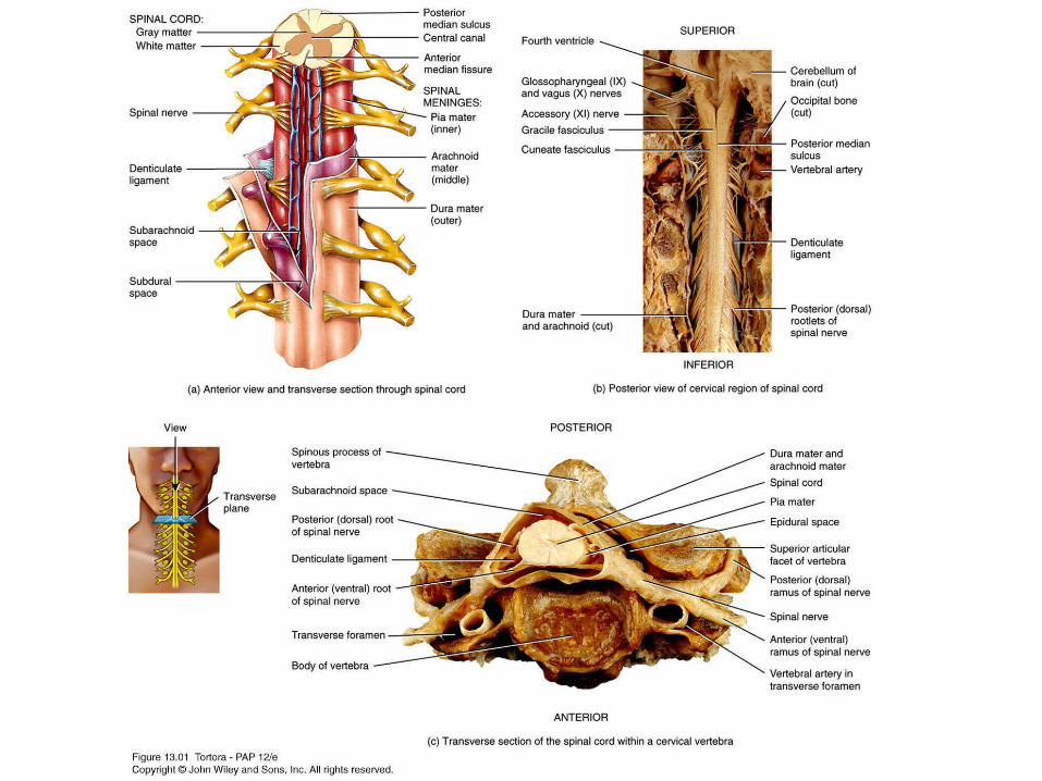
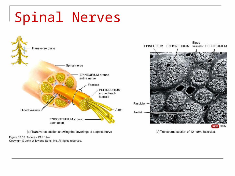
13_01
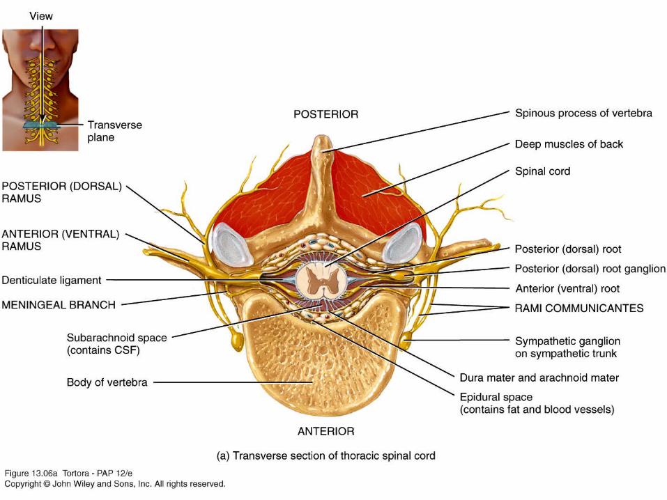
13_06a
External Anatomy of the Spinal Cord Two enlargements: cervical and lumbar Conus medullaris Filum terminale Cauda equina Posterior (dorsal root) & anterior(ventral) root Posterior (dorsal root) ganglion Spinal nerve
Copyright 2009, John Wiley & Sons, Inc.

13_02
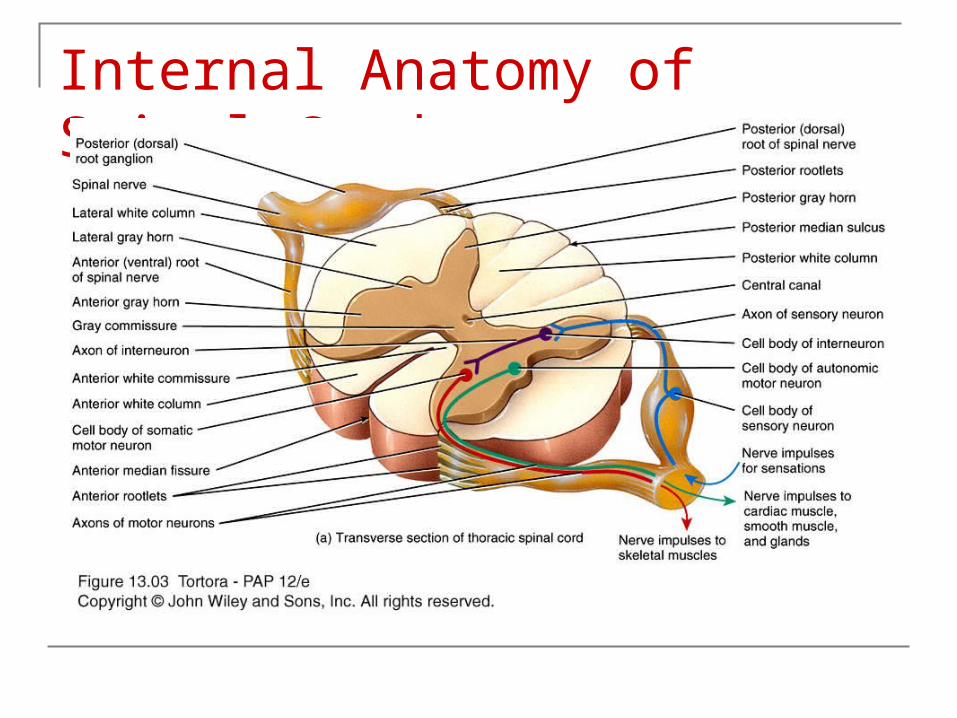
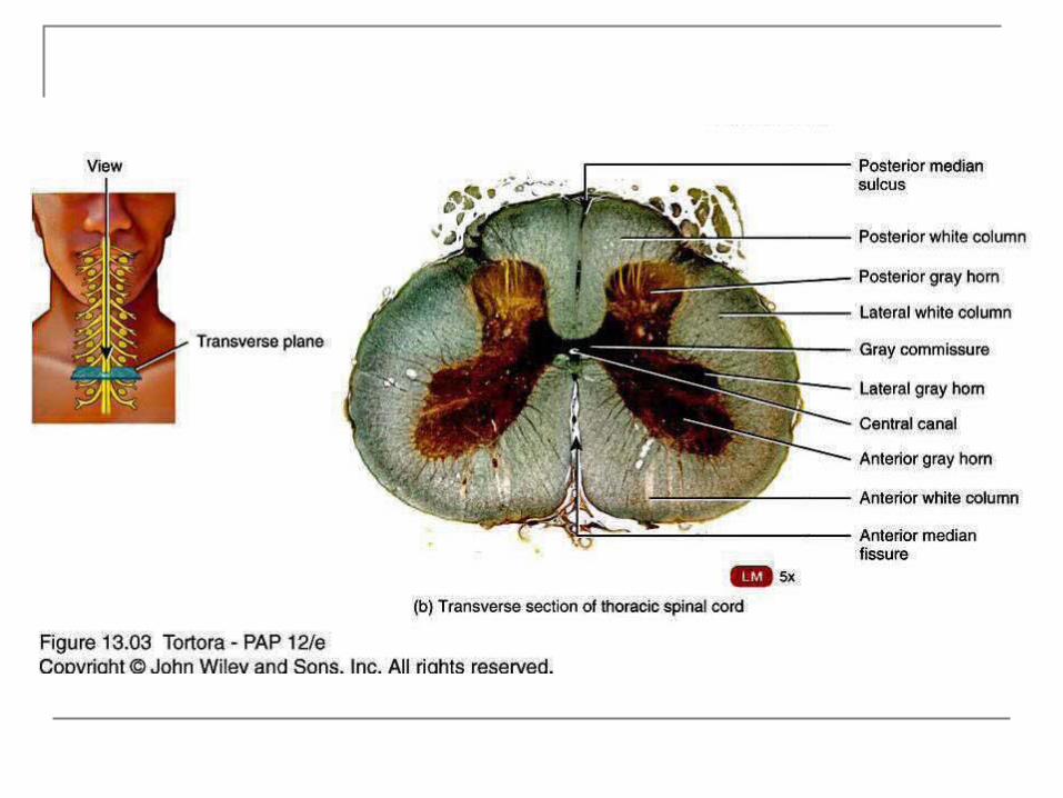
Internal Anatomy of the Spinal Cord Partially divide spinal cord into right and left
Anterior median fissure Posterior median sulcus
Gray matter Divided into horns Anterior, posterior & lateral gray horns
White matter Columns Anterior, posterior & lateral white columns
Central canal
Copyright 2009, John Wiley & Sons, Inc.
Internal Anatomy of the Spinal CordLook for (see next slide)
1. Gray commissure
2. Central canal
3. Anterior, posterior and lateral gray horns
4. Anterior, posterior and lateral white columns
Copyright 2009, John Wiley & Sons, Inc.
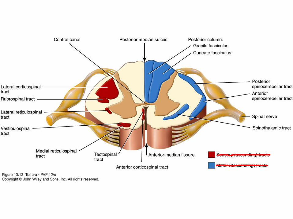
Internal Anatomy of Spinal Cord
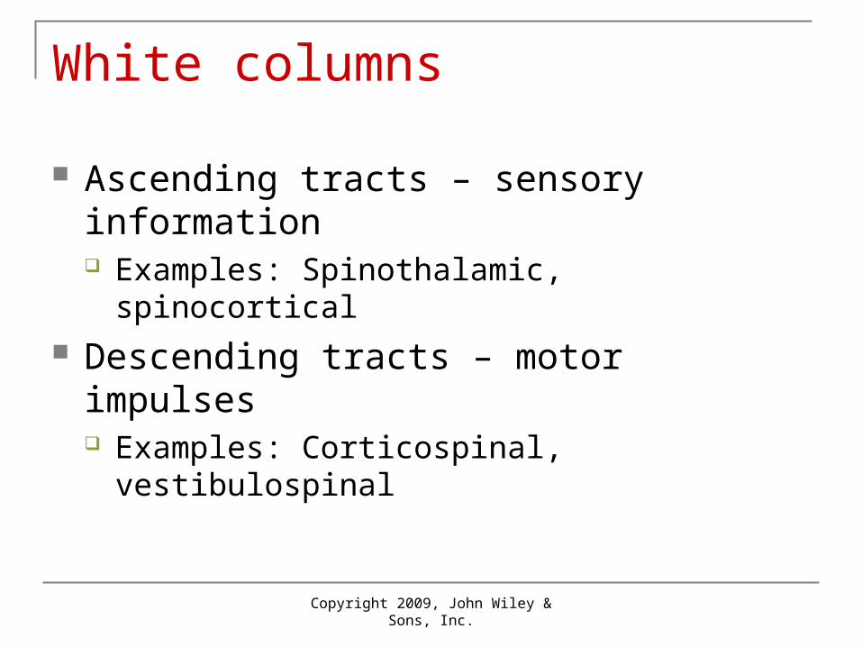
White columns
Ascending tracts – sensory information Examples: Spinothalamic, spinocortical
Descending tracts – motor impulses Examples: Corticospinal, vestibulospinal
Copyright 2009, John Wiley & Sons, Inc.
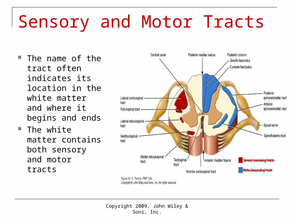
Sensory and Motor Tracts
The name of the tract often indicates its location in the white matter and where it begins and ends
The white matter contains both sensory and motor tracts
Copyright 2009, John Wiley & Sons, Inc.
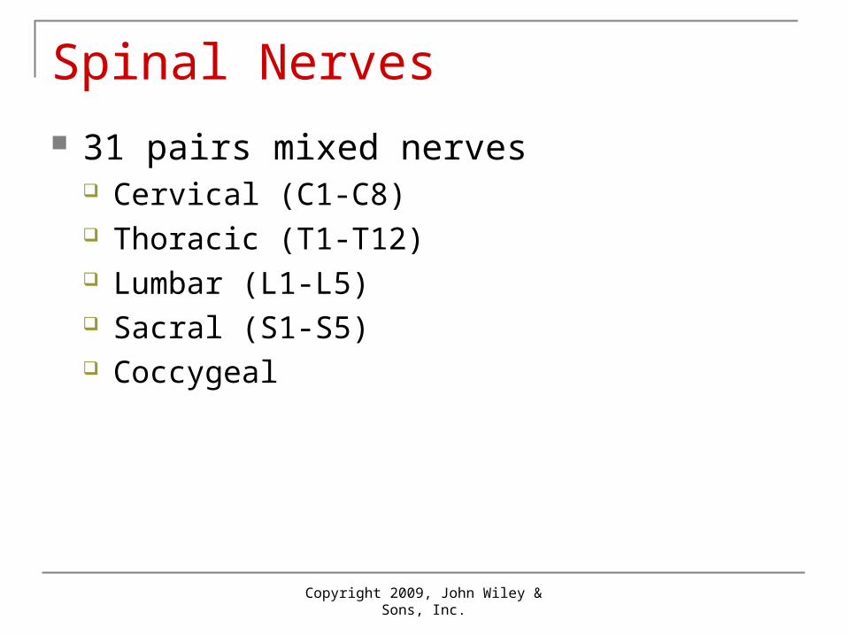
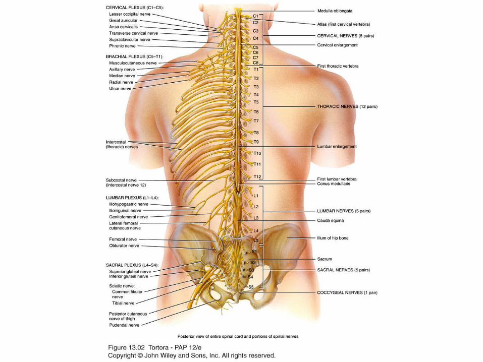
Spinal Nerves 31 pairs mixed nerves
Cervical (C1-C8) Thoracic (T1-T12) Lumbar (L1-L5) Sacral (S1-S5) Coccygeal
Copyright 2009, John Wiley & Sons, Inc.
13_02
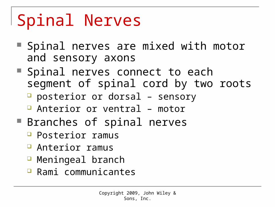
Spinal Nerves Spinal nerves are mixed with motor and sensory
axons Spinal nerves connect to each segment of spinal
cord by two roots posterior or dorsal – sensory Anterior or ventral – motor
Branches of spinal nerves Posterior ramus Anterior ramus Meningeal branch Rami communicantes
Copyright 2009, John Wiley & Sons, Inc.
Spinal Nerves
Distribution of Spinal Nerves
Plexus Anterior rami of spinal nerves, except T2-T12,
form networks of nerves Emerging from a plexus are nerves bearing
names that typically describe the general regions they supply or route they follow
Copyright 2009, John Wiley & Sons, Inc.
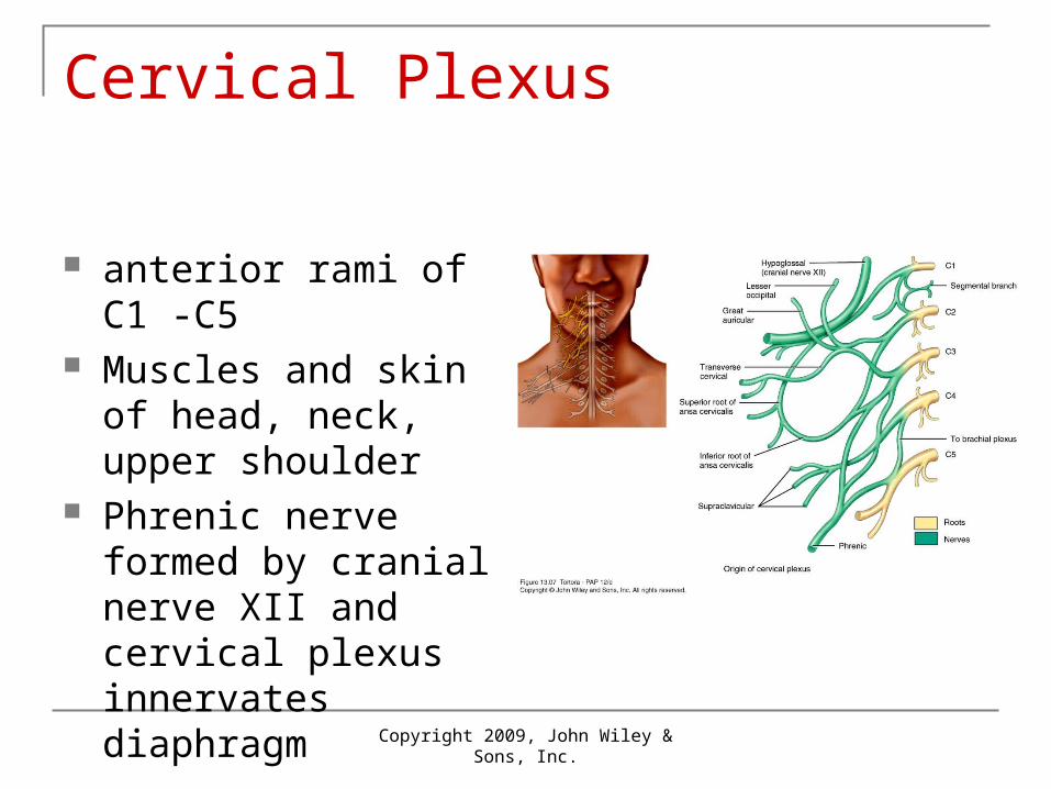
Cervical Plexus
Copyright 2009, John Wiley & Sons, Inc.
anterior rami of C1 -C5 Muscles and skin of
head, neck, upper shoulder
Phrenic nerve formed by cranial nerve XII and cervical plexus innervates diaphragm
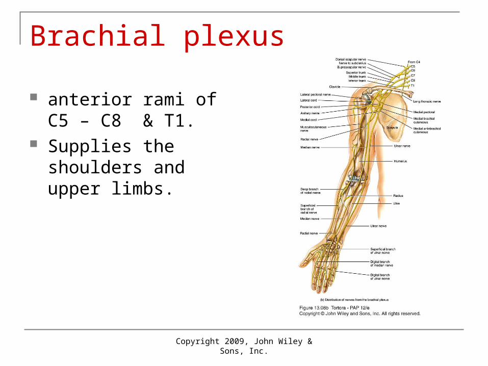
Brachial plexus
anterior rami of C5 – C8 & T1.
Supplies the shoulders and upper limbs.
Copyright 2009, John Wiley & Sons, Inc.
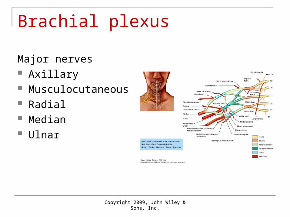
Brachial plexus
Major nerves Axillary Musculocutaneous Radial Median Ulnar
Copyright 2009, John Wiley & Sons, Inc.
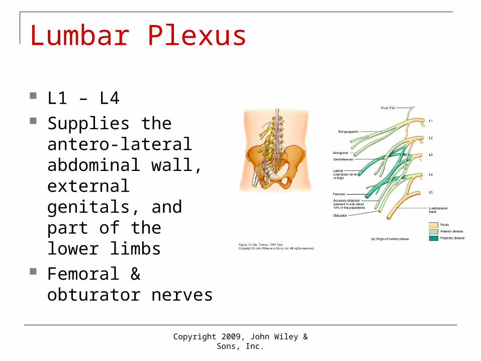
Lumbar Plexus
L1 – L4 Supplies the antero-
lateral abdominal wall, external genitals, and part of the lower limbs
Femoral & obturator nerves
Copyright 2009, John Wiley & Sons, Inc.
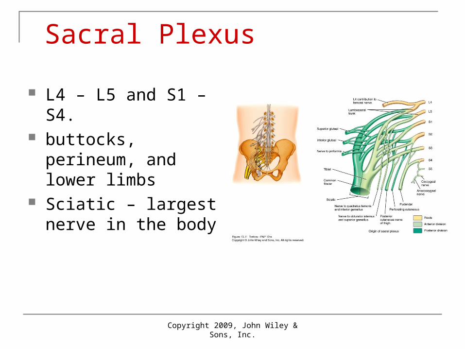
Sacral Plexus
L4 – L5 and S1 – S4. buttocks, perineum,
and lower limbs Sciatic – largest nerve
in the body
Copyright 2009, John Wiley & Sons, Inc.
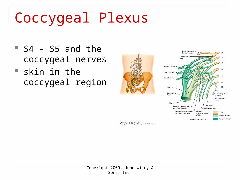
Coccygeal Plexus
S4 – S5 and the coccygeal nerves
skin in the coccygeal region
Copyright 2009, John Wiley & Sons, Inc.
T2 – T12
Anterior rami do not form plexus Intercostal / thoracic nerves Distributed directly to structures they supply
in intercostal spaces
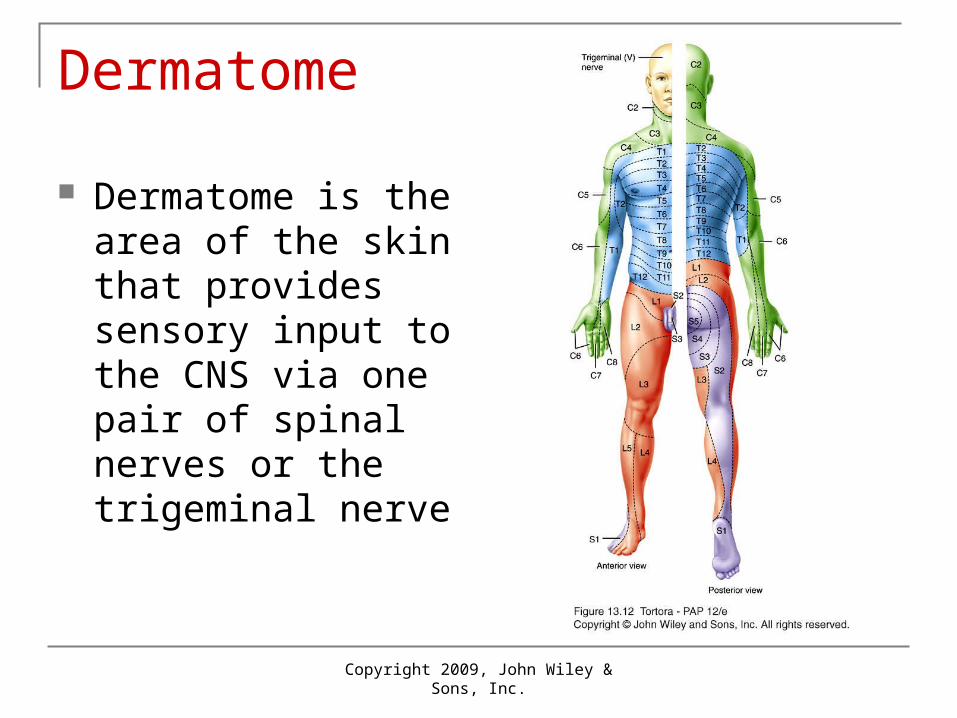
Dermatome
Dermatome is the area of the skin that provides sensory input to the CNS via one pair of spinal nerves or the trigeminal nerve
Copyright 2009, John Wiley & Sons, Inc.
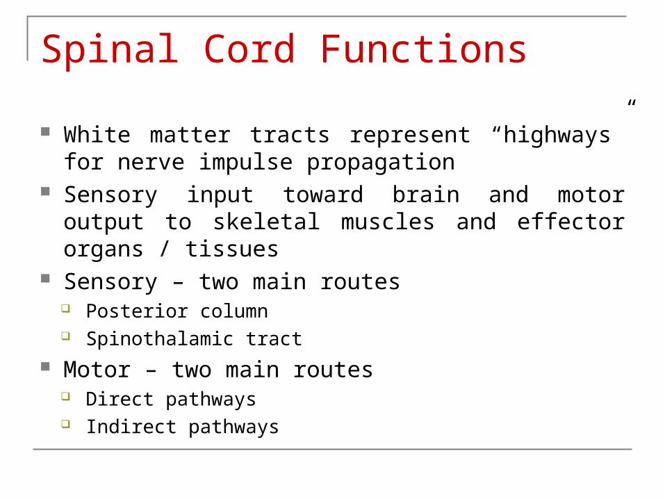
Spinal Cord Functions
White matter tracts represent “highways” for nerve impulse propagation
Sensory input toward brain and motor output to skeletal muscles and effector organs / tissues
Sensory – two main routes Posterior column Spinothalamic tract
Motor – two main routes Direct pathways Indirect pathways
13_13
Spinal Cord Functions
Integrating center for spinal reflexes Integration occurs in gray matter Reflex
Fast, automatic, and predictable involuntary response to a stimulus
May occur as muscle contractions or glandular secretions Occurs in response to changes in environment May be spinal or cranial and somatic or autonomic
(visceral) When the integration takes place in the spinal cord it is a
spinal reflex
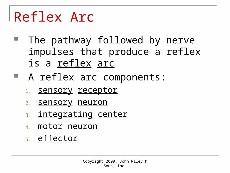
Reflex Arc The pathway followed by nerve impulses
that produce a reflex is a reflex arc A reflex arc components:
1. sensory receptor
2. sensory neuron
3. integrating center
4. motor neuron
5. effector
Copyright 2009, John Wiley & Sons, Inc.
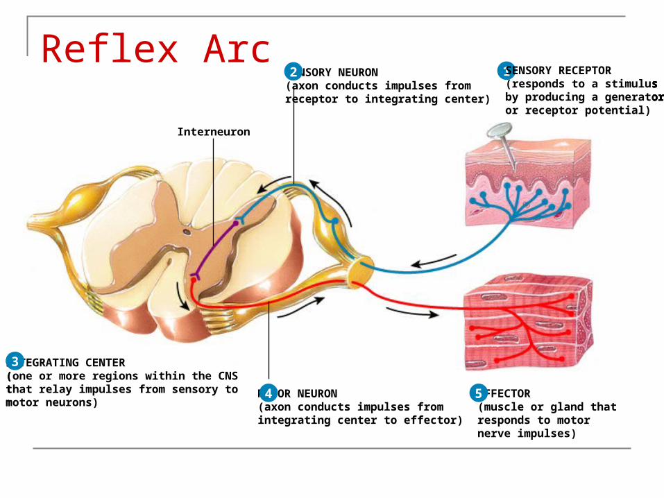
1 SENSORY RECEPTOR(responds to a stimulusby producing a generatoror receptor potential)
1SENSORY NEURON(axon conducts impulses from receptor to integrating center)
SENSORY RECEPTOR(responds to a stimulusby producing a generatoror receptor potential)
2 1SENSORY NEURON(axon conducts impulses from receptor to integrating center)
SENSORY RECEPTOR(responds to a stimulusby producing a generatoror receptor potential)
INTEGRATING CENTER(one or more regions within the CNSthat relay impulses from sensory tomotor neurons)
Interneuron
2
3
1SENSORY NEURON(axon conducts impulses from receptor to integrating center)
SENSORY RECEPTOR(responds to a stimulusby producing a generatoror receptor potential)
INTEGRATING CENTER(one or more regions within the CNSthat relay impulses from sensory tomotor neurons)
MOTOR NEURON(axon conducts impulses fromintegrating center to effector)
Interneuron
2
3
4
1SENSORY NEURON(axon conducts impulses from receptor to integrating center)
SENSORY RECEPTOR(responds to a stimulusby producing a generatoror receptor potential)
INTEGRATING CENTER(one or more regions within the CNSthat relay impulses from sensory tomotor neurons)
MOTOR NEURON(axon conducts impulses fromintegrating center to effector)
EFFECTOR(muscle or gland thatresponds to motornerve impulses)
Interneuron
2
3
4 5
Reflex Arc
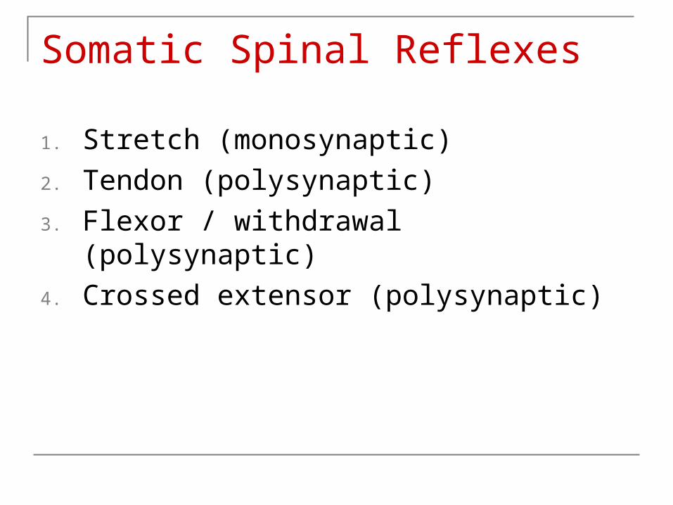
Somatic Spinal Reflexes
1. Stretch (monosynaptic)
2. Tendon (polysynaptic)
3. Flexor / withdrawal (polysynaptic)
4. Crossed extensor (polysynaptic)
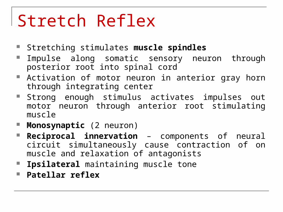
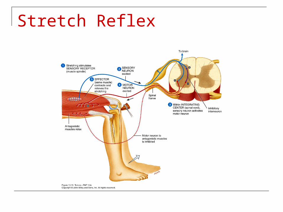
Stretch Reflex Stretching stimulates muscle spindles Impulse along somatic sensory neuron through posterior root into
spinal cord Activation of motor neuron in anterior gray horn through
integrating center Strong enough stimulus activates impulses out motor neuron
through anterior root stimulating muscle Monosynaptic (2 neuron) Reciprocal innervation – components of neural circuit
simultaneously cause contraction of on muscle and relaxation of antagonists
Ipsilateral maintaining muscle tone Patellar reflex
Stretch Reflex
Tendon Reflex
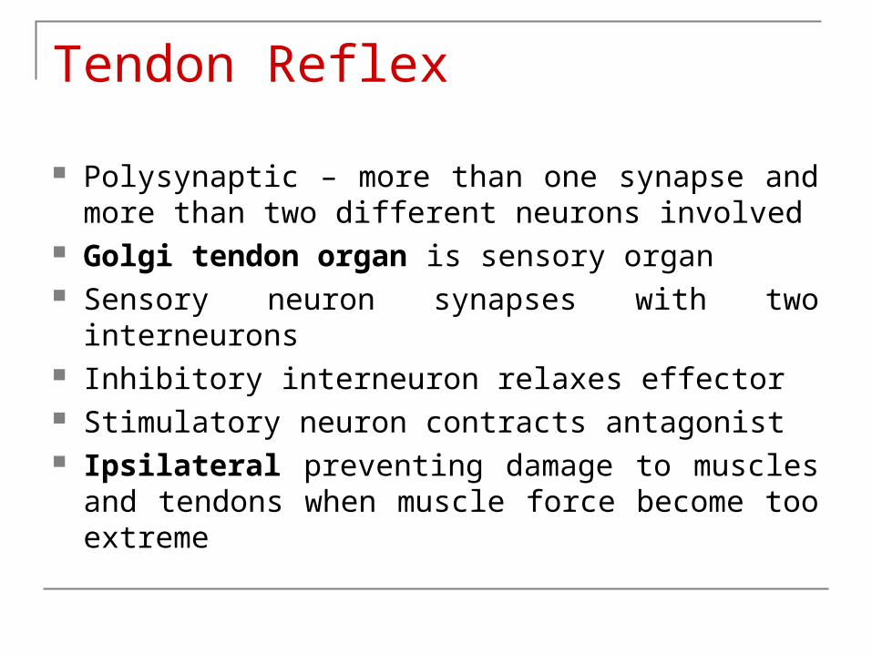
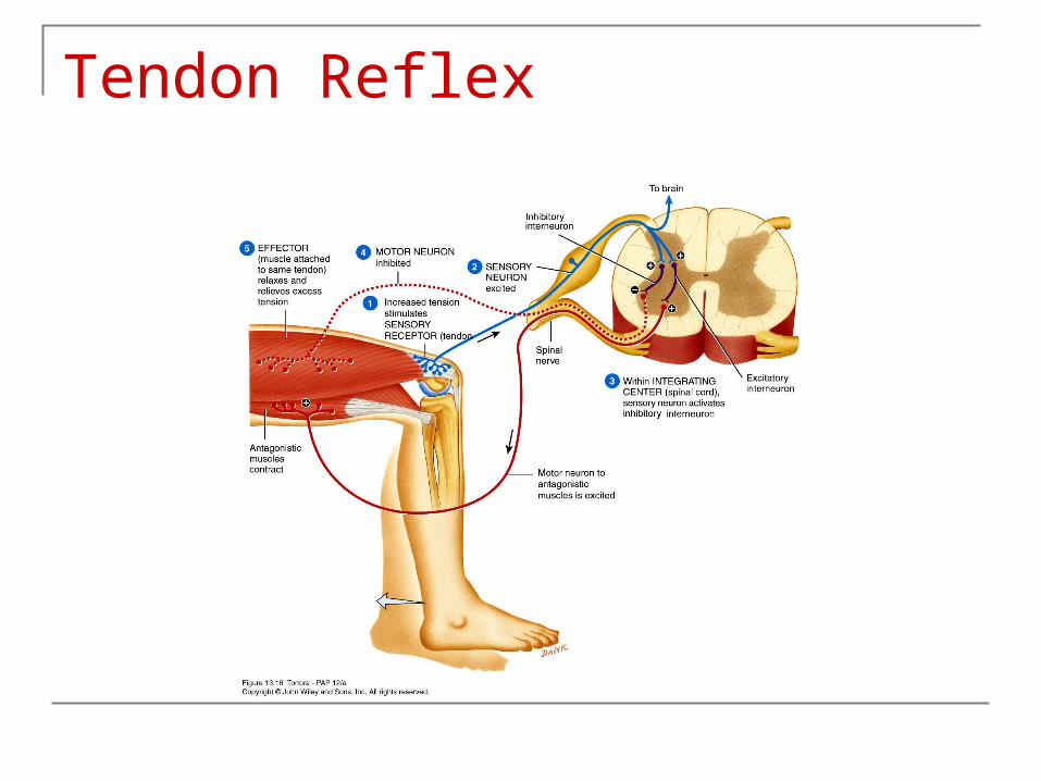
Polysynaptic – more than one synapse and more than two different neurons involved
Golgi tendon organ is sensory organ Sensory neuron synapses with two interneurons Inhibitory interneuron relaxes effector Stimulatory neuron contracts antagonist Ipsilateral preventing damage to muscles and
tendons when muscle force become too extreme
Tendon Reflex
Flexor (withdrawal) Reflex
Ipsilateral and polysynaptic Inter-segmental reflex arc Reciprocal innervation Single sensory neuron activates several
motor neurons Stimulates more than one effector
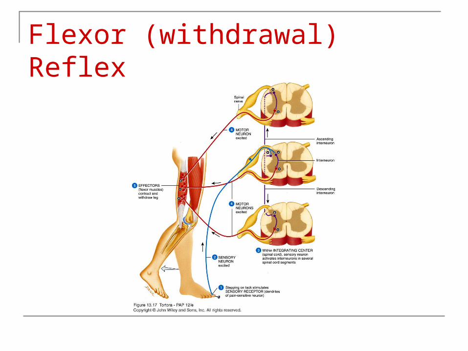
Flexor (withdrawal) Reflex
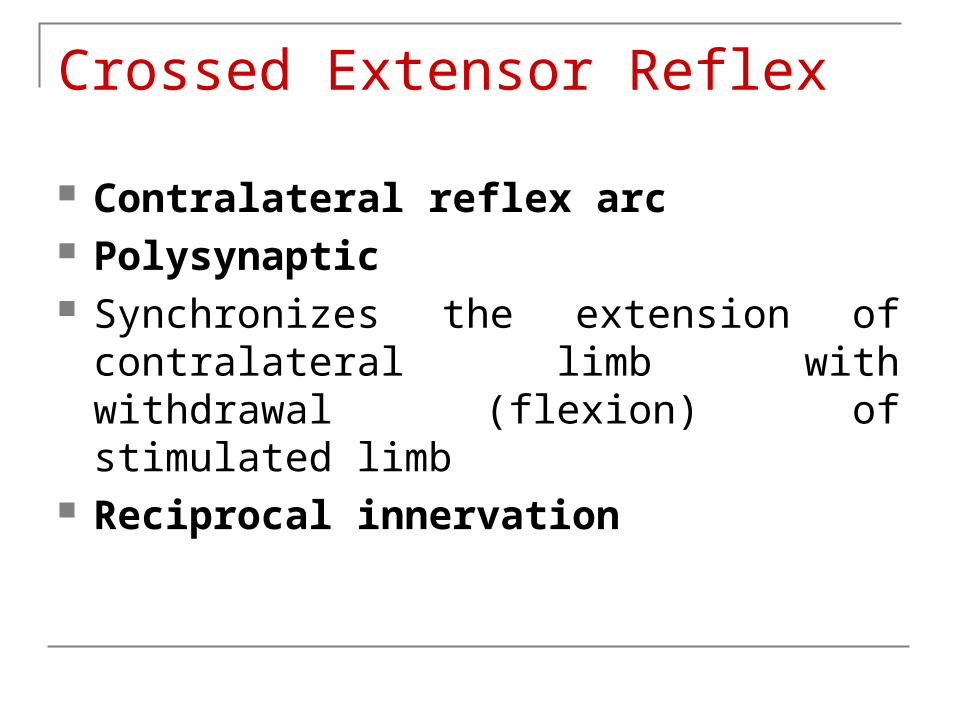
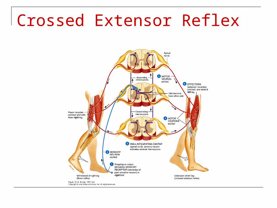
Crossed Extensor Reflex
Contralateral reflex arc Polysynaptic Synchronizes the extension of contralateral
limb with withdrawal (flexion) of stimulated limb
Reciprocal innervation
Crossed Extensor Reflex
Reflexes and Diagnosis
1. Patellar reflex Absent in damage to sensory or motor nerves or
integrating center in 2nd, 3rd, 4th lumbar Absent in chronic DM and neurosyphilis Exaggerated in disease or injury involving certain
descending motor tracts from brain
Reflexes and Diagnosis
2. Achilles reflex – plantar flexion through contraction of gastrocnemius and soleus
Absence indicates damage nerves of posterior leg or neurons of lumbo-sacral region
DM, neurosyphilis, alcoholism, subarachnoid hemorrhage
Exaggerated Cervical cord compression or lesion of motor tracts of
1st and 2nd sacral segments
Reflexes and Diagnosis
3. Babinski sign – gentle stroking of the lateral outer margin of the sole
Great toe dorsiflexes / extends with fanning of other toes
Normal in children under 1 ½ due to incomplete myelination corticospinal tract
Abnormal after 1 ½ indicating interruption of corticospinal tract
Negative Babinski – normal response is plantar felxion – curling under of toes
Reflexes and Diagnosis
4. Abdominal reflex – contraction of muscles that compress abdominal wall in response to stroking side of abdomen
Causes contraction moving umbilicus toward stimulus
Absence indicates lesions of corticospinal tract Multiple sclerosis
5. Absence of normal pupillary light reflex – associated with brain injury or damage
Copyright 2009, John Wiley & Sons, Inc.
End of Chapter 13
Copyright 2009 John Wiley & Sons, Inc.All rights reserved. Reproduction or translation of this work beyond that permitted in section 117 of the 1976 United States Copyright Act without express permission of the copyright owner is unlawful. Request for further information should be addressed to the Permission Department, John Wiley & Sons, Inc. The purchaser may make back-up copies for his/her own use only and not for distribution or resale. The Publishers assumes no responsibility for errors, omissions, or damages caused by the use of theses programs or from the use of the information herein.


