Embed Size (px)
Citation preview

Chapter 12 The Lymphatic System
Biology 110
Tri-County Technical College
Pendleton, SC
Lymphatic Components
• Lymphatic vessels, lymph nodes, & various lymphatic organs and tissues
• Has two semi-independent parts– Meandering network of lymphatic vessels– Various scattered lymphoid tissues and organs
• Vessels transport fluids that escaped from blood vascular system back to blood
• Organs house phagocytic cells/lymphocytes essential in body defense & resistance to disease

Lymph System Visual

Vessels and “Ebb and Flow”
• Vessels pick up excess tissue fluid (lymph) and return it to bloodstream
• Lymphatics form one-way system and lymph flows ONLY toward heart
• Lymph capillaries spiderweb between tissue cells and blood capillaries in loose CT
• Absorb leaked fluid (mostly water & small dissolved proteins
• Lymph capillaries remarkably permeable

Lymph Flow Visual
Vessels, cont.
• All lymph vessels drain into 2 large vessels– RIGHT lymphatic duct drains right arm and right side
of head and thorax– THORACIC duct drains rest of body
• BOTH empty into Subclavian vein on their side of body
• Vessels are thin-walled and larger ones have “valves
• Is low pressure but pumpless system

Lymph System Visual

Vessels, cont.
• Lymph transported by same mechanisms that aid return of venous blood– Milking action of skeletal muscles and pressure
changes in thorax during breathing– Smooth muscle in walls of larger lymphatic
vessels contracts rhythmically which helps “pump” lymph
– Valves in larger vessels prevent backflow

Lymph Nodes• Nodes are located along lymphatic vessels• Lymph filtered as passes through 1000s of
nodes• Large clusters found in inguinal, axillary,
and cervical regions of body• Nodes may enlarge (become swollen)
during active infection• Nodes composed of soft reticular
connective tissue

We still talking about nodes…?
• Macrophages located within nodes– Engulf and destroy bacteria/viruses/foreign
substances in lymph before its returned to blood
• Plasma cells for antibody production also located in nodes
• Collections of lymphocytes (WBC type) also in nodes and respond to foreign substances in lymph stream

Nodes, nodes, and nodes
• Nodes composed of outer cortex containing collections of lymphocytes called follicles– Follicles have germinal centers = generate plasma
cells that release antibodies– Rest of cortical cells = lymphocytes in transit
• Inner medulla area contains phagocytic macrophages
• Enters cortex of node via afferent lymphatic vesselssinusesexits node at HILUS via efferent lymphatic vessels

Node Structure Visual

Lymphatic Organs
• Spleen, thymus, tonsils, & Peyer’s patches
• Spleen located in left hypochondriac region
• Filters/cleanses blood of bacteria, viruses, and other debris
• Most important function=destroy worn-out RBCs and recycle iron to make hemoglobin– Some iron secreted in bile

Organs, cont.
• Spleen also stores platelets and acts as blood reservoir (so does liver)
• Produces blood in the fetus• In adults, spleen produces only lymphocytes• Meanwhile, back at the ranch, and not wanting to
waste space, I am practicing my typing skills, and if I may say so, not doing too badly for a over the hill ex-long haired bearded hippy….
Thymus Gland
• Functions at peak levels during youth
• Is lymphatic mass found in throat overlying heart
• Produces the hormone THYMOSIN that functions in programming lymphocytes so they can perform protective roles in body
• NOT active during adulthood

Say ahh……!!!!
• Tonsils are small masses of lymphatic tissue that ring pharynx where they are found in the mucosa
• Trap and remove bacteria/other foreign pathogens entering throat
• May become swollen and red = tonsillitis• My mom treated with warm salt water
gargle, but I have switched to aspirin

Peyer’s Patches
• Found in walls of small intestines• In ideal location to capture/destroy bacteria that
could cause enteritis• Tonsils & Peyer’s patches part of “mucosa-
associated lymphatic tissue” (MALT)• Acts as sentinel to protect upper respiratory and
the digestive tract from never-ending attacks of foreign matter entering those cavities

Lymph Organs Visual

Antibodies and more…• B lymphocytes (B cells) produce antibodies
and oversee humoral immunity
• T lymphocytes (T cells) constitute cell-mediated arm of immunity
• Antibodies called Immunoglobulins (Igs)
• GMADE
• Heavy and light chains; Variable and Constant regions

Antibody Visual

Antibodies, cont.
• IgG is most abundant is ONLY type that can cross placenta
• Only IgM and IgG fix complement
• IgA found only in mucus (major role in preventing pathogens from gaining entry into body
• IgE “troublemaker” involved in allergies

What it are…?

Antibody Action
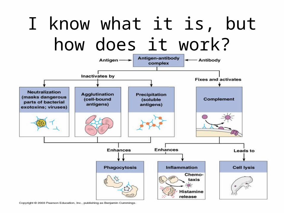
• Inactive antigens by complement fixation, neutralization, agglutination, and precipitation
• Complement fixation and neutralization most important to body function
• Complement is chief weapon against cellular antigens (bacteria/mismatched blood cells)

Complement Action Visual
I know what it is, but how does it work?

Acquired Immunity Visual
Autoimmune Disorders
• Body produces antibodies (autoantibodies) and sensitized T cells that attack and damage its own tissues
• Graves disease-thyroid gland produces excess thyroxine
• Multiple sclerosis, Myasthenia gravis, Juvenile (I) diabetes mellitus, Systemic lupus erythematosus, Glomerulonephritis, and Rheumatoid arthritis (p. 396)

Same Coin but Two Sides