Embed Size (px)
Citation preview

CHAPTER 11 Membranes
– The function of biological membranes – The structure and composition of membranes – Dynamics of membranes – Structure and function of membrane proteins – Transport across biological membranes
Key topics
1

Lipids aggregate into structures in water
• Structures formed depend on type of lipid, concentration
• Micelles • Liposomes • Bilayers
2
Micelle
• Forms in the solution of amphipathic molecules that have larger head than tail (Fatty acids, Sodium dodecyl sulfate)
• Each micelle has from a few dozen to a few thousand lipid molecules
• Aggregation occurs when the concentration of molecules is higher than a certain threshold
3
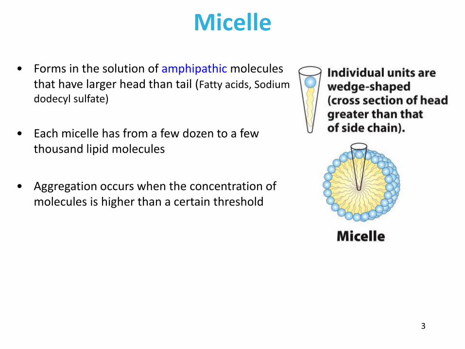
Vesicle (Liposome)
• Small bilayers will spontaneously seal into spherical vesicles
• Vesicle membranes can contain artificially inserted proteins
• The central aqueous cavity can enclose dissolved molecules
• They are useful artificial carriers of molecules (e.g., drugs)
• Vesicles fuse readily with cell membranes or with each other
4

Membrane Bilayer • consists of two leaflets of lipid monolayers
– Hydrophilic head groups interact with water – Hydrophobic fatty acid tails are packed inside – One leaflet faces the cytoplasm – Another leaflet faces the extracellular space or
the inside of membrane-enclosed organelle
5
What are membranes? • Complex lipid-based structures that form pliable sheets
• Composed of a variety of lipids and proteins
• Some membrane lipids and proteins are glycosylated
• All cells have a cell membrane, which separates the cell from its surrounding
• Eukaryotic cells have various internal membranes that divide the internal space into compartments
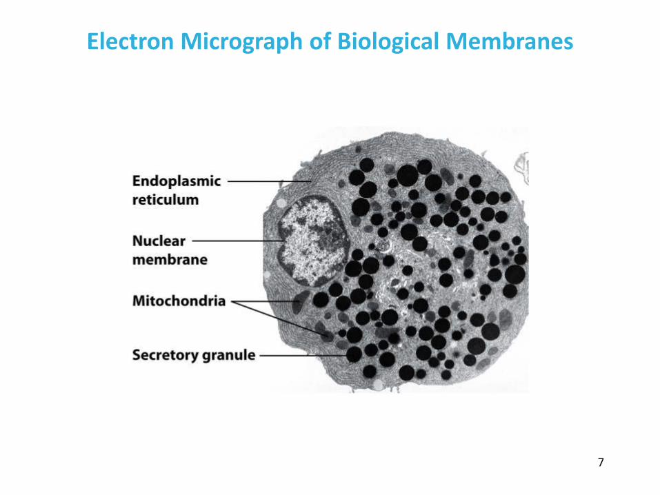
Electron Micrograph of Biological Membranes
7

Functions of Membranes
• Define the boundaries of the cell • Allow import and export
– Selective import of nutrients (e.g., lactose) – Selective export of waste and toxins (e.g., antibiotics)
• Retain metabolites and ions within the cell • Sense external signals and transmit information into the cell
• Provide compartmentalization within the cell
– separate energy-producing reactions from energy-consuming ones – keep proteolytic enzymes away from important cellular proteins
• Produce and transmit nerve signals • Store energy as a proton gradient • Support synthesis of ATP
8

Common Features of Membranes
• Sheet-like flexible structure, 30–100 Å (3–10 nm) thick • Main structure is composed of two leaflets of lipids (bilayer)
– With the exception of archaebacteria: monolayer of bifunctional lipids
• Form spontaneously in aqueous solution and are stabilized by noncovalent forces, especially hydrophobic effect
• Protein molecules span the lipid bilayer • Asymmetric
– Some lipids are found preferably “inside” – Some lipids are found preferably “outside” – Carbohydrate moieties are always outside the cell – Electrically polarized (inside negative ~ –60mV)
• Fluid structures: two-dimensional solution of oriented lipids
9
Fluid Mosaic Model of Membranes
• Proposed in 1972 by Singer and Nicholson (UCSD)
• Lipids form a viscous, two-dimensional solvent into which proteins are inserted and integrated more or less deeply
• Integral proteins are firmly associated with the membrane, often spanning the bilayer
• Peripheral proteins are weakly associated and can be removed easily
– Some are noncovalently attached
– Some are linked to membrane lipids
10

11

The Composition of Membranes
• Lipid composition of membranes is different in different organisms, tissues, and organelles
• Ratio of lipid to protein varies • Type of phospholipid varies
• Abundance and type of sterols varies: prokaryotes lack sterols • Cholesterol predominant in the plasma membrane, virtually
absent in mitochondria • Galactolipids abundant in plant chloroplasts but almost
absent in animals
12

Membrane composition is highly variable in different organisms
13

Membrane Structure in Archaea
• Unique glycerol chirality in phospholipids – L-glycerol in archaea – D-glycerol in bacteria
• Unique fatty acids – Branched isoprene chains in archaea – Unbranched fatty acid chains in bacteria
• Unique linkages – Ether linkages in archaea – Ester linkages in bacteria
• Membrane topology – Monolayer in some archaea – Bilayer in all bacteria
14

Lipid Monolayer in Archaea
• Sulfolobus solfataricus and relatives – Volcanic hot springs – Temperatures 75–80°C – Acidic environment: pH 2–3
• Better membrane stability by
– Isoprenoid tetraethers with unique alcohols
15

Membrane composition is highly variable in different organelles
16

Membrane bilayers are asymmetric
• Two leaflets have different lipid compositions • Outer leaflet is often more positively charged • Phosphatidylserine outside has a special meaning
– Platelets: Activates blood clotting – Other cells: Marks the cell for destruction
17
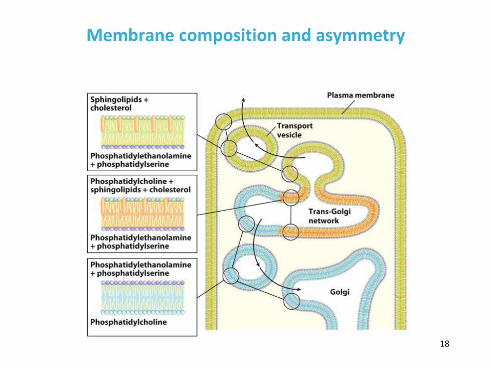
Membrane composition and asymmetry
18

Functions of Proteins in Membranes
• Receptors: detecting signals from outside – Light (opsin) – Hormones (insulin receptor) – Neurotransmitters (acetylcholine receptor) – Pheromones (taste and smell receptors)
• Channels, gates, pumps – Nutrients (maltoporin) – Ions (K-channel) – Neurotransmitters (serotonin reuptake protein)
• Enzymes – Lipid biosynthesis (some acyltransferases) – ATP synthesis (F0F1 ATPase/ATP synthase)
19

Three Types of Membrane Proteins
20

Peripheral Membrane Proteins
• Associate with the polar head groups of membranes • Relatively loosely associated with membrane
– through ionic interactions with the lipids or aqueous domains of integral membrane proteins
• Removed by disrupting ionic interactions either with high salt or change in pH
• Purified peripheral membrane proteins are no longer associated with any lipids
21

Integral Membrane Proteins • Span the entire membrane • Have asymmetry like the membrane
– Different domains in different compartments
• Tightly associated with membrane – Hydrophobic stretches in the protein interact with the hydrophobic regions
of the membrane
• Removed by detergents that disrupt the membrane • Purified integral membrane proteins still have phospholipids associated
with them
22

Six Types of Integral Membrane Proteins
23
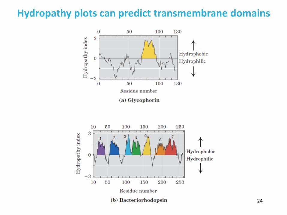
Hydropathy plots can predict transmembrane domains
24

Amino acids in membrane proteins cluster in distinct regions
• Transmembrane segments are predominantly hydrophobic • Tyr and Trp cluster at nonpolar/polar interface • Charged amino acids are only found in aqueous domains
25

Membrane proteins also contain β-sheets
26

Lipid Anchors
• Some membrane proteins are lipoproteins • They contain a covalently linked lipid molecule
– Long-chain fatty acids – Isoprenoids – Sterols – Glycosylated phosphatidylinositol (GPI)
• The lipid part can become part of the membrane • The protein is now anchored to the membrane
– reversible process – allows targeting of proteins – Proteins with GPI anchors are found only on the outer face of plasma
membrane
27
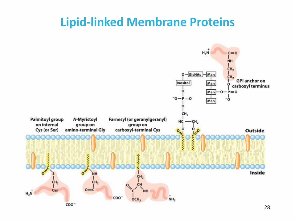
Lipid-linked Membrane Proteins
28

Farnesylation of Proteins
• Proteins can be targeted to the inner leaflet of the plasma membrane by farnesylation
• Primary sequence of the protein contains a signature for farnesylation: CaaX
– C is a conserved Cys – “a” is usually an aliphatic amino acid – “X” is Met, Ser, Glu or Ala
• This reaction is catalyzed by farnesyl transferase • Nonfarnesylated proteins do not go to the membrane and are inactive
– Promising cancer therapy (onco-Ras)
29

Cell membranes are asymmetric
• Every component of the membrane exhibits asymmetry • Lipids
– Outer and inner leaflets have different lipid compositions • Proteins
– Individual peripheral membrane proteins are only associated with one side of the membrane
– Integral membrane proteins have different domains on different sides of the membrane.
– Specific lipid modification of proteins targets the protein to a specific leaflet • Carbohydrates
– Only on the outside of cells
30

Physical Properties of Membranes
• Dynamic and flexible structures • Can exist in various phases and undergo phase transitions • Not permeable to large polar solutes and ions • Permeable to small polar solutes and nonpolar compounds • Permeability can be artificially increased by chemical treatment
– When we want to get DNA into the cell
31

Membrane Phases • Depending on their composition and the temperature, lipid bilayer can be in
gel or fluid phase – Gel phase: individual molecules do not move around – Fluid phase: individual molecules can move around
• Heating causes phase transition from the gel to fluid • Under physiological conditions, membranes are more fluid-like than gel-like
– Must be fluid for proper function
32

Organisms can adjust the membrane composition
• Membrane fluidity is determined mainly by the fa composition • More fluid membranes require shorter and more unsaturated fa
* Remember differences in TM of fa
• At higher temperatures cells need more saturated fa: To maintain integrity
• At lower temperatures cells need more unsaturated fa: To maintain fluidity
33

Sterols and hopanols increase membrane rigidity and permeability
• Cell membranes of many eukaryotes contain sterols
– Cholesterol in animals – Phytosterols in plants – Ergosterol in fungi
• Cell membranes of aerobic
prokaryotes contain hopanols
OH
OH
OHO OH
OHOH OH
NH2
OH
OH
cholesterol
ergosterol
one particular hopanol 34

Membrane Dynamics: Lateral Diffusion
Individual lipids undergo fast lateral diffusion within the leaflet
35

Membrane Dynamics: Transverse Diffusion
Spontaneous flips from one leaflet to another are rare because the charged head group must transverse the hydrophobic tail region of the membrane
36

Membrane Diffusion: Flippase
• Flippases catalyze transverse diffusion • Flippases use energy of ATP to move lipids against the concentration gradient
37

Study of Membrane Dynamics
• Fluorescence Recovery After Photobleaching (FRAP) allows us to monitor lateral lipid diffusion by monitoring the rate of fluorescence return
• From the rate of return of lipids, the diffusion coefficient of a lipid in the leaflet can be determined
• Rates of lateral diffusion are high (up to 1 µm/sec) – a lipid can circumnavigate E.coli cell in one second

Movement of a Single Lipid in 56 ms
39

Membrane Rafts
• Lipid distribution in a single leaflet is not random or uniform • Lipid rafts
– contain clusters of glycosphingolipids with longer-than-usual tails – are more ordered – contain specific doubly or triply acylated proteins – allow segregation of proteins in the membrane
40

Caveolin forces membrane curvature
41 Caveola are small invaginations in the plasma membrane

Other Modes of Membrane Curvature
42

Membrane Fusion
• Membranes can fuse with each other without losing continuity • Fusion can be spontaneous or protein-mediated • Examples of protein-mediated fusion are
– Entry of influenza virus into the host cell – Release of neurotransmitters at nerve synapses

Neurotransmitter Release
44

Transport Across Membranes
• Cell membranes are permeable to small nonpolar molecules that passively diffuse through the membrane
• Passive diffusion of polar molecules involves desolvation and thus has a high activation barrier
• Transport across the membrane can be facilitated by proteins that provide an alternative diffusion path
• Such proteins are called transporters or permeases
45

Types of Transport
46

Polar solutes need alternative paths to cross cell membranes
47

Three Classes of Transport Systems
48

Glucose Transporter in the Membrane
49

Model for Glucose Transport
50

There are multiple glucose transporters
• A Na+-glucose symporter and a glucose uniporter operate on opposite sides of epithelial cells
• Cells can also have asymmetry, with distinct proteins confined to one side
51

Bicarbonate transporter is an antiporter
Maintains the electrochemical potential across the membrane
52
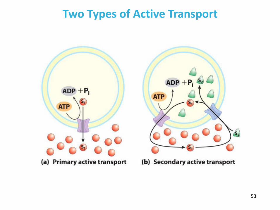
Two Types of Active Transport
53
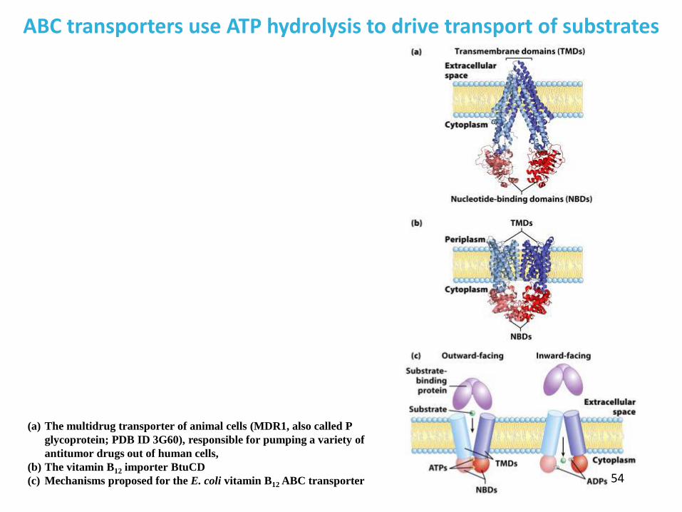
ABC transporters use ATP hydrolysis to drive transport of substrates
54
(a) The multidrug transporter of animal cells (MDR1, also called P glycoprotein; PDB ID 3G60), responsible for pumping a variety of antitumor drugs out of human cells,
(b) The vitamin B12 importer BtuCD (c) Mechanisms proposed for the E. coli vitamin B12 ABC transporter

Proton Transport and Chemical Energy of ATP
• Energy of ATP hydrolysis can be used to drive protons through the membrane – pH control in the cell by F-type ATPase
• Energy of the proton gradient can be used to synthesize ATP – in chloroplast and mitochondrial membranes by ATP synthase
55

Proton driven ATPases can function in both directions
The VoV1 H+ ATPase uses ATP to pump protons into vacuoles and lysosomes, creating their low internal pH. Fo F1 ATPase/ATP synthase of mitochondria has an integral domain, Fo (orange) and a peripheral domain, F1
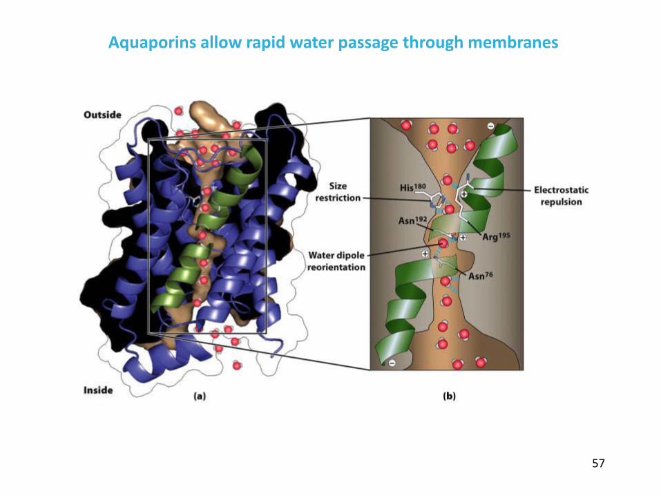
Aquaporins allow rapid water passage through membranes
57

Ion channels maintain gradients for active transport
58

59
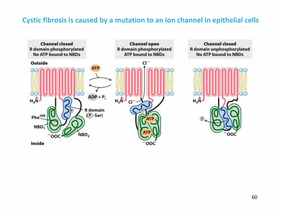
Cystic fibrosis is caused by a mutation to an ion channel in epithelial cells
60

• membranes are composed of various lipids and proteins • phospholipids form a selectively permeable bilayer • properties of the bilayer depend on the lipid composition, which varies
strongly from – organism to organism – tissue to tissue – organelle to organelle
• membrane proteins play a variety of structural and functional roles, especially in the transport of solutes across the membrane
• Active transport of solutes across membranes requires ATP but can be accomplished in many different ways
In this chapter, we learned
61