Embed Size (px)
Citation preview

404 Section 5 Shock and Resuscitation
Scene Size-up
Scene Safety Ensure scene safety and address hazards. Standard precautions should include a minimum of gloves and eye protection. Consider the number of patients, the need for additional help/ALS, and cervical spine stabilization.
Mechanism of Injury (MOI)/Nature of Illness (NOI)
Determine the MOI/NOI. Observe the scene and look for indicators of the MOI such as falls, motor vehicle crashes, gunshot wounds, or stabbings. Observe the patient for signs of NOI such as urticaria, chest pain, or fever.
Primary Assessment
Form a General Impression
Airway and Breathing
Determine level of consciousness and fi nd and treat any immediate life threats. Observe overall appearance of patient and body position. Determine priority of care based on the MOI/NOI. If the patient has a poor general impression, call for ALS assistance. A rapid scan will help you identify and manage life threats.
If a cervical spine injury is suspected, open the airway using a modifi ed jaw-thrust and ensure the airway is patent. A patient with an altered level of consciousness may need emergency airway man-agement; consider inserting a properly sized oropharyngeal or nasopharyngeal airway. Assess for gurgling, stridor, or rales. Quickly assess the chest for DCAP-BTLS, accessory muscle use, intercostal and abdominal muscle use, and treat any threats to life. Provide high-fl ow oxygen at 15 L/min and evaluate the depth and rate of the respiratory cycle, providing ventilatory support as needed.
Circulation
Transport Decision
Evaluate distal pulse rate and quality; observe skin color, temperature, and condition; look for life-threatening bleeding and treat accordingly. If distal pulses are not palpable, assess for a central pulse. Place the patient in a supine (Trendelenburg’s) or shock position. Prevent heat loss by using blankets. Serious bleeding must be treated at once.
If the patient has an airway or breathing problem, signifi cant external bleeding, or signs and symptoms of internal bleeding, consider rapid transport. Suspected shock patients or those with a suspicious MOI should go to a trauma center. ALS providers can treat these patients with intravenous fl uids to support circulation (shock) problems. If anaphylactic shock is suspected, determine if the patient has a prescribed EpiPen auto-injector before leaving the scene. Do not delay transport to manage non–life-threatening injuries, instead treat en route to the hospital.g ii
History Taking
Investigate Chief Complaint
Investigate the chief complaint. Identify signs and symptoms and pertinent negatives. Be alert for injuries and life threats. Observe for signs and symptoms of shock. Monitor patient for change in mental status. If possible, ask OPQRST and SAMPLE questions.
NOTE: The order of the steps in this section differs depending on whether the patient is conscious or unconscious. The following order is for a conscious patient. For an unconscious patient, perform a primary assessment, perform a rapid full-body scan, obtain vital signs, and if possible, obtain the past medical history from a family member, bystander, or emergency medical identifi cation device.
78286_CH10_Printer.indd 40478286_CH10_Printer.indd 404 2/23/10 11:06:10 PM2/23/10 11:06:10 PM

Chapter 10 Shock 405
Secondary Assessment
Physical Examinations
Vital Signs
Perform a systematic full-body scan beginning with the head, looking for DCAP-BTLS. Assessment should be rapid if the patient has a poor general impression. Inspect, palpate, and auscultate the chest, focusing on the respiratory effort and adequacy of ventilation. Perform a thorough neurologic examination assessing the pupils, motor response, and sensory response in all extremities. Assess the musculoskeletal system for DCAP-BTLS. Assess the abdomen for signs of internal bleeding. Log roll and inspect the posterior torso for injuries.
Obtain baseline vital signs, monitoring trends. Repeat every 5 to 15 minutes depending on patient impression. Vitals signs should include blood pressure, pulse rate and quality, respiration rate and quality, and skin assessment for perfusion. Note patient’s level of consciousness. Use pulse oximetry, if available, to assess the patient’s perfusion status. nt
Reassessment
NOTE: Although the steps below are widely accepted, be sure to consult and follow your local protocols.
Interventions
Communication and Documentation
Consider cervical spine precautions. Repeat the primary assessment and reassess vital signs and chief complaint. Check interventions and treatment rendered. Airway control using adjuncts may be necessary. Assist breathing as required, administering high-fl ow oxygen. Control all bleeding and circulation problems. Support the cardiovascular system and treat for shock early. Patients on a backboard should be placed in the Trendelenburg’s position by raising the foot end of the backboard 6" to 12" . Prevent body heat loss by placing blankets under and over the patient. Splint bone or joint injuries. Do not give the patient anything by mouth. Do not delay transport.
Contact medical control with a radio report. Include a thorough description of the MOI/NOI and the position the patient was found in. Include treatments performed and patient response. Follow local protocols. Be sure to document any changes in patient status and the time. Document the reasoning for your treatment and the patient’s response.
78286_CH10_Printer.indd 40578286_CH10_Printer.indd 405 2/23/10 11:06:15 PM2/23/10 11:06:15 PM
406 Section 5 Shock and Resuscitation
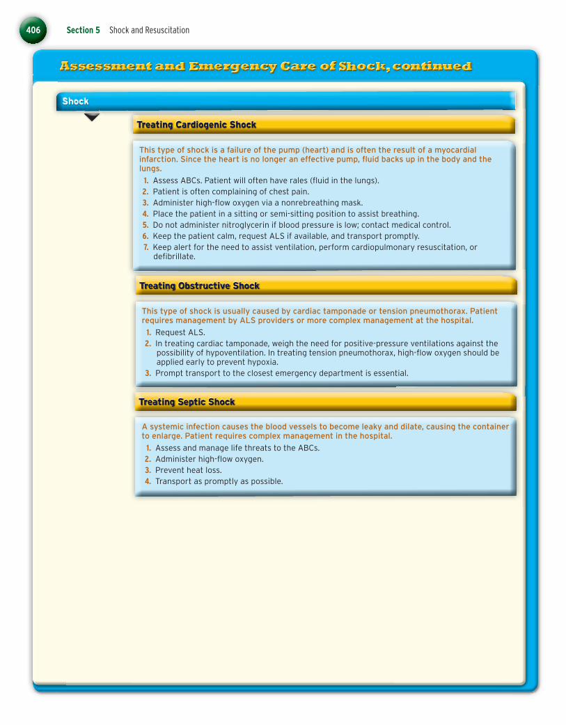
Treating Cardiogenic Shock
This type of shock is a failure of the pump (heart) and is often the result of a myocardial infarction. Since the heart is no longer an effective pump, fl uid backs up in the body and the lungs.
1. Assess ABCs. Patient will often have rales (fl uid in the lungs). 2. Patient is often complaining of chest pain. 3. Administer high-fl ow oxygen via a nonrebreathing mask. 4. Place the patient in a sitting or semi-sitting position to assist breathing. 5. Do not administer nitroglycerin if blood pressure is low; contact medical control. 6. Keep the patient calm, request ALS if available, and transport promptly. 7. Keep alert for the need to assist ventilation, perform cardiopulmonary resuscitation, or
defi brillate.
Shock
Treating Obstructive Shock
This type of shock is usually caused by cardiac tamponade or tension pneumothorax. Patient requires management by ALS providers or more complex management at the hospital.
1. Request ALS. 2. In treating cardiac tamponade, weigh the need for positive-pressure ventilations against the
possibility of hypoventilation. In treating tension pneumothorax, high-fl ow oxygen should be applied early to prevent hypoxia.
3. Prompt transport to the closest emergency department is essential.
Treating Septic Shock
A systemic infection causes the blood vessels to become leaky and dilate, causing the container to enlarge. Patient requires complex management in the hospital.
1. Assess and manage life threats to the ABCs. 2. Administer high-fl ow oxygen. 3. Prevent heat loss. 4. Transport as promptly as possible.
78286_CH10_Printer.indd 40678286_CH10_Printer.indd 406 2/23/10 11:06:23 PM2/23/10 11:06:23 PM
Chapter 10 Shock 407
Shock
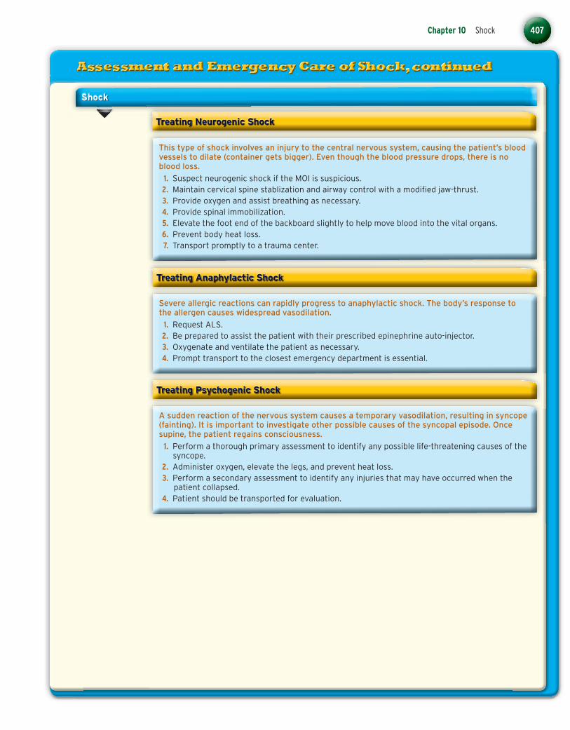
Treating Neurogenic Shock
This type of shock involves an injury to the central nervous system, causing the patient’s blood vessels to dilate (container gets bigger). Even though the blood pressure drops, there is no blood loss.
1. Suspect neurogenic shock if the MOI is suspicious. 2. Maintain cervical spine stablization and airway control with a modifi ed jaw-thrust. 3. Provide oxygen and assist breathing as necessary. 4. Provide spinal immobilization. 5. Elevate the foot end of the backboard slightly to help move blood into the vital organs. 6. Prevent body heat loss. 7. Transport promptly to a trauma center.
Treating Anaphylactic Shock
Severe allergic reactions can rapidly progress to anaphylactic shock. The body’s response to the allergen causes widespread vasodilation.
1. Request ALS. 2. Be prepared to assist the patient with their prescribed epinephrine auto-injector. 3. Oxygenate and ventilate the patient as necessary. 4. Prompt transport to the closest emergency department is essential.
A sudden reaction of the nervous system causes a temporary vasodilation, resulting in syncope (fainting). It is important to investigate other possible causes of the syncopal episode. Once supine, the patient regains consciousness.
1. Perform a thorough primary assessment to identify any possible life-threatening causes of the syncope.
2. Administer oxygen, elevate the legs, and prevent heat loss. 3. Perform a secondary assessment to identify any injuries that may have occurred when the
patient collapsed. 4. Patient should be transported for evaluation.
Treating Psychogenic Shock
78286_CH10_Printer.indd 40778286_CH10_Printer.indd 407 2/23/10 11:06:28 PM2/23/10 11:06:28 PM

408 Section 5 Shock and Resuscitation
Shock
Treating Hypovolemic Shock
This type of shock is caused by a loss of blood or body fl uids. It should be suspected fi rst whenever a patient presents with signs and symptoms of shock. Blood loss may be external or internal secondary to a traumatic injury. Body fl uids can be lost due to burns, excessive vomiting, or diarrhea.
1. Management of a patient with hypovolemic shock focuses on preventing further blood or fl uid loss.
2. Manage threats to the ABCs. 3. Control external bleeding with direct pressure, pressure dressings, and tourniquets. 4. Internal bleeding is diffi cult to manage. Splinting injured extremities may slow blood loss. 5. Place the patient on a long backboard. The Trendelenburg’s or the shock position may used to
assist with perfusion. 6. High-fl ow oxygen should be administered all hypovolemic patients. 7. Prompt transport to a trauma center is required. Do not delay transport.
Treating Respiratory Insuffi ciency
Patients in shock as a result of respiratory insuffi ciency require immediate airway maintenance and oxygen.
1. Clear obstructions and suction airway as required. 2. Give supplemental oxygen and assist ventilations if necessary. 3. Transport promptly.
78286_CH10_Printer.indd 40878286_CH10_Printer.indd 408 2/23/10 11:06:33 PM2/23/10 11:06:33 PM