Embed Size (px)
Citation preview

July 12, 2017 0:48 World Scientific Review Volume - 9.75in x 6.5in ws-rv975x65 page 1
Chapter 1
Robot Autonomy for Surgery
Michael Yip and Nikhil Das
University of California, San Diego,9500 Gilman Drive, La Jolla, CA, 92093
{yip, nrdas}@ucsd.edu
Autonomous surgery involves having surgical tasks performed by a robot operat-ing under its own will, with partial or no human involvement. There are severalimportant advantages of automation in surgery, which include increasing preci-sion of care due to sub-millimeter robot control, real-time utilization of biosignalsfor interventional care, improvements to surgical efficiency and execution, andcomputer-aided guidance under various medical imaging and sensing modalities.While these methods may displace some tasks of surgical teams and individualsurgeons, they also present new capabilities in interventions that are too difficultor go beyond the skills of a human. In this chapter, we provide an overviewof robot autonomy in commercial use and in research, and present some of thechallenges faced in developing autonomous surgical robots.
1. Introduction
Robot autonomy refers to the performance of tasks where part or all of the task
is completed by an intelligent robotic system. Since the use of robotics in surgery
in the mid-1980s, applying autonomy to surgery has been a continuing effort for
engineers and researchers. As with conventional human-performed surgery, surg-
eries performed autonomously through robotic systems require these systems to be
safe and precise in their movements, to adequately sense the environment and the
patient, and to make decisions and adapt to changes in the environment under dif-
ferent situations. There is a large spectrum of autonomous behaviors that can be
observed in surgical procedures: some are far easier to implement and encourage the
use of (such as tremor reduction), while others are much more forward-looking, such
as autonomous cardiac ablation in the beating heart, where a clinician must rely
on the robot entirely to creating continuous lesions in the heart. The technologies
associated with these works are often a combination of multiple areas of research
and technology: robot design and control, medical image integration and real-time
signal processing, and artificial intelligence and machine learning.
In this chapter, we will consider the following topics in order:
1
arX
iv:1
707.
0308
0v1
[cs
.RO
] 1
0 Ju
l 201
7

July 12, 2017 0:48 World Scientific Review Volume - 9.75in x 6.5in ws-rv975x65 page 2
2 M. Yip and N. Das
(1) define and distinguish the levels of autonomy for surgical robotics,
(2) consider the potential benefits of autonomy in surgery,
(3) describe systems for surgical autonomy in commercial use,
(4) describe research efforts in hardware and software toward surgical automation,
(5) detail current challenges in research and development for automated surgery,
and
(6) discuss ethical and legal concerns involved in automating surgery.
We conclude with future directions for robot autonomy in surgery.
While we will highlight specific technologies and developments of interest and
importance to surgical autonomy, this chapter is not meant to provide a complete
report on all efforts on surgical autonomy, since an exhaustive search would bring up
thousands of examples including efforts that may or may not be considered automa-
tion; instead, we will use the topics above to frame the topic of surgical autonomy
and explore key strategies, innovations, and considerations in its implementation.
2. Scope of Automation
The spectrum of automation in surgical robotics relates to the level of dependence
on the human surgeon to guide the procedure, as compared to the robot guiding
itself. These levels are direct control, shared control, supervised autonomy, and full
autonomy. Fig. 1 illustrates this range of autonomy and provides examples of
commercial systems at each level. Here we define the different automation methods
at a high level, though many different commercial systems may fall within two or
more categories.
In direct control, human surgeons are in complete control of the surgical tasks by
either manually controlling or teleoperating the robotic manipulators. In a teleoper-
ation scheme, the surgeon sits at a user control console with joysticks and a camera
feed, including access to any other pertinent biosignals or imaging modalities placed
in the same display, to remotely operate the tools. The robot directly obeys the
human surgeon’s commands and therefore does not move by itself; however, one can
consider some form of robot automation involved in the mapping of complex robot
joints velocities into tool velocities, a complex mapping that is masked from the hu-
man operator. This level of autonomy can be found in most teleoperated systems.
Fig. 1. Spectrum of autonomy in robotic surgery and examples of systems. There are currently
no examples of surgical systems that can operate without any level of human involvement.

July 12, 2017 0:48 World Scientific Review Volume - 9.75in x 6.5in ws-rv975x65 page 3
Robot Autonomy for Surgery 3
The da Vinci Surgical System, developed by Intuitive Surgical Inc. in 2000 was the
first FDA-approved robotic system for laparoscopic surgery and is an example of
such a system. The surgical instruments working on the patient are teleoperated
by a human surgeon sitting at a console in a non-sterile section of the operating
room.1 Indeed, the da Vinci system is capable of fully autonomous behavior as we
will see in future sections, but is currently not approved for autonomous motions.
The next level of autonomy is shared control. As the name suggests, shared con-
trol is where different types of surgical tool motions are simultaneously distributed
in their control between the human and robot.2,3 The Steady Hand robot, developed
at Johns Hopkins University, is a system designed for sub-millimeter manipulation
tasks such as retinal microsurgery.4 The Steady Hand system allows the human
surgeon to control all movements of the surgical instrument but gradually applies a
counteractive force proportional to the force sensed by the tool tip.5 The net motion
of the robot manipulator is thus a based on a combination of the human’s move-
ments and the robot’s application of force feedback to correct for tremor, making
the Steady Hand an example of a shared control system.
At the intermediate level is supervised autonomy, where the robot executes sur-
gical tasks with some autonomy but under supervision of a human surgeon. The
human oversees the operation and issues high-level commands while the robot exe-
cutes the motions. This enables the surgeon to still remain in charge of the decision-
making process, but with less involvement in the execution. The CyberKnife system,
invented at Stanford University and now built by Accuray,2 generates and executes
preoperative plans to deliver radiotherapy to tumors located on the patient’s body.
The human surgeon adjusts the automatically generated plan prior to execution
and ensures the system performs the task safely during the procedure.6
At the end of the spectrum is full autonomy, where a robotic system completely
supplants the intraoperative role of the surgeon. Such a system will autonomously
plan and execute entire surgical operations. As surgical procedures are complex and
intricacies of the operation depend on patient-specific information, full autonomy
in surgery is currently not likely to be realized in practice in any near future.
However, it is a potential goal in automated healthcare7 in that it could significantly
improve the consistency and quality of care to the patient population, as well as
be distributed to remote areas for improving patient access to high-quality surgical
care.
While full autonomy of entire surgical procedures is currently far-fetched even
from a technological point of view, entire procedures may be decomposed into
smaller surgical tasks, such as suturing or resection, that are more feasible to au-
tomate. Section 7 discusses recent approaches to surgical automation of certain
surgical tasks in research. Similar to human language, surgical activities can be
described systematically in terms of the constituent parts, i.e., the basic motions
that the surgeons perform with their instruments or hands.8 Thus, decomposition
of surgical procedures may be taken a step further by identifying the primitive

July 12, 2017 0:48 World Scientific Review Volume - 9.75in x 6.5in ws-rv975x65 page 4
4 M. Yip and N. Das
surgically-relevant motions (called surgemes) that make up the execution of a given
task.8
3. Advantages of Autonomy in Surgery
There are many advantages to automating parts or all of a surgery. Human surgeons,
while great at making high-level decisions regarding the care of the patient, are less
capable at performing precision tasks, especially in the face of fatigue and under
hand tremors. This causes the variability of fine motor tasks and, by extension,
surgical care, to be large between experts and novices, and from metropolitan areas
to remote communities. With many of these surgeons overworked to high levels of
fatigue, they are less attentive and more prone to human error. Autonomous surgery
provides a consistency and quality to a treatment unaffected by these issues.
A second major advantage of automating robots for performing surgery is that a
robotic system can provide significantly greater dexterity in its tools than a human-
controlled tool.9 Increasing dexterity of tools has been shown to be effective in
minimally-invasive procedures. Such advantages can already be observed in current
robotic systems such as the da Vinci Surgical System, which affords greater dexterity
during laparoscopic surgery through its wristed manipulators.10,11 Additionally,
endoscopic robots and catheters, including the Hansen Medical Sensei System,12
allow the clinician to position the tip of a catheter without concerning her or himself
with the manipulation of a catheter handle. This avoids learning the complex
mappings from the proximal handle to the distal tip of catheter, which can take
years to master.13 In these systems, another compelling benefit is that the surgeon
no longer needs to be in the same room and thus can avoid stray radiation from
X-ray fluoroscopy.13,14
Medical imaging, including X-ray fluoroscopy, MRI, CT, and ultrasound, pro-
vide important subsurface and volumetric information of a patient’s anatomy and
can be used both preoperatively to plan or intraoperatively to guide a procedure.15
The potential benefits from incorporating medical imaging as real-time feedback
for a surgical procedure cannot be understated; its use in automating the biopsy
and delivery of therapy has been shown to improve surgical precision and accu-
racy, reduce margins, avoid sensitive tissues and nerves, and improve consistency of
treatments.16–19
While robotic systems benefit humans in terms of mechanical ability, a great deal
of research is still needed before they can provide judgment and planning capabilities
that approach human cognition. Preoperative planning is better suited for human
surgeons. Automation in current surgical robotic systems thus potentially provides
advantages including the ability to execute predefined motions in complex envi-
ronments accurately and tirelessly, to restrict paths or positions of instruments to
satisfy safety constraints, or to respond quickly to changing environments based on
sensors or commands.14 Systems such as the CyberKnife and ACROBOT demon-

July 12, 2017 0:48 World Scientific Review Volume - 9.75in x 6.5in ws-rv975x65 page 5
Robot Autonomy for Surgery 5
Table 1. Advantages and disadvantages of humans and autonomous robotic systems
Human Robot
Advantages Good judgement Good mechanical precision
Adaptable and able to improvise Untiring and stableAble to use qualitative information Can work in hazardous environments
Easy to train Multimodal sensory integration
Easy communication with humans
Disadvantages Limited mechanical precision No judgementProne to fatigue, tremor, inattention No qualitative abilities
Cannot work in hazardous environments Limited in haptic sensation
Limited quantitative abilities Expensive
Table 2. Robotic systems with various levels of autonomy
Name Branch of surgery Level of Autonomy
da Vinci Surgical System General minimally-invasive Direct control
EndoBot General minimally-invasive Direct, shared control, supervised
Trauma Pod General Direct controlSensei Robotic System Cardiac Direct control
NeuroMate Neurosurgery Supervised
Probot Urologic SupervisedACROBOT Orthopedic Shared control
RIO Orthopedic Shared controlPrecision Freehand Sculptor Orthopedic Shared control
ROBODOC Orthopedic Supervised
CyberKnife Radiosurgery Supervised
strate some of these advantages. Table 1 summarizes advantages and disadvantages
of the capabilities of human surgeons and automated robotic surgical systems.
Autonomy in surgery may also benefit administratively by increasing hospital
throughput. There is a greater consumption of skilled surgical staff during a teleop-
erated robotic surgery with the da Vinci Surgical System compared with traditional
laparoscopic surgery.20,21 During a robotic surgery, the team involved in the in-
traoperative procedures includes bedside assistants standing near the patient who
perform assistive and often routine tasks including tissue retraction, staple applica-
tion, suction, and irrigation. Since the bedside assistant must perform these tasks
manually, the assistant does not experience the same benefits of the primary sur-
geon sitting at a surgical console where 4D stereoscopic and egocentric displays22
and tremor filtering10,11 may be available. Not only does this degrade the bed-
side assistants’ capabilities and places them in potentially awkward ergonomics, it
also results in poor fine-motor coordination between them and the surgeons. Au-
tomation of the routine assistive tasks alleviates the bedside assistants from the
disadvantages they face and could allow redistribution of these skilled surgical staff
to other areas of the hospital.
July 12, 2017 0:48 World Scientific Review Volume - 9.75in x 6.5in ws-rv975x65 page 6
6 M. Yip and N. Das
4. Application Areas
Surgical automation can provide several significant benefits over manual surgeries,
from increased precision and accuracy, improved consistency in treatments, greater
dexterity and access to tissues, more effective utilization of medical imaging, etc.
Despite these many benefits, the adoption of autonomy in the surgical domain is
still at its infancy.
Surgical procedures can be considered in three automation-relevant stages:23
(1) information analysis and acquisition
(2) plan generation, and
(3) execution of surgical action.
The information analysis and acquisition stage may include preoperative imaging
such as CT or MRI scans and the identification of supplementary landmarks. Au-
tomation of this stage may use image processing and pattern recognition algorithms
to assist in the location of unique features that may be used as reference points.23
The plan generation stage involves making decisions based on the information gath-
ered in the first stage. Automated systems may suggest trajectories or insertion
points to the surgeon during this stage. The execution stage is when the robot
carries out the trajectories either autonomously or with partial human assistance.
To be able to carry out a surgical plan autonomously, the system must be able to
correlate the landmarks identified in the first stage to the patient in the operating
room.24 Procedures for which each of these stages may be automated are suitable
candidates to incorporate autonomy in clinical practice. This section describes ex-
amples in different branches and types of surgery for which various surgical stages
are automated.
4.1. Orthopedic Surgery
One of the most important considerations for autonomy is to match a precomputed
treatment plan based on preoperative medical images with anatomical features on
the patient in the operating room. Between the preoperative and intraoperative
stages, patient anatomies may shift, and they may continue to shift over the course
of the operation. Thus, it not surprising that orthopedics was among the first areas
to utilize automation because bones can generally be considered non-deformable
and are thus easier to manipulate.25 Autonomous execution of orthopedic surgical
tasks, such as milling predefined areas in bone as a preparatory step for implant
surgery, is consequently more accurate and consistent to preoperative plans than
work with soft tissues.
The ROBODOC system, sold by THINK Surgical Inc.,26 is used for Total Knee
Arthroplasty procedures, which requires the formation of cavities in the bone prior
to placing the prosthetic implant. Prior to the ROBODOC cavity formation, ti-
tanium locator pins are implanted in the patient’s leg which are used as fiducial

July 12, 2017 0:48 World Scientific Review Volume - 9.75in x 6.5in ws-rv975x65 page 7
Robot Autonomy for Surgery 7
markers or supplementary landmarks for the information acquisition (via CT scan)
stage.27 Based on these CT scans and fiducial markers, the software system gen-
erates motion plans that the surgeon verifies and supervises during execution.27
Clinical trials demonstrate that compared to manually performing the procedure,
automated robotic cavity sculpting with the ROBODOC leaved a smoother surface
which is more consistent with the preoperative plan.27 Systems like the ROBODOC
are often referred to as surgical CAD/CAM systems, alluding to high-precision
computer-aided design (CAD) and manufacturing (CAM) of a part, which are anal-
ogous to the planning stage and the execution stage respectively.28
Fig. 2. MAKO/Stryker Surgical System29 is an example of a shared control system, as the robot
applies constrains to the surgeon’s actions when approaching the boundaries of the safe region.
Another recent example of automation in othopedic surgery includes the
MAKO/Stryker Surgical System30 (Fig. 2), which operates on a different principle
than the ROBODOC. While the ROBODOC acts nearly like a milling machine on
an autonomous route, the MAKO System acts as a hand-held robotic arm, that
knows internally the cutting boundaries of the bone but do not drive the milling;
instead, the surgeon drives the milling tool around until the tool reaches a vir-
tual fixture, virtual structures that constrain the surgeon’s movements to a certain
volume.31 These virtual fixtures serve as a guide to the clinician. There is little
technological reason as to why the surgeon needs to hold the robot tool, but it more
comes down to the FDA approval process and how much of the surgical procedure
was deemed safe to hand over to a computer agent (see Section 9.3). Regardless of
the technology, the basic principles of these technologies rest on the fact that bone
is stiff and nondeformable, and thus preoperative plans can be registered without
fail and without loss of accuracy to intraoperative anatomy.

July 12, 2017 0:48 World Scientific Review Volume - 9.75in x 6.5in ws-rv975x65 page 8
8 M. Yip and N. Das
4.2. Neurosurgery
Automation has also been applied to minimally-invasive neurosurgery for opera-
tions such as deep brain stimulation and stereotactic electroencephalography.2 As
is the case with bones in orthopedic surgery, the skull provides a rigid frame of refer-
ence during surgery. Unfortunately, brain tissue is soft and requires intraoperative
imaging to account for changes in the tissue during the operation.
Frameless stereotaxy, the precise localization of target sites inside a patient us-
ing 3D coordinates without fixing the patient to a rigid frame,32 is possible with
the NeuroMate system by Renishaw using an ultrasound probe to locate fiducial
markers placed on the patient’s head which are compared with CT or MRI scan
images.33,34 The planning software locates the markers and plans the trajectory
based on the high-resolution preoperative images.34 During the actual operation,
an ultrasound transmitter and receiver system captures the same points as located
during the preoperative imaging and planning stage.33 Triangulation of the mark-
ers is performed intraoperatively using a time-of-flight method, in which a signal
processing unit estimates distances by measuring the time delay between each ul-
trasound transmitter-receiver pair.33 If the same procedure were performed us-
ing a frame-based method, i.e. the patient’s head is mechanically fixed in place,
this ultrasound-based intraoperative localization step is ignored as fixation of the
patient’s head suggests the preoperative images are still aligned with the current
position of the patient.33,34 The NeuroMate is thus able to use the preoperative
imaging to generate a plan and position itself for frameless stereotaxy in preparation
for further procedures such as deep brain stimulator implantation. The NeuroMate
ultimately can not only lessen the need for a human surgical tool holder assistant
but also provide safer and more cost-efficient assistance.33 Trials demonstrate that
the frameless stereotaxy method does not perform as well as frame-based stereotaxy,
but still is able to achieve an error of less than 2 mm for 3D tracking.33
4.3. Radiosurgery
Autonomous execution of motion plans is also used in stereotactic radiosurgery, a
procedure that uses focused beam of radiation for tumor treatment.6 Contactless
radiotherapy systems typically fire multiple intersecting beams of radiation from
various angles to maximize radiation dosage to the target site while minimizing
radiation to healthy tissue, known as isocentric targeting.35
The CyberKnife system differs from other radiotherapy systems like the Gamma
Knife36 by generating motion plans for nonisocentric targeting, which promotes ho-
mogeneous dosage over irregular targets.6,35 During the information analysis and
acquisition stage, a surgeon defines the regions of interest from CT or MRI scans.
The motion planner named CARABEAMER represents these regions of interest as
3D volumes and generates a plan to maximize dosage the target regions.6 During the
execution of the surgical plan, the patient is not mechanically fixed to a frame. The

July 12, 2017 0:48 World Scientific Review Volume - 9.75in x 6.5in ws-rv975x65 page 9
Robot Autonomy for Surgery 9
CyberKnife system provides frameless treatment using image-guided stereotactic
targeting.32 Using periodic X-ray images, the CyberKnife tracks patient movement
using fiducial markers. Unlike the NeuroMate, however, the CyberKnife does not re-
quire implanted fiducial markers, but instead uses external fiducial markers to track
patient movement such as a snugly-fit vest with optical markers to track motion
due to breathing. The internal target locations are updated in real-time based on a
correlation function to the external markers, located using the X-ray images.37 The
CyberKnife system ultimately is able to provide a contactless radiotherapy treat-
ment38 based on an automatically-generated surgical plan and real-time updating
through image-guided targeting.
4.4. Brachytherapy
Automation for image-guided procedures is useful for precise tasks such as needle
steering for brachytherapy source placement.39,40 Brachytherapy is a procedure
used for cancer treatment involving placing radioactive sources, or seeds, near tu-
mors.16 Aligned with the three automation-relevant stages of surgery, the clinical
workflow for brachytherapy treatment involves image acquisition of the patient,
radiation dose and seed placement planning, and seed placement via needles.40
These stages have been automated in an experimental environment using the
MRBot, a MRI-compatible system developed at Johns Hopkins University,41 for
prostate brachytherapy.40 Image acquisition may be performed using either MRI
or CT imaging modalities. A special marker is installed on the tip of the robot to
facilitate in aligning the frame of the world to the frame of the images. Planning
software analyzes the images by identifying organ locations and determining needle
patterns, seed placement locations, and radiation dosages. MRI guidance was used
to place needles through tissue phantoms and non-homogeneous bovine tissue and
to verify the needle placement prior to seed delivery.16,40 The average error for
needle placement in tissue phantoms is 0.72 mm, demonstrating the precision of the
planning software and robotic system.16
Current research seeks to improve image-guided needle steering to account for
uncertainties due to patient differences and non-homogeneous tissues.17 This ap-
proach uses a Markov decision process, a popular model for random systems in
which a robot must make decisions, to maximize the probability the needle reaches
the goal location.
4.5. Telesurgery
Telesurgery refers to the use of robotic systems by a human surgeon to assist a
geographically-separated patient42 and is useful for remote or dangerous environ-
ments such as the battlefield, space, and underserved regions of the world.9 When
the patient is in a location inaccessible to the medically trained humans, automation
of surgically-relevant tasks is crucial.

July 12, 2017 0:48 World Scientific Review Volume - 9.75in x 6.5in ws-rv975x65 page 10
10 M. Yip and N. Das
The Trauma Pod system, founded by the US military-technology agency
DARPA,43 is a deployable robotic system that is a contained surgical suite with
a da Vinci-like system, robotically controlled imaging, and a robot assistant, and is
designed for the treatment of soldiers in critical condition and out of range of a com-
bat hospital.44 Without the availability of human assistants, the Trauma Pod must
autonomously handle all primary and assistive tasks, including preoperative image
acquisition and intraoperative tool changes.44 The entire Trauma Pod system is
divided into subsystems which facilitate the automation of the surgical stages. The
Patient Registration Subsystem (PRS) generates a 3D model of the patient which
is used to safely maneuver the manipulators around the patient.45 The Patient
Imaging Subsystem handles the image acquisition of the patient through X-ray flu-
oroscopy device that is moved in a grid pattern above the patient.44,45 The Resource
Monitoring Subsystem (RMS) records intraoperative clinically-relevant events and
protocols, such as supply usage, fluids, and event times of surgical procedures.44
The actual surgical operation is not performed autonomously, but instead a remote
surgeon interacts with the Trauma Pod through the User Interface Subsystem at
a surgical master console, which may also provide a 3D visualization of a simu-
lated reconstruction of the operating room generated with the Simulator Subsys-
tem (SIM).44 The SIM is capable of high-resolution collision detection and real-time
model updating at a 30 Hz rate.44 Via verbal commands, the human surgeon com-
mands the Scrub Nurse Subsystem (SNS) to handle subsystem calibration, deliver
supplies, and exchange surgical tools,44 tasks similar to a human scrub nurse. Thus,
while the Trauma Pod does not perform the surgical intervention itself, the Trauma
Pod successfully automates the three stages of surgical procedures as a bedside
assistant for tool changing during telesurgeries. The PRS handles the information-
gathering stage for the operating environment. The Supervisory Control Subsystem
serves as the task planner, managing the high-level control of the subsystems and
scheduling the execution of all assistive events.45 The SNS, PIS, and RMS execute
the assistive tasks such as tool swapping and patient monitoring.44
4.6. Intracardiac Surgery
For research purposes, intraoperative surgical tasks are often considered in isolation,
i.e. preoperative information-gathering and planning stages are less emphasized. In-
stead, more emphasis is placed on technology for intraoperative care, being able to
adapt to the environment during the execution stage or to autonomously perform
the intraoperative task with more accuracy or speed than manual performance. For
example, intracardiac surgery involves procedures in the heart, a challenging envi-
ronment due to the narrow space and high-signal disturbances due to heartbeats.
Cardiac ablation procedures, a common treatment for arrhythmia, requires scarring
tissue to correct abnormal heartbeats. Manual control of the ablation catheters is
difficult for even well-trained surgeons, and thus control of such catheters is a suit-
able candidate for automation.
July 12, 2017 0:48 World Scientific Review Volume - 9.75in x 6.5in ws-rv975x65 page 11
Robot Autonomy for Surgery 11
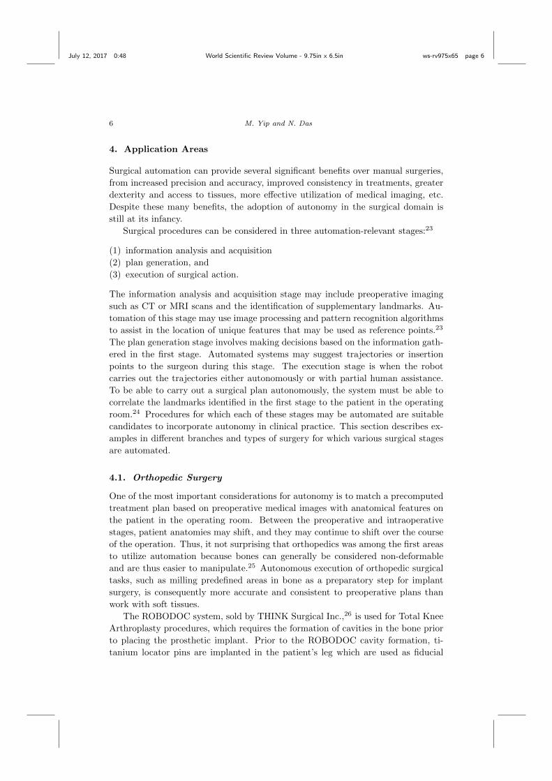
The task of autonomous control for catheters robots (called continuum robots
due to their continuously flexible body) has been studied for use in cardiac abla-
tion procedures46 (Figure 3). Because maintaining direct contact with the tissue
surfaces is critical to treatment success, a robot is used to trace a heart wall with
constant applied force in the presence of beating disturbances. The challenge with
these types of robots (both catheter robots and endoscopic robots) is that that the
control mappings from proximal to distal ends change arbitrarily due to how the
devices are constrained, thus making them unsuitable for traditional physics model-
based robot controllers to work with. A viable strategy is to learn on-the-fly how to
control these devices by estimating the robot Jacobian mappings in real-time which
may be used to determine how much to change an actuator for a desired change
in the tool position. Compared with manual control of the continuum robot, the
automated control can successfully traced desired paths by dragging the catheter
around the heart,46 a task too challenging for human to control through a manual
catheter (they typically use point-by-point ablations) . This is an example of where
robot controlled devices can present a new, potentially more effective method of
treatment. Ultrasound image guidance has also been proposed using intracardiac
echography, can can be controlled in an autonomous fashion to keep important fea-
tures in view,47 while others have considered fluoroscopic image guidance for guiding
catheters in vasculature.48 However, these works are still in the research phase, and
most continuum robotic systems in use for surgery are directly teleoperated.49
5. Autonomy in Commercially-Available Surgical Systems
Some degree of autonomy in the form of shared control and supervised autonomy is
available on commercial surgical systems. The current state of autonomy employed
(a) (b)
Fig. 3. (a) Continuum robot system for cardiac ablation and (b) Simulated beating heart en-
vironment for testing path-tracing tasks.46 Automating treatments using robotic catheters andendoscopes are still in their infancy, challenged by the complexities of robot control and limited
sensing.
July 12, 2017 0:48 World Scientific Review Volume - 9.75in x 6.5in ws-rv975x65 page 12
12 M. Yip and N. Das
on these commercial systems requires a high level of preoperative or intraoperative
human involvement, and the tasks which are automated are typically tasks that
can be routinized. These commercial systems have been successfully implemented
in clinical practices because they combine the computational and mechanical advan-
tages of robotic systems with the contextual and high-level intelligence of a human
surgeon.
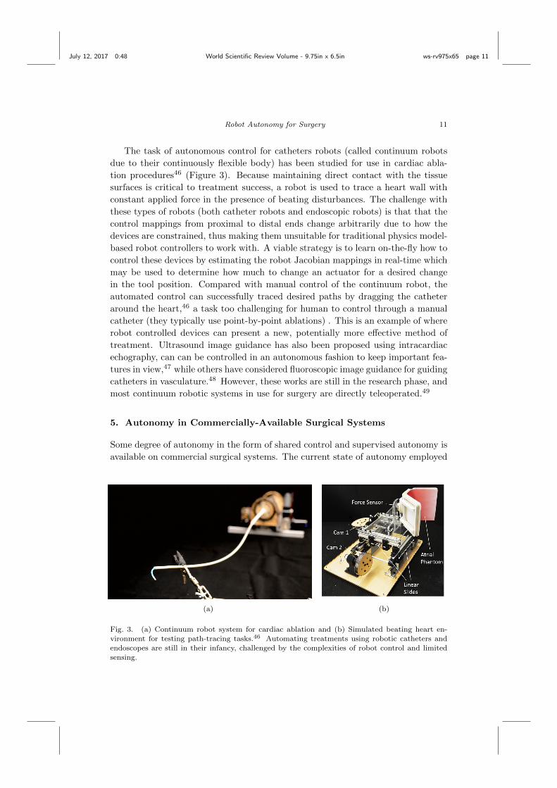
The Probot, developed in 1991 at Imperial College London, is one of the first
instances of supervised autonomy in surgery (Fig. 4(a)). The robot was used for
transurethral prostate resection. After appropriate positioning of the system by a
human, the Probot could autonomously remove conical segments of tissue while the
human surgeon controlled an emergency stop switch.50,51 Playing a passive role as
the robot performed actions autonomously caused uneasiness for the surgeons, so a
system in which the surgeon plays a more active role in the operation was desired.52
The ACROBOT (Active Constraint Robot), which began development in 1991
at Imperial College and later marketed by the Acrobot Company Ltd.,52 is one of
the first examples of shared autonomy in robotic surgery (Fig. 4(b)). Unlike the
Probot, the human surgeon is in control of the ACROBOT during operation. The
(a) (b)
Fig. 4. (a) Probot system, a supervised autonomous system which carries out autonomous
prostate resection motions. (b) ACROBOT system, a shared control system which constrainssurgeon-controlled movements of the surgical tool to a safe region. Reproduced by permission of
Dr. Brian Davies, Imperial College London.

July 12, 2017 0:48 World Scientific Review Volume - 9.75in x 6.5in ws-rv975x65 page 13
Robot Autonomy for Surgery 13
ACROBOT is designed for precise bone cutting prior to knee replacement surgery.
The ACROBOT is used as an intelligent surgeon-controlled tool that constrains
movements within a predefined safe region.52 If the surgeon attempts to move out
of the safe region, the surgeon feels some resistance in the controller prohibiting fur-
ther erroneous motions,53 similar to virtual fixtures deployed on the Mako Surgical
system by Stryker.30
The ROBODOC and CyberKnife systems are examples of supervised autonomy,
which are used for orthopedic surgery and stereotactic radiosurgery, respectively.
Both systems utilize preoperative imaging to create motion plans and can execute
preoperative plans without human interruption excluding emergency situations. A
difference between the ROBODOC and CyberKnife is in how the patient registration
problem is addressed. Patient registration is the process of relating preoperative
images to the position of the patient,24 which is a crucial step when executing
preoperative image-based plans on the actual patient. The ROBODOC system
requires the patient to be rigidly held in place and preoperative images are utilized to
generate and execute surgical plans. A position monitor discontinues the operation
if the patient leg moves.45 The CyberKnife system uses real-time imaging and
adjustment to track the position of the patient to account for movement, such as
that due to respiration.2,37
6. Research Platforms for Surgical Automation
Automation in surgery is a developing field, and thus an active area of research.
Open source platforms exist to facilitate research in automation in surgical robotics.
These platforms are useful to develop and test new automation techniques in sim-
ulated or artificial environments before progressing to in vivo clinical trials.
The da Vinci Research Kit (dVRK) is based on the first generation da Vinci
Surgical System and enables control-level access to the system.54 Either a retired
first-generation da Vinci system or a subset of the components provided by Intu-
itive Surgical, Inc., may be used for the hardware of the research kit. The subset
includes two Master Tool Manipulators, two Patient Side Manipulators (PSMs), a
High Resolution Stereo Viewer, and a footpedal tray. The full system addition-
ally includes a third PSM, an Endoscopic Camera Manipulator, and passive Setup
Joints55 (Fig. 5. All computation is centralized on a high-performance PC while
multiple FPGAs handle the I/O communication. Centralizing computation and
distributing I/O enable flexible reconfigurations of the system, such as converting
a bimanual teleoperation system into two independent unilateral systems.54 The
da Vinci System uses components of the Surgical Assistant Workstation (SAW)
software package for low-level I/O, joint-level and high-level control, and teleoper-
ation.54 SAW attempts to standardize the interfaces with preexisting open-source
standards such as OpenIGTLink used for image-guided therapy.56 Current research
has used the dVRK for debridement, pattern cutting,57 and ultrasound transducer
placement58 tasks.

July 12, 2017 0:48 World Scientific Review Volume - 9.75in x 6.5in ws-rv975x65 page 14
14 M. Yip and N. Das
Fig. 5. The da Vinci Research Kit provides a stripped down, open-access hardware and softwareinterface for researching autonomy in surgical scenarios. The major advantage of this system
is that the hardware matches the clinical model and thus provides a standardized platform for
development as well as an easy facilitator towards translation into future da Vinci systems.
The Raven II, developed at the University of Washington and currently managed
by Applied Dexterity, Inc, is an open-architecture system for laparoscopic surgery
and is designed to facilitate research in surgery (Fig. 6). Compared to the dVRK,
the Raven II system is designed to encourage collaboration among a network of
researchers.59 This system includes two cable-driven laparoscopic arms with wristed
graspers. Unlike the da Vinci system, the Raven II is designed to be portable and
small, enabling two Raven II’s to be mounted on both sides of the surgery site
allowing a total of four arms in the same space. Having two separate systems on
the same surgical site enables two independent surgeons to collaborate on the same
surgery. The users teleoperate the system through the PHANTOM Omni haptic
device controller. The surgeon interfaces with the system over the internet using a
TCP/UDP protocol59 enabling the user to operate the system from anywhere on
the globe. The Raven II system has been used to automate tasks such as tumor
ablation60 and debridement.61
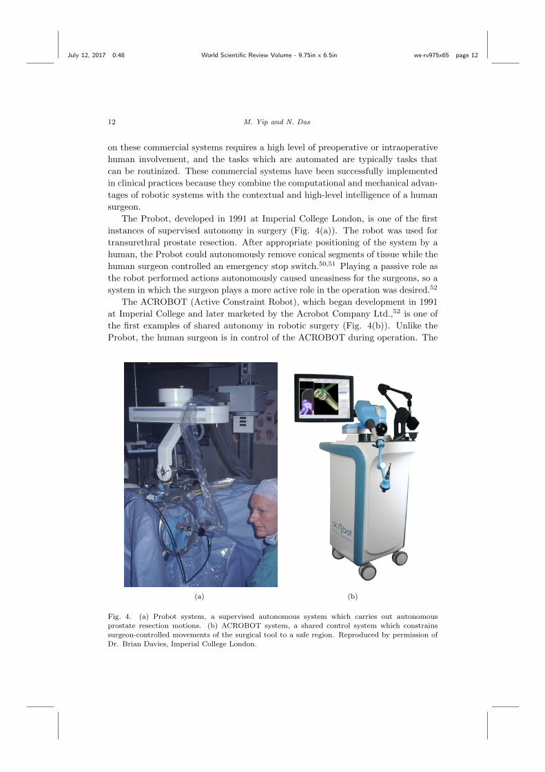
The MiroSurge system is in development at the German Aerospace Center specif-
ically for use as a research platform for multiple endoscopic surgical applications62
(Fig. 7). The majority of the system’s components, such as position sensors and
motor brakes, are on the manipulators, which reduces the external components.

July 12, 2017 0:48 World Scientific Review Volume - 9.75in x 6.5in ws-rv975x65 page 15
Robot Autonomy for Surgery 15
The robot manipulators are lightweight and allow ceiling or wall mounting, en-
abling adaptability to the operating room. Unlike the da Vinci system which has a
physically fixed remote-center-of-motion for each manipulator, the MiroSurge ma-
nipulators use null space constraints to artificially enforce fulcrum point constraints.
Null space constraints enable changes to the joint angles of the manipulator with
certain points of the arm fixed in space. The advantage of using null space con-
straints is the MiroSurge has the ability to arbitrarily define the fulcrum point,
thereby increasing the system’s versatility. While capable of autonomous surgery,
this system has seen little use outside of DLR’s research team, though its capabilities
surpass those present in the daVinci system and the Raven II.
One of the key ingredients to the continued development and success of surgical
autonomy on robotic platforms is the open-sourced nature of the research platforms
and the common testbeds of use. Both the daVinci and the Raven II has been used
by over a dozen institutions worldwide, who provide full schematics to the hardware
and software for their base systems through online, version-controlled repositories.
Another approach is to use highly-engineered industrial robotic arms as a tool for
exploring automation without concerning oneself with the open-source maintained
and buggy nature of the above systems. The Kuka Lightweight Robot Arm, one
of the most popular industrial robot arms in the world, has been used successfully
for surgical automation in microanastamosis tasks. While not cleared for surgical
Fig. 6. Two surgeons in University of Washington collaborate on a surgical task using a Raven
II surgical system located at the University of California, Santa Cruz.59
July 12, 2017 0:48 World Scientific Review Volume - 9.75in x 6.5in ws-rv975x65 page 16
16 M. Yip and N. Das
Fig. 7. MiroSurge system developed at DLR with table-mounted manipulators.62
tasks, the affordance of a industrial-robot-precision arm offers great advantages for
researchers interested in the higher-level autonomy, as demonstrated in Shademan
et al.63
7. Approaches to Automation
Various techniques have been employed to automate surgical tasks. The techniques
described in this section involve a human in a preplanning stage, utilize control the-
ory to follow a human during the operation, or use machine learning techniques to
learn behaviors or motions from human-provided examples. Many of the techniques
described here are also highlighted in other sections as features such as visual ser-
voing or haptic constraints can be found in both research and commercial systems.
7.1. Predefining Motions or Constraints
The simplest form of surgical automation is predefining and executing a sequence
of steps without updating the motion plan in response to a different environment.
Minimally-invasive suturing was accomplished by executing a predefined and fixed
sequence of steps using the EndoBot (Fig. 8), a system designed to allow direct,
shared, and supervised autonomous control of the manipulators.3,11 While this
method may work for controlled environments and tasks, execution of steps without
a method to adapt to the environment will generally fail in real surgical situations if
there is movement. Nevertheless, defining a fixed sequence of steps may be beneficial
July 12, 2017 0:48 World Scientific Review Volume - 9.75in x 6.5in ws-rv975x65 page 17
Robot Autonomy for Surgery 17
(a) (b)
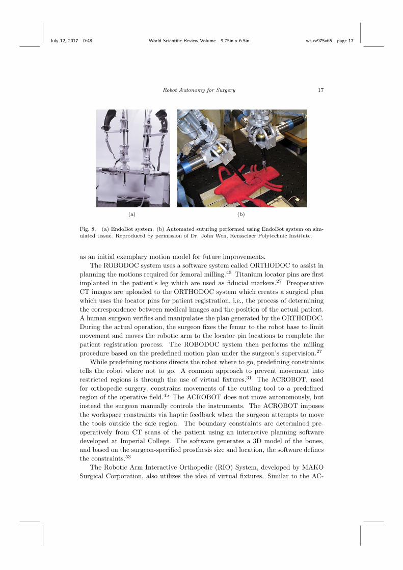
Fig. 8. (a) EndoBot system. (b) Automated suturing performed using EndoBot system on sim-
ulated tissue. Reproduced by permission of Dr. John Wen, Rensselaer Polytechnic Institute.
as an initial exemplary motion model for future improvements.
The ROBODOC system uses a software system called ORTHODOC to assist in
planning the motions required for femoral milling.45 Titanium locator pins are first
implanted in the patient’s leg which are used as fiducial markers.27 Preoperative
CT images are uploaded to the ORTHODOC system which creates a surgical plan
which uses the locator pins for patient registration, i.e., the process of determining
the correspondence between medical images and the position of the actual patient.
A human surgeon verifies and manipulates the plan generated by the ORTHODOC.
During the actual operation, the surgeon fixes the femur to the robot base to limit
movement and moves the robotic arm to the locator pin locations to complete the
patient registration process. The ROBODOC system then performs the milling
procedure based on the predefined motion plan under the surgeon’s supervision.27
While predefining motions directs the robot where to go, predefining constraints
tells the robot where not to go. A common approach to prevent movement into
restricted regions is through the use of virtual fixtures.31 The ACROBOT, used
for orthopedic surgery, constrains movements of the cutting tool to a predefined
region of the operative field.45 The ACROBOT does not move autonomously, but
instead the surgeon manually controls the instruments. The ACROBOT imposes
the workspace constraints via haptic feedback when the surgeon attempts to move
the tools outside the safe region. The boundary constraints are determined pre-
operatively from CT scans of the patient using an interactive planning software
developed at Imperial College. The software generates a 3D model of the bones,
and based on the surgeon-specified prosthesis size and location, the software defines
the constraints.53
The Robotic Arm Interactive Orthopedic (RIO) System, developed by MAKO
Surgical Corporation, also utilizes the idea of virtual fixtures. Similar to the AC-

July 12, 2017 0:48 World Scientific Review Volume - 9.75in x 6.5in ws-rv975x65 page 18
18 M. Yip and N. Das
ROBOT, the RIO system provides haptic feedback on the controller when the sur-
geon attempts to move outside the predefined safe volume.30 However, unlike the
ACROBOT, the RIO system can keep track of changes in tissues and update the
virtual model of the patient’s knee based on fiducial markers.45 This flexibility to
account for changes even enables intraoperative revisions of preoperative plans.30
7.2. Visual Tracking and Servoing
Visual servoing defines the use of images in a feedback loop to automate the robotic
system. Thus, a significant number of efforts in automating surgery are intrinsically
directly related to or an extension of visual servoing problems. One task to highlight
is the positioning of tools relative to underlying soft tissues, which is difficult to
do as it requires both tracking of the anatomy and maintaining registration to
the underlying anatomy.64 Thus, integration of real-time imaging, particularly
ultrasound, provides a useful means for visualizing tissue locations.65 Often, it
can be difficult to discern tissues from instruments, and thus ridged instrument
markers may be used to facilitate locating the instrument in 3D space, where the
ridges assist in identifying the instrument’s position and orientation with a single
ultrasound image.66
Another area that has garnered considerable investigation in visual servoing for
surgery is autonomous endoscopic camera movement. Since endoscopic cameras
for minimally invasive surgery are usually manually moved by an assistant or the
primary surgeon, this task of endoscopic control is a low-reaching goal. Surgeons
typically position the camera such that the area being worked on is in clear or
panoptic view. One study has shown autonomously adjusting the camera can re-
duce the number of camera corrections per hour by a factor of approximately 7 when
using an autonomous camera control system versus manual control.67 Autonomous
endoscopes typically move according to the surgeon’s intent, but inferring the sur-
geon’s intent is a challenge in itself. One method to measure the motions that may
indicate the surgeon’s intent is through instrument tracking, which works under the
assumption that the location of the tools indicate the location of the surgeon’s at-
tention.68 The AESOP system, a robotic endoscope developed by Computer Motion
typically controlled by voice commands,69 was used as a testbed for an autonomous
instrument tracking system, where color is used to distinguish tissue from instru-
ments and the camera is moved planarly to keep the instruments fixed in the field
of view.70 Similar to the AESOP system, the ViKY Robotic Endoscope Holder can
be controlled via voice command. One research group has used the ViKY system
to track the surgical instruments.71
Another method of automation involves tracking instruments or the surgeon
and moving the robotic instruments accordingly. This is useful when the movement
of the tracked targets originates from a human, thereby making the autonomous
movement a direct extension of the surgeon’s skill. An alternative to instrument
tracking for inferring the surgeon’s intent is to track the surgeon’s gaze.72 Eye
July 12, 2017 0:48 World Scientific Review Volume - 9.75in x 6.5in ws-rv975x65 page 19
Robot Autonomy for Surgery 19
(a) (b)
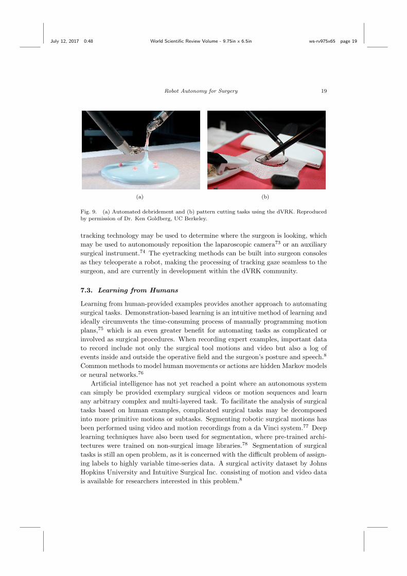
Fig. 9. (a) Automated debridement and (b) pattern cutting tasks using the dVRK. Reproducedby permission of Dr. Ken Goldberg, UC Berkeley.
tracking technology may be used to determine where the surgeon is looking, which
may be used to autonomously reposition the laparoscopic camera73 or an auxiliary
surgical instrument.74 The eyetracking methods can be built into surgeon consoles
as they teleoperate a robot, making the processing of tracking gaze seamless to the
surgeon, and are currently in development within the dVRK community.
7.3. Learning from Humans
Learning from human-provided examples provides another approach to automating
surgical tasks. Demonstration-based learning is an intuitive method of learning and
ideally circumvents the time-consuming process of manually programming motion
plans,75 which is an even greater benefit for automating tasks as complicated or
involved as surgical procedures. When recording expert examples, important data
to record include not only the surgical tool motions and video but also a log of
events inside and outside the operative field and the surgeon’s posture and speech.8
Common methods to model human movements or actions are hidden Markov models
or neural networks.76
Artificial intelligence has not yet reached a point where an autonomous system
can simply be provided exemplary surgical videos or motion sequences and learn
any arbitrary complex and multi-layered task. To facilitate the analysis of surgical
tasks based on human examples, complicated surgical tasks may be decomposed
into more primitive motions or subtasks. Segmenting robotic surgical motions has
been performed using video and motion recordings from a da Vinci system.77 Deep
learning techniques have also been used for segmentation, where pre-trained archi-
tectures were trained on non-surgical image libraries.78 Segmentation of surgical
tasks is still an open problem, as it is concerned with the difficult problem of assign-
ing labels to highly variable time-series data. A surgical activity dataset by Johns
Hopkins University and Intuitive Surgical Inc. consisting of motion and video data
is available for researchers interested in this problem.8

July 12, 2017 0:48 World Scientific Review Volume - 9.75in x 6.5in ws-rv975x65 page 20
20 M. Yip and N. Das
Some autonomous systems have successfully performed isolated surgical tasks
based on a human-provided exemplary dataset. Trajectory smoothing of human-
provided motion examples enabled faster and smoother trajectory executions on
suture knot-tying tasks on an in-house laparoscopic workstation79 compared to a
human.80 Faster trajectories were achieved by iteratively updating the parame-
ters of a controller function based on the error of a target trajectory derived from
the human-provided examples.80 The EndoPAR system, developed at the Tech-
nical University of Munich as a ceiling-mounted experimental surgical platform,
autonomously performed knot-tying tasks using recurrent neural networks using a
database of 25 expert trajectories.81 Recurrent neural networks are a class of mod-
els that can approximate dynamical systems, such as the set of motions involved
in suture knot-tying.81 Learning by observation (LBO) techniques were employed
to automate multilateral subtasks including debridement and pattern cutting57 as
shown in Fig. 9. To achieve autonomous debridement and pattern cutting, the LBO
approach first involved segmenting human-provided motion examples into funda-
mental motions, such as penetration, grasping, and retraction, from which a finite
state machine (FSM) is defined. An FSM is an abstract model composed of the
steps involved in the task at hand. The parameters of the FSM are adjusted based
on repeated executions of the motion sequences represented by the FSM.57 Finally,
autonomous microanastamosis was demonstrated using learning from demonstra-
tion techniques on the KUKA LWR platform.63
While instrument motion and video recordings may be the most direct modality
to represent human-performed surgical examples, other sensor modalities may pro-
vide context-specific information based on human behavior during surgical tasks.
The concept of perceptual docking, developed at Imperial College London, enables
robotic systems to learn behaviors of a human based on eye tracking (Fig. 10(a)),
such as visual search and information seeking behaviors.82 Additionally, the fixa-
tion point of the surgeon can be used to update a model of safety boundaries in the
operative field without any prior knowledge of the anatomy.83 Fig. 10(b) shows
an example safety boundary and conical pathways whose positions depend on the
surgeon’s fixation point.
8. Challenges
Despite success in automation of complicated but isolated surgical tasks in a re-
search setting, bringing these automation techniques to the operating room is not
straightforward as real-world challenges are often ignored in controlled laboratory
experiments. This section outlines some of the many challenges associated with
automating surgical tasks and some methods that are used to compensate for these
difficulties.

July 12, 2017 0:48 World Scientific Review Volume - 9.75in x 6.5in ws-rv975x65 page 21
Robot Autonomy for Surgery 21
(a) (b)
Fig. 10. (a) Eye tracking system integrated into da Vinci Surgical System’s surgical console. (b)
Model of safety boundaries to constrain motion of surgical tools. Conical pathways are positioneddepending on the the surgeon’s fixation point.83
8.1. Uncertainties with Environment
Involuntary movement of the patient make the operative field a changing environ-
ment. This is a challenge for automation because vital structures may be at risk
when instruments are in close proximity. Preoperative plans may become obsolete
when the patient moves. One approach to respond to this issue is to mechanically
fix the patient to restrict intraoperative movement as is done with the ROBODOC
system. A disadvantage of this technique is that the pins or frames to fix the patient
in place are often invasive84 and require an additional preoperative step.27
An alternative approach is to accommodate for the changing environment. Ac-
commodation requires the autonomous systems to either track movements or be
flexible to unknown environments. The CyberKnife system is capable of adapting
to patient movement using respiratory tracking using fiducial markers.2 Compen-
sating for smaller-scale involuntary movement can provide a more stable image,
such as compensation for a beating heart in intracardiac surgery. Heartbeat com-
pensation may be performed using a high-speed camera to track points of reference,
using a synchronization algorithm to offset the camera output to make the points
of reference appear stationary to the surgeon or to move robotic manipulators in
synchrony with the beats.85–87
Uncertainties may also arise when working with vision systems. Visual obsta-
cles such as organs, instruments, and electrocautery smoke may occlude areas of
interest88 in endoscopic cameras that provide difficulty for vision-based systems
and surgeons that rely on direct vision. Alternative real-time vision modalities may
thus be useful, such as ultrasound imaging, which for instance may be used for
intracardiac surgery when blood prevents direct vision;89 however, even then there
is masking and shadowing effects from non-tissue (air, or instrument) materials, as
well as generally poor image resolutions. MR and CT guidance provides other chal-
lenges, including visual artifacts from devices, and insufficient imaging bandwidth

July 12, 2017 0:48 World Scientific Review Volume - 9.75in x 6.5in ws-rv975x65 page 22
22 M. Yip and N. Das
to capture breathing and heartbeat in real-time. These uncertainties will modify
the treatment approach and the treatment trajectories themselves.
8.2. Uncertainties with Device
Force sensing capabilities will be beneficial to autonomous surgical systems for au-
tonomously identifying if a collision between a tool and a tissue (or tool) has oc-
curred off-screen, which can be particularly dangerous as the teleoperating surgeon
has no line-of-sight or haptic feedback.90 However, even for current teleoperated
surgical systems such as the da Vinci system, surgeons do not receive full haptic
force feedback.21 Part of the challenge in supplying full haptic feedback is outfit-
ting the surgical instruments with appropriate force feedback sensors,91 but this
requires careful design of the sensors or tools.92 Integration of appropriate force
sensors in manipulators is an active area of research, with approaches including soft
and deformable grippers93 and miniature uniaxial force sensors.94 Forces may also
be estimated without direct force sensors based on the difference between true and
desired manipulator positions, but the precision of such methods may not be high
enough for delicate work.91
In addition to sensing insufficiencies, modeling and controlling robots with com-
plex configurations may be difficult. A promising technology for robotic surgery is
continuum manipulators, structures that can bend continuously along their length95
producing motion similar to that of an elephant’s trunk. Continuum manipulators
provide increased manipulability, which is advantageous for navigating through nar-
row or complicated passages. Surgical applications for continuum manipulators in-
clude endoscopes, colonoscopes,96 and catheters,97 and most surgical methods in
the past two decades have moved increasingly towards a minimally invasive proce-
dure using continuum devices. A disadvantage of continuum manipulators is they
are difficult to model and control. Recent research shows a promising implemen-
tation of a control scheme for a continuum manipulator for cardiac ablation tasks,
even without a completely known model for the manipulator.46
8.3. Uncertainties with Procedure
The easiest tasks to automate are those that may be routinized. One current ob-
stacle for autonomy in surgical planning is there are generally no well-defined rules
stating the optimal corrections to perform on a human or allowances for certain
surgical tasks. For example, there is no consensus among expert surgeons on the
range of acceptable knee-to-hip angles in knee replacement surgery.52 Thus, in the
interest of routinizing surgical tasks, task-specific specifications or constraints must
be agreed upon for the autonomous system to satisfy.
Randomized clinical trials is a necessity to reduce uncertainty with surgical tech-
niques and protocols.98 Clinical trials have been used to reject new and promising
surgical techniques and to determine safe protocols, such as appropriate safety mar-
July 12, 2017 0:48 World Scientific Review Volume - 9.75in x 6.5in ws-rv975x65 page 23
Robot Autonomy for Surgery 23
gins for melanoma excision.99,100 Following a methodological approach in accepting
surgical techniques and defining protocols should develop a set of standardizations
in surgery and thus potentially lead to a routinization of certain procedures.
9. Ethics and Legality
Beyond the technical challenges, ethical and legal issues may provide another ob-
stacle in bringing automation to the operating room. As current practices of au-
tomation in surgery utilize robotics more as a tool than as an entirely independent
agent, the ethical and legal issues described in this section will become increasingly
more significant as systems increase in their level of autonomy.
9.1. Ensuring Patient Safety
Classically, physicians take the Hippocratic Oath, to which the phrase ”do no harm”
is often mistakenly attributed.101 Nevertheless, ”do no harm” roughly summarizes
the message the oath intends to deliver. The Declaration of Geneva more explicitly
binds physicians to the promotion of patient health with the words ”The health of
my patient will be my first consideration”.102 To ensure that autonomous surgi-
cal systems satisfy the duty to prioritize the health of the patient, there must be
guarantees of safety in place.
As autonomy in surgical robotics is still in infancy, human surgeons still play
a large role in ensuring the safety of the patient. Human experts are still required
for generating and verifying preoperative plans. The ROBODOC system can exe-
cute plans autonomously, but the plans are generated, optimized, and supervised
by human surgeons.25 Similarly, the CyberKnife system is capable of processing
preoperative CT images and generating motion plans to reach all target sites, but a
human must first review and edit the plans prior to execution.38 Thus, with human
verification of surgical plans, autonomous surgical systems may vicariously satisfy
the requirement to prioritize patient health.
Robotic systems may also be mechanically or programmatically designed to in-
trinsically incorporate safety measures, such as the RIO arm which is designed such
that failure of any component does not cause an unsafe environment.30 Motion
constraints can be employed to ensure surgical safety by setting active assists, such
as no-fly zones, to prohibit dangerous or harmful movements.103 Examples in prac-
tice include the ACROBOT which restricts movements to a predefined volume,52
the Precision Freehand Sculptor which retracts its rotary blade near prohibited re-
gions,104 and the da Vinci Surgical System’s manipulators whose remote center of
motion prevents harm at the trocar point.
9.2. Culpability
Four commonly utilized guiding principles for medical ethics are
July 12, 2017 0:48 World Scientific Review Volume - 9.75in x 6.5in ws-rv975x65 page 24
24 M. Yip and N. Das
(1) Respect for Patient Autonomy,
(2) Justice,
(3) Nonmaleficence, and
(4) Beneficence.105
The first two guiding principles ensure that the treatments are well-suited to the pa-
tient’s specific situation and preferences and that medical resources are distributed
in a just manner. The last two principles are to not intentionally harm the pa-
tient and to take actions that promote patient welfare.106 While the Principles
for Patient Autonomy and Justice are more concerned with patient and hospital
management, the Principles of Nonmaleficence and Beneficence are the principles
more related to intraoperative surgical autonomy as they summarize the primary
constraints for automating surgery: to take actions that improve the health of the
patient without harming the patient.
Enforcing the principles of Nonmaleficence and Beneficence is not straightfor-
ward because autonomy in robotics yields concerns regarding culpability in the
event of errors, such as collisions in autonomous vehicles.107 Ambiguity arises when
assigning blame of the cause of the error. The potential assignees include the de-
signers of the system, the user of the system, or the system itself.
Autonomy for surgical robotics faces this concern in the event of surgical compli-
cations or controversial procedures. While current practices of autonomy in surgery
utilize robotics more as an intelligent tool than as an entirely independent agent,
increasing the level of autonomy of the surgical system raises the question about
who is in charge of the surgical operation.52 In the case of surgical errors, the hu-
man surgeon who used or authorized the use of the robot, the hospital, the robot
designer, and the insurer are included as potential culpable entities.108 Addition-
ally, euthanasia and abortion are controversial procedures for clinicians as they may
be considered to conflict with the clinician’s effort to promote patient health. If an
autonomous robot performs these procedures, a question that remains is whether
the clinician still satisfies the ethical guiding principles.
Long distance telemedicine or telesurgery, while currently not common, further
complicates the issue of culpability when surgical errors occur when the surgical staff
and patient are in different jurisdictions,7,109 such as if a surgery is performed in an
underserved region and is overseen by a surgeon in another part of the world. Not
only may there be a conflict of law or socially acceptable practices among different
regions, but the parties involved in the litigation may attempt to select the court
system most advantageous to their own agendas.109 Finally, the issue of appropriate
licenses to operate for clinicians teleoperating into remote areas brings into question
legal concerns. To avoid these issues, clear delineation of accountability must be
established prior to any operations that cross borders.7
July 12, 2017 0:48 World Scientific Review Volume - 9.75in x 6.5in ws-rv975x65 page 25
Robot Autonomy for Surgery 25
9.3. Approval for Use
A surgical system must have approval from the Food and Drug Administration
(FDA) prior to marketing in the US. Surgical robotic systems typically fall under the
Class II category for FDA regulation, where Class I includes devices with minimal
potential harm and Class III includes riskier or life-sustaining devices. To market a
new surgical robotic device, a 510(k) premarket notification must be submitted to
the FDA to illustrate how the new device is substantially equivalent to a predicate,
or legally marketed, device.
Introducing completely novel automation techniques into the market is more
difficult without an equivalent predicate system. If no equivalent predicate device
is found, the system is marked as Class III and a premarket approval application
must be submitted, which is a more involved process. Acquiring 510(k) approval is
more feasible if small steps toward automation are taken. For instance, while the
CyberKnife system was the first to introduce frameless stereotactic radiosurgery
to treat tumors anywhere on the body, Accuray initially established substantial
equivalence to a predicate system called Varian Clinac 600SR. Subsequent 510(k)
notifications for later versions of the CyberKnife referred to preceding CyberKnife
versions to introduce features such as respiratory tracking.
10. Conclusion and Future Directions
As techniques in automation improve in the field of robotics, so will robot autonomy
for surgery. While systems such as the ROBODOC and CyberKnife demonstrate
supervised execution of surgical plans in practice, perfecting and implementing the
more complex and involved tasks shown in research settings is the next step in en-
hancing autonomy in surgery. Introducing the more complex tasks into the clinical
setting is a joint effort, involving engineering teams to design techniques and sys-
tems for automation, clinicians to guide and verify the design of new systems, and
industries to bring new techniques and systems into practice. These participants of
the efforts toward surgical autonomy must work together to overcome the challenges
mentioned in this chapter associated with robotics, the operating environment, and
marketing new systems.
In the pursuit of automation in surgery, we must exercise caution that the ob-
jective is not to remove the human from the surgical team but to (1) enhance the
efficacy of a surgery, and (2) generate new surgical approaches. Furthermore, while
dedicating minimal level of research to preoperative planning for complicated pro-
cedures precludes hope for full autonomy, improving automation of intraoperative
tasks is a step toward introducing more supervised autonomy in the operating room.
Supervised autonomy may be the most promising level of autonomy for surgery as
it takes advantage of mechanical systems’ skill at executing precise motions and
of human surgeons’ abilities to provide high-level supervisory and interventional
support.
July 12, 2017 0:48 World Scientific Review Volume - 9.75in x 6.5in ws-rv975x65 page 26
26 M. Yip and N. Das
References
1. M. Talamini, K. Campbell, and C. Stanfield, Robotic Gastrointestinal Surgery: EarlyExperience and System Description, Journal of Laparoendoscopic & Advanced Sur-gical Techniques. 12(4), (2002).
2. G. Moustris, S. Hiridis, K. Deliparaschos, and K. Konstantinidis, Evolution of au-tonomous and semi-autonomous robotic surgical systems : a review of the literature,The International Journal of Medical Robotics and Computer Assisted Surgery. 7(4),375–392, (2011). doi: 10.1002/rcs.
3. H. Kang and J. Wen. EndoBot : a Robotic Assistant in Minimally Invasive Surgeries.In International Conference on Robotics and Automation, pp. 2031–2036, (2001).ISBN 0780364759.
4. R. Taylor, P. Jensen, L. Whitcomb, A. Barnes, D. Stoianovici, Z. Wang, andL. Kavoussi, A Steady-Hand Robotic System for Microsurgical Augmentation, TheInternational Journal of Robotics Research. 18(12), 1201–1210, (1999).
5. A. Kapoor, R. Kumar, and R. Taylor. Simple Biomanipulation Tasks with ”SteadyHand” Cooperative Manipulator. In Medical Image Computing and Computer As-sisted Intervention Conference, pp. 141–148, (2003). ISBN 3540204628.
6. R. Z. Tombropoulos, J. R. Adler, and J.-c. Latombe, CARABEAMER: A treatmentplanner for a robotic radiosurgical system with general kinematics, Medical ImageAnalysis. 3(3), 237–264, (2011).
7. N. Sharkey and A. Sharkey, Robotic Surgery: On the Cutting Edge of Ethics, Com-puter. 46(1), 56–64, (2013).
8. Y. Gao, S. S. Vedula, C. E. Reiley, N. Ahmidi, B. Varadarajan, H. C. Lin, L. Tao,L. Zappella, B. Bejar, D. D. Yuh, C. C. G. Chen, R. Vidal, S. Khudanpur, andG. D. Hager. JHU-ISI Gesture and Skill Assessment Working Set ( JIGSAWS ): ASurgical Activity Dataset for Human Motion Modeling. In Workshop on Modelingand Monitoring of Computer Assisted Interventions, pp. 1–10, (2014).
9. R. H. Taylor, A. Menciassi, G. Fichtinger, and P. Dario. Medical Robotics andComputer-Integrated Surgery. In Springer Handbook of Robotics, pp. 1199–1222.Springer, 1st edition, (2008).
10. J. Heemskerk, R. Zandbergen, J. G. Maessen, J. W. M. Greve, and N. D. Bouvy, Ad-vantages of advanced laparoscopic systems, Surgical Endoscopy. 20, 730–733, (2006).doi: 10.1007/s00464-005-0456-3.
11. H. Kang and J. Wen. Robotic Assistants Aid Surgeons During Minimally Inva-sive Procedures. In IEEE Engineering in Medicine and Biology Conference, numberFebruary, pp. 94–104, (2001).
12. P. Gomes, Robotics and Computer-Integrated Manufacturing Surgical robotics : Re-viewing the past , analysing the present , imagining the future, Robotics and Com-puter Integrated Manufacturing. 27(2), 261–266, (2011). ISSN 0736-5845. doi: 10.1016/j.rcim.2010.06.009. URL http://dx.doi.org/10.1016/j.rcim.2010.06.009.
13. K. R. J. Chun, B. Schmidt, B. Kokturk, R. Tilz, A. Furnkranz, M. Konstantinidou,E. Wissner, A. Metzner, F. Ouyang, and K.-h. Kuck, Catheter Ablation New Devel-opments in Robotics, Herz. 33(8), 586–589, (2008). doi: 10.1007/s00059-008-3180-7.
14. B. Davies. A review of robotics in surgery. In Proceedings of the Institution of Me-chanical Engineers, vol. 214, pp. 129–140, (2000).
15. L. F. Boogerd, H. J. Handgraaf, C. Boonstra, A. L. Vahrmeijer, and C. J. VanDe Velde, Image-guided surgery, Surgical Oncology: Theory and MultidisciplinaryPractice. (2016).
16. M. Muntener, A. Patriciu, D. Petrisor, D. Mazilu, H. Bagga, L. Kavoussi, K. Cleary,

July 12, 2017 0:48 World Scientific Review Volume - 9.75in x 6.5in ws-rv975x65 page 27
Robot Autonomy for Surgery 27
and D. Stoianovici, Magnetic Resonance Imaging Compatible Robotic System forFully Automated Brachytherapy Seed Placement, Urology. 68(6), 1313–1317, (2006).doi: 10.1016/j.urology.2006.08.1089.
17. R. Alterovitz, M. Branicky, and K. Goldberg, Uncertainty for Image-guided MedicalNeedle Steering, The International Journal of Robotics Research. 27(11), 1361–1374,(2008). doi: 10.1177/0278364908097661.
18. J. Vargo, J. Townsend, S. Sullivan, M. Detamore, and B. Andrews, Modern Applica-tions of Computer Bioengineering in Maxillofacial Surgery: Image Guided SurgicalNavigation and CAD/CAM Custom Implants, Journal of Computer Engineering andInformation Technology. 6, 2–5, (2017). doi: 10.4172/2324-9307.1000164.
19. M. Abayazid, G. J. Vrooijink, S. Patil, R. Alterovitz, and S. Misra, Experi-mental evaluation of ultrasound-guided 3D needle steering in biological tissue,International Journal of Computer Assisted Radiology. 9, 931–939, (2014). doi:10.1007/s11548-014-0987-y.
20. R. E. Link, S. B. Bhayani, and L. R. Kavoussi, A Prospective Comparison of Roboticand Laparoscopic Pyeloplasty, Annals of Surgery. 243(4), 486–491, (2006). doi: 10.1097/01.sla.0000205626.71982.32.
21. A. R. Lanfranco, A. E. Castellanos, J. P. Desai, and W. C. Meyers, Robotic Surgery- A Current Perspective, Annals of Surgery. 239(1), 14–21, (2003). doi: 10.1097/01.sla.0000103020.19595.7d.
22. G. H. Ballantyne, Robotic surgery, telerobotic surgery, telepresence, and telementor-ing: Review of early clinical results, Surgical Endoscopy. 16, 1389–1402, (2002). doi:10.1007/s00464-001-8283-7.
23. D. Manzey, G. Strauss, C. Trantakis, T. Lueth, S. Rottger, J. Bahner-Heyne, A. Di-etz, and J. Meixensberger, Automation in Surgery: A Systematic Approach, SurgicalTechnology International. 18, (2009).
24. G. Eggers and J. Mu, Image-to-patient registration techniques in head surgery, In-ternational Journal of Oral and Maxillofacial Surgery. 35, 1081–1095, (2006). doi:10.1016/j.ijom.2006.09.015.
25. R. D. Howe and Y. Matsuoka, Robotics for Surgery, Annual Review of BiomedicalEngineering. 1, 211–240, (1999).
26. N. A. Netravali, M. Borner, and W. L. Bargar. The Use of ROBODOC in Total Hipand Knee Arthroplasty. In Computer-Assisted Musculoskeletal Surgery, pp. 219–234.(2016). ISBN 9783319129433. doi: 10.1007/978-3-319-12943-3.
27. E. H. Spencer, The ROBODOC Clinical Trial: A Robotic Assistant for Total HipArthroplasty, Orthopaedic Nursing1. 15(1), 9–14, (1996).
28. P. Kazanzides, G. Fichtinger, G. D. Hager, A. M. Okamura, L. L. Whitcomb, andR. H. Taylor, Surgical and Interventional Robotics: Core Concepts, Technology,and Design., IEEE robotics & automation magazine / IEEE Robotics & AutomationSociety. 15(2), 122–130, (2008). ISSN 1070-9932. doi: 10.1109/MRA.2008.926390.
29. J. P. van der List, H. Chawla, and A. D. Pearle, Robotic-Assisted Knee Arthroplasty:An Overview, The American Journal of Orthopedics. (June), (2016).
30. B. Hagag, R. Abovitz, H. Kang, B. Schmitz, and M. Conditt. RIO: Robotic-ArmInteractive Orthopedic System MAKOplasty : User Interactive Haptic Orthope-dic Robotics. In Surgical Robotics: Systems Applications and Visions, pp. 219–246.(2011). ISBN 9781441911261. doi: 10.1007/978-1-4419-1126-1.
31. L. B. Rosenberg. Virtual Fixtures: Perceptual Tools for Telerobotic Manipulation.In Proceedings of the IEEE Virtual Reality Annual Symposium, pp. 76–82, (1993).ISBN 0780313631.
32. G. Barnett, M. Linskey, J. Adler, J. Cozzens, W. Friedman, M. Heilburn, L. Lunsford,

July 12, 2017 0:48 World Scientific Review Volume - 9.75in x 6.5in ws-rv975x65 page 28
28 M. Yip and N. Das
M. Schulder, and A. Sloan, Stereotactic radiosurgery - an organized neurosurgery-sanctioned definition, Journal of Neurosurgery. 106, 1–5, (2007).
33. Q. H. Li, L. Zamorano, A. Pandya, R. Perez, J. Gong, and F. Diaz, The Applica-tion Accuracy of the NeuroMate Robot A Quantitative Comparison with Framelessand Frame-Based Surgical Localization Systems, Computer Aided Surgery. 7, 90–98,(2002). doi: 10.1002/igs.10035.
34. T. Varma and P. Eldridge, Use of the NeuroMate stereotactic robot in a framelessmode for functional neurosurgery, The International Journal of Medical Robotics andComputer Assisted Surgery. 2, 107–113, (2006). doi: 10.1002/rcs.
35. S. Dieterich and I. C. Gibbs, The CyberKnife in Clinical Use: Current Roles, FutureExpectations, Frontiers of Radiation Therapy and Oncology. 43, 181–194, (2011).
36. C. Lindquist, Gamma Knife Radiosurgery, Seminars in Radiation Oncology. 5, 197–202, (1995).
37. S. Sayeh, J. Wang, W. T. Main, W. Kilby, and C. R. Maurer. Respiratory MotionTracking for Robotic Radiosurgery. In Treating Tumors that Move with Respiration,pp. 15–29. (2007).
38. D. Camarillo, T. Krummel, and J. Salisbury, Robotic technology in surgery: past,present, and future, The American Journal of Surgery. 188, 2–15, (2004). doi: 10.1016/j.amjsurg.2004.08.025.
39. N. Hungr, J. Troccaz, N. Zemiti, and N. Tripodi. Design of an Ultrasound-GuidedRobotic Brachytherapy Needle- Insertion System. In IEEE Engineering in Medicineand Biology Conference, pp. 250–253, (2009). ISBN 9781424432967.
40. J. A. Cunha, I.-c. Hsu, J. Pouliot, M. Roach III, K. Shinohara, J. Kurhanewicz,G. Reed, and D. Stoianovici, Toward adaptive stereotactic robotic brachytherapyfor prostate cancer: Demonstration of an adaptive workflow incorporating inverseplanning and an MR stealth robot, Minimally Invasive Therapy. 19, 189–202, (2010).doi: 10.3109/13645706.2010.497000.
41. D. Stoianovici, D. Song, D. Petrisor, D. Ursu, D. Mazilu, M. Mutener, M. Schar,and A. Patriciu. ”MRI Stealth” robot for prostate interventions. In MinimallyInvasive Therapy & Allied Technologies, pp. 241–248, (2011). doi: 10.1080/13645700701520735.
42. T. Haidegger, J. Sandor, and Z. Benyo, Surgery in space: the future ofrobotic telesurgery, Surgical Endoscopy. 25, 681–690, (2011). doi: 10.1007/s00464-010-1243-3.
43. S. Romaniuk and F. Grice, The Future of US Warfare. (Taylor and Francis, 2017).44. P. Garcia, J. Rosen, M. Noakes, G. Elbert, M. Treat, T. Ganous, M. Hanson, and
J. Manak, Trauma Pod : a semi-automated telerobotic surgical system, The Inter-national Journal of Medical Robotics and Computer Assisted Surgery. 5, 136–146,(2009). doi: 10.1002/rcs.
45. J. Rosen, B. Hannaford, and R. Satava, Surgical Robotics: Systems Applications andVisions. (Springer Science & Business Media, 2011).
46. M. C. Yip, J. A. Sganga, and D. B. Camarillo, Autonomous Control of Contin-uum Robot Manipulators for Complex Cardiac Ablation Tasks, Journal of MedicalRobotics Research. 2(1), 1–13, (2017). doi: 10.1142/S2424905X17500027.
47. P. M. Loschak, L. J. Brattain, and R. D. Howe. Automated pointing of cardiacimaging catheters. In Robotics and Automation (ICRA), 2013 IEEE InternationalConference on, pp. 5794–5799. IEEE, (2013).
48. J. Jayender, M. Azizian, and R. V. Patel, Autonomous image-guided robot-assistedactive catheter insertion, IEEE Transactions on Robotics. 24(4), 858–871, (2008).
49. J. Burgner-Kahrs, D. C. Rucker, and H. Choset, Continuum robots for medical

July 12, 2017 0:48 World Scientific Review Volume - 9.75in x 6.5in ws-rv975x65 page 29
Robot Autonomy for Surgery 29
applications: A survey, IEEE Transactions on Robotics. 31(6), 1261–1280, (2015).50. S. J. Harris, F. Arambula-Cosio, Q. Mei, R. D. Hibberd, B. L. Davies, J. E. A.
Wickham, M. S. Nathan, and B. Kundu, The Probot an active robot for prostateresection, Proceedings of the Institution of Mechanical Engineers. 211(4), 317–325,(1997).
51. Q. Mei, S. J. Harris, M. S. Nathan, R. D. Hibberd, J. E. A. Wickham, and B. Davies.PROBOT - A Computer Integrated Prostatectomy System. In Visualization inBiomedical Computing, pp. 581–590, (1996).
52. F. Rodriguez y Baena and B. Davies, Robotic surgery: from autonomous systems tointelligent tools, Robotica. 28, 163–170, (2010). doi: 10.1017/S0263574709990427.
53. M. Jakopec, S. J. Harris, F. Rodriguez, P. Gomes, and B. L. Davies. Acrobot: a”Hands-on” Robot for Total Knee Replacement Surgery. In Advanced Motion Con-trol, pp. 116–120, (2002). ISBN 0780374797.
54. Z. Chen, A. Deguet, R. Taylor, S. DiMaio, G. Fischer, and P. Kazanzides, An Open-Source Hardware and Software Platform for Telesurgical Robotics Research, TheMIDAS Journal. pp. 1–11, (2013).
55. Z. Chen, A. Deguet, R. H. Taylor, and P. Kazanzides. Software Architecture of theda Vinci Research Kit. In IEEE International Conference on Robotic Computing, pp.180–187, (2017). ISBN 9781509067244. doi: 10.1109/IRC.2017.69.
56. P. Kazanzides, S. P. Dimaio, A. Deguet, B. Vagvolgyi, M. Balicki, C. Schneider,R. Kumar, A. Jog, B. Itkowitz, C. Hasser, and R. H. Taylor, The Surgical AssistantWorkstation (SAW) in Minimally-Invasive Surgery and Microsurgery, The MIDASJournal. pp. 1–9, (2010).
57. A. Murali, S. Sen, B. Kehoe, A. Garg, S. Mcfarland, S. Patil, W. D. Boyd, S. Lim,P. Abbeel, and K. Goldberg, Learning by Observation for Surgical Subtasks: Mul-tilateral Cutting of 3D Viscoelastic and 2D Orthotropic Tissue Phantoms, Interna-tional Conference on Robotics and Automation. pp. 1202–1209, (2015).
58. O. Mohareri and S. Salcudean. da Vinci Auxiliary Arm as a Robotic Surgical As-sistant for Semi-Autonomous Ultrasound Guidance During Robot-Assisted Laparo-scopic Surgery. In The Hamlyn Symposium on Medical Robotics, pp. 45–46, (2014).ISBN 9780956377654.
59. B. Hannaford, J. Rosen, D. W. Friedman, H. King, P. Roan, L. Cheng, D. Glozman,J. Ma, S. N. Kosari, and L. White, Raven-II : An Open Platform for Surgical RoboticsResearch, IEEE Transactions on Biomedical Engineering. 60(4), 954–959, (2013).
60. D. Hu, Y. Gong, B. Hannaford, and E. J. Seibel. Semi-autonomous SimulatedBrain Tumor Ablation with RavenII Surgical Robot using Behavior Tree. In In-ternational Conference on Robotics and Automation, pp. 3868–3875, (2015). ISBN9781479969234.
61. B. Kehoe, G. Kahn, J. Mahler, J. Kim, A. Lee, A. Lee, K. Nakagawa, S. Patil, W. D.Boyd, P. Abbeel, and K. Goldberg. Autonomous Multilateral Debridement with theRaven Surgical Robot. In International Conference on Robotics and Automation, pp.1432–1439, (2014). ISBN 9781479936854.
62. U. Hagn, R. Konietschke, A. Tobergte, M. Nickl, S. Jorg, B. Kubler, G. Passig,M. Groger, F. Frohlich, U. Seibold, L. Le-Tien, A. Albu-Schaffer, A. Nothhelfer,F. Hacker, M. Grebenstein, and G. Hirzinger, DLR MiroSurge: a versatile systemfor research in endoscopic telesurgery, International Journal of Computer AssistedRadiology. 5, 183–193, (2010). doi: 10.1007/s11548-009-0372-4.
63. A. Shademan, R. S. Decker, J. D. Opfermann, S. Leonard, A. Krieger, and P. C. Kim,Supervised autonomous robotic soft tissue surgery, Science translational medicine. 8(337), 337ra64–337ra64, (2016).
July 12, 2017 0:48 World Scientific Review Volume - 9.75in x 6.5in ws-rv975x65 page 30
30 M. Yip and N. Das
64. M. C. Yip, D. G. Lowe, S. E. Salcudean, R. N. Rohling, and C. Y. Nguan, Tissuetracking and registration for image-guided surgery, IEEE transactions on medicalimaging. 31(11), 2169–2182, (2012).
65. M. Vitrani, G. Morel, and T. Ortmaier. Automatic Guidance of a Surgical Instrumentwith Ultrasound Based Visual Servoing. In International Conference on Robotics andAutomation, pp. 508–513, (2005). ISBN 078038914X.
66. J. Stoll, P. Novotny, R. Howe, and P. Dupont. Real-time 3D Ultrasound-based Servo-ing of a Surgical Instrument. In International Conference on Robotics and Automa-tion, pp. 613–618, (2006). ISBN 0780395050.
67. K. Omote, H. Feussner, A. Ungeheuer, K. Arbter, G.-q. Wei, J. Siewert, andG. Hirzinger, Self-Guided Robotic Camera Control for Laparoscopic Surgery Com-pared with Human Camera Control, The American Journal of Surgery. 177, 321–324,(1999).
68. A. Pandya, L. A. Reisner, B. King, N. Lucas, A. Composto, M. Klein, and R. D. Ellis,A Review of Camera Viewpoint Automation in Robotic and Laparoscopic Surgery,Robotics. 3, 310–329, (2014). doi: 10.3390/robotics3030310.
69. G. Wei, K. Arbter, and G. Hirzinger, Real-Time Visual Servoing for LaparoscopicSurgery, IEEE Engineering in Medicine and Biology Conference. 16, 40–45, (1997).
70. C. Lee, Y. Wang, D. Uecker, and Y. Wang. Image Analysis for Automated Track-ing in Robot-Assisted Endoscopic Surgery. In International Conference on PatternRecognition, pp. 88–92, (1994).
71. S. Voros, G.-P. Haber, J.-F. Menudet, J.-A. Long, and P. Cinquin, ViKY RoboticScope Holder: Initial Clinical Experience and Preliminary Results Using InstrumentTracking, IEEE Transactions on Mechatronics. 15(6), 879–886, (2010).
72. G. Gras and G.-Z. Yang. Intention recognition for gaze controlled robotic minimallyinvasive laser ablation. In Intelligent Robots and Systems (IROS), 2016 IEEE/RSJInternational Conference on, pp. 2431–2437. IEEE, (2016).
73. S. Ali, L. Reisner, B. King, A. Cao, G. Auner, M. Klein, and A. Pandya, Eye gazetracking for endoscopic camera positioning: An application of a hardware/softwareinterface developed to automate AESOP, Studies in Health Technology and Infor-matics. 132, 4–7, (2007).
74. D. P. Noonan, G. P. Mylonas, A. Darzi, and G. Yang. Gaze Contingent ArticulatedRobot Control for Robot Assisted Minimally Invasive Surgery. In International Con-ference on Intelligent Robotics and Systems, pp. 22–26, (2008). ISBN 9781424420582.
75. S. Schaal. Learning From Demonstration. In Conference on Neural Information Pro-cessing Systems, pp. 1040–1046, (1997).
76. M. Kaiser and R. Dillmann. Building Elementary Robot Skills from Human Demon-stration. In International Conference on Robotics and Automation, pp. 2700–2705,(1996).
77. H. Lin, I. Shafran, T. Murphy, A. Okamura, D. Yuh, and G. Hager. AutomaticDetection and Segmentation of Robot-Assisted Surgical Motions. In Medical ImageComputing and Computer Assisted Intervention Conference, pp. 802–810, (2005).
78. A. Murali, A. Garg, S. Krishnan, F. T. Pokorny, P. Abbeel, T. Darrell, andK. Goldberg. TSC-DL: Unsupervised Trajectory Segmentation of Multi-Modal Sur-gical Demonstrations with Deep Learning. In International Conference on Roboticsand Automation, (2016).
79. M. C. Cavu, F. Tendick, M. Cohn, and S. S. Sastry, A Laparoscopic Telesurgi-cal Workstation, IEEE Transactions on Robotics and Automation. 15(4), 728–739,(1999).
80. J. V. D. Berg, S. Miller, D. Duckworth, H. Hu, A. Wan, X.-y. Fu, K. Goldberg,

July 12, 2017 0:48 World Scientific Review Volume - 9.75in x 6.5in ws-rv975x65 page 31
Robot Autonomy for Surgery 31
and P. Abbeel. Superhuman Performance of Surgical Tasks by Robots using Itera-tive Learning from Human-Guided Demonstrations. In International Conference onRobotics and Automation, pp. 2074–2081, (2010). ISBN 9781424450404.
81. H. Mayer, F. Gomez, D. Wierstra, I. Nagy, A. Knoll, and J. Schmidhuber. A Systemfor Robotic Heart Surgery that Learns to Tie Knots Using Recurrent Neural Net-works. In International Conference on Intelligent Robotics and Systems, pp. 1521–1537, (2006).
82. G. Yang, L. Dempere-Marco, X.-p. Hu, and A. Rowe, Visual search: psychophysicalmodels and practical applications, Image and Vision Computing. 20(4), 291–305,(2002).
83. G. Mylonas, K. Kwok, A. Darzi, and G. Yang. Gaze-Contingent Motor Channellingand Haptic Constraints for Minimally Invasive Robotic Surgery. In Medical ImageComputing and Computer Assisted Intervention Conference, pp. 676–683, (2008).
84. R. H. Taylor, B. D. Mittelstadt, H. A. Paul, W. Hanson, P. Kazanzides, J. F. Zuhars,B. Williamson, B. L. Musits, E. Glassman, and W. L. Bargar, An Image-DirectedRobotic System for Precise Orthopaedic Surgery, IEEE Transactions on Roboticsand Automation. 10(3), 261–275, (1994).
85. Y. Nakamura, K. Kishi, and H. Kawakami. Heartbeat Synchronization for RoboticCardiac Surgery. In International Conference on Robotics and Automation, pp. 2014–2019, (2001). ISBN 0780364759.
86. R. Ginhoux, J. A. Gangloff, M. E. de Mathelin, L. Soler, M. M. Arenas Sanchez,and J. Marescaux. Beating Heart Tracking in Robotic Surgery Using 500 Hz VisualServoing, Model Predictive Control and an Adaptive Observer. In IEEE InternationalConference on Robotics and Automation, (2004). ISBN 0780382323.
87. A. Ruszkowski, C. Schneider, O. Mohareri, and S. Salcudean. Bimanual teleopera-tion with heart motion compensation on the da vinci R© research kit: Implementationand preliminary experiments. In Robotics and Automation (ICRA), 2016 IEEE In-ternational Conference on, pp. 4101–4108. IEEE, (2016).
88. M. Groeger, K. Arbter, and G. Hirzinger. Motion Tracking for Minimally InvasiveRobotic Surgery. In Medical Robotics, pp. 117–148. I-Tech Education and Publishing,(2008). ISBN 9783902613189.
89. J. Cannon, R. Howe, P. Dupont, J. Triedman, G. Marx, and P. del Nido, Applicationof Robotics in Congenital Cardiac Surgery, Pediatric Cardiac Surgery Annual of theSeminars in Thoracic and Cardiovascular Surgery. 6, 72–83, (2003).
90. G. Sung and I. Gill, Robotic Laparoscopy Surgery: A Comparison of the da Vinciand Zeus Systems, Urology. 58(6), 893–898, (2001).
91. A. M. Okamura, Haptic Feedback in Robot-Assisted Minimally Invasive Surgery,Current Opinion in Urology. 19(1), 102–107, (2009).
92. O. Mohareri, C. Schneider, and S. Salcudean. Bimanual Telerobotic Surgery WithAsymmetric Force Feedback: A da Vinci Surgical System Implementation. In IEEEInternational Conference on Intelligent Robots and Systems, pp. 4272–4277, (2014).ISBN 9781479969340.
93. B. S. Homberg, R. K. Katzschmann, M. R. Dogar, and D. Rus. Haptic Identificationof Objects using a Modular Soft Robotic Gripper. In International Conference onIntelligent Robotics and Systems, pp. 1698–1705, (2015). ISBN 9781479999941.
94. M. C. Yip, S. G. Yuen, R. D. Howe, S. Member, A. F. Sensing, and H. Annuloplasty,A Robust Uniaxial Force Sensor for Minimally Invasive Surgery, IEEE Transactionson Biomedical Engineering. 57(5), 1008–1011, (2010).
95. G. Robinson and J. Davies. Continuum Robots - A State of the Art. In InternationalConference on Robotics and Automation, pp. 2849–2854, (1999).

July 12, 2017 0:48 World Scientific Review Volume - 9.75in x 6.5in ws-rv975x65 page 32
32 M. Yip and N. Das
96. G. Chen, M. T. Pham, and T. Redarce, Sensor-based guidance control of a continuumrobot for a semi-autonomous colonoscopy, Robotics and Autonomous Systems. 57(6-7), 712–722, (2009). ISSN 0921-8890. URL http://dx.doi.org/10.1016/j.robot.
2008.11.001.97. Y. Bailly and Y. Amirat. Modeling and Control of a Hybrid Continuum Active
Catheter for Aortic Aneurysm Treatment. In International Conference on Roboticsand Automation, pp. 24–29, (2005). ISBN 078038914X.
98. J. Rosenberg, A. Fischer, and E. Haglind, Current controversies in colorectal surgery:the way to resolve uncertainty and move forward, Colorectal Disease. 14(3), 266–269,(2012).
99. J. C. Hall, B. Mills, H. Nguyen, and J. L. Hall, Methodologic standards in surgicaltrials, Surgery. 119(4), 466–472, (1996).
100. A. Hauschild, F. Rosien, and S. Lischner, Surgical Standards in the Primary Care ofMelanoma Patients, Onkologie. 26, 218–222, (2003).
101. H. Markel, ”I Swear by Apollo” - On Taking the Hippocratic Oath, The New EnglandJournal of Medicine. 350, 2026–2029, (2004).
102. WHO, World Medical Association Declaration of Helsinki: Ethical Principles forMedical Research Involving Human Subjects, World Medical Association. 79(June1964), (2001).
103. R. Taylor, A Perspective on Medical Robotics, Proceedings of the IEEE. 94(9), 1–14,(2006).
104. G. Brisson, T. Kanade, A. Digioia, and B. Jaramaz. Precision Freehand Sculpting ofBone. In Medical Image Computing and Computer Assisted Intervention Conference,pp. 105–112, (2004).
105. T. Beauchamp and J. Childress, Principles of biomedical ethics. (Oxford UniversityPress, 2001), 4th edition.
106. M. Anderson, S. L. Anderson, and C. Armen, MedEthEx: A Prototype MedicalEthics Advisor, Proceedings of the 18th Conference on Innovative Applications ofArtificial Intelligence. pp. 1759–1765, (2006).
107. A. Hevelke and N. Julian, Responsibility for Crashes of Autonomous Vehicles: AnEthical Analysis, Science and Engineering Ethics. 21, 619–630, (2015).
108. G. A. Bekey. Current Trends in Robotics : Technology and Ethics. In Robot ethics:the ethical and social implications of robotics, pp. 17–34. The MIT Press, Cambridge,(2012).
109. B. M. Dickens and R. J. Cook, Legal and ethical issues in telemedicine and robotics,International Journal of Gynecology and Obstetrics. 94, 73–78, (2006).

July 12, 2017 0:48 World Scientific Review Volume - 9.75in x 6.5in ws-rv975x65 page 33