Embed Size (px)
Citation preview
Hyperkinetic Movement Disorders: Differential Diagnosis and Treatment, First Edition. Edited by Alberto Albanese
and Joseph Jankovic. © 2012 Blackwell Publishing Ltd. Published 2012 by Blackwell Publishing Ltd.
3
CHAPTER 1
Distinguishing Clinical Features of Hyperkinetic DisordersAlberto Albanese1 and Joseph Jankovic 21 Fondazione IRCCS Istituto Neurologico Carlo Besta, Università Cattolica del Sacro Cuore, Milan, Italy2 Parkinson’s Disease Center and Movement Disorders Clinic, Department of Neurology,Baylor College of Medicine, Houston, TX, USA
Introduction
Movement abnormalities can be dichotomized into the two broad categories of hypokinetic and hyperkinetic syndromes. The hallmark of hypoki-nesias is the loss of voluntary and automatic movements (akinesia), which is combined with slowness (bradykinesia) and stiffness or increased muscle tone (rigidity) in akinetic-rigid or parkin-sonian syndromes [1]. In contrast, hyperkinesias are manifested by abnormal, uncontrollable, and unwanted movements. This term should not be confused with “hyperkinetic disorders” used in ICD 10 [2] to describe a behavioral abnormality – typically labeled attention deficit disorder with hyperactivity, occurring particularly in children and often associated with attention deficit and a tendency to move from one activity to another without completing any one. This is often associated with disorganized, ill-regulated, and scattered activity and thinking. This is not the only inconsistency between terminology in adult and childhood disorders, and efforts have been recently undertaken to unify the nosology and diagnostic recommendations in pediatric and adult movement disorders [3].
Hyperkinetic movement disorders include six main phenotypic categories, which can appear in
isolation or in variable combinations: tremor, chorea, tics, myoclonus, dystonia, and stereotypies. In addition to these six categories there are other abnormalities of motor control that are also included within the field of movement disorders, such as akathisia, amputation stumps, ataxia, athetosis, ballism, hyperekplexia, mannerisms, myorhythmia, restlessness, and spasticity. The term “dyskinesia” is commonly used to indicate any or a combination of abnormal involuntary movements, such as tardive or paroxysmal dyskinesias or levodopa-induced dyskinesia, but more specific phenomenological categorization should be used whenever possible. In addition, there is a large and important group of peripherally-induced movement disorders, exemplified by hemifacial spasm [4], although any hyperkinetic movement disorder can be triggered or induced by peripheral injury [5].
Some conditions combine hypokinetic and hyperkinetic features, as exemplified by the coexistence of bradykinesia and tremor in Parkinson disease (PD) often referred to by the oxymora “gait disorder with acceleration” [6] or “shaking palsy” [7]. Probably the best examples of coexistent hyper- and hypokinesia is levodopa-induced dyskinesia in patients with PD and chorea or dystonia in patients with Huntington disease, many of whom have an underlying hypokinesia [8].
Albanese_c01.indd 3Albanese_c01.indd 3 12/24/2011 6:01:55 AM12/24/2011 6:01:55 AM
COPYRIG
HTED M
ATERIAL
4 Chapter 1
We describe here the hallmark features and phenomenology of the main hyperkinetic disorders, which are listed according to the time of their medical recognition.
Historical background
The importance of recognizing the appropriate phenomenology, not only as a guide to diagnosis but also as a means to study the pathophysiology of the disorder, is highlighted by the following statement attributed to Sir William Osler: “To study the phenomenon of disease without books is to sail an uncharted sea, while to study books without patients is not to go to sea at all” [9].
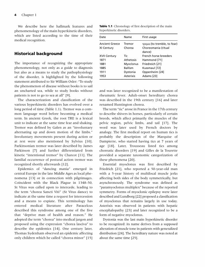
The characterization and classification of the various hyperkinetic disorders has evolved over a long period of time (Table 1.1). Tremor was a com-mon language word before becoming a medical term. In ancient Greek, the root TRE is a lexical unit to indicate at the same time fear and shaking. Tremor was defined by Galen as an “involuntary alternating up and down motion of the limbs.” Involuntary movements present during action or at rest were also mentioned by Sylvius [10]. Parkinsonian tremor was later described by James Parkinson [7] and further differentiated from kinetic “intentional tremor” by Charcot [11]. The familial occurrence of postural action tremor was recognized shortly afterwards [12].
Epidemics of “dancing mania” emerged in central Europe in the late Middle Ages as local phe-nomena [13] or in connection with pilgrimages. Coincident with the Black Plague in 1348–50, St Vitus was called upon to intercede, leading to the term “chorea Sancti Viti” (St Vitus dance) to indicate at the same time a request for intercession and a means to expiate. This terminology has entered medical literature after Paracelcus described this syndrome among one of the five that “deprive man of health and reason.” He adopted the term “chorea” into medical jargon and proposed using the expression “chorea lasciva” to describe the epidemics [14]. One century later, Thomas Sydenham observed an epidemic affecting only children which he called “chorea minor” [15]
and was later recognized to be a manifestation of rheumatic fever. Adult-onset hereditary chorea was described in the 19th century [16] and later renamed Huntington chorea.
The term “tic” arose in France in the 17th century to describe shivers in horses, particularly of certain breeds, which affect primarily the muscles of the pelvic region, pelvic limbs, and tail [17]. The word was later used by French doctors by analogy. The first medical report on human tics is probably the description of the Marquise of Dampierre, who started having tics at 7 years of age [18]. Later, Trousseau listed tics among choreatic disorders [19] and Gilles de la Tourette provided a separate taxonomic categorization of these phenomena [20].
Essential myoclonus was first described by Friedrich [21], who reported a 50-year-old man with a 5-year history of multifocal muscle jerks affecting both sides of the body symmetrically, but asynchronously. The syndrome was defined as “paramyoclonus multiplex” because of the reported symmetry. Forms of myoclonic epilepsy were later described and Lundborg [22] proposed a classification of myoclonus that remains largely in use today. Asterixis was observed in patients with hepatic encephalopathy [23] and later recognized to be a form of negative myoclonus.
Dystonia was the last main hyperkinetic disorder to be recognized: its name derives from a supposed alteration of muscle tone in patients with generalized distribution [24]. The hereditary nature was noted at about the same time [25].
Table 1.1 Chronology of first description of the main hyperkinetic disorders.
Date Name First usage
Ancient Greece Tremor τρεμω (to tremble, to fear) XI Century Chorea Choreomania (ritual
dance)XVII Century Tic French horse breeders1871 Athetosis Hammond [71]1881 Myoclonus Friedreich [21]1885 Ballism Kussmaul [72]1911 Dystonia Oppenheim [24]1953 Asterixis Adams [23]
Albanese_c01.indd 4Albanese_c01.indd 4 12/24/2011 6:01:55 AM12/24/2011 6:01:55 AM
Distinguishing Clinical Features of Hyperkinetic Disorders 5
Phenomenology and classification
Although at first sight involuntary movements resemble each other, each hyperkinetic disorder has a specific phenomenology (signature) that can be identified by direct observation of the patient or videotaped examination. Duration, rhythmicity, topography, and other features must be carefully analyzed and noted in order to make a specific phenomenological diagnosis [26] (Table 1.2).
TremorTremor is an involuntary, rhythmic, oscillation of a body region about a joint axis. It is usually produced by alternating or synchronous contrac-tions of reciprocally innervated agonistic and antagonistic muscles that generate a relatively symmetric velocity in both directions about a midpoint of the movement [27, 28]. The oscillation produced by tremor can be represented by a sinusoidal curve; it is generated by rhythmical discharges in an oscillating neuronal network and maintained by feedback and feed-forward loops. The resulting movement is patterned and rhythmic, characteristics that distinguish tremor from other hyperkinesias [29].
Tremor varies when different voluntary movements are performed or postures are held: it is labeled as a rest tremor, postural tremor, or action tremor according to the condition of greatest severity. Intention tremor, typically associated
with cerebellar dysfunction, is characterized by the worsening of tremor on approach to a target, as in a finger-to-nose maneuver. The typical rest tremor of PD has a frequency of 4 to 6 Hz, and is most prominent distally. Its characteristic appearance in the hand is also referred to as a pill-rolling tremor. Parkinsonian rest tremor also typically involves the chin, jaw, and legs, but almost never involves the neck. Indeed, head oscillation should suggest essential tremor or dystonic tremor rather than PD. True rest tremor, however, disappears during complete rest, such as sleep, and is reduced or disappears with voluntary mus-cle contraction, or during movement. Postural tremor is present with the maintenance of a particular posture, such as holding the arms outstretched in front of the body. It is commonly seen in physiological and essential tremor. Re-emergent tremor refers to a postural tremor that occurs after a variable latency period during which time no observable postural tremor is present [30]. This typically occurs in the setting of PD, and most likely represents a parkinsonian rest tremor that has been “reset” during the maintenance of a posture [31].
Task-specific tremor occurs only during execu-tion of a particular task, such as writing, and is considered by many to be a variant of dystonic tremor. Dystonic tremor may occur in the setting of dystonia, and is a rhythmic, oscillation-like, dystonic movement [32]. Position-specific tremors only occur when the affected body part is placed in
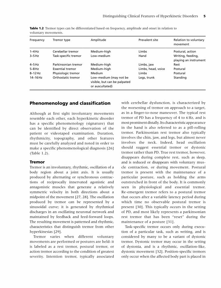
Table 1.2 Tremor types can be differentiated based on frequency, amplitude and onset in relation to voluntary movements.
Frequency Tremor type Amplitude Prevalent site Relation to voluntary movement
1–4 Hz Cerebellar tremor Medium–high Limbs Postural, action3–5 Hz Task-specific tremor Low–medium Hand Writing, feeding,
playing an instrument4–5 Hz Parkinsonian tremor Medium–high Limbs, jaw Rest5–8 Hz Essential tremor Medium–high Limbs, head, voice Postural8–12 Hz Physiologic tremor Medium Limbs Postural14–16 Hz Orthostatic tremor Low–medium (may not be
visible, but can be palpated or auscultated)
Legs, trunk Standing
Albanese_c01.indd 5Albanese_c01.indd 5 12/24/2011 6:01:55 AM12/24/2011 6:01:55 AM

6 Chapter 1
a particular position or posture. Orthostatic tremor is an example of a position-specific tremor, and refers to a fast (14–16 Hz) tremor, mainly affecting the trunk and legs, that occurs after standing for a certain period of time [33].
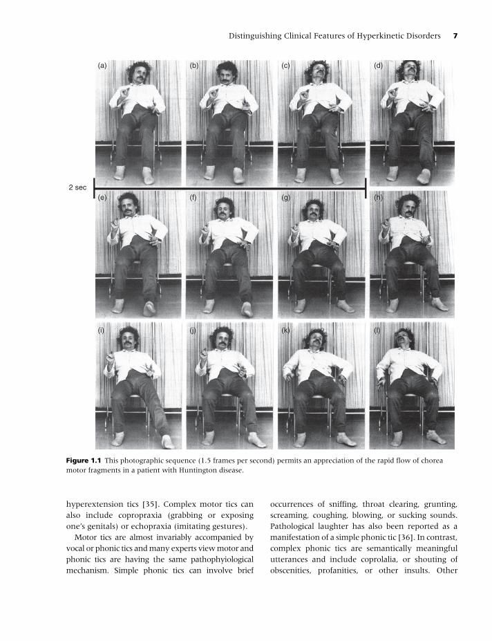
ChoreaChorea is an irregular, unpredictable, involuntary random-appearing sequence of one or more, discrete, involuntary jerk-like movements or movement fragments. Movements appear ran-dom due to the variability in timing, duration, direction, or anatomic location. Each movement may have a distinct start and end point, although these may be difficult to identify since movements are often strung together, one immediately following or overlapping another. Movements may, therefore, appear to flow randomly from one muscle group to another, and can involve trunk, neck, face, tongue, and extremities. Infrequent and mild chorea may appear as isolated, small-amplitude brief movements. It may resem-ble restless, fidgety, or anxious behavior. When chorea is more severe, it may appear to be almost continuous, flowing from one site of the body to another (Figure 1.1).
Although chorea may be worsened by movement, it usually does not stop with attempted relaxation. Chorea is distinguished from tremor and dystonia by its lack of rhythmicity and predictability. Chorea may be difficult to differentiate from myoclonus, but the latter is more intermittent rather than continuous. Chorea is typically a fluent disorder involving contiguous body parts in variable order and direction. It may be associated with hypotonia, hung-up and pendular reflexes, and motor impersistence (inability to maintain a sustained contraction). Examples of impersistence include an inability to maintain prolonged tongue protru-sion or handgrip (“milkmaid grip”). The term “parakinesia” refers to the incorporation of the involuntary movements into semipurposeful movements, in a semiconscious attempt to camouflage the chorea. Examples of parakinesia include touching one’s face, adjusting glasses, and other mannerisms that often served to delay the recognition of the involuntary movement.
Ballism is characterized by high amplitude, almost violent, movements that mainly involve the proximal limb joints. It is considered an extreme phenomenological expression of the spectrum of chorea that affects proximal joints such as shoulder or hip. This leads to large amplitude movements of the limbs, sometimes with a flinging or flailing quality. As patients recover from acute ballism, frequently associated with a stroke in the contralateral subthalamic nucleus, the ballistic movements often gradually evolve into chorea or dystonia (see Chapters 10 and 11).
TicsTics are repeated, individually recognizable, intermittent movements or movement fragments that are almost always briefly suppressible and are usually associated with the awareness of an urge to perform the movement, the so-called “premonitory sensation.” Motor tics often result in either a simple jerk-like movement such as a blink, facial grimace, head jerk, or shoulder shrug, or more complex, stereotyped, semivoluntary, intermittent movements. Tics are usually abrupt in onset, fast and brief (clonic tics), slow and sustained (dystonic tics), or manifested by sudden cessation of movement because of isometric muscle contractions (tonic tics), or inhibition of voluntary movement (blocking tics). The duration of each tic movement is characteristic of that tic, and the duration does not generally vary between different repetitions [34]. Tics can occur during all stages of sleep.
Characteristic features include predictability of both the nature of the movement and its onset, suggestibility, exacerbation during excitement or stress and also after stress (rebound), and brief voluntary suppressibility. Complex motor tics may resemble normal motor acts or gestures, but are generally inappropriately intense and timed [34]. The movements can appear purposeful, such as touching, throwing, hitting, jumping, and kicking, or non-purposeful, such as head shaking or trunk bending. Occasionally tics can be so severe as to cause neurological sequels, with reports of compressive cervical myelopathy resulting from recurrent head thrusting and violent neck
Albanese_c01.indd 6Albanese_c01.indd 6 12/24/2011 6:01:55 AM12/24/2011 6:01:55 AM
Distinguishing Clinical Features of Hyperkinetic Disorders 7
hyperextension tics [35]. Complex motor tics can also include copropraxia (grabbing or exposing one’s genitals) or echopraxia (imitating gestures).
Motor tics are almost invariably accompanied by vocal or phonic tics and many experts view motor and phonic tics are having the same pathophyiological mechanism. Simple phonic tics can involve brief
occurrences of sniffing, throat clearing, grunting, screaming, coughing, blowing, or sucking sounds. Pathological laughter has also been reported as a manifestation of a simple phonic tic [36]. In contrast, complex phonic tics are semantically meaningful utterances and include coprolalia, or shouting of obscenities, profanities, or other insults. Other
2 sec
(a) (b) (c) (d)
(i) (j) (k) (l)
(e) (f) (g) (h)
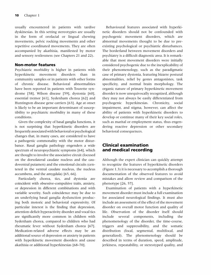
Figure 1.1 This photographic sequence (1.5 frames per second) permits an appreciation of the rapid flow of chorea motor fragments in a patient with Huntington disease.
Albanese_c01.indd 7Albanese_c01.indd 7 12/24/2011 6:01:55 AM12/24/2011 6:01:55 AM
8 Chapter 1
complex phonic tics include echolalia (repeating someone else’s words or phrases) and palilalia (repeating one’s own utterances, particularly the last syllable, word, or phrase in a sentence). Rarely, tics may be continuous and disabling, resulting in a so-called “tic status” [37] or in severe, self-injurious, even life-threatening behaviors, so called “malignant Tourette syndrome” [38]. Because of the broad expression of Tourette syndrome, manifested not only by motor and phonic tics but by a variety of behavioral comorbidities (such as attention deficit with hyperactivity, obsessive-compulsive disorder, and impulsivity), the management depends on establishing an appropriate hierarchy of the various symptoms and targeting the therapeutic strategies to the most troublesome problems [39]. (See Chapters 12 and 13).
AthetosisAthetosis is a slow, continuous, involuntary writhing movement that (1) prevents the maintenance of a stable posture; (2) involves continuous smooth movements that appear to be random and are not composed of recognizable movement fragments; (3) typically involves the distal extremities (hands or feet) more than the proximal and can also involve the face, neck, and trunk; and (4) may worsen with attempts at movement or posture, but can also occur at rest.
Athetosis rarely occurs in isolation but is much more commonly associated with chorea and dystonia. In fact, it is considered a variant of distal chorea or dystonia. Phenomenologically, athetosis is at the opposite end of ballism, resulting in a slow, gentle, and distal motion, resembling slow chorea. The recognition of athetosis often leads to consideration of cerebral palsy or paroxysmal choreoathetosis. Pseudoathetosis refers to a severe distal sensory loss syndrome whereby involuntary, slow, writhing movements are due to loss of proprioception [40].
MyoclonusMyoclonus consists of repeated, often non-rhythmic, brief shock-like jerks due to the sudden involuntary contraction or relaxation of one or more muscles.
These “lightning-like” movements differ from epileptic myoclonus and do not affect consciousness [41]. Myoclonus may be synchronous (several mus-cles contracting simultaneously), spreading (several muscles contracting in a predictable sequence), or asynchronous (several muscles contracting with var-ying and unpredictable relative timing). When myo-clonus affects more than one muscle in an apparently random and varying pattern it is called multifocal; it is called generalized when many muscles through the body are involved simultaneously. Myoclonus is characterized by a sudden unidirectional movement due to agonist contraction (positive myoclonus) or by sudden brief muscle relaxation (negative myo-clonus) [42]. The latter is exemplified by asterixis, which typically presents in patients with hepatic and other encephalopathy.
The distinction between myoclonus and other involuntary disorders – particularly tics, chorea, and different varieties of jerks – is not always clear. Tics are usually associated with a generalized, conscious, urge or local premonitory sensation to move and a feeling of relief of tension after the movement. In addition, many tics are suppressible, in contrast to myoclonus. Brief muscle movements in dystonia are often associated with dystonic posturing. Mild chorea may be difficult to distin-guish from myoclonus. Sometimes myoclonus is rhythmic and can resemble tremor. When myo-clonus is repeated rhythmically it is also called “myoclonic tremor”, but this is a misnomer as rhythmical myoclonus, such as palatal myoclonus [43], is caused by contractions of agonists only, not alternating contractions of antagonist muscles as seen in tremor.
Myoclonus can be caused or worsened by movement and can sometimes occur during sleep. Myoclonus can be categorized as action myoclonus, postural myoclonus, or rest myoclonus on the basis of the condition in which it is observed [44]. It can also be categorized on the basis of the presumed anatomic origin as cortical, subcortical, brainstem, propriospinal, or spinal. Myoclonus may coexist with dystonia (as in myoclonus-dystonia syndrome) or with tremor (as in essential myoclonus) [45]. (See Chapters 14, 15, and 16).
Albanese_c01.indd 8Albanese_c01.indd 8 12/24/2011 6:01:58 AM12/24/2011 6:01:58 AM
Distinguishing Clinical Features of Hyperkinetic Disorders 9
DystoniaIn dystonia, involuntary sustained or intermittent muscle contractions cause twisting and repetitive movements, abnormal postures, or both. The com-bination of postures and dystonic movements is typical of dystonia [46].
Dystonic postures are repeated and particular patterns or postures are characteristic of each patient at a given point in time. Similar dystonic postures may occur in different patients. Postures can be sustained, particularly at the peak of dystonic movements, or may occur during very brief intervals. Dystonic postures are often triggered by attempts at voluntary movement or voluntary posture, and in some cases they are triggered only in particular body positions or by particular movements as may occur in task-specific dystonia. With the exception of certain seizure disorders [47], dystonic movements or postures are not typically seen during sleep, possibly due to inhibition of movements by spinal mechanisms [48]. Postures tend to occur at intervals determined by voluntary movement and can be sustained for variable lengths of time. Relaxation may be impaired so that the dystonic posture may be maintained well beyond the end of the attempted voluntary movement that triggered it. There may be multiple dystonic postures in the same patient, so that different dystonic postures may be combined.
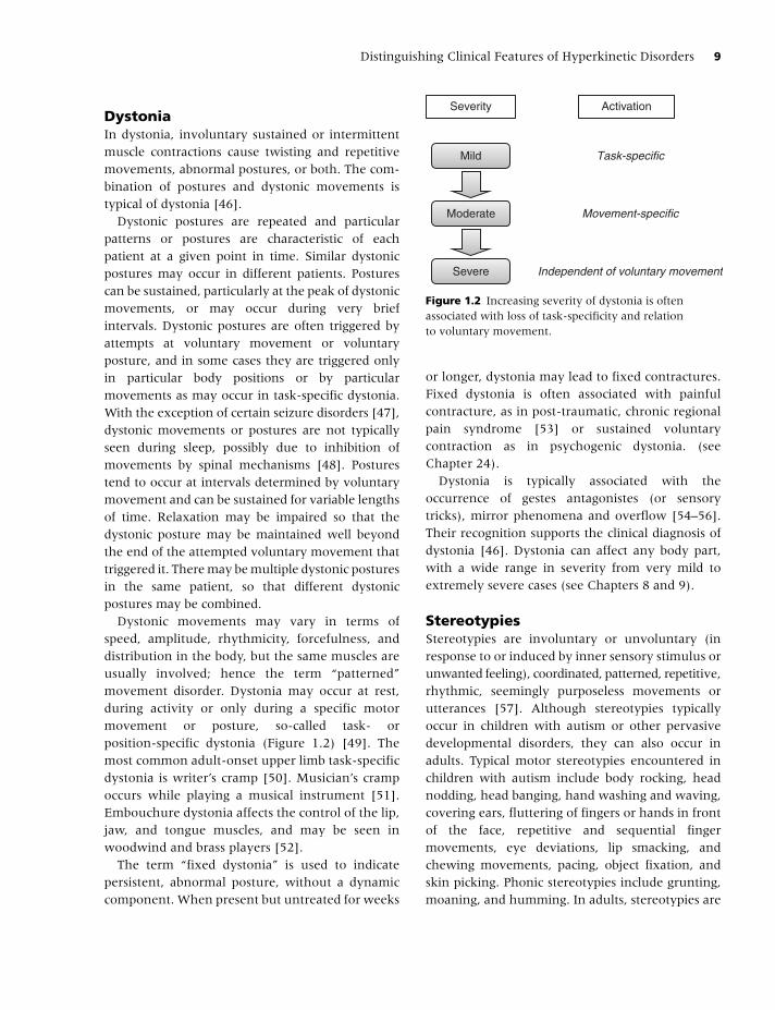
Dystonic movements may vary in terms of speed, amplitude, rhythmicity, forcefulness, and distribution in the body, but the same muscles are usually involved; hence the term “patterned” movement disorder. Dystonia may occur at rest, during activity or only during a specific motor movement or posture, so-called task- or position-specific dystonia (Figure 1.2) [49]. The most common adult-onset upper limb task-specific dystonia is writer’s cramp [50]. Musician’s cramp occurs while playing a musical instrument [51]. Embouchure dystonia affects the control of the lip, jaw, and tongue muscles, and may be seen in woodwind and brass players [52].
The term “fixed dystonia” is used to indicate persistent, abnormal posture, without a dynamic component. When present but untreated for weeks
Severity Activation
Mild
Moderate Movement-specific
Task-specific
Independent of voluntary movementSevere
Figure 1.2 Increasing severity of dystonia is often associated with loss of task-specificity and relation to voluntary movement.
or longer, dystonia may lead to fixed contractures. Fixed dystonia is often associated with painful contracture, as in post-traumatic, chronic regional pain syndrome [53] or sustained voluntary contraction as in psychogenic dystonia. (see Chapter 24).
Dystonia is typically associated with the occurrence of gestes antagonistes (or sensory tricks), mirror phenomena and overflow [54–56]. Their recognition supports the clinical diagnosis of dystonia [46]. Dystonia can affect any body part, with a wide range in severity from very mild to extremely severe cases (see Chapters 8 and 9).
StereotypiesStereotypies are involuntary or unvoluntary (in response to or induced by inner sensory stimulus or unwanted feeling), coordinated, patterned, repetitive, rhythmic, seemingly purposeless movements or utterances [57]. Although stereotypies typically occur in children with autism or other pervasive developmental disorders, they can also occur in adults. Typical motor stereotypies encountered in children with autism include body rocking, head nodding, head banging, hand washing and waving, covering ears, fluttering of fingers or hands in front of the face, repetitive and sequential finger movements, eye deviations, lip smacking, and chewing movements, pacing, object fixation, and skin picking. Phonic stereotypies include grunting, moaning, and humming. In adults, stereotypies are
Albanese_c01.indd 9Albanese_c01.indd 9 12/24/2011 6:01:58 AM12/24/2011 6:01:58 AM
10 Chapter 1
usually encountered in patients with tardive dyskinesias. In this setting stereotypies are usually in the form of orofacial or lingual chewing movements, pelvic rocking movements and other repetitive coordinated movements. They are often accompanied by akathisia, manifested by motor and sensory restlessness (see Chapters 21 and 22).
Non-motor featuresPsychiatric morbidity is higher in patients with hyperkinetic movement disorders than in community samples or in patients with other forms of chronic disease. Behavioral abnormalities have been reported in patients with Tourette syn-drome [58], Wilson disease [59], dystonia [60], essential tremor [61], Sydenham chorea [62] and Huntington disease gene carriers [63]. Age at onset is likely to be an important determinant of suscep-tibility to psychiatric morbidity in many of these conditions.
Given the complexity of basal ganglia functions, it is not surprising that hyperkinetic disorders are frequently associated with behavioral or psychological changes that, in many cases, are considered to have a pathogenic commonality with the motor distur-bance. Basal ganglia pathology engenders a wide spectrum of neuropsychiatric symptoms [64], which are thought to involve the associative circuit (focused on the dorsolateral caudate nucleus and the cau-doventral putamen) and the emotional circuits (cen-tered in the ventral caudate nucleus, the nucleus accumbens, and the amygdala) [65, 66].
Particularly chorea, tics, and dystonia are coincident with obsessive-compulsive traits, anxiety, or depression in different combinations and with variable severity. Such coincidence may be due to an underlying basal ganglia dysfunction produc-ing both motoric and behavioral expressivity. Of particular interest is the finding that depression, attention-deficit hyperactivity disorder and vocal tics are significantly more common in children with Sydenham chorea, compared to children who had rheumatic fever without Sydenham chorea [67]. Medication-related adverse effects may be an additional source of depression or anxiety in patients with hyperkinetic movement disorders and cause akathisia or additional hyperkinesias [68–70].
Behavioural features associated with hyperki-netic disorders should not be confounded with psychogenic movement disorders, which are abnormal movements thought to be due to pre-existing psychological or psychiatric disturbances. The borderland between movement disorders and psychiatry is a difficult diagnostic area. It is remark-able that most movement disorders were initially considered psychogenic due to the inexplicability of their phenomenology, such as the paradigmatic case of primary dystonia, featuring bizarre postural abnormalities, relief by gestes antagonistes, task specificity, and normal brain morphology. The organic nature of primary hyperkinetic movement disorder is now unequivocally recognized, although they may not always be easily differentiated from psychogenic hyperkinesias. Chronicity, social impairment, and stigma, however, can affect the ability of patients with hyperkinetic disorders to develop or continue many of their key social roles, such as marital or employment status, thus engen-dering reactive depression or other secondary behavioral consequences.
Clinical examination and medical recording
Although the expert clinician can quickly attempt to recognize the features of hyperkinetic disorders (Figure 1.3) it is necessary to accomplish a thorough documentation of the observed features to avoid mistakes and allow review and comparison of the phenotype [26, 57].
Examination of patients with a hyperkinetic movement disorder must include a full examination for associated neurological findings. It must also include an assessment of the effect of the movement disorder on overall motor function and quality of life. Observation of the disorder itself should include several components, including the phenomenology of the disorder, the time-course, triggers and suppressibility, and the somatic distribution (focal, segmental, multifocal, and generalized). The phenomenology should be described in terms of duration, speed, amplitude, jerkiness, repeatability, or stereotyped quality, and
Albanese_c01.indd 10Albanese_c01.indd 10 12/24/2011 6:01:58 AM12/24/2011 6:01:58 AM
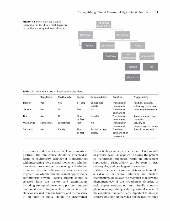
Distinguishing Clinical Features of Hyperkinetic Disorders 11
the number of different identifiable movements or postures. The time-course should be described in terms of rhythmicity, whether it is intermittent with intervening more normal movement, whether movements are sustained or ongoing, and whether there are discrete submovements or movement fragments or whether the movement appears to be continuously flowing. Possible triggers should be assessed from the history and examination, including attempted movement, posture, rest, and emotional state. Suppressibility can be tested in clinic or assessed from the history, and the presence of an urge to move should be determined.
Distractibility evaluates whether unrelated mental or physical tasks (as opposed to asking the patient to voluntarily suppress) result in movement suppression. Distractibility can be seen in tics, stereotypies, and psychogenic movements.
Given the patient’s consent, it is valuable to take a video of the clinical interview and medical examination. This allows the examiner to review the phenomenology of the hyperkinetic disorder, to seek expert consultation and visually compare phenomenology changes during natural course of the condition. It is particularly important to show as clearly as possible on the video clip the features listed
Table 1.3 General features of hyperkinetic disorders.
Regularity Rhythmicity Speed Suppressibility Duration Triggerability
Tremor Yes Yes 1–16 Hz Sometimes briefly
Transient or permanent
Position, posture, voluntary movement
Chorea No No Fast No Transient or permanent
Voluntary movement
Tics No No Slow or fast
Usually Transient or permanent
Sensory stimuli, stress, thoughts
Myoclonus Sometimes Sometimes Fast No Transient or permanent
Sensory or proprioceptive stimuli
Dystonia No
Rarely Slow or fast
Partial or only briefly
Transient, permanent or paroxysmal
Specific motor tasks
Abnormalmovements
Rhythmic
Tremor
Arrhythmic
Sustained
Dystonia
Rapid
Nonsuppressible
Chorea
Myoclonus
Suppressible
Tics
Figure 1.3 Flow chart for a quick orientation in the differential diagnosis of the five main hyperkinetic disorders.
Albanese_c01.indd 11Albanese_c01.indd 11 12/24/2011 6:01:58 AM12/24/2011 6:01:58 AM
12 Chapter 1
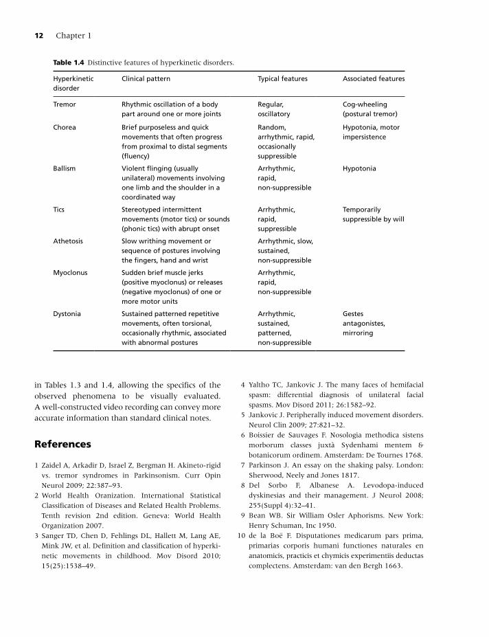
in Tables 1.3 and 1.4, allowing the specifics of the observed phenomena to be visually evaluated. A well-constructed video recording can convey more accurate information than standard clinical notes.
References
1 Zaidel A, Arkadir D, Israel Z, Bergman H. Akineto-rigid
vs. tremor syndromes in Parkinsonism. Curr Opin
Neurol 2009; 22:387–93.
2 World Health Oranization. International Statistical
Classification of Diseases and Related Health Problems.
Tenth revision 2nd edition. Geneva: World Health
Organization 2007.
3 Sanger TD, Chen D, Fehlings DL, Hallett M, Lang AE,
Mink JW, et al. Definition and classification of hyperki-
netic movements in childhood. Mov Disord 2010;
15(25):1538–49.
4 Yaltho TC, Jankovic J. The many faces of hemifacial
spasm: differential diagnosis of unilateral facial
spasms. Mov Disord 2011; 26:1582–92.
5 Jankovic J. Peripherally induced movement disorders.
Neurol Clin 2009; 27:821–32.
6 Boissier de Sauvages F. Nosologia methodica sistens
morborum classes juxtà Sydenhami mentem &
botanicorum ordinem. Amsterdam: De Tournes 1768.
7 Parkinson J. An essay on the shaking palsy. London:
Sherwood, Neely and Jones 1817.
8 Del Sorbo F, Albanese A. Levodopa-induced
dyskinesias and their management. J Neurol 2008;
255(Suppl 4):32–41.
9 Bean WB. Sir William Osler Aphorisms. New York:
Henry Schuman, Inc 1950.
10 de la Boë F. Disputationes medicarum pars prima,
primarias corporis humani functiones naturales en
anatomicis, practicis et chymicis experimentiis deductas
complectens. Amsterdam: van den Bergh 1663.
Table 1.4 Distinctive features of hyperkinetic disorders.
Hyperkinetic disorder
Clinical pattern Typical features Associated features
Tremor Rhythmic oscillation of a body part around one or more joints
Regular, oscillatory
Cog-wheeling (postural tremor)
Chorea Brief purposeless and quick movements that often progress from proximal to distal segments (fluency)
Random, arrhythmic, rapid, occasionally suppressible
Hypotonia, motor impersistence
Ballism Violent flinging (usually unilateral) movements involving one limb and the shoulder in a coordinated way
Arrhythmic, rapid, non-suppressible
Hypotonia
Tics Stereotyped intermittent movements (motor tics) or sounds (phonic tics) with abrupt onset
Arrhythmic, rapid, suppressible
Temporarily suppressible by will
Athetosis Slow writhing movement or sequence of postures involving the fingers, hand and wrist
Arrhythmic, slow, sustained, non-suppressible
Myoclonus Sudden brief muscle jerks (positive myoclonus) or releases (negative myoclonus) of one or more motor units
Arrhythmic, rapid, non-suppressible
Dystonia Sustained patterned repetitive movements, often torsional, occasionally rhythmic, associated with abnormal postures
Arrhythmic, sustained, patterned, non-suppressible
Gestes antagonistes, mirroring
Albanese_c01.indd 12Albanese_c01.indd 12 12/24/2011 6:01:59 AM12/24/2011 6:01:59 AM
Distinguishing Clinical Features of Hyperkinetic Disorders 13
11 Charcot JM. Leçons sur les maladies du système
nerveux. Paris: A. Delahaye 1872.
12 Dana CL. Hereditary tremor: a hitherto undescribed
form of motor neurosis. Am J Med Sci 1887;
94:386–93.
13 Waller J. A forgotten plague: making sense of dancing
mania. Lancet 2009; 21(373):624–5.
14 von Hohenheim P. Von den Krankheiten, so die
Vernunfft berauben. Basel: Adam of Bodenstein.
1567.
15 Sydenham T. Schedula monitoria de novae febris
ingressu. London: G. Kettilby. 1686.
16 Huntington G. On chorea. Medical and Surgical
Reporter (Philadelphia) 1872; 26:317–21.
17 DeLahunta A, Glass E. Veterinary neuroanatomy and
clinical neurology 3rd ed. St. Louis: Saunders Elsevier.
2008.
18 Itard JMG. Mémoire sur quelques fonctions involon-
taires des appareils de la locomotion, de la préhension
et de la voix. Archives Générales de Médecine 1825;
8:385–407.
19 Trousseau A. Clinique médicale de l’Hôtel-Dieu de
Paris 5 edn. Paris: J.-B. Baillière 1877.
20 Gilles de la Tourette GE. La maladie des tics convulsifs.
La Semaine Médicale 1899; 19:153–6.
21 Friedreich N. Neuropathologische Beobachtung beim
Paramyoklonus multiplex. Virchows Arch Path Anat
1881; 86:421–9.
22 Lundborg H. Die progressive Myoklonus-Epilepsie
(Unverricht’s Myoklonie). Upsala: Almqvist & Wiksell
1903.
23 Adams RD, Foley JM. The neurological disorders
associated with liver disease. Res Publ Assoc Res Nerv
Ment Dis 1953; 32:198–237.
24 Oppenheim H. Über eine eigenartige Krampfkrankheit
des kindlichen und jugendlichen Alters (Dysbasia
lordotica progressiva, Dystonia musculorum defor-
mans). Neurolog Centralb 1911; 30:1090–7.
25 Flatau E, Sterling W. Progressiver Torsionspasm bie
Kindern. Z ges Neurol Psychiat 1911; 7:586–612.
26 Jankovic J, Lang AE. Movement disorders: diagnosis
and assessment. In: Bradley WG, Daroff RB, Fenichel
GM, Jankovic J, editors. Neurology in clinical practice,
5th edn. Philadelphia, PA: Butterworth-Heinemann
(Elsevier) 2008; pp 293–325.
27 Hallett M. Classification and treatment of tremor.
JAMA 1991; 28(266):1115–17.
28 Deuschl G, Bain P, Brin M. Consensus statement of the
Movement Disorder Society on Tremor. Ad Hoc
Scientific Committee. Mov Disord 1998; 13(Suppl 3):
2–23.
29 Jankovic J, Fahn S. Physiologic and pathologic
tremors. Diagnosis, mechanism, and management.
Ann Intern Med 1980; 93:460–5.
30 Jankovic J, Schwartz KS, Ondo W. Re-emergent
tremor of Parkinson’s disease. J Neurol Neurosurg
Psychiat 1999; 67:646–50.
31 Fekete R, Jankovic J. Revisiting the relationship
between essential tremor and Parkinson’s disease.
Mov Disord 2011; 26:391–8.
32 Lalli S, Albanese A. The diagnostic challenge of
primary dystonia: evidence from misdiagnosis. Mov
Disord 2010; 15:1619–26.
33 Piboolnurak P, Yu QP, Pullman SL. Clinical and
neurophysiologic spectrum of orthostatic tremor: case
series of 26 subjects. Mov Disord 2005; 20:1455–61.
34 Jankovic J. Tourette syndrome. Phenomenology and
classification of tics. Neurol Clin 1997; 15:267–75.
35 Krauss JK, Jankovic J. Severe motor tics causing
cervical myelopathy in Tourette’s syndrome. Mov
Disord 1996; 11:563–6.
36 Cavanna AE, Ali F, Leckman JF, Robertson MM.
Pathological laughter in Gilles de la Tourette syn-
drome: an unusual phonic tic. Mov Disord 2010;
15(25):2233–9.
37 Kovacs N, Herold R, Janszky J, Komoly S, Nagy F. Tics
status: a movement disorder emergency: observations.
J Neurol 2011; 258:143–5.
38 Cheung MY, Shahed J, Jankovic J. Malignant Tourette
syndrome. Mov Disord 2007; 15:1743–50.
39 Jankovic J, Kurlan R. Tourette syndrome: evolving
concepts. Mov Disord 2011; 26:1149–56.
40 Spitz M, Costa Machado AA, Carvalho Rdo C, Maia FM,
Haddad MS, Calegaro D, et al. Pseudoathetosis: report
of three patients. Mov Disord 2006; 21:1520–2.
41 Halliday AM. The electrophysiological study of
myoclonus in man. Brain 1967; 90:241–84.
42 Shibasaki H, Hallett M. Electrophysiological studies of
myoclonus. Muscle Nerve 2005; 31:157–74.
43 Deuschl G, Wilms H. Clinical spectrum and physiology
of palatal tremor. Mov Disord 2002; 17(Suppl 2),
S63–S66.
44 Caviness JN, Brown P. Myoclonus: current concepts
and recent advances. Lancet Neurol 2004; 3:598–607.
45 Dijk JM, Tijssen MA. Management of patients with myo-
clonus: available therapies and the need for an
evidence-based approach. Lancet Neurol 2010; 9:1028–36.
46 Albanese A, Lalli S. Is this dystonia? Mov Disord 2009;
24:1725–31.
47 Meierkord H, Fish DR, Smith SJ, Scott CA, Shorvon SD,
Marsden CD. Is nocturnal paroxysmal dystonia a form
of frontal lobe epilepsy? Mov Disord 1992; 7:38–42.
Albanese_c01.indd 13Albanese_c01.indd 13 12/24/2011 6:01:59 AM12/24/2011 6:01:59 AM
14 Chapter 1
48 Fish DR, Sawyers D, Smith SJ, Allen PJ, Murray NM,
Marsden CD. Motor inhibition from the brainstem is
normal in torsion dystonia during REM sleep. J Neurol
Neurosurg Psychiat 1991; 54:140–4.
49 Fahn S. Concept and classification of dystonia. Adv
Neurol 1988; 50:1–8.
50 Pont-Sunyer C, Marti MJ, Tolosa E. Focal limb
dystonia. Eur J Neurol 2010; 17(Suppl 1):22–7.
51 Jankovic J, Ashoori A. Movement disorders in
musicians. Mov Disord 2008; 30(23):1957–65.
52 Frucht SJ, Fahn S, Greene PE, O’Brien C, Gelb M,
Truong DD, et al. The natural history of embouchure
dystonia. Mov Disord 2001; 16:899–906.
53 Schrag A, Trimble M, Quinn N, Bhatia K. The
syndrome of fixed dystonia: an evaluation of 103
patients. Brain 2004; 127:2360–72.
54 Albanese A. The clinical expression of primary
dystonia. J Neurol 2003; 250:1145–51.
55 Sitburana O, Jankovic J. Focal hand dystonia, mirror
dystonia and motor overflow. J Neurol Sci 2008;
15(266):31–3.
56 Sitburana O, Wu LJ, Sheffield JK, Davidson A,
Jankovic J. Motor overflow and mirror dystonia.
Parkinsonism Relat Disord 2009; 15:758–61.
57 Fahn S, Jankovic J, Hallett M. Principles and practice
of movement disorders. Philadelphia, PA: Churchill
Livingstone Elsevier 2011.
58 Gaze C, Kepley HO, Walkup JT. Co-occurring
psychiatric disorders in children and adolescents
with Tourette syndrome. J Child Neurol 2006; 21:
657–64.
59 Brewer GJ. Behavioral abnormalities in Wilson’s
disease. Adv Neurol 2005; 96:262–74.
60 Jahanshahi M. Behavioral and psychiatric manifesta-
tions in dystonia. Adv Neurol 2005; 96:291–319.
61 Louis ED. Behavioral symptoms associated with
essential tremor. Adv Neurol 2005; 96:284–90.
62 Maia DP, Teixeira AL, Jr., Quintao Cunningham MC,
Cardoso F. Obsessive compulsive behavior, hyperactiv-
ity, and attention deficit disorder in Sydenham chorea.
Neurology 2005; 24:1799–801.
63 van Duijn E, Kingma EM, van der Mast RC.
Psychopathology in verified Huntington’s disease gene
carriers. J Neuropsychiat Clin Neurosci 2007; 19:441–8.
64 Saint-Cyr JA, Taylor AE, Nicholson K. Behavior and
the basal ganglia. Adv Neurol 1995; 65:1–28.
65 de Olmos JS, Heimer L. The concepts of the ventral
striatopallidal system and extended amygdala. Ann N
Y Acad Sci 1999; 29(877):1–32.
66 Garcia-Cairasco N, Miguel EC, Rauch SL, Leckman JF.
Current controversies and future directions in basal
ganglia research. Integrating basic neuroscience and
clinical investigation. Psychiatr Clin North Am 1997;
20:945–62.
67 Marques-Dias MJ, Mercadante MT, Tucker D,
Lombroso P. Sydenham’s chorea. Psychiat Clin North
Am 1997; 20:809–20.
68 Kane JM, Fleischhacker WW, Hansen L, Perlis R,
Pikalov A, III, Assuncao-Talbott S. Akathisia: an
updated review focusing on second-generation
antipsychotics. J Clin Psychiat 2009; 70:627–43.
69 Sinclair LI, Christmas DM, Hood SD, Potokar JP,
Robertson A, Isaac A, et al. Antidepressant-induced
jitteriness/anxiety syndrome: systematic review.
Br J Psychiat 2009; 194:483–90.
70 Madhusoodanan S, Alexeenko L, Sanders R,
Brenner R. Extrapyramidal symptoms associated with
antidepressants–a review of the literature and an
analysis of spontaneous reports. Ann Clin Psychiat
2010; 22:148–56.
71 Hammond WA. A treatise of diseases of the nervous
system. New York: Appleton Century Crofts 1871.
72 Kussmaul A. Ziemssen’s Handbuch der Speciellen
Pathologie und Therapie. Leipzig: Vogel 1885.
Albanese_c01.indd 14Albanese_c01.indd 14 12/24/2011 6:01:59 AM12/24/2011 6:01:59 AM