Embed Size (px)
Citation preview

Chapter 1
Experimental protocols for and studies of theeffects of surface passivation and water isotopeson the gliding speed of microtubules propelled
by kinesin-1
By
Andy Maloney

Table of Contents
List of Figures . . . . . . . . . . . . . . . . . . . . . . . . . . . . . . . . iii
1 Introduction 11.1 Kinesin . . . . . . . . . . . . . . . . . . . . . . . . . . . . . . . . . . . 21.2 Microtubules . . . . . . . . . . . . . . . . . . . . . . . . . . . . . . . 51.3 Gliding motility assay . . . . . . . . . . . . . . . . . . . . . . . . . . 61.4 Conclusion . . . . . . . . . . . . . . . . . . . . . . . . . . . . . . . . . 7
References a
ii
List of Figures
1.1 Gliding motility assay cartoon. . . . . . . . . . . . . . . . . . . . . . 11.2 Kinesin molecule cartoon. . . . . . . . . . . . . . . . . . . . . . . . . 31.3 Cartoon of a kinesin molecule walking. . . . . . . . . . . . . . . . . . 41.4 Microtubule cartoon. . . . . . . . . . . . . . . . . . . . . . . . . . . . 5
iii
Chapter 1
Introduction
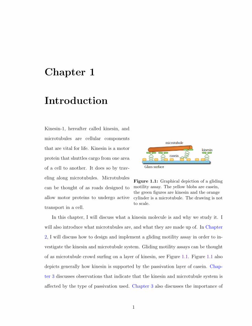
Figure 1.1: Graphical depiction of a glidingmotility assay. The yellow blobs are casein,the green figures are kinesin and the orangecylinder is a microtubule. The drawing is notto scale.
Kinesin-1, hereafter called kinesin, and
microtubules are cellular components
that are vital for life. Kinesin is a motor
protein that shuttles cargo from one area
of a cell to another. It does so by trav-
eling along microtubules. Microtubules
can be thought of as roads designed to
allow motor proteins to undergo active
transport in a cell.
In this chapter, I will discuss what a kinesin molecule is and why we study it. I
will also introduce what microtubules are, and what they are made up of. In Chapter
2, I will discuss how to design and implement a gliding motility assay in order to in-
vestigate the kinesin and microtubule system. Gliding motility assays can be thought
of as microtubule crowd surfing on a layer of kinesin, see Figure 1.1. Figure 1.1 also
depicts generally how kinesin is supported by the passivation layer of casein. Chap-
ter 3 discusses observations that indicate that the kinesin and microtubule system is
affected by the type of passivation used. Chapter 3 also discusses the importance of
1

temperature stabilization and how without it, obtaining stable gliding speeds are not
achievable.
Figure 1.1 does not show the water molecules that are in the system when in-
vestigating the gliding motility assay. If I was to depict the water molecules in this
picture, then you would not be able to see any of the proteins since there would be
a lot of water molecule obscuring the proteins. Water is an important component
in biomolecular interactions and plays a significant role in the microtubule and ki-
nesin system. Chapter 4 discusses the results of some experiments I did that involved
changing the isotope of water and measuring gliding motility speeds. It also discusses
future experimental work that will allow further investigation to the importance of
water interactions in the kinesin and microtubule experiment.
Finally, Chapter 5 discusses open science. This dissertation and all the notebook
entries associated with its research was done openly and in the public domain. I
discuss my experiences with open science and some success stories from participating
in open science.
1.1 Kinesin
Kinesin is a dimeric motor protein that can be thought of as having four distinct
areas to the molecule, see Figure 1.2. Figure 1.2 is a cartoon of a kinesin molecule
and is also Koch Lab’s kinesin mascot called Kiney. The first major component to
the kinesin molecule are the motor domains also known as the “heads”. The motor
domains are depicted as Kiney’s feet. They are where chemical energy (in the form of
ATP) is converted to mechanical work through hydrolysis. This statement is a very
interesting one which is not fully understood and is a major area of study with the
kinesin molecule ([Cross 2000], [Coppin 1997], [Kikkawa 2001]).
2

Figure 1.2: Cartoon of a kinesin molecule.This is our mascot known as Kiney. Themotor domains are depicted as Kiney’s feet.The motor linker links the motor domainstogether. The stalk or coiled coil is depictedas Kiney’s torso and the cargo bindingdomains are depicted as Kiney’s eyebrows.
Kinesin walks along a microtubule
([Carter 2006], [Vale 2000]) in much the
same way humans walk: we place one
foot in front of the other in order to take
a step. The difference between how we
walk and how kinesin manages to walk
along a microtubule is that we have a
brain that sends signals to our legs in
order for them to take a step. Kinesin
does not have a central processing cen-
ter and thus needs a different method for
communication between the two motor
domains to signal steps. This commu-
nication comes from the linkage of the
two motor domains via the motor linker
which is also known in the literature as
the “neck linker”. The motor linker can be thought of as a stretchy polymer of amino
acids that connects the two motor domains together. The stretchiness of the poly-
mer is a key component to how kinesin sends signals to each of the motor domains
([Guydosh 2006], [Yildiz 2008]). The trailing motor domain of a kinesin takes a step
by first docking the motor linker, see Figure 1.3. This docking results in strain along
the motor linker that is a signal for the leading motor domain that the trailing motor
is going to take a step. These steps are gated by nucleotide presence and nucleotide
states within the motor [Herskowitz 2010]. Along with each step is the exclusion of
water between the motor domain and the microtubule. I will discuss this in greater
detail in Chapter 4.
Figure 1.2 shows the stalk or coiled coil region of kinesin as Kiney’s torso. This is
3

Figure 1.3: Cartoon of Kiney taking a step. When a motor domain takes a step, themotor linker docks to the motor domain. Steps are governed by nucleotide states in themotor domain and are depicted as a different coloring in Kiney’s shoes. For a thoroughdiscussion of how kinesin steps, please see [Herskowitz 2010].
where the two protein chains of kinesin coil around each other and terminate in the
cargo binding domain depicted as Kiney’s eyebrows. Kinesin is a motor protein that
transports cellular cargo ([Duncan 2006], [Muresan 2000], [Nakata 2003]). It travels
along microtubules in one direction taking 8 nm steps [Svoboda 1993] using one ATP
molecule per step [Coy 1999]. Kinesin actively transports cargo from one area of the
cell to another and can do so by traveling along the microtubule at about 1 µm/s
[Herskowitz 2010]. This is essential for cellular function. For instance, sciatic nerves
can be nearly a meter long. All components generated in cells are done in their
nucleus. If diffusion was the only form of transportation for these items, it would
take thousands of years for proteins made in the nucleus to be transported to the end
of the nerve a meter away. If kinesin were to transport the same items, it would take
about 2 weeks.
If the kinesin microtubule system is affected by mutations in kinesin, then this
can lead to a variety of diseases ([Goldstein 2001], [Hurd 1996], [Goshima 2003]).
One such disease causes loss of motor function due to accumulation of cellular cargo
transported by kinesin which is lethal. Other mutations that affect humans in-
clude Alzheimer’s disease [Stokin 2005]. Also, the kinesin and microtubule sys-
tem has been suggested to be used as components in microdevices. Some work
has been done to incorporate kinesin and microtubules in devices ([Moorjani 2003],
[van den Heuvel 2005], [Korten 2008]).
There are several types of kinesin molecules as well as many different types of
motor proteins [Sack 1999]. This thesis attempts to illuminate experimental proce-
4

dures necessary in order to obtain good signal to noise data in order to investigate
the kinesin and microtubule system.
1.2 Microtubules
Figure 1.4: Cartoon of a microtubule.α-tubulin and β-tubulin form the tubulinsubunit of a microtubule (red+green balls).Tubulin will oligomerize into a microtubulecreating protofilaments, the orange section ofballs. Microtubules are made up of 13protofilaments, are hollow and are about 25nm in diameter.
Microtubules are one type of road pro-
duced in cells that kinesin and other mo-
tor proteins walk on [Kis 2002]. They
are polymers made up of subunits called
tubulin. Tubulin in turn is made up
of two different proteins called α-tubulin
and β-tubulin. α-tubulin and β-tubulin
combine together to form a single tubulin
subunit via a GTP molecule. The bond
between α-tubulin and β-tubulin is fairly
robust [Desai 1997].
A very interesting thing about microtubules is that they are ”polarized” with a
”plus” and a ”minus” end. These definitions do not refer to the charge of the mi-
crotubule. What this terminology is referring to is how a microtubule polymerizes.
Free tubulin heterodimers will oligomerize into small segments of a protofilament such
that the subunits of tubulin line up in a regular fashion, i.e. (α-β)-(α-β)-(α-β)- etc.
This is done through GTP hydrolysis. These oligomers will then polymerize into a
microtubule [Mozziconacci 2008]. The regularity of how tubulin stacks together “po-
larizes” the microtubule with an end that polymerizes quickly (the plus end, beta
tubulin end) and an end that polymerizes more slowly (the minus end, the alpha
tubulin end). This polarization definition is important because kinesin “walks” along
the microtubule from the minus to plus end and takes steps on the β-tubulin subunit.
5

The bond between tubulin subunits is not as strong as the bond between the α and
β-tubulin subunits. This allows the microtubule to undergo depolymerization when
the cell needs to reduce the length of a microtubule. In order to prevent depoly-
merization, I use a chemical called Taxol to stabilize the tubulin-tubulin interactions
([Nogales 1995], [Salmon 1984]). A brief description of Taxol is given in Chapter 2.
Microtubules are hollow and have an outer diameter of about 25 nm and an inner
diameter of 15 nm [Mozziconacci 2008]. One thing to note about Figure 1.4 is that
all the protofilaments in the microtubule are lined up in nice neat rows. This is not
the case in nature nor is it the case when I polymerize microtubules in a thermal
cycler. There does exist some helicity to the protofilaments and it turns out that
one can change the number of protofilaments, and thus the helicity, by changing the
chemicals that the tubulin is polymerized in. When cells polymerize microtubules,
they predominantly have 13 protofilaments. A protein produced in the cells called
doublecortin is used to regulate microtubules protofilaments [Moores 2004]. When I
polymerize microtubules in glycerol, studies have shown that the predominant number
of protofilaments is 14 [Ray 1993]. I have not measured the number of microtubule
protofilaments from the microtubules that I have polymerized. In my experiments, I
have seen microtubules vary in length from 2 µm to 40 µm.
Without microtubules, kinesin would have no structure on which to perform active
transport. They are essential to cellular structure as well as for cellular replication
[Hyman 1987].
1.3 Gliding motility assay
The experimental procedure used in this dissertation is the gliding motility assay.
Figure 1.1 shows a basic depiction of what the gliding motility assay is. A glass
substrate is first passivated with a protein called casein. I will discuss in depth what
6

casein is and its importance in Chapter 3. For now, it is a layer of protein on the
glass that is necessary in order to make a gliding motility assay work. Kinesin, the
green entities, are then added to the assay. Kinesin orients itself on the casein such
that its motor domains are pointing into solution. The motor domains then attach
to a microtubule in solution and walk along it. This propels the microtubule in
solution and is similar to how crowd surfing looks. I then visualize the motion of the
microtubules through fluorescence.
In the above description, I did not explicitly mention the water that both the
kinesin and microtubules are in. Water plays an important role in biomolecular
interactions and I will discuss some of the experiments conducted to understand how
these interactions occur in this system in Chapter 4.
1.4 Conclusion
Kinesin and microtubules are essential for biological functions. Understanding the
mechanochemistry of how kinesin hydrolyzes ATP in order to do work is a broad
question. It requires the reproduction of consistent experimental tools in order to
obtain data that is of sufficient quality. In Chapter 2, I discuss in great detail the
experimental procedures required to produce consistent samples of kinesin and mi-
crotubules. Producing consistent tools should be the most important thing to an
experimentalist.
7

References
[Carter 2006] Carter, N. J., & Cross, R. A. (2006). Kinesin’s moonwalk. Current
opinion in cell biology, 18(1), 61-7. doi: 10.1016/j.ceb.2005.12.009. Click on the
number to return to that page. 3
[Coppin 1997] Coppin, C. M. (1997). The load dependence of kinesin’s mechanical
cycle. Proceedings of the National Academy of Sciences, 94(16), 8539-8544. doi:
10.1073/pnas.94.16.8539. Click on the number to return to that page. 2
[Coy 1999] Coy, D. L. (1999). Kinesin takes one 8-nm step for each ATP
that it hydrolyzes. Journal of Biological Chemistry, 274(6), 3667-3671. doi:
10.1074/jbc.274.6.3667. Click on the number to return to that page. 4
[Cross 2000] Cross, R. A., Crevel, I., Carter, N. J., Alonso, M. C., Hirose, K., &
Amos, L. A. (2000). The conformational cycle of kinesin. Philosophical transac-
tions of the Royal Society of London. Series B, Biological sciences, 355(1396),
459-64. doi: 10.1098/rstb.2000.0587. Click on the number to return to that
page. 2
[Desai 1997] Desai, A., & Mitchison, T. J. (1997). Microtubule polymerization
dynamics. Annual review of cell and developmental biology, 13, 83-117. doi:
10.1146/annurev.cellbio.13.1.83. Click on the number to return to that
page. 5
a

[Duncan 2006] Duncan, J. E., & Goldstein, L. S. B. (2006). The genetics of ax-
onal transport and axonal transport disorders. PLoS Genetics, 2(9), e124. doi:
10.1371/journal.pgen.0020124. Click on the number to return to that page.
4
[Goldstein 2001] Goldstein, L. S. (2001). Kinesin molecular motors: trans-
port pathways, receptors, and human disease. Proceedings of the National
Academy of Sciences of the United States of America, 98(13), 6999-7003. doi:
10.1073/pnas.111145298. Click on the number to return to that page. 4
[Goshima 2003] Goshima, G., & Vale, R. D. (2003). The roles of microtubule-based
motor proteins in mitosis: comprehensive RNAi analysis in the Drosophila S2
cell line. The Journal of cell biology, 162(6), 1003-16. doi: 10.1083/jcb.200303022.
Click on the number to return to that page. 4
[Guydosh 2006] Guydosh, N. R., & Block, S. M. (2006). Backsteps induced by nu-
cleotide analogs suggest the front head of kinesin is gated by strain. Proceedings
of the National Academy of Sciences of the United States of America, 103(21),
8054-9. doi: 10.1073/pnas.0600931103. Click on the number to return to
that page. 3
[Herskowitz 2010] Herskowitz, L. J. (2010). Kinetic and statistical mechanical model-
ing of DNA unzipping and kinesin mechanochemistry. PhD dissertation. The Uni-
versity of New Mexico, http://hdl.handle.net/1928/12079. Click on the number
to return to that page. 3, 4
[Hurd 1996] Hurd, D. D., & Saxton, W. M. (1996). Kinesin mutations cause mo-
tor neuron disease phenotypes by disrupting fast axonal transport in Drosophila.
Genetics, 144(3), 1075-85. Click on the number to return to that page. 4
b

[Hyman 1987] Hyman, A. A., & White, J. G. (1987). Determination of cell division
axes in the early embryogenesis of Caenorhabditis elegans . The Journal of Cell
Biology, 105(11), 2123-2135. doi: 10.1083/jcb.105.5.2123. Click on the number
to return to that page. 6
[Kikkawa 2001] Kikkawa, M., Sablin, E. P., Okada, Y., Yajima, H., Fletterick, R.
J., & Hirokawa, N. (2001). Switch-based mechanism of kinesin motors. Nature,
411(6836), 439-45. doi: 10.1038/35078000. Click on the number to return to
that page. 2
[Kis 2002] Kis, A., Kasas, S., Babic, B., Kulik, A., Benoıt, W., Briggs, G., et al.
(2002). Nanomechanics of microtubules. Physical Review Letters, 89(24), 1-4. doi:
10.1103/PhysRevLett.89.248101. Click on the number to return to that
page. 5
[Korten 2008] Korten, T., & Diez, S. (2008). Setting up roadblocks for kinesin-1:
mechanism for the selective speed control of cargo carrying microtubules. Lab on
a chip, 8(9), 1441-7. doi: 10.1039/b803585g. Click on the number to return
to that page. 4
[Moores 2004] Moores, C. A., Perderiset, M., Francis, F., Chelly, J., Houdusse, A., &
Milligan, R. A. (2004). Mechanism of microtubule stabilization by doublecortin.
Molecular cell, 14(6), 833-9. doi: 10.1016/j.molcel.2004.06.009. Click on the
number to return to that page. 6
[Moorjani 2003] Moorjani, S. G., Jia, L., Jackson, T. N., & Hancock, W. O. (2003).
Lithographically patterned channels spatially segregate kinesin motor activity
and effectively guide microtubule movements. Nano Letters, 3(5), 633-637. doi:
10.1021/nl034001b. Click on the number to return to that page. 4
c

[Mozziconacci 2008] Mozziconacci, J., Sandblad, L., Wachsmuth, M., Brunner, D.,
& Karsenti, E. (2008). Tubulin dimers oligomerize before their incorporation into
microtubules. PLoS One, 3(11), e3821. doi: 10.1371/journal.pone.0003821. Click
on the number to return to that page. 5, 6
[Muresan 2000] Muresan, V. (2000). One axon, many kinesins: What’s the logic?.
Journal of neurocytology, 29(11-12), 799-818. doi: 10.1023/A:1010943424272.
Click on the number to return to that page. 4
[Nakata 2003] Nakata, T., & Hirokawa, N. (2003). Microtubules provide directional
cues for polarized axonal transport through interaction with kinesin motor head.
The Journal of cell biology, 162(6), 1045-55. doi: 10.1083/jcb.200302175. Click
on the number to return to that page. 4
[Nogales 1995] Nogales, E., Wolf, S. G., Khant, I. A., Luduena, R. F., & Downing,
K. H. (1995). Structure of tubulin at 6.5 Aand location of the taxol-binding site.
Nature, 375(6530), 424-7. doi: 10.1038/375424a0. Click on the number to
return to that page. 6
[Ray 1993] Ray, S., Meyhofer, E., Milligan, R., & Howard, J. (1993). Kinesin follows
the microtubule’s protofilament axis. The Journal of Cell Biology, 121(5), 1083-
1093. doi: 10.1083/jcb.121.5.1083. Click on the number to return to that
page. 6
[Sack 1999] Sack, S., Kull, F. J., & Mandelkow, E. (1999). Motor proteins of the ki-
nesin family. Structures, variations, and nucleotide binding sites. European jour-
nal of biochemistry / FEBS, 262(1), 1-11. doi: 10.1046/j.1432-1327.1999.00341.x.
Click on the number to return to that page. 4
d

[Salmon 1984] Salmon, E. D., & Wolniak, S. M. (1984). Taxol stabilization of mi-
totic spindle microtubules: analysis using calcium-induced depolymerization. Cell
motility, 4(3), 155-167. Click on the number to return to that page. 6
[Stokin 2005] Stokin, G. B., Lillo, C., Falzone, T. L., Brusch, R. G., Rockenstein,
E., Mount, S. L., et al. (2005). Axonopathy and transport deficits early in the
pathogenesis of Alzheimer’s disease. Science (New York, N.Y.), 307(5713), 1282-
8. doi: 10.1126/science.1105681. Click on the number to return to that
page. 4
[Svoboda 1993] Svoboda, K., Schmidt, C. F., Schnapp, B. J., & Block, S. M. (1993).
Direct observation of kinesin stepping by optical trapping interferometry. Nature,
365(6448), 721-7. doi: 10.1038/365721a0. Click on the number to return to
that page. 4
[Vale 2000] Vale, R. D., & Milligan, R. A. (2000). The way things move: looking
under the hood of molecular motor proteins. Science (New York, N.Y.), 288(5463),
88-95. doi: 10.1126/science.288.5463.88. Click on the number to return to
that page. 3
[van den Heuvel 2005] van den Heuvel, M. G. L., Butcher, C. T., Lemay, S. G., Diez,
S., & Dekker, C. (2005). Electrical docking of microtubules for kinesin-driven
motility in nanostructures. Nano letters, 5(2), 235-41. doi: 10.1021/nl048291n.
Click on the number to return to that page. 4
[Yildiz 2008] Yildiz, A., Tomishige, M., Gennerich, A., & Vale, R. D. (2008). In-
tramolecular strain coordinates kinesin stepping behavior along microtubules.
Cell, 134(6), 1030-41. doi: 10.1016/j.cell.2008.07.018. Click on the number
to return to that page. 3
e