Embed Size (px)
Citation preview

BIOCHEMISTRY 2019
1
Chapter 1
CELL AND MARKER ENZYMES
Cell is defined as structural and functional unit of life.
Cell membrane has two major components Lipids and Proteins
Lipids can be Phospholipids, Glycolipids, Cholesterol, fatty acids. All are Amphipathic in nature Proteins can be Integral proteins or Peripheral proteins
Fluidity of cell membrane increases on addition of unsaturated fatty acid with cis double bonds
Major phospholipids of cell membrane Glycerophospholipids
The aminophospholipids in cell membrane are located in the inner leaflet, while Choline containing phospholipids
are located on outer molecular layer
Most common steroid in membrane is Cholesterol. It is present in cell membrane on the outer leaflet
Filamentous material found on cytoplasmic pits which helps in Pinocytosis is Clathrin
Proteins synthesized on ER bound ribosomes are destined for ER membrane, Golgi membrane, cell membrane,
Lysosomal enzymes, and secretory proteins
Proteins synthesized on free cytosolic ribosomes are destined for Nucleus, Mitochondria, Peroxisomes, and
Cytosolic proteins. Most of the glycosylation reactions occur in the Golgi apparatus
Zellweger’s syndrome occurs due to deficiency of Peroxisomes.
Cytoskeleton (cell filaments) consists of Microfilaments Intermediate filaments and Microtubules
In the dividing cells spindle is formed by Microtubules
Exchange of cytoplasmic molecules between two cells are through regions known as Gap Junctions
Composition of cell membrane [% by weight]
Membrane Protein % Lipid % Remarks
Myelin 20 75 Lipid = 75%
Erythrocyte 49 43 Protein to lipid around 50 %
Hepatocyte plasma membrane 54 39 Protein to lipid around 50
Outer mitochondrial membrane 50 46 Protein to lipid around 50
Inner mitochondrial membrane 75 23 Protein = 75%
Intracellular organelles, their functions and Marker enzymes
Organelle /Fraction Marker Enzyme Major Functions
Mitochondria Glutamate dehydrogenase Citric acid cycle, oxidative phosphorylation
Endoplasmic reticulum Glucose-6-phosphatase Membrane-bound ribosome RER is a major site of
protein synthesis
SER: Synthesis of various lipids
Oxidation of many xenobiotics (cytochrome P-450)
Golgi apparatus Galactosyl transferase Intracellular sorting of proteins
Glycosylation and Sulfating reactions
Lysosome Acid phosphatase Site of many hydrolases (enzymes catalyzing degradative reactions)
Peroxisome Catalase
Uric acid oxidase
Degradation of long chain fatty acids & amino acids
Production and degradation of hydrogen peroxide
Nucleus DNA Site of chromosomes
Site of DNA-directed RNA synthesis (transcription)
Plasma membrane Na-K-ATPase
5’-Nucleotidase
Transport of molecules in and out of cells
Intercellular adhesion and communication
Cytoskeleton Nil (Not applicable) Microfilaments, microtubules, intermediate filaments
– impart strength to the cell
Cytosol Lactate dehydrogenase Site of glycolysis, fatty acid synthesis, HMP
Chapter 2

BIOCHEMISTRY 2019
2
FREE RADICALS AND ANTIOXIDANTS
Any chemical species with a single unpaired electron is called free radical. It is a highly reactive species. There are
two highly reactive species or free radicals produced in the human body
Oxygen based free radicals (ROS) Nitrogen based free radicals (RNI)
Singlet oxygen, Superoxide, hydroxyl radical, hydrogen
peroxide, hypochlorite
NO, nitrates and nitrites
Produced in various metabolic reactions NO Produced by NO synthase, which in turn is rapidly
converted to nitrates and nitrites
Result in:
DNA damage
Protein damage
Membrane lipid peroxidation
Free radical mediated membrane lipid peroxidation is a
pathogenic mechanism in:
Aging
Cancer
Inflammation
Atherosclerosis
NO synthase is present in:
Endothelium (eNOS): NO produced by this is
responsible for vasodilatation. hence NO is also
called endothelium derived relaxing factor
(EDRF)
Cholinergic neurons (nNOS)
Induced at the site of inflammation (iNOS)
Antioxidants: take care of normal inevitable production
of ROS
Antioxidant Enzymes:
SOD
Peroxidase
Catalase
Antioxidant molecules:
Tocopherol: 1st line of antioxidant defense
Ascorbate: 2nd line of antioxidant defense in all cells
except RBC
Glutathione: 2nd line of antioxidant defense in RBC
Selenium
Beta carotene
Urate
BIOCHEMISTRY 2019
3
Xenobiotic Metabolism Xenobiotics are foreign molecules. They are not a part of human metabolism. We acquire these xenobiotics from
environment in the form of:
1. Pollutants
2. Carcinogens
3. Food additives
4. Drugs
These Xenobiotics have to be effectively detoxified. It is done in liver in two phases
Phase I reaction (Hydroxylation): Catalyzed by cytochrome P450 Monooxygenase. A hydroxyl group is added to
the xenobiotic. In this process the iron in cytochrome P450 gets oxidized to ferric. It is reduced back to ferrous by
cytochrome P450 Reductase. These 2 enzymes Cytochrome P450 Monooxygenase and Cytochrome P450 Reductase
are present in liver endoplasmic reticulum and hence called microsomal enzyme system.
Phase II reaction (Conjugation): Hydroxylated Xenobiotic is conjugated by
1. Glycine
2. Glutathione
3. Glucuronate: UDP Glucuronate is the donor
4. Acetylation: Acetyl CoA is the donor of acetyl group
5. Methylation: S – Adenosyl Methionine is the donor of methyl group
6. Sulfation: PAPS (3 – Phospho Adenosyl – 5 Phospho Sulfate) is the donor of sulfate group
BIOCHEMISTRY 2019
4
Chapter 3
CARBOHYDRATES: Chemistry and metabolism
Aldehyde or Ketone are derivatives of polyhydric alcohols or compounds which yield these derivatives on hydrolysis.
Classification
Monosaccharides :
Contain only one Sugar unit ; cannot be hydrolysed to more simple sugars.
Depending on the functional group they can be Aldose or Ketose
Aldose Ketose
Triose Glyceraldehyde Dihydroxy acetone
Tetrose Erythrose, Arbinose Erythrulose
Pentose Ribose, Xylose Ribulose, Xylulose
Hexose Glucose, Galactose, Mannose Fructose
D-Fructose has optical rotation of (minus) - 92 and therefore it is laevorotatory
The predominant sialic acid found in glycoproteins and gangliosides is the 9 carbon sugar is N Acetyl Neuraminic
acid and Neuraminic acid is derivative of N Acetyl mannose and pyruvic acid
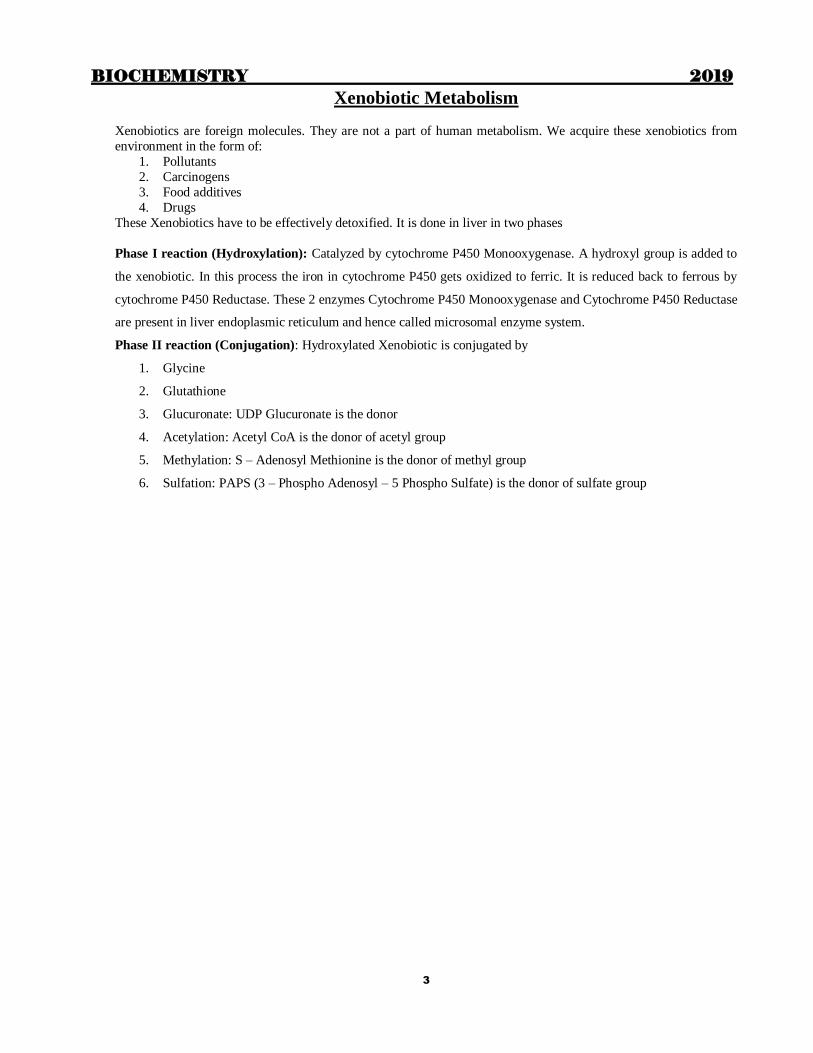
Isomerism in Carbohydrates
Anomerism: Difference in the orientation of H and OH around anomeric carbon (C1 for aldoses and C2 for ketoses)
. e.g. Alpha and beta anomers D and L forms: Based on orientation of OH around penultimate carbon. Also for cyclic structures the orientation of
ring.
Epimerism: Difference in the orientation of H and OH around a single carbon other than anomeric and penultimate
carbon
• Mannose is the epimer of glucose with respect to C2 while Galactose is C4 epimer;
• Similarly Allose is C3 epimer and L Idose is C5 epimer
• d and l forms- Depending on the direction of rotation of polarised light
Important derivatives of monosaccharides are :
Class Examples Functions
1. Glycosides Digitalis Cardiac drugs
2. Amino sugars Glucosamine Glycosaminoglycan (GAG)
3. Deoxy sugar Deoxyribose, DNA, mucin
4. Uronic acid Glucuronic acid, Iduronic acid GAG
5. Fucose Mucin Glycoprotein

BIOCHEMISTRY 2019
5
Disaccharides:
Contains 2 monosaccharide units joined together by a glycosidic linkage
Eg:- Maltose = Glucose + Glucose in α 1, 4 linkage
Lactose = Galactose + Glucose in β 1, 4 linkage
Sucrose = Glucose + Fructose in α, β 1, 2 linkage
They can be Reducing or Non reducing
Different disaccharides of glucose: Glucose + Glucose
Cellobiose β 1,4
Maltose α 1,4
Isomaltose α 1,6
Trehalose α 1, 1
Note: intestinal brush border contains enzymes: For disaccharides (Disaccharidases): Lactase (β –
galactosidase), Sucrase, Maltase, Isomaltase (α 1, 6 Glucosidase), Limit Dextrinase, Trehalase
For Peptides: Entero peptidases, Amino peptidases, Carboxy peptidases, Endo peptidases, Di peptidases
For nucleotides and Nucleosides: Phosphatases, Nucleosidases
Oligosaccharides : Contain 3-10 monosaccharide units
Polysaccharides : More than 10 monosaccharide units
Can be Homoglycans or (Homopolysaccharide) (Contain same type of monosaccharide units) or Heteroglycans or (Heteropolysaccharide) More than one type of monosaccharide units
Homoglycans: Starch, Glycogen, Cellulose, Inulin, Chitin
Heteroglycans: Mucopolysaccharides
Starch: Amylose + Amylopectin
Amylose: Soluble: Glucose units in α 1, 4 linkages: No branches
Amylopectin: Insoluble: Glucose units in α 1, 4 linkage: Branches with α 1,6 linkage
Glycogen: Same as amylopectin but more branched and compact. Spherical molecule with 21 ηm diameter & 12
concentric layers. 13 Glucose residues in each chain with α 1.4 linkage. Generally branching at every 5th glucose
residue with α 1, 6 linkage.
Cellulose: Glucose in β-1, 4
linkages; Extensive H bonds between and within the chains rigid fibrils Inulin: Fructose in β-1, 2 linkages. If taken orally it is not hydrolyzed & constitutes dietary fiber. If given I.V it is
not metabolized or degraded or taken up by cells and exclusively filtered. Used in clearance test
Chitin: N-acetyl Glucosamine (GluNac) in β-1, 4 links; Extensive H bonding; Seen in Exoskeleton of insects and
crustaceans
Dextran: α Glucose units in 16, 14 and 13 linkages; Plasma volume expanders
Agar: polymer of galactose

BIOCHEMISTRY 2019
6
Heteroglycans
Mucopolysaccharides (GAG): Contains aminosugars and uronic acids
Glycosaminoglycans Sugars Location
Hyaluronic acid GlcNAc,GlcUA Synovial fluid, vitreous humor, loose connective
Chondroitin SO4 GalNAc,GlcUA Cartilage, bone, cornea
Keratan SO4 GlcNAc, Gal Cornea Loose connective tissue
Heparin GlcN,IdUA Mast cells Present in liver, lung, spleen,
monocytes
Heparan sulfate GlcN, GlcUA Skin fibroblasts, aortic wall
Dermatan sulfate GalNAc,IdUA,
(GlcUA)
Wide distribution
OXIDATION AND REDUCTION REACTIONS AND TESTS IN CARBOHYDRATES:
Oxidation of sugars
Oxidation at C1 Gluconic acid (aldonic); Oxidation at C6 Glucuronic acid (uronic)
Oxidation at both C1&C6Glucaric (Aldaric)
Reduction of carbohydrates
Glucose Sorbitol. Sorbitol is one of the reasons for long term complications of diabetes
Fructose can be reduced to either Sorbitol or Mannitol because of the keto group at C2
Galactose Dulcitol (galactitol). Responsible for premature cataract in galactosemics
Non reducing sugars: Polysaccharides, aminosugars and glycosides are non reducing
Sucrose and trehalose are non reducing disaccharides
Mannose Mannitol. Mannitol is :
Mild osmotic diuretic. Maintains GFR in ARF, Used for forced diuresis in poisonings.
Reduces cerebral edema, given in Head injury, Stroke, before and after brain surgery.
Reduces intraocular tension, given in Acute congestive glaucoma, before and after ocular surgery
Different tests for carbohydrates
Molisch test- General test for carbohydrates
Benedict’s test and Fehling’s test : for reducing sugars
Barfoeds test, Moore’s test- to differentiate between monosaccharides and disaccharides
Feulgen stain- for deoxy sugars: used to detect DNA – Reaction of deoxy sugars with Schiff reagent
COMPLEX CARBOHYDRATES WITH PROTEINS
1. Glycoproteins:-
Protein molecule on which oligosaccharide chain (upto 15 subunits) is attached by N or O glycosidic bond
This process is called glycosylation or glycation and the complex is called glycosylated or glycated or glyco protein
It is most common post translational modification and almost every protein is glycated
The oligosaccharides are:
o Hexoses

BIOCHEMISTRY 2019
7
Mannose
Galactose
o Acetyl Hexosamines (amino sugars)
Gal Nac
Glu Nac
o Sialic acids – NANA (predominat sialic acid)
o Pentoses
Arabinose
Xylose
o Methyl pentoses
o L – Fucose (6 deoxy - -L-galactose)
2. GAGs – Glycosaminoglycans (Mucopolysaccharides)
Heteropolysaccharides containing
i. amino sugars
1. Glucosamine
2. N Acetyl Glucosamine
3. Galactosamine (Chondrosamine)
4. N Acetyl Galactosamine
5. Galactose residues may be sulfated
ii. Uronic acids
1. Glucuronic acid
2. Iduronic acid
If attached to a protein core: - Proteoglycans (mucoproteins)
Large No. of OH groups & negative charge hold large qty of water & occupy space.
Negative charges also repel & keep the chains apart.
Due to above 2 properties proteoglycan chains can easily slide over each other and have
cushioning & lubricating functions
Provide ground (packing) substance of connective tissue
Due to high viscosity, also present in mucus and synovial fluid
GLUCOSE TRANSPORTERS
Facilitative Bidirectional Transporters
Transporter Specified Tissue location Functions
GLUT 1 Brain, Kidney, Colon, Placenta, RBCs, Retina Uptake of Glc
GLUT 2 Liver, β Cell of pancreas, serosal
surface of intestinal cells Km high. Low affinity for Glc.
GLUT 3 Brain, Neurons, Placenta High affinity for Glc.
GLUT 4 Heart and Skeletal muscle, adipose tissue Insulin dependent uptake
GLUT 5 Small intestine, testis sperms, kidney Fructose transporter- Low affinity to Glc
Sodium dependent unidirectional Transporter
SGLT 1
SGLT 2
Small intestine- High affinity
Kidney- Low affinity Active uptake of Glc
BIOCHEMISTRY 2019
8
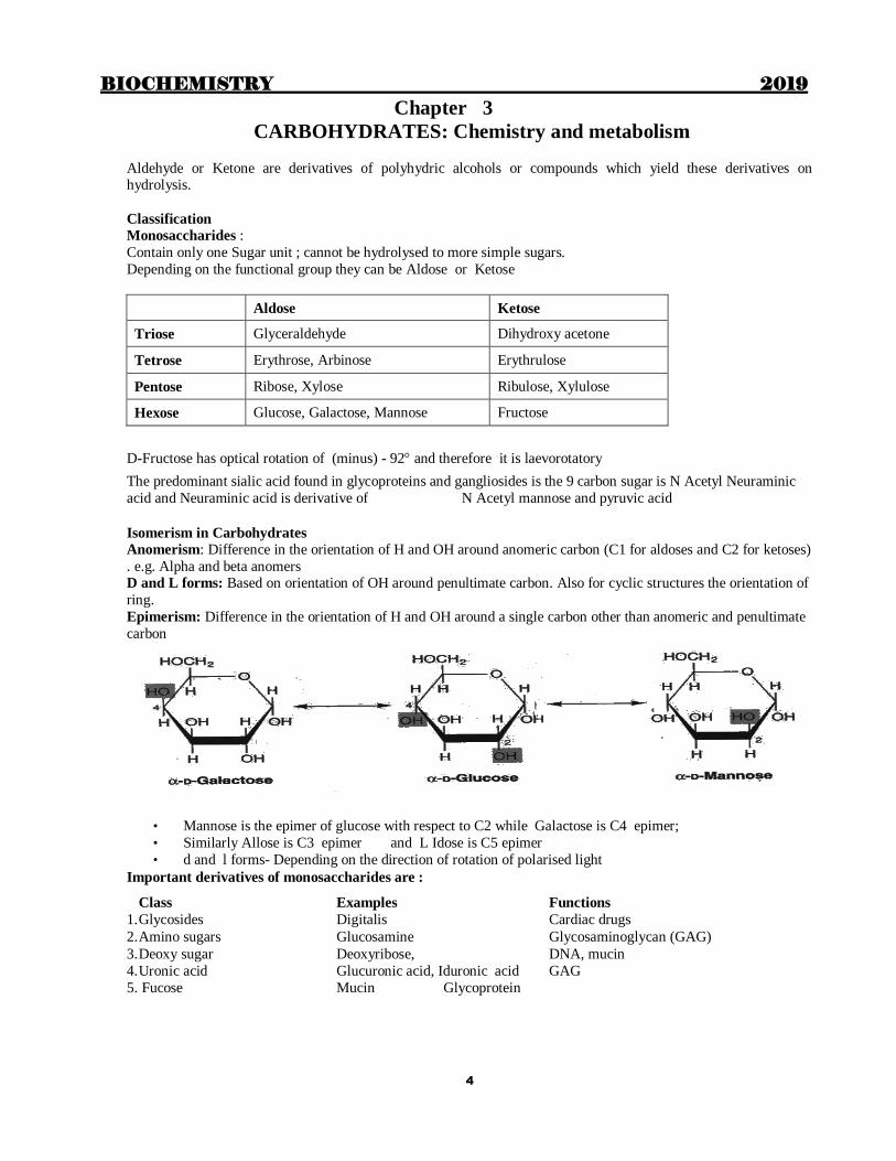
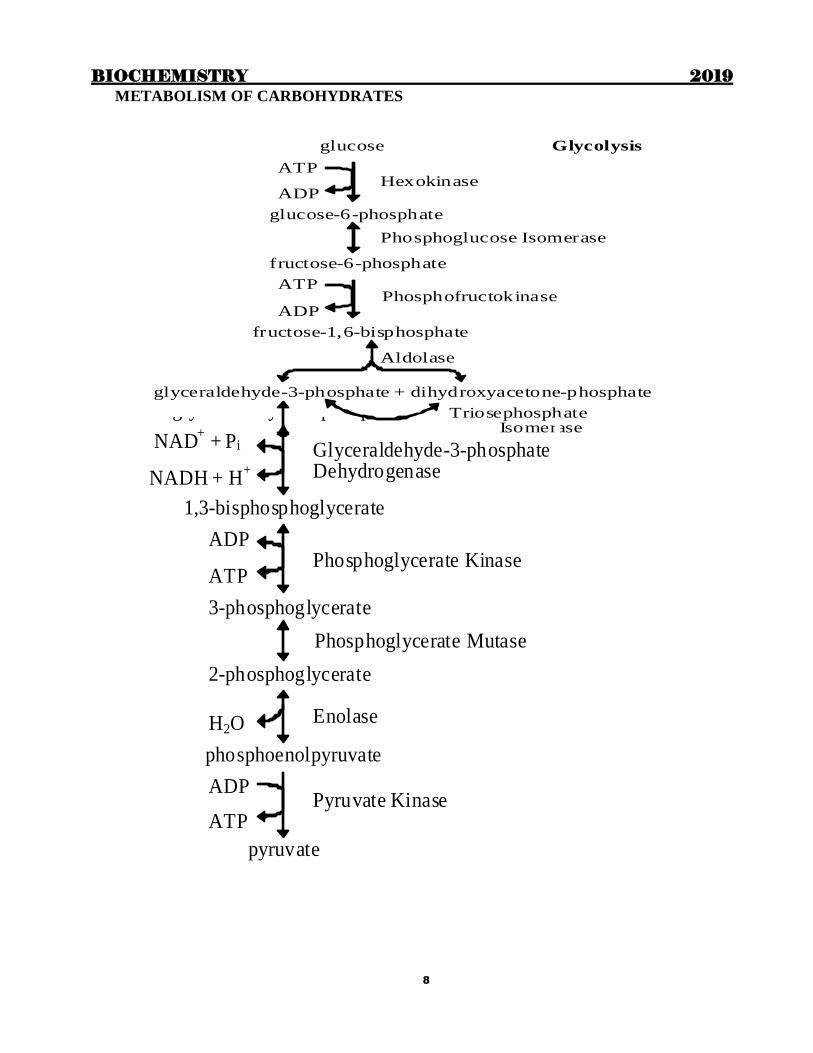
METABOLISM OF CARBOHYDRATES
Hexokinase
Phosphofructokinase
glucose Glycolysis
ATP
ADP
glucose-6-phosphate
Phosphoglucose Isomerase
fructose-6-phosphate
ATP
ADP
fructose-1,6-bisphosphate
Aldolase
glyceraldehyde-3-phosphate + dihydroxyacetone-phosphate
Triosephosphate Isomerase
Glycolysis continued Glyceraldehyde-3-phosphate Dehydrogenase
Phosphoglycerate Kinase
Enolase
Pyruvate Kinase
glyceraldehyde-3-phosphate
NAD+ + Pi
NADH + H+
1,3-bisphosphoglycerate
ADP
ATP
3-phosphoglycerate
Phosphoglycerate Mutase
2-phosphoglycerate
H2O
phosphoenolpyruvate
ADP
ATP
pyruvate
BIOCHEMISTRY 2019
9
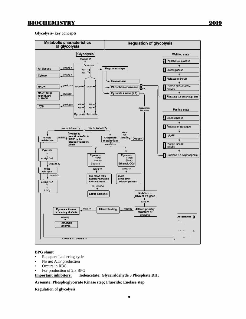
Glycolysis- key concepts
BPG shunt
• Rapaport-Leubering cycle
• No net ATP production
• Occurs in RBC • For production of 2,3 BPG
Important inhibitors: Iodoacetate: Glyceraldehyde 3 Phosphate DH;
Arsenate: Phosphoglycerate Kinase step; Fluoride: Enolase step
Regulation of glycolysis

BIOCHEMISTRY 2019
10
Enzyme Activator Inhibitor
HK G-6-P
GK Insulin Glucagon
PFK Insulin, AMP,
F-6-P, F-2,6-BP
Glucagon, ATP, Citrate, Low pH,
cAMP
Pyruvate kinase Insulin, F-1,6-BP Glucagon, ATP, cAMP, Alanine
PDH CoA, NAD+ Acetyl CoA, NADH
Fnergy yield- Anaerobic glycolysis
Enzyme Source ATP per Glucose
HK -1
PFK -1
1,3 BPG kinase ATP 1x2=2
Pyruvate kinase ATP 1x2=2
Total 2 ATP
Energy yield- Aerobic glycolysis
Enzyme Source ATP / glucose
HK -1
PFK -1
Glyceral-3P DH NADH 3x2=6
1,3 BPG kinase ATP 1x2=2
Pyruvate kinase ATP 1x2=2
Total 8 ATP
Energetic of Aerobic glycolysis+TCA cycle
• Glycolysis 8 ATP
• PDH 6 ATP (2 x NADH)
• TCA cycle 24 ATP (2x 12)
• Total = 38 ATP
TCA cycle Oxidation of Pyruvate to acetyl CoA
Multienzyme complex (Pyruvate Dehydrogenase Complex)
Present in inner mitochondrial membrane. Made up of
3 enzymes: PDH, DHL Transacetylase , DHL Dehydrogenase
5 Coenzymes: TDP (Thiamine, Vitamin B1), Lipoamide (lipoic acid), CoASH (Pantothenate vitamin B5),
FAD (Riboflavin vitamin B2), NAD (Niacin vitamin B3)
Analogous to αKG DH complex of TCA cycle & αKA Decarboxylase complex in branched chain amino acid
metabolism
All the three are thymine dependent enzymes and carry out oxidative decarboxylation.
Other example of multienzyme complex fatty acid synthase complex
Overall reaction: Pyr + CoASH + NAD+ → CO2 + Acetyl CoA + NADH + H+
BIOCHEMISTRY 2019
11
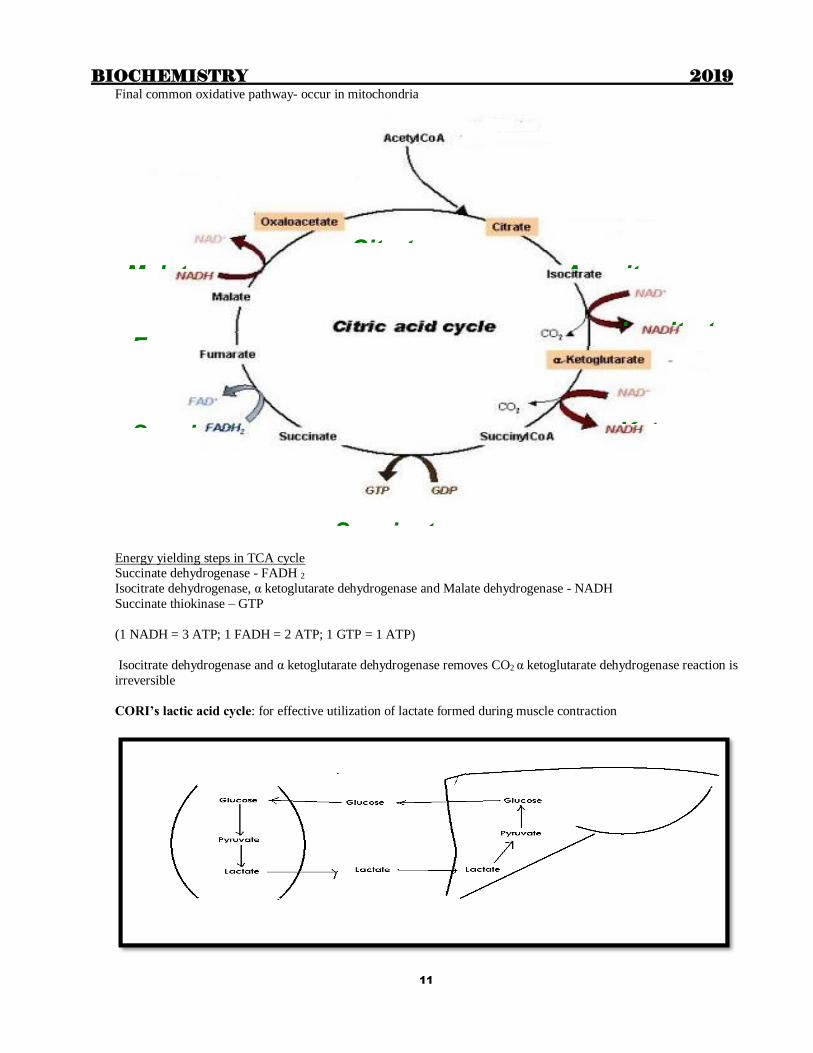
Final common oxidative pathway- occur in mitochondria
Energy yielding steps in TCA cycle Succinate dehydrogenase - FADH 2
Isocitrate dehydrogenase, α ketoglutarate dehydrogenase and Malate dehydrogenase - NADH
Succinate thiokinase – GTP
(1 NADH = 3 ATP; 1 FADH = 2 ATP; 1 GTP = 1 ATP)
Isocitrate dehydrogenase and α ketoglutarate dehydrogenase removes CO2 α ketoglutarate dehydrogenase reaction is
irreversible
CORI’s lactic acid cycle: for effective utilization of lactate formed during muscle contraction
Citrate Aconitase
Keto
Isocitrate
Succinate
Fumar
Succinat
Malate

BIOCHEMISTRY 2019
12

BIOCHEMISTRY 2019
13
HMP shunt pathway: Other names are
• Pentose phosphate pathway; Phosphogluconate pathway
• Dickens-Horecker pathway
• Warburg-Lipmann- Dickens-Horecker shunt
Sites
• Liver, Adipose tissue, RBC, Adrenal cortex, Mammary glands, Testes, ovary, Lens of the eye
2 phases
• Oxidative phase: Irreversible; Produce NADPH
Operates in those organs where NADPH is required for fatty acid, cholesterol or steroid synthesis
Also in tissues where NADPH is required to prevent oxidative damage
• Non-oxidative phase: Reversible; Produce pentoses
Operates in all tissues Pentoses can be produced in all tissues
Oxidative phase Steps are irreversible
• 2 steps produce NADPH
– G-6-PD (Glucose-6-phosphate dehydrogenase)
– 6-phosphogluconate dehydrogenase
• First pentose formed is RIBULOSE-5-Phosphate
• Rate limiting enzyme is G-6-PD
Non-oxidative phase
Produces pentoses
‘Transketolase reaction’
(Transketolases transfer a 2 C unit [glycolaldehyde]from a ketose to an aldose) Transketolase requires thiamine pyrophosphate (TPP), a derivative of vitamin B1. as a
coenzyme
G-6-PD deficiency
Most common enzyme deficiency
Trasmitted as X-linked recessive
Hemolysis when exposed to certain drugs [Eg: Antimalarials, Sulpha drugs etc]
Ingestion of Fava beans also precipitate hemolysis
- Met hemoglobinemia
Metabolism of glycogen
Glycogenesis (Synthesis of glycogen)
Degradation of Glycogen

BIOCHEMISTRY 2019
14
Regulation of Glycogen metabolism
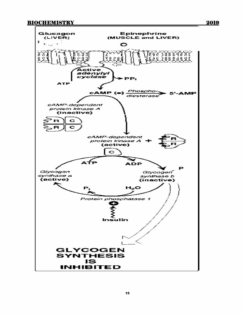
BIOCHEMISTRY 2019
15

BIOCHEMISTRY 2019
16
Importance of glycogen
1. Major storage form of glucose
Liver – 5% of tissue concentration (1/4 of total body glycogen)
Muscle – 0.7 % of tissue concentration (3/4 of total body glycogen) 2. Muscle glycogen – Provides glucose for its own use (lacks Glucose 6 Phosphatase)
Depletion occurs after prolonged vigorous exercise
3. Liver glycogen – Maintains Blood glucose for 10 – 12 hours in between meals.
4. Highly branched structure of glycogen provides multiple sites for glycogenolysis.
Rapid release of glucose in need
Glycogen Storage Diseases Glycogen storage disorders are inborn errors due to defect in enzmes concerned with metabolism of Glycogen.
Glycogen gets deposited in Liver, spleen, muscles etc.
Characteristic features are hepatosplenomegaly, fasting hypoglycemia, decreased exercise tolerance.
Glycogenosis Name Cause of Disorder
Type 0 Deficiency of Glycogen Synthase
Type 1 Von Glerke’s disease Deficiency of glucose – 6 phosphatase
Type II Pompe’s disease (1-3% glycogen is
degraded in lysosomes) Deficiency of lysosomal -14 and 1 6-
glucosidase (acid maltase)
Type III a Limit dextrinosis, Forbes’ or cori’s
disease
Deficiency of debranching enzyme in Liver
and Muscle
Type III b Limit dextrinosis Deficiency of debranching enzyme only in
liver
Type IV Amylopectinosis, Andersen’s disease Deficiency of Branching Enzyme
Type V Myophosphorylase deficiency,
McArdle’s syndrome Deficiency of muscle phosphorylase
Type VI Her’s disease Deficiency of liver phosphaorylase
Type VII Tarui’s disease Deficiency of phosphofructokinase in
muscle and erythrocytes
Type VIII Deficiency of liver phosphorylase kinase
Type IX Deficiency of liver & muscle phosphorylase
kinase
Type X
Deficiency of cAMP dependent Protein
Kinase A in both muscle and liver but
affects only liver
Type 0
• Glycogen synthase deficiency
• Hypoglycemia, hyperketonemia, early death
Type I
• I a – von Gierke’s
• I b – E.R glucose 6 PO4 transporter
Fatal GSD: Cardiac failure in GSD- Pompe’s disease, Anderson’s disease

BIOCHEMISTRY 2019
17
Fructose metabolism

BIOCHEMISTRY 2019
18
Summary of fructose metabolism
Metabolism of galactose

BIOCHEMISTRY 2019
19
Galactosemia
Gal-1-P uridyl transferase deficiency
Cataract is due to accumulation of dulcitol (from galactose)
Other features due to toxic effects of Galactose-1-P
– MR, Jaundice, Hepatomegaly, Renal damage
– Hypoglycemia
– Galactosuria, aminoaciduria
Galactokinase deficiency
• Gal-1-P not formed : No toxic effects
• Galactose accumulates: cataract
•Social smile not developed
MUCOPLOYSACCHARIDOSIS
Disease Defective enzyme GAG in urine
Hurler
Hunter
Sanfilippo
Morquis
Scheie’s
Maroteaux
Slys
L Iduronidase
Iduronate Sulfatase
NAc Glucosaminidase
NGal Sulfatase
L Iduronidase
NAc Gal 4 sulfatase
Beta Glucuronidase
DS, HS
DS, HS
HS
KS,CS
DS
DS
DS, HS
Insulin
Secreted as Pre-pro insulin (109 a.a)
Coverted to Pro insulin (86 a.a) in E.R
B chain-C peptide-A chain
Cleaved in Golgi apparatus
C peptide 33 a.a
Insulin
Composed of 51 amino acids arranged in two polypept ide chains, designated A and B, which
are l inked together by two disulfide br idges. The insulin molecule a lso con tains an
in tramolecular disulfide br idge between amino acid r esidues of the A chain
2 chains: A (21 a.a) B (30 a.a) One intrachain (A6A11) S-S bonds
2 interchain (A7B7 and A20B19)
Species variation is restricted to a.a 8,9,10 of A chain and C terminal of B chain
Number of a.a in C-peptide also varies according to species
Metabolic effects of insulin
Carbohydrates: In the liver and muscle, increases glycogen syn thesis. In the muscle
and adipose, increases glucose uptake by increasing the number of glucose transporter s
(GLUT-2). . In liver , in sul in decreases the p roduct ion of glucose through the inhibi t ion
of glycogenolysis and gluconeogenesis.
Lipids: o Inhibi t hormone sensi tive l ipase by dephosphoryl a t ion .
o Increased tr iacylgl ycerol syn thesis : increases the tr ansport and metabol ism of
glucose in to adipocytes, provid ing glycerol 3-phosphate for TAG syn thesis.
Also increases the lipoprotein lipase in adipose t issue by increasing the
enzyme's syn thesis, thus providing fa tty acids for esteri fica t ion
o In liver , in sulin promotes the conversion of glucose to tr iacylgl ycerols
Protein: st imulates the en try of amino acids into cel ls, and protein syn thesis.

BIOCHEMISTRY 2019
20
Metabol ic effects of Glucagon
Carbohydrate: increase the breakdown of l iver (not muscle) glycogen and increase
gluconeogenesis.
Lipids: Glucagon activates lipolysis in adipose. So glucagon is ketogenic.
Proteins: Glucagon increases uptake of amino acids by the l iver , r esult ing in increased
avai labi l ity of carbon skeletons for gluconeogenesis. Decreases plasma levels of amino
acids
Metabolic syndrome (X) , Reaven syndrome: NCEP criteria
Central obesity, Elevated BP
High TG, Low HDL, Elevated FBS

BIOCHEMISTRY 2019
21
Chapter 4
LIPIDS Important points to remember
Neutral lipids: Triglycerides, Cholesterol and Cholesteryl esters are uncharged and thus neutral lipids All phospholipids are derived from simplest phospholipid Phosphatidic acid
Phospholipids contain in addition to fatty acids and alcohol A phosphoric acid residue
Based on whether the alcohol is glycerol or sphingosine, the phospholipids are classified respectively as
Glycerophospholipids or Sphingophospholipids
Plasmalogens which constitute 10% of total phospholipids of brain and muscle has Ether linkage instead of ester
linkage at Sn1 Neonatal Respiratory distress syndrome results from deficiency of Dipalmitoyl lecithin (surfactant)
Cardiolipin, a major lipid of the inner mitochondrial membrane is Diphosphatidyl glycerol
The alcohol present in sphingophospholipids and glycolipids is Sphingosine (Glycerol is not present)
Ceramide is formed by combination of Sphingosine and fatty acid
Sphingomyelins consist of Ceramide (Sphingosine + fatty acid), phosphoric acid and choline Cerebrosides are glycolipids containing Ceramide and either glucose or galactose
Gangliosides are composed of Sphingosine, a long chain fatty acid, and oligosaccharide chain containing sialic acid
and glucose
The simplest ganglioside present in human body GM3
Fatty acids, glycerol and steroids & other alcohols eg. Cetyl alcohol, Dolichol are called Derived lipids
Most of the naturally occurring unsaturated fatty acids have double bonds in Cis configuration
Oleic acid with 18 carbons, one double bond at 9th position from carboxyl carbon can be represented as
18 :1; 9 or 9, 18 : 1 (belongs to 9 series)
Linoleic acid and Arachidonic acid belong to the 6 series
Linolenic acid is an example of 3 fatty acid
Eicosanoids (derived from C20 PUFAs) are Prostaglandins, Thromboxanes, Leukotrienes & Lipoxins
Common feature of all prostaglandins is Cyclopentane ring
Common to all thromboxanes is Cyclopentane ring interrupted with an oxygen atom
Common feature of Leukotrienes & Lipoxins is Absence of cyclopentane ring and presence of 3 & 4 conjugated
double bonds respectively
All steroids contain Cyclo Pentano Perhydro Phenanthrene ring (CPP ring) Chemical features of cholesterol are Cyclopentano perhydro phenanthrene ring; 27 Carbon atoms; OH at position 3;
Double bond between C5 and C6
Lipids that contain both hydrophilic & hydrophobic groups are called Amphipathic lipids
Examples of amphipathic lipids are Phospholipids, cholesterol, fatty acids, bile salts
Examples of totally hydrophobic lipids are Triacylglycerol and cholesteryl ester
Lipids- Classification
Simple lipids: Esters of fatty acids with an alcohol
Esters of fatty acids with glycerol- Fats
Fats which exist as liquid at room temperature- Oils
Esters of fatty acids with higher molecular weight monohydric alcohols- waxes
Compound lipids: Contain groups in addition to f.a and alcohol Phospholipids, Glycolipids, Sulfolipids etc
Derived lipids: Derived from any of the above:
Fatty acids
Saturated fatty acids
Acetic (2) SCFA
Butyric (4) Milk, Butter SCFA
Caproic (6) Milk, Butter SCFA
Caprylic (8) MCFA
Capric (10) Coconut oil MCFA Lauric (12) Coconut oil MCFA
Myristic (14) Coconut oil MCFA

BIOCHEMISTRY 2019
22
Palmitic (16) Body fat LCFA
Stearic (18) Body fat LCFA
(Very long chain= more than 24 C)
Unsaturated fatty acids
No of C and No & position of Double Bonds
Omega Number
Common name Occurrence
Monoenoic acids (one double bond)
16:1; 9 7 Palmitoleic In nearly all fats
18:1; 9 9 Oleic Possibly the most common fatty acid in natural fats
18:1; 9 9 Elaidic Hydrogenated and ruminant fats
Dienoic Acids (two double bonds)
18:2; 9,12 6 Linoleic Corn, peanut, cottonseed, soybean, many plant oils
Trienoic acids (three double bonds)
18:3; 6,9,12 6 -Linolenic Some plants, eg. Oil of evening primrose, borage oil; minor fatty acid in animals
18:3; 9,12, 15 3 -Linolenic Frequently found with linoleic acid but particularly in linseed oil
Tetraenoic acids (four double bonds)
20:4; 5,8,11,14 6 Arachidonic Found in animal fats and in peanut oil, important component of phospholipids in animals
Pentaenoic acids (five double bonds)
20:5; 5,8,11,14, 17 3 Timnodonic Important component of fish oils, e.g. Cod liver, mackerel, menhaden, salmon oils
Hexaenoic acids (six double bonds)
20:6; 4,7,10,13,16,19 3 Cervonic Fish oil, phospholipids in brain
ω 9 fatty acids eg: Oleic, Elaidic Note that Elaidic is a Trans fatty acid
Phospholipids
Phosphatidates: those which contain phosphatidic acid [1, 2 diacyl glycerol to which phosphoric acid is added at 3rd position]
Nitrogenous base is attached to phosphoric acid
Cardiolipin is Diphosphatidyl glycerol
Lecithin- Phosphatidyl choline
Cephalin- Phosphatidyl ethanolamine
Plasmalogens
• Similar to phosphatidate; an aliphatic long chain unsaturated alcohol is attached in ether linkage to the first
OH of glycerol
• Phosphoric acid is attached to Nitrogenous base
Plasmalogen
BIOCHEMISTRY 2019
23
Sphingolipids
• They contain ceramide (fatty acid + sphingosine in amide linkage)
– Phosphosphingosides: have phosphoric acid Eg: Sphingomyelin
– Glycosphingolipids: Ceramide + carbohydrates Eg: Glucocerebroside
Gangliosides
• Ceramide-oligosaccharides with at least one NANA attached to them
• GM3 is simpler than GM1 G = Ganglioside M = monosialo
• 1,2,3 etc – based on electrophoretic migration
Antioxidants: Act either by preventing the chain of events which lead to damage from starting or by breaking the
chain of events
Preventive: reduce the rate of chain initiation: catalase, peroxidase, Se, EDTA, DTPA (di ethylene triamine penta acetate)
Chain breaking: Interfere with chain propagation: SOD, Viamin E
Iodine number: No: of grams of Iodine taken up by 100g of fat. Directly proportional to the content or degree of un
saturation.
Saponification number : the number of mg of KOH required to saponify1g fat. Inversely proportional to molecular
weight anf thus is measure of average molecular size of fatty acids
Higher Saponification number indicates Short chain fatty acids. butter (230 – 240; coconut oil (250-260)
Reichert – Meissl (RM) number defined as ml of 0.1 N KOH to neutralize soluble volatile fatty acids in 5 g fat is a
measure of Volatile fatty acids like butyric, caproic and caprylic acid. RM number is highest for butter (range 25-30)
Acid number defined as mg of KOH to neutralize free fatty acids in 1 g fat is used to check decomposition and
bacterial contamination of fats and oil
Essential fatty acids
Linoleic, α linolenic and Arachidonic acid
Linoleic and α linolenic acid cannot be synthesized in the body. Arachidonic acid can be formed from Linoleic acid
Action of phospholipases on Lecithin produces PL A1 Fatty acid + 2 acyl glycerophosphoryl choline
PLA2 Lysolecithin + fatty acid
PL C 1, 2 diacyl glycerol + Phosphoryl choline PL D Phosphatidic acid + Choline

BIOCHEMISTRY 2019
24
OXIDATION OF FATTY ACIDS
Mainly β-oxidation
Fatty acids are “activated” first: Fatty acid Fatty acyl CoA (enzyme: Acyl CoA synthetase)
Requires 2 high energy bonds (ATP AMP)
Only step in degradation of fatty acids which require energy
Takes place in E.R, Peroxisomes or on the outer mitochondrial membrane
Long chain fatty acyl CoA cannot penetrate IMM : So it combines with carnitine (Enzyme- CAT-I) to form acyl
carnitine. Inside the mitochondria, acyl CoA is regenerated by CAT II
Carnitine : β hydroxyl γ trimethyl ammonium butyrate Formed from Lys and Met I Liver and kidney
Note: Short chain fatty acids (SCFA) & Medium Chain Fatty acids (MCFA) < 12 carbons do not require
carnitine system
.

BIOCHEMISTRY 2019
25
Beta oxidation of fatty acids with even number of C atoms produce acetyl CoA molecules
Energy yield from beta oxidation
Initial activation requires 2 ATP
Each cycle (which removes one acetyl CoA) produce 5 ATP (1 NADH + 1 FADH2)
Each acetyl CoA will produce 12 ATP in TCA cycle
Oxidation of odd chain fatty acids
During beta oxidation of odd chain fatty acid, 2C units (Acetyl CoA) are successively removed till a 3C compound (Propionyl CoA) is formed

BIOCHEMISTRY 2019
26
Oxidation of very long chain fatty acids
Peroxisomal beta oxidation: For very long chain fatty acids- does not produce ATP
β oxidation of unsaturated fatty acids occurs in mitochondria till Δ3 or 4 cis acyl CoA forms. Then isomerases are
required to convert it to a Δ2 Trans enoyl compound
- Oxidation:
For Phytanic Acid, which cannot be metabolized by β – oxidation. Phytanic acid is found in ruminant fat, meat and
milk. Takes place in brain. Does not generate energy. End product is propionyl CoA
Refsum Disease: deficiency of Phytanoyl CoA Hydroxylase leads to defective - oxidation & deposition of
Phytanic Acid in Brain resulting in neurodegeneration
ω Oxidation
for MCFA due to deficiency of MC acyl dehydrogenase (1: 40000). Takes place in endoplasmic reticulum

BIOCHEMISTRY 2019
27
Cytochrome P450 Monooxygenase introduces a COOH group at ω position. Di-carboxylic acid then undergoes β –
oxidation from both ends forming Di-carboxylic acids (Succinate & Adipate) in the end. Deficiency of medium
chain acyl dehydrogenase leads to dicarboxylic aciduria.
Defects in oxidation of fatty acids
Carnitine deficiency: Occur in preterm babies, hemodialysis
Hypoglycemia, lipid accumulation, muscle weakness
Treatment: oral carnitine
CAT –I (CPT-I) deficiency: Affects liver; Reduced ketogenesis, hypoglycemia
CAT-II deficiency: Affects skeletal muscles (affects liver in severe cases) Medium chain acyl CoA DH deficiency : most common inborn error of fa t ty acid oxidation
Leads to non ketot ic hypogl ycemia, and dicarboxyl ic aciduria
Affects mainly in fants
Jamaican vomiting sickness: Eating unripe fruit of akee tr ee: contains hypogl ycin -
Inact ivates medium and short chain acyl CoA dehydrogenase
Refsum’s disease : defect ive a lpha oxidat ion of phytan ic acid; leads to neurological disorder
Zellweger’s syndrome : (cerebrohepatorena l syndrome)- Defect ive peroxisomal beta oxidation
– due to defect ive protein targett ing
Ketogenesis:
Acetoacetyl CoA+ Acetyl CoA HMG CoA [HMG CoA synthase]
HMG CoA Acetoacetate+ Acetyl CoA [HMG CoA lyase]
Acetoacetate is converted to acetone or β OH butyrate Ketogenesis occurs in liver mitochondria only.
Ketogenesis
- Ketone bodies are produced in liver

BIOCHEMISTRY 2019
28
- Due to increased breakdown of FA (Starvation & Diabetes mellitus)
- Acetoacetate is the 1st ketone body to be synthesized
- β – Hydroxybutyrate is present in maximum concentration
- both are organic acids causing metabolic acidosis (Ketoacidosis)
- all are excreted in urine (Ketonuria)
- Acetone is volatile, exhaled in breath and responsible for acidotic or ketotic
breath
- Rothera’s test detects ketone bodies in urine (acetoacetate and acetone)
Ketone bodies are utilized by : Muscle, Heart, Brain
Ketolysis occurs in extrahepatic tissues with the help of enzyme Succinyl CoA- Acetoacetate-CoA transferase. Rate
of ketolysis increases with increase in blood level until it saturates the oxidative machinery.
Lipotropic factors:
Required for normal mobilization of fat from liver. Deficiency causes fatty liver
Eg: Choline, Lecithin, Methionine, Vitamin E, Se, Omega 3 fatty acids
Fatty acid synthase is a multienzyme complex having 7 enzymes and acyl carrier protein. 7 enzymes are:
Ketoacyl synthase, Acetyl transacylase, Malonyl transacylase, Hydratase, Enoyl reductase, Ketoacyl reductase and
thiesterase
Acetyl CoA carboxylase is not its part
Acetyl CoA carboxylase converts acetyl CoA to malonyl CoA. It is inhibited by long chain acylCoA
Acetyl CoA is formed in cytoplasm. It cannot diffuse freely into cytosol. So it is converted to citrate and diffuses
out with the help of Tricarboxylate transporter. And in cytosol, Acetyl CoA is released by the action of ATP-
Citrate lyase
Microsomal fatty acid elongase system elongates saturated and unsaturated fatty acyl CoA: Malonyl CoA donates C
(not acetyl CoA). Uses NADPH, but can also use NADH as coenzyme
Acetl CoA carboxylase (AcetylCoA Malonyl CoA) is the rate limiting step in de novo synthesis of fatty acids.
Allosterically activated by citrate.
Inhibited by: Feedback Long chain acyl CoA
Covalent modification Phosphorylation
Prostaglandins
Originally isolated from prostate: Present in all tissues. Most potent biologically active substances: Local hormones
Considered to be derivatives of 20C cyclic saturated fatty acid:- Prostanoic acid
According to substituent groups in the ring, named as A, B, D etc
Numbering is given based on the number of double bonds in side chain (PGE1, PGE2 etc)
Series 2 is most common [C13-14 trans and C5-6 cis]
PG with Double ring structure is PGI and is known as Prostacyclin
Synthesis of prostaglandins
Series 1 Linoleic acid: Series 2 Arachidonic acid: Series 3 Eicosa penta enoic acid
COX: Activated by catecholamines: Inhibited by NSAIDs
COX-1 : Constitutive form mediates gastric, renal and platelet functions
COX-2: Inducible form Mediates inflammatory response
Corticosteroids inhibit the transcription of COX-2
Aspirin inhibits COX-1 and 2 : Coxibs selectively inhibit COX-2
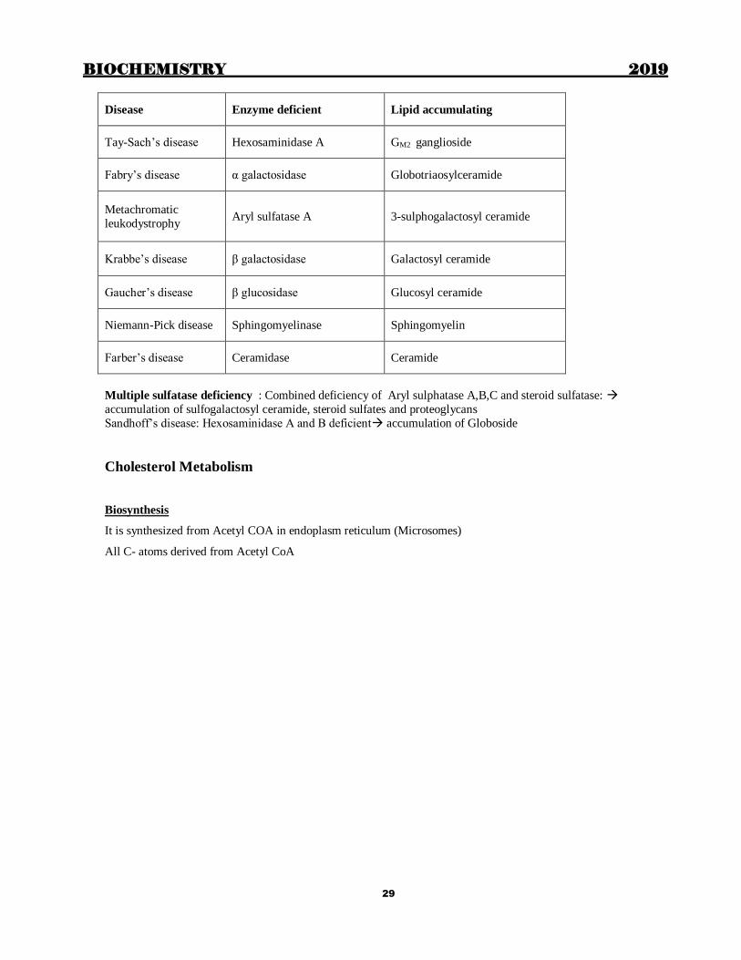
Lipid storage diseases (Sphingolipidoses)
Due to defect in the catabolism of sphingolipids
All types except Fabry’s disease have Mental retardation
BIOCHEMISTRY 2019
29
Disease Enzyme deficient Lipid accumulating
Tay-Sach’s disease Hexosaminidase A GM2 ganglioside
Fabry’s disease α galactosidase Globotriaosylceramide
Metachromatic
leukodystrophy Aryl sulfatase A 3-sulphogalactosyl ceramide
Krabbe’s disease β galactosidase Galactosyl ceramide
Gaucher’s disease β glucosidase Glucosyl ceramide
Niemann-Pick disease Sphingomyelinase Sphingomyelin
Farber’s disease Ceramidase Ceramide
Multiple sulfatase deficiency : Combined deficiency of Aryl sulphatase A,B,C and steroid sulfatase:
accumulation of sulfogalactosyl ceramide, steroid sulfates and proteoglycans
Sandhoff’s disease: Hexosaminidase A and B deficient accumulation of Globoside
Cholesterol Metabolism
Biosynthesis
It is synthesized from Acetyl COA in endoplasm reticulum (Microsomes)
All C- atoms derived from Acetyl CoA

BIOCHEMISTRY 2019
30
Step I: Formation of mevalonate. Similar to formation of Ketone bodies in mitochondria
Main Regulatory enzyme: HMG CoA reductase
- Induced by insulin
- Stimulated by insulin and inhibited by glucagon by covalent modification - Allosteric inhibitors:
Cholesterol
Bile acids
Mevalonate
- Competitive inhibitors
Hypocholesterolemic drugs i.e. statins
Rate limiting step in cholesterol synthesis: HMG CoA reductase (HMG CoA Mevalonate)
Inhibited by mevalonate, cholesterol. Cholesterol and its metabolites causes repression of HMG CoA reductase via
activation of sterol regulatory element binding protein (SREBP) transcription factor. Also regulated by covalent
modification: Dephosphorylated form is active

BIOCHEMISTRY 2019
31
Important products of cholesterol are steroids
Cholesterol breakdown:
Cholesterol is excreted in Bile. It is a major constituent of bile stones
Total 1g/ Day
i. 600 mg as cholesterol (60 %) Coprostanol is the major end product formed from cholesterol by
intestinal bacteria
ii. 400 mg as bile acids & salts (40 %)
Synthesis of bile salts
Cholesterol
7 α – Hydroxylase
7 α – Hydroxy Cholesterol
Cholyl CoA Chenodeoxy Cholyl CoA
Taurocholate GlycocholateTaurocheno–
deoxycholate
Glycocheno–
deoxycholate
Deconjugation & Dehydrogenation
Deoxycholic acid Lithocholic acid
Conjugation with Taurine or GlycinePrimary
bile acids
(Liver)
Secondary
bile acids
(Intestine)

BIOCHEMISTRY 2019
32
Lipoproteins
Lipids are non polar in nature and hence insoluble in water. They are transported in plasma (aqueous medium) as
lipoproteins
- Lipid component of lipoproteins: o Surface lipids
Amphipathic/ hydrophilic
Cholesterol
Phospholipids
o Core lipids
Non polar/ hydrophophic
Cholesterol ester
Triglycerides
- Protein component of lipoproteins (Apoproteins)
o Integral proteins: embedded in the surface membrane and cannot be
separated from the lipoprotein Apo A
A I: Cofactor for LCAT
A II: inhibit LPL
A IV
Note: Apo A is peripheral protein in Chylomicrons
Apo B
B 48
B 100: Ligand for LDL receptor
o Peripheral proteins: present on the surface and can be interchanged between
various lipoproteins
Apo C
C I : inhibits CETP
C II: activator (cofactor) for LPL
C III: Inhibit LPL
Apo E Ligand for LRP receptor and also LDL receptor
Recently Apo D has been identified
Apo B 48 is the integral apoprotein of chylomicron synthesized by intestinal cells
Apo B100 is the integral apoprotein of VLDL synthesized from Liver

BIOCHEMISTRY 2019
33
The gene for both is same except that in intestine there is premature termination due to introduction of a stop
signal by RNA editing enzyme converting CAA (codon) for glutamine to UAA (stop codon)
Apo B 100 is one of the longest single polypeptide chain known, having 4536 Amino acids. Apo B 48 is 48 % of
Apo B 100
Lipoprotein Source Density Protein
%
Lipid
%
Main lipid
component
Electrophoresis
on Agarose
Integral
protein
Chylomicron Intestine TG Origin B 48
VLDL Liver TG Pre β B 100
LDL VLDL Cholesterol β B 100
HDL Liver +
Intestine
Phospholipid
s Cholesterol
α A
FFA/
Albumin
Adipose
Tissue
FFA
Note: Free fatty acid is a misnomer. Fatty acid is always associated with some protein
Plasma: Albumin
Plasma membrane: Membrane fatty acid transport protein
Intracellular: fatty acid binding protein (Z protein)
Non-esterified or un-esterified fatty acid is a more appropriate term
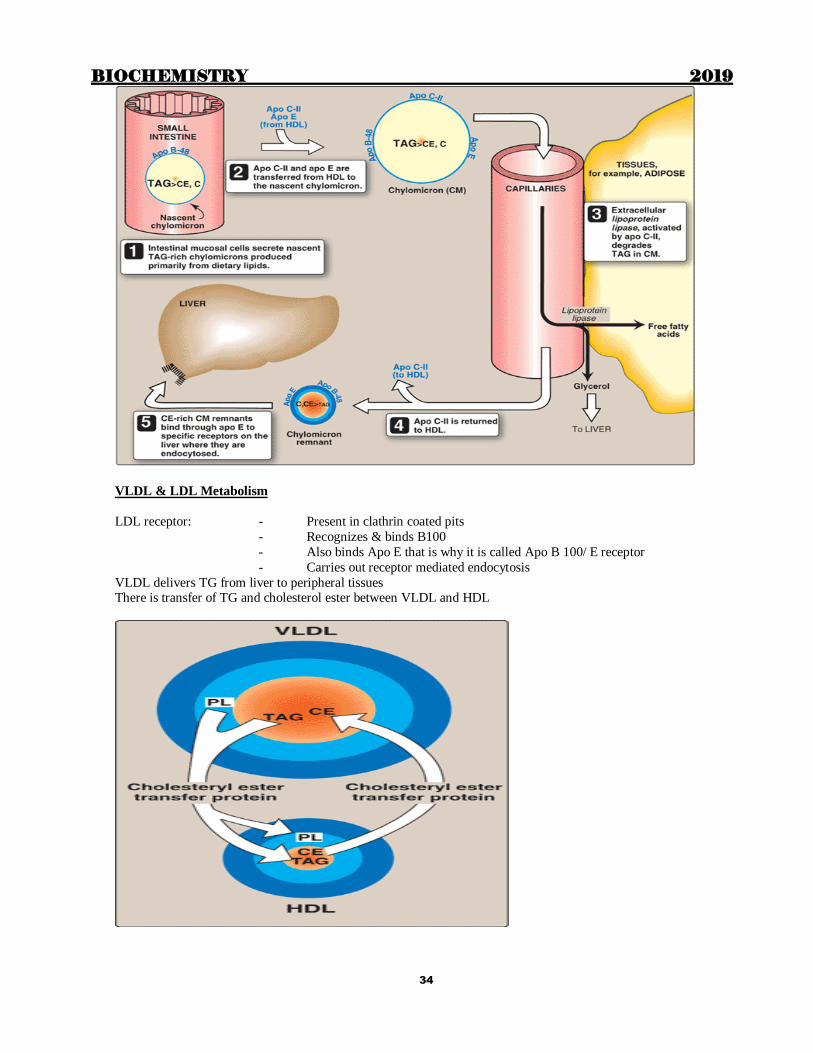
Chylomicron metabolism
LPL
- Attached to the capillary membrane through heparin releasable heparan sulphate
(HRHS)
- Hepatic lipase is also releasable by heparin (HRHL)
- Stimulated by Insulin and Apo CII
- Adipose Tissue (high Km)
- Skeletal muscle (low Km) - Heart (maximum concentration of LPL)
LRP receptor:
- Also present in skeletal muscle apart from liver
- Binds Apo E
- Also releasable by heparin
TG transfer protein required for assembly of B48/ B100 + phospholipids + Triglycerides
Chylomicrons deliver dietary lipids (TG, Cholesterol, cholesterol ester and fat soluble vitamins) to peripheral tissues
BIOCHEMISTRY 2019
34
VLDL & LDL Metabolism
LDL receptor: - Present in clathrin coated pits
- Recognizes & binds B100
- Also binds Apo E that is why it is called Apo B 100/ E receptor
- Carries out receptor mediated endocytosis
VLDL delivers TG from liver to peripheral tissues There is transfer of TG and cholesterol ester between VLDL and HDL

BIOCHEMISTRY 2019
35
BIOCHEMISTRY 2019
36
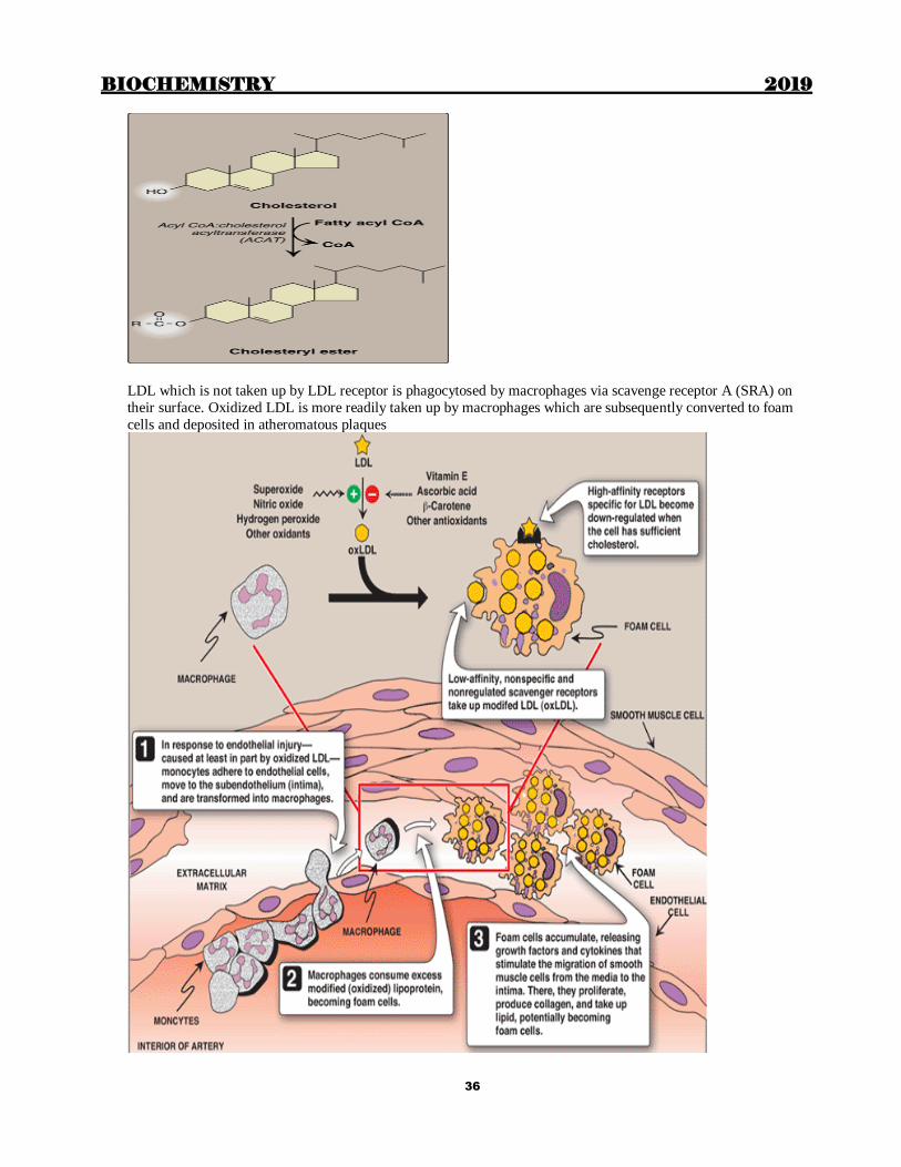
LDL which is not taken up by LDL receptor is phagocytosed by macrophages via scavenge receptor A (SRA) on
their surface. Oxidized LDL is more readily taken up by macrophages which are subsequently converted to foam
cells and deposited in atheromatous plaques
BIOCHEMISTRY 2019
37
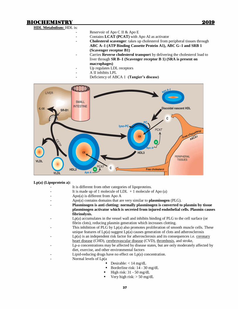
HDL Metabolism: HDL is:
- Reservoir of Apo C II & Apo E
- Contains LCAT (PCAT) with Apo AI as activator
- Cholesterol scavenger: takes up cholesterol from peripheral tissues through
ABC A–1 (ATP Binding Cassette Protein A1), ABC G–1 and SRB 1
(Scavenger receptor B1)
- Carries Reverse cholesterol transport by delivering the cholesterol load to
liver through SR B–1 (Scavenger receptor B 1) (SRA is present on
macrophages) - Up regulates LDL receptors
- A II inhibits LPL - Deficiency of ABCA 1 (Tangier’s disease)
Lp(a) (Lipoprotein a):
- It is different from other categories of lipoproteins.
- It is made up of 1 molecule of LDL + 1 molecule of Apo (a)
- Apo(a) is different from Apo A
- Apo(a) contains domains that are very similar to plasminogen (PLG).
- Plasminogen is anti clotting: normally plasminogen is converted to plasmin by tissue
plasminogen activator which is secreted from injured endothelial cells. Plasmin causes
fibrinolysis.
- Lp(a) accumulates in the vessel wall and inhibits binding of PLG to the cell surface (or
fibrin clots), reducing plasmin generation which increases clotting.
- This inhibition of PLG by Lp(a) also promotes proliferation of smooth muscle cells. These
unique features of Lp(a) suggest Lp(a) causes generation of clots and atherosclerosis - Lp(a) is an independent risk factor for atherosclerosis and its consequences i.e. coronary
heart disease (CHD), cerebrovascular disease (CVD), thrombosis, and stroke,
- Lp-a concentrations may be affected by disease states, but are only moderately affected by
diet, exercise, and other environmental factors
- Lipid-reducing drugs have no effect on Lp(a) concentration.
- Normal levels of Lp(a
Desirable: < 14 mg/dL
Borderline risk: 14 - 30 mg/dL
High risk: 31 - 50 mg/dL
Very high risk: > 50 mg/dL

BIOCHEMISTRY 2019
38
During electrophoresis, free fatty acids move to the position of Albumin: Chylomicrons remain at the origin. Other
fractions move in the region of globulins
HDL – α lipoprotein
VLDL- Pre β lipoprotein
{IDL- Broad β lipoprotein}
LDL- β lipoprotein
Lipoprotein Apolipoproteins
Chylomicron-Nascent B-48, A
Chylomicron- circulation B-48, A, E,C
Chylomicron remnant B-48, E
VLDL B-100, E,C
VLDL remnant (IDL) B-100, E
LDL B-100
HDL A-I
Apo C-II acts as cofactor for lipoprotein lipase; Apo A-II and Apo C-III inhibits lipoprotein lipase
Apo A-I acts as cofactor for LCAT
Apo C-I inhibits CETP
Dietary triglycerides are transported as chylomicrons

BIOCHEMISTRY 2019
39
Lipoprotein
lipase Hydrolyse TAG Glycerol +fatty acids
Attached to capillary endothelium by chains of heparin sulphate Active in many tisuues, not in liver
Heart lipoprotein lipase has a low Km
Not normally found in blood, but may be released after injecting heparin
Phospholipids and apo C-II are required as cofactors
Hepatic lipase: Bound to sinusoidal surface of liver cells, released by heparin. Involved in metabolism of
chylomicron remnants and HDL.
LDL receptors are most abundant in hepatocytes. Binds apo B100 and apo E. They are located in Clathrin coated
pits. Can take up LDL and Chylomicron remnants which is then internalized for lysosomal degradation. Free
receptors return to membrane surface.
Lipoprotein (a) Lp (a)
Attached to apo B100 by S-S
Homology with plasminogen: Interferes with plasminogen activation
Inhibits fibrinolysis: Strong association with MI

BIOCHEMISTRY 2019
40
Hyperlipoproteinemias
Type LP elevated Metabolic defect TG Cholesterol
Type I
Familial LPL def CM
LPL deficiency or
Apo CII N
Type II A Familial hypercholesterolaemia
LDL LDL receptor defect N
Type II B LDL,VLDL Excess of apo B
Type III
Familial hypolipoproteinaemia
Broad beta
VLDL &CM Abnormal apo E
Type IV
Familial
hypertriglycerolaenaemia
VLDL
Over production of VLDL.
Associated with glucose
intolerance & hyperinsulinaemia
LDL,
HDL
Type V LP elevated Metabolic defect TAG Cholesterol
Familial hyper alpha
lipoproteinemia HDL Not known N
Hepatic lipase
deficiency
TG rich HDL &
VLDL remnants LPL deficient N
Familial LCAT deficiency Nascent discoidal HDL
Absence of LCAT N
Familial lipoprotein (a) excess LP(a) Abnormal LP (a)
HYPERLIPOPROTEINEMIAS
Type II A (Primary familial hypercholesterolemia)
Most common
Due to LDL receptor defect
LDL receptor deficiency
Defective binding of B-100 to LDL receptor
Eg: 3500th Arg Gln (familial defective apo-B or B-3500)
Defective internalisation of receptor-LDL complex
LDL very high; Strong predisposition to develop MI;Death in second decade
TYPE II B
Excess production of Apo-B (LDL and VLDL are elevated) thus Increased cholesterol and TG
Risk of MI (usually from 3rd decade)
SECONDARY HYPERLIPIDEMIAS
Cholesterol and triglycerides increased: occurs in Diabetes mellitus, Nephrotic syndrome, Hypothyroidism
Increased cholesterol, Normal TG: Biliary obstruction
Hypertriglyceridemia with normal cholesterol: Alcoholism, Pregnancy, Oral contraceptive
WOLMANN’S DISEASE
Lysosomal storage disease: Lysosomal acid lipase (Cholesterol ester hydrolase) is deficient

BIOCHEMISTRY 2019
41
Autosomal recessive
Late onset form is called Cholesteryl Ester Storage Disease (CESD)
Perilipin
Protein involved in the formation of lipid droplets in adipose tisse. Inhibts lipolysis by preventing access of lipases
to the stored TAG. When needed, it targets Hormone sensitive lipase to the lipid droplets
METABOLIC SYNDROME (SYNDROME X) (REAVEN SYNDROME)
Diagnostic criteria
Central obesity: Waist 40 (m)/ 35 (f) or above
Elevated BP.> 130/85 mm Hg
High TG > 150 mg/dl;
Low HDL < 40 (m); <50 (f) mg/dl
Elevated FBS > 100 mg/dl
3 out of 5 criteria is diagnostic

BIOCHEMISTRY 2019
42
Chapter 5
AMINOACIDS AND PROTEINS
Proteins are formed by polymerization of amino acids. Human amino acids are present as L α – amino acids All
proteins contain only the same 20 L amino acids
All amino acids are structurally similar to L-Glyceraldehyde & amino group is attached to –Carbon
All amino acids are in L form (all carbohydrates are in D form except L- Fucose and L - Iduuronate). They are optically active and may be d (dextrorotatory) or l (levorotatory) at pH 7, EXCEPT Glycine, which is optically
inactive
– Carbon is asymmetric (chiral) to which are attached:-
i. H atom;.
ii. Carboxyl group (–COOH)
iii. Amino group (–NH2):
There are certain non amino acids e.g.
a. GABA (Gamma Amino Butyric Acid): It is an inhibitory neurotransmitter
NH 2 – CH 2 – CH 2 – CH 2 – COOH
b. β – Alanine: NH 2 – CH 2 – CH 2 – COOH
iv. R – Group: the 4th group which is different in different amino acids
Properties of R group
1. Determine physical properties of amino acids e.g. polarity, acidity, basicity etc.
2. R group undergoes various post translational modifications.
3. R groups determine proteins folding & confirmation.
Important points to remember
All amino acids:
- Except glycine have at least 1 asymmetric carbon and thus exhibit optical activity
- Have no net charge at isoelectric pH (PI) , therefore exists as zwitterions
- Have positive charge at pH less than PI and have negative charge at pH greater than PI
- Do not absorb visible light and therefore give colourless solutions
All amino acids absorb light of wave length less than 240 nm except aromatic amino acids which absorb
light of 280 nm
Most effective amino acid that can act as a buffer at physiologic pH is Histidine due to its imidazole group
At Isoelectric pH, the amino acids exists as Zwitter ions .The amino acid has both positive and negative
charges same (Electrically neutral)
BIOCHEMISTRY 2019
43
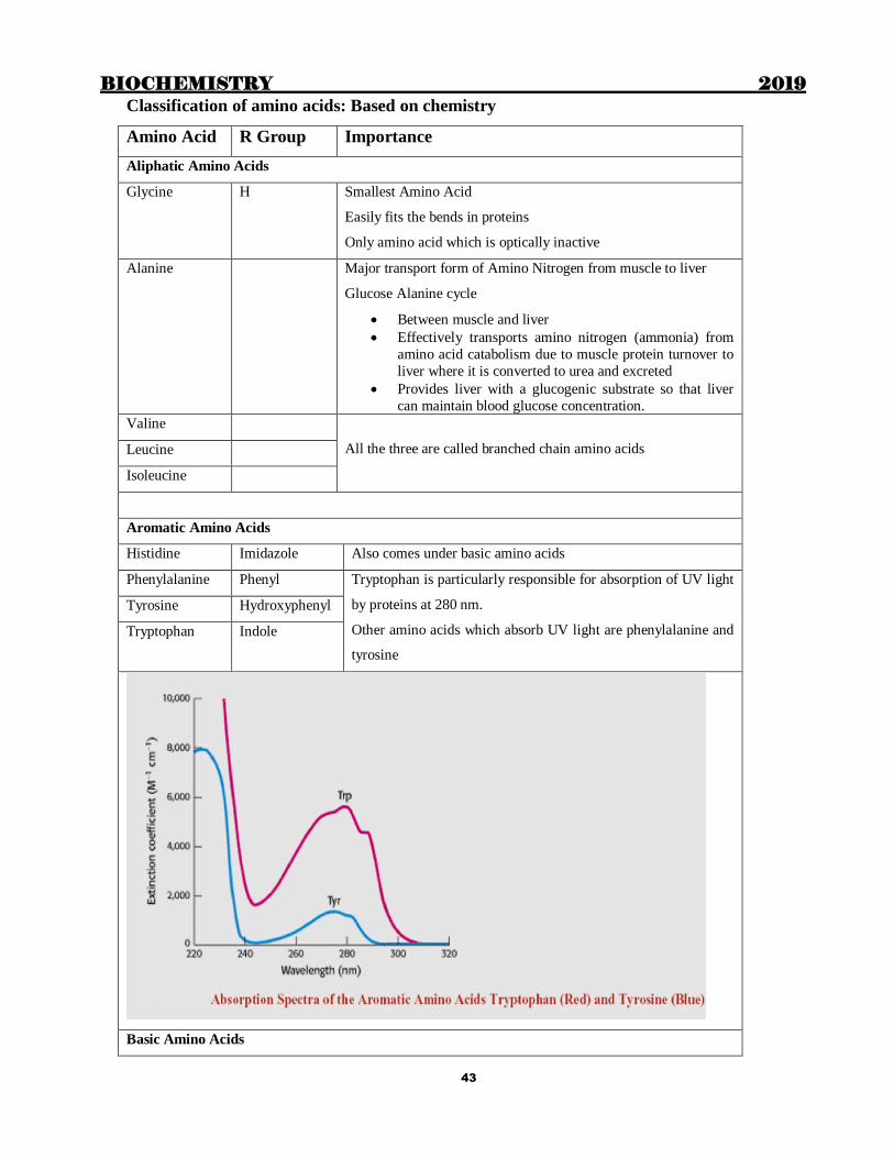
Classification of amino acids: Based on chemistry
Amino Acid R Group Importance
Aliphatic Amino Acids
Glycine H
Smallest Amino Acid
Easily fits the bends in proteins
Only amino acid which is optically inactive
Alanine Major transport form of Amino Nitrogen from muscle to liver
Glucose Alanine cycle
Between muscle and liver
Effectively transports amino nitrogen (ammonia) from
amino acid catabolism due to muscle protein turnover to
liver where it is converted to urea and excreted
Provides liver with a glucogenic substrate so that liver
can maintain blood glucose concentration.
Valine
All the three are called branched chain amino acids Leucine
Isoleucine
Aromatic Amino Acids
Histidine Imidazole Also comes under basic amino acids
Phenylalanine Phenyl Tryptophan is particularly responsible for absorption of UV light
by proteins at 280 nm.
Other amino acids which absorb UV light are phenylalanine and
tyrosine
Tyrosine Hydroxyphenyl
Tryptophan Indole
Basic Amino Acids

BIOCHEMISTRY 2019
44
Histidine Imidazole also comes under aromatic amino acids
R groiup of Histidine can act as acid or base at pH 7.0. This
helps histidine residues in protein to have buffering action.
Lysine Epsilon (ε)
amino group
ε NH2 group forms Schiff base with aldehyde group in:
Retinal (Vitamin A)
Collagen cross linkages
It also forms amide bond with carboxyl group of:
Biotin in various carboxylases
Arginine Guanidium/
Guanido
Strongest basic amino acid
Acidic Amino Acids
Glutamate γ – carboxyl Acidic & basic R groups form salt bridges in
hemoglobin & other proteins
Vitamin K dependent γ – carboxylation of glutamate
residues in factor II, VII, IX and X is responsible for
their activation.
Aspartate β – carboxyl
Amides of Acidic Amino Acids
Glutamine (Amide of Glutamate) Aspargine and glutamine are neither acidic nor basic
Aspargine (Amide of Aspartate)
Amino Acids with OH groups
Serine OH group of serine, Threonine & tyrosine get phosphorylated or
dephosphorylated changing the activity/ confirmation of protein.
This is called covalent modification of proteins
Serine and threonine also get glycosylated in various proteins.
Threonine
Tyrosine
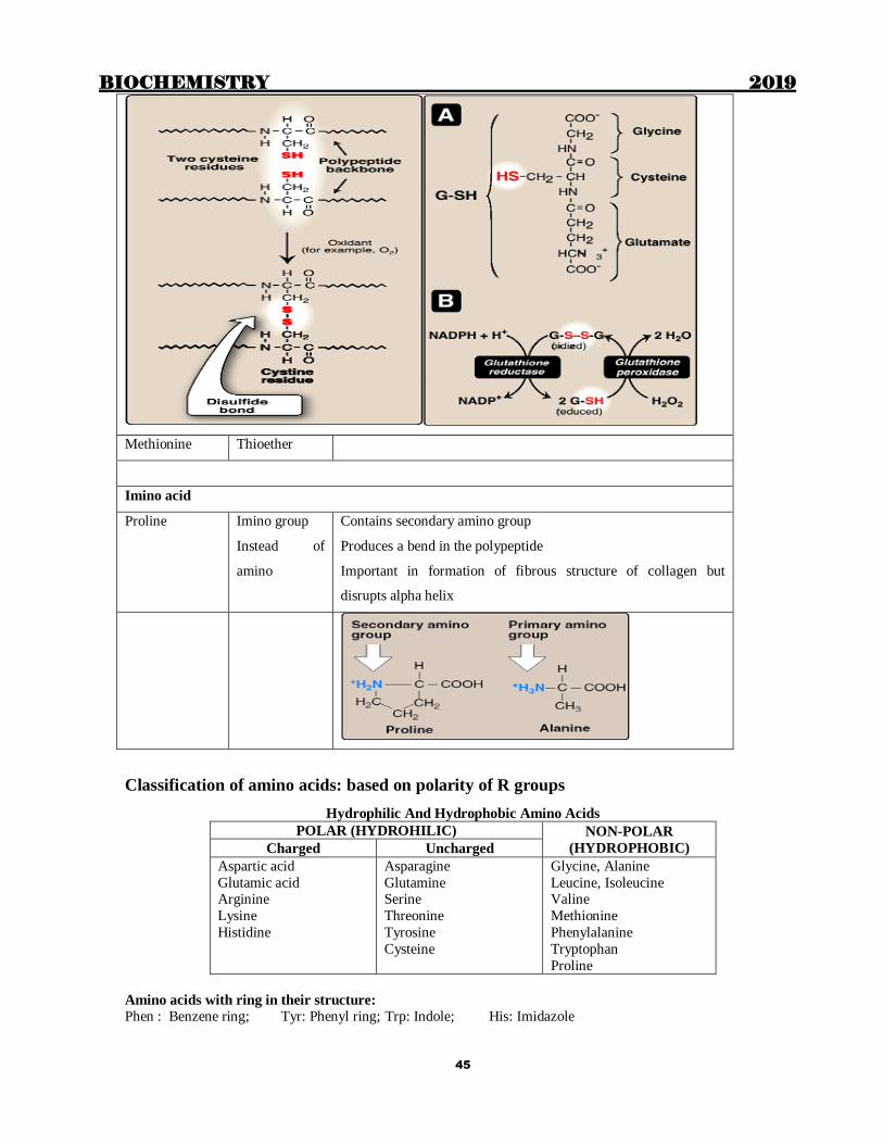
Sulphur containing amino acids
Cysteine Sulfhydryl 2 molecules of cysteine form cystine, which is:-
2nd most common post translational modification
Responsible for formation of various inter-chain and
intra-chain disulfide bonds or linkages or bridges
Responsible for glutathione acting as an antioxidant.
BIOCHEMISTRY 2019
45
Methionine Thioether
Imino acid
Proline Imino group
Instead of
amino
Contains secondary amino group
Produces a bend in the polypeptide
Important in formation of fibrous structure of collagen but
disrupts alpha helix
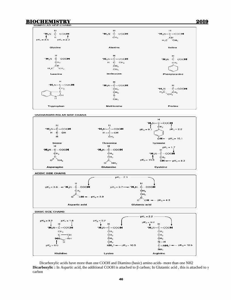
Classification of amino acids: based on polarity of R groups
Hydrophilic And Hydrophobic Amino Acids
POLAR (HYDROHILIC) NON-POLAR
(HYDROPHOBIC) Charged Uncharged
Aspartic acid
Glutamic acid Arginine
Lysine
Histidine
Asparagine
Glutamine Serine
Threonine
Tyrosine
Cysteine
Glycine, Alanine
Leucine, Isoleucine Valine
Methionine
Phenylalanine
Tryptophan
Proline
Amino acids with ring in their structure:
Phen : Benzene ring; Tyr: Phenyl ring; Trp: Indole; His: Imidazole
BIOCHEMISTRY 2019
46
Dicarboxylic acids have more than one COOH and Diamino (basic) amino acids- more than one NH2
Dicarboxylic : In Aspartic acid, the additional COOH is attached to β carbon; In Glutamic acid , this is attached to γ
carbon
BIOCHEMISTRY 2019
47
Basic amino acids
Arginine- Complicated structure/ Extra amino group attached to δ carbon
Lysine-Extra is ε amino group
Most basic amino acid- Arginine
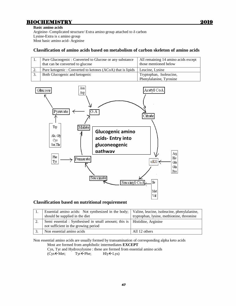
Classification of amino acids based on metabolism of carbon skeleton of amino acids
1. Pure Glucoogenic : Converted to Glucose or any substance
that can be converted to glucose
All remaining 14 amino acids except those mentioned below
2. Pure ketogenic : Converted to ketones (ACoA) that is lipids Leucine, Lysine
3. Both Glucogenic and ketogenic Tryptophan, Isoleucine,
Phenylalanine, Tyrosine
Classification based on nutritional requirement
1. Essential amino acids: Not synthesized in the body;
should be supplied in the diet
Valine, leucine, isoleucine, phenylalanine,
tryptophan, lysine, methionine, threonine
2. Semi essential : Synthesised in small amount; this is
not sufficient in the growing period Histidine, Arginine
3. Non essential amino acids All 12 others
Non essential amino acids are usually formed by transamination of corresponding alpha keto acids
Most are formed from amphibolic intermediates EXCEPT
Cys, Tyr and Hydroxylysine : these are formed from essential amino acids
(CysMet; TyrPhe; HlyLys)
Glucogenic amino acids- Entry into gluconeogenic pathway
BIOCHEMISTRY 2019
48
Abbreviations of amino acids
Name 3 letter Single letter
Glycine Gly G
Alanine Ala A
Valine Val V
Leucine Leu L Isoleucine Ile I
Serine Ser S
Threonine Thr T
Tyrosine Tyr Y
Cysteine Cys C
Methionine Met M
Aspartic acid Asp D
Asparagine Asn N
Glutamic acid Glu E
Glutamine Gln Q
Arginine Arg R Lysine Lys K
Histidine His H
Phenyl alanine Phe F
Tryptophan Trp W
Proline Pro P
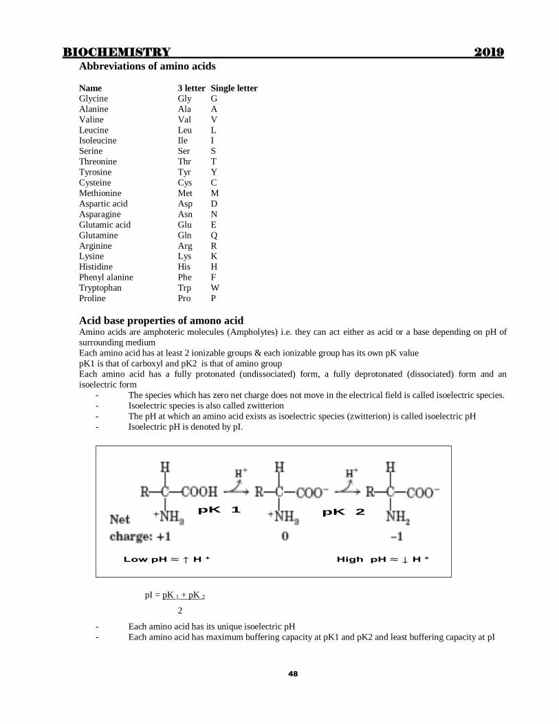
Acid base properties of amono acid Amino acids are amphoteric molecules (Ampholytes) i.e. they can act either as acid or a base depending on pH of
surrounding medium
Each amino acid has at least 2 ionizable groups & each ionizable group has its own pK value
pK1 is that of carboxyl and pK2 is that of amino group
Each amino acid has a fully protonated (undissociated) form, a fully deprotonated (dissociated) form and an
isoelectric form
- The species which has zero net charge does not move in the electrical field is called isoelectric species.
- Isoelectric species is also called zwitterion
- The pH at which an amino acid exists as isoelectric species (zwitterion) is called isoelectric pH
- Isoelectric pH is denoted by pI.
pI = pK 1 + pK 2
2
- Each amino acid has its unique isoelectric pH
- Each amino acid has maximum buffering capacity at pK1 and pK2 and least buffering capacity at pI
Low pH ≈ ↑ H + High pH ≈ ↓ H +
pK 1 pK 2
BIOCHEMISTRY 2019
49
PI of Histidine = 9.3+6 = 7.6
2
a. At physiological pH free histidine has zero charge.
b. When combined in proteins, its R- group at physiological pH is ½ dissociated so acts
as buffer.
Isoelectric point – important feature
pH at which the amino amino acid has NO NET charge
Applicable to proteins also
At isoelectric point,
Does not migrate in electric field Solubility is minimum
Buffering capacity minimum
Precipitability is maximum
Non Standard amino acids GABA (Gamma Amino Butyric Acid)
β Alanine Only naturally occuring β amino acid. Present in
Pantothenic acid-- CoA
β Alanyl dipeptides
Carnosine (skeletal muscle) – His+β alanine
Anserine (skeletal muscle) – N-methyl carnosine
Homocarnosine (brain)- GABA+Histidine
Formed from Pyrimidines, Carnosine or Anserine
Transamination produces malonate semialdehyde
Metabolized into-Acetic acid
Ornithine, Citrulline, Arginosuccinate
Hydroxyproline and hydroxylysine
21st and 22nd amino acids Selenocysteine (Sec)
21st amino acid. Formed in the body from Serine
Coded by UGA. Incorporated into protein with the help of SECIS element (Selenocysteine insertion sequence
element)
Present in – Thioredoxin reductase; Glutathione peroxidase; Deiodinase;
–Selenoprotein-P (an antioxidant glycoprotein present in mammalian blood)
Pyrrolysine (Pyl)
22nd amino acid. Coded by UAG (amber codon). Incorporated with the help of PYLIS element.
Detection of amino acids & proteins
Ninhydrin- can detect microgram quantities of amino acids
–Forms Ruhemann’s purple colour with all amino acids EXCEPT Proline which gives yellow colour
–Used to detect fingerprints
Fluorescamine- detect picogram quantities of amino acids- Most sensitive method
Colour reactions of proteins and amino acids Xanthoproteic test: Aromatic amino acids
Millon’s test : Tyrosine, Phenylalanine
Aldehyde test: Tryptophan (Hopkins-Cole test)
Acree-Rosenheim reaction : Tryptophan
Sakaguchi’s test : Arginine

BIOCHEMISTRY 2019
50
Pauly’s test : Histidine, Tyrosine
Sulphur test: Cysteine, Cystine
Biuret test: general test for proteins. At least 2 peptide bonds required
STRUCTURE OF PROTEIN - Different levels of protein structure
Primary structure
Includes a.a sequence and position of disulfide bonds. Maintained by the covalent bonds of peptide linkages. Peptide
bond- partial double bond character. Distance 1.32 Ao
Secondary structure
Relationship between residues which are 3-4 a. a apart H bonds
α helix, β pleated sheet, collagen helix
Alpha helix
Commonly right handed. Abundant in Hb and Mb
Absent in chymotrypsin
Pro and Hyp do not allow its formation
Collagen: Triple helix
Glycine induces bends in the alpha helix.
β pleated sheet
Chains fully extended.Stabilised by H bonds.
Tertiary structure -AA far apart. Bonds- Hydogen, Hydrophobic, Ionic, van der Waal’s
Quarternary – only when protein has 2 or more subunits
Stabilized by non-covalent interactions Domain- Compact globular functional unit of a protein
Amino acid residues which favor the secondary structure of proteins
Alpha helix Beta pleated sheet Turns
Ala, Cys, Leu, Met, Glu, Gln
His, Lys,
Arg
Ile, Val,
Phe, Tyr, Trp, Thr,
Arg
Gly, Ser, Pro
Asp, Asn
Arg
Protein sequencing- detecting the amino acid sequence : Sanger
Sanger’s reagent
Fluoro DinitroBenzene.Used for identification of N-terminal amino acid
End group analysis N-terminal a.a identified by Sanger’s reagent
C-terminal by Carboxypeptidase A and B
– Carboxypeptidase A will not act if C-terminal is Arg, Lys or Pro
– Carboxypeptidase B will act only if penultimate residue is Pro
Edman’s reagent
Phenyl isothiocyanate. Used for Sequential analysis of amino acids from N-terminal
Dansyl chloride
Combines with N-terminal amino acid. Used to assess the number of polypeptide chains
Cyanogen bromide
Hydrolyses peptide bonds following Mehionine
“Sequenator”: Automated instrument for protein sequencing
Ingram’s technique
Protein finger printing
Helps to identify qualitative abnormalities in proteins
Digestion by Trypsin followed by Chromatography and peptide mapping
SOME IMPORTANT PROTEINS:
BIOCHEMISTRY 2019
51
Chaperones
Heat shock proteins which help in proper folding of proteins
Different classes (hsp 70kDa, hsp 60 kDa)
Prevent misfolding
Unfold misfolded regions
Keeps protein unfolded to pass through membranes
Prevents inappropriate interactions with other proteins
Chaperonins are hsp60 chaperones
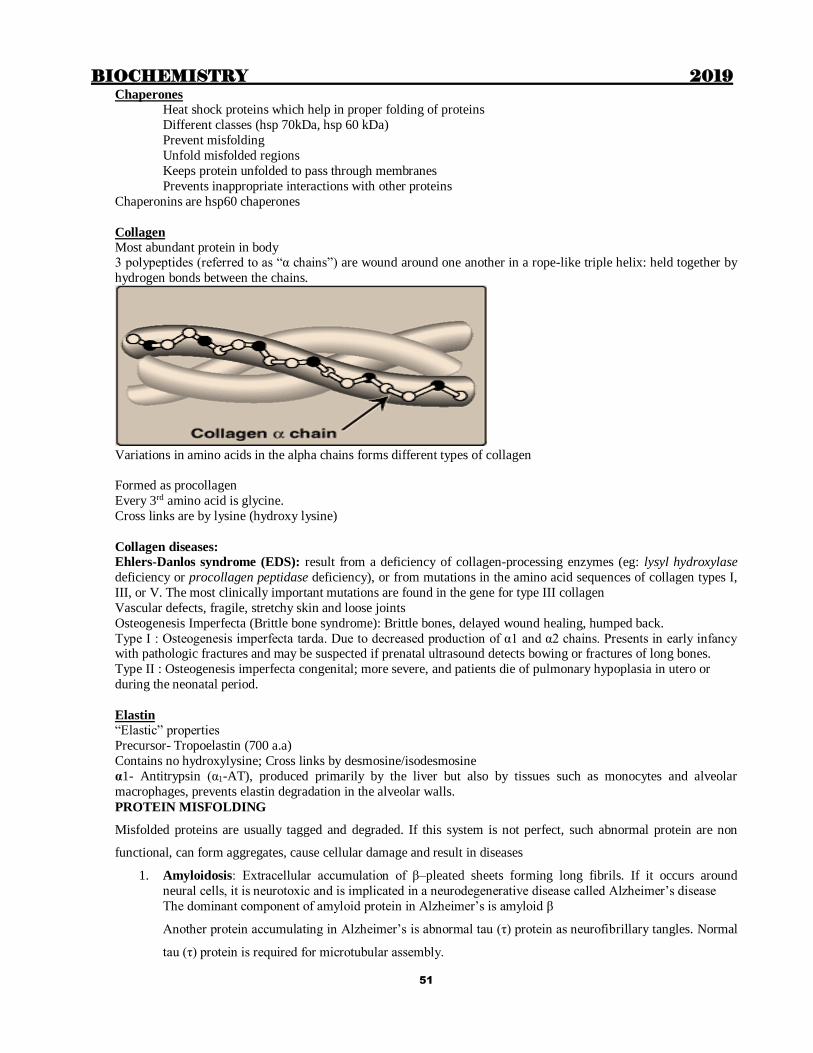
Collagen
Most abundant protein in body 3 polypeptides (referred to as “α chains”) are wound around one another in a rope-like triple helix: held together by
hydrogen bonds between the chains.
Variations in amino acids in the alpha chains forms different types of collagen
Formed as procollagen
Every 3rd amino acid is glycine.
Cross links are by lysine (hydroxy lysine)
Collagen diseases:
Ehlers-Danlos syndrome (EDS): result from a deficiency of collagen-processing enzymes (eg: lysyl hydroxylase
deficiency or procollagen peptidase deficiency), or from mutations in the amino acid sequences of collagen types I,
III, or V. The most clinically important mutations are found in the gene for type III collagen
Vascular defects, fragile, stretchy skin and loose joints
Osteogenesis Imperfecta (Brittle bone syndrome): Brittle bones, delayed wound healing, humped back.
Type I : Osteogenesis imperfecta tarda. Due to decreased production of α1 and α2 chains. Presents in early infancy with pathologic fractures and may be suspected if prenatal ultrasound detects bowing or fractures of long bones.
Type II : Osteogenesis imperfecta congenital; more severe, and patients die of pulmonary hypoplasia in utero or
during the neonatal period.
Elastin
“Elastic” properties
Precursor- Tropoelastin (700 a.a)
Contains no hydroxylysine; Cross links by desmosine/isodesmosine
α1- Antitrypsin (α1-AT), produced primarily by the liver but also by tissues such as monocytes and alveolar
macrophages, prevents elastin degradation in the alveolar walls.
PROTEIN MISFOLDING
Misfolded proteins are usually tagged and degraded. If this system is not perfect, such abnormal protein are non
functional, can form aggregates, cause cellular damage and result in diseases
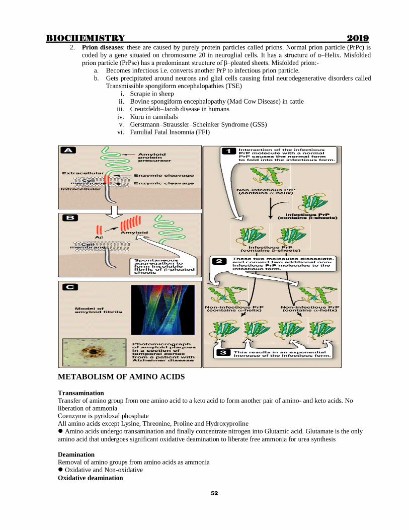
1. Amyloidosis: Extracellular accumulation of β–pleated sheets forming long fibrils. If it occurs around
neural cells, it is neurotoxic and is implicated in a neurodegenerative disease called Alzheimer’s disease
The dominant component of amyloid protein in Alzheimer’s is amyloid β
Another protein accumulating in Alzheimer’s is abnormal tau (τ) protein as neurofibrillary tangles. Normal
tau (τ) protein is required for microtubular assembly.
BIOCHEMISTRY 2019
52
2. Prion diseases: these are caused by purely protein particles called prions. Normal prion particle (PrPc) is
coded by a gene situated on chromosome 20 in neuroglial cells. It has a structure of α–Helix. Misfolded
prion particle (PrPsc) has a predominant structure of β–pleated sheets. Misfolded prion:-
a. Becomes infectious i.e. converts another PrP to infectious prion particle.
b. Gets precipitated around neurons and glial cells causing fatal neurodegenerative disorders called
Transmissible spongiform encephalopathies (TSE)
i. Scrapie in sheep
ii. Bovine spongiform encephalopathy (Mad Cow Disease) in cattle
iii. Creutzfeldt–Jacob disease in humans
iv. Kuru in cannibals
v. Gerstmann–Straussler–Scheinker Syndrome (GSS) vi. Familial Fatal Insomnia (FFI)
METABOLISM OF AMINO ACIDS
Transamination
Transfer of amino group from one amino acid to a keto acid to form another pair of amino- and keto acids. No
liberation of ammonia Coenzyme is pyridoxal phosphate
All amino acids except Lysine, Threonine, Proline and Hydroxyproline
Amino acids undergo transamination and finally concentrate nitrogen into Glutamic acid. Glutamate is the only
amino acid that undergoes significant oxidative deamination to liberate free ammonia for urea synthesis
Deamination
Removal of amino groups from amino acids as ammonia
Oxidative and Non-oxidative
Oxidative deamination
BIOCHEMISTRY 2019
53
All ammonia concentrated as glutamate. Oxidatively deaminated in liver and kidney. Enzyme is Glutamate
dehydrogenase. Ammonia is liberated
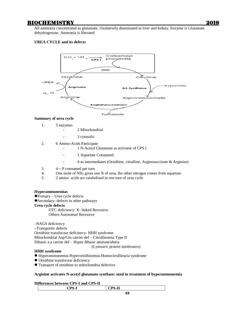
UREA CYCLE and its defects
Summary of urea cycle
1. 5 enzymes
- 2 Mitochondrial
- 3 cytosolic
2. 6 Amino Acids Participate
- 1 N-Acetyl Glutamate as activator of CPS I
- 1 Aspartate Consumed
- 4 as intermediates (Ornithine, citrulline, Arginosuccinate & Arginine)
3. 4 ~ P consumed per turn
4. One mole of NH3 gives one N of urea, the other nitrogen comes from aspartate
5. 2 amino acids are catabolised in one turn of urea cycle
Hyperammonemias
Primary – Urea cycle defects
Secondary- defects in other pathways
Urea cycle defects
OTC deficiency: X- linked Recessive
Others Autosomal Recessive
–NAGS deficiency
–Transporter defects
Ornithine translocase deficiency- HHH syndrome
Mitochondrial Asp/Glu carrier def – Citrullinemia Type II
Dibasic a.a carrier def – Hyper dibasic aminoaciduria
(Lysinuric protein intolerance)
HHH syndrome
Hyperammonemia-Hyperornithinemia-Homocitrullinuria syndrome
Ornithine translocase deficiency
Transport of ornithine to mitochondria defective
Arginine activates N-acetyl glutamate synthase: used in treatment of hyperammonemia
Differences between CPS-I and CPS-II
CPS-I CPS-II

BIOCHEMISTRY 2019
54
• Urea synthesis
• Mitochondria
• N donor is ammonia
• Dependent on N-acetyl glutamate
• Pyrimidine synthesis
• Cytosol
• N donor is Gln
• Not dependent
Biochemical Basis of Ammonia intoxication
Decreased energy charge due to: Decreased NADH/ NAD or NADPH/ NADP ratio
Decreased TCA cycle intermediate α – keto glutarate.
Decreased ATP
Synthesis of inhibitory neurotransmitter GABA Local brain alkalosis
INDIVIDUAL AMINO ACID METABOLISM and their metabolic defects
A) AROMATIC AND HETEROCYCLIC AMINO ACIDS

BIOCHEMISTRY 2019
55
Hawkinsinuria
Hawkinsin is an unusual metabolite of Tyrosine
Autosomal Dominant form of tyrosinuria
Transient tyrosinemia
Excretion of PHPPA,PHPAA, Hawkinsin
Hypertyrosinemias
Type I (hepatorenal)
Fumaryl acetoacetate hydrolase def
Hypoglycemia
Liver failure
Tyrosine
Phenyl alanine
DOPA p-OH Phenyl
pyruvate
Homogentisic acid Catecholamines
Phe Hydroxylase
Tyrosine Hydroxylase

BIOCHEMISTRY 2019
56
Renal tubular dysfunction, rickets
Cabbage like odor
Early death
Type II (Oculocutaneous)
Tyrosine transaminase deficiency
– M.R, Palmar keratosis
– Corneal lesions, Photophobia
Type III
Deficiency of parahydroxy phenyl pyruvate hydroxylase
Normal liver function
Intermittent ataxia, Seizures Drowsiness, mild M.R
All types have Autosomal Recessive inheritance
Catecholamines
Dopamine, Epinephrine, Norepinephrine
Formed from Tyrosine
DOPA is an intermediate in formation of Catecholamines and Melanin
Catecholamine synthesis: Tyrosine is converted to DOPA by tyrosine hydroxylase
Melanin synthesis: TyrDOPA by Tyrosinase
Phenylketonuria (PKU)
Enzyme: Phe hydroxylase
Metabolic abnormalities
Mental retardation, Hyopigmentation,Mousy odour
Tests: Guthrie test, Ferric chloride test, Phe load test, HPLC for Phe
Diet: Restriction to be started within 3 weeks
Continued throughout life
Alkaptonuria
Enzyme : Homogentisate oxidase
C/F : Blackening of urine, Ochronosis Tests : Ferric chloride test (Transient blue-green), Benedict’s test
Diet : Restrict Phe and Tyr
Drug : Nitisone [2-(2-nitro-4-trifluoromethylbenzoyl)-1,3-cyclohexanedione]
Garrod’s tetrad: Cystinuria, Alkaptonuria,Pentosuria, Albinism

BIOCHEMISTRY 2019
57
Disorder of Histidine metabolism - Histidinemia
• AR due to deficiency of Histidase
• MR and delayed speech development
• Increased excretion of imidazole pyruvic acid in urine
• Sweat urocanic acid absent
Disorder of Tryptophan - Blue diaper syndrome
Trp malabsorption
Trp acted upon by bacteria: Trp Indican Indicanuria
Indican in urine is oxidised to Indigo blue which stains diaper
B) BRANCHED CHAIN AMINO ACID DEFECTS
Maple Syrup Urine Disease (MSUD)
Enzyme: Branched chain α keto acid dehydrogenase , Decarboxylation is defective
Mental Retardation, Convulsions, Acidosis, Coma , Death within 1 year
Tests : DNPH test, Rothera’s test
Diet: Restrict branched chain amino acids
C) METABOLISM OF SULPHUR CONTAINING AMINO ACIDS:
METHIONINE
Homocystinuria
Note: Homocysteinuria is Defect of methionine metabolism not of cysteine
AR
C/F: M.R, Skeletal abnormalities, Ectopia lentis Cyanide nitroprusside test – Magenta colour
Diet: Low Met
Normal HS in blood 5-15 μ mol/L
Increase of 5 μ mol/L in serum increases the risk of IHD equivalent to increase of 20 mg/dl of cholesterol
Thiolates LDL Aggregation of LDL
Also activates Hageman’s factor intravascular thrombosis
CYSTEINE

BIOCHEMISTRY 2019
58
Cystinuria
Cystinuria is a condition characterized by the buildup of cystine crystals or stones in the kidneys and bladder.
Cystine is an amino acid, one of the building blocks of proteins. As the kidneys filter blood to create urine, cystine is
normally absorbed back into the bloodstream. People with cystinuria cannot properly reabsorb cystine into their
bloodstream and the amino acid accumulates in their urine. As urine becomes more concentrated in the kidneys, the
excess cystine forms crystals. As these crystals become larger, they form stones that may lodge in the kidneys or in
the bladder. Sometimes cystine crystals combine with calcium molecules in the kidneys to form larger stones. These
crystals and stones can create blockages in the urinary tract and reduce the ability of the kidneys to eliminate waste
through urine. The stones also provide sites where bacteria may cause infections.
Cystinuria affects approximately 1 in 10,000 people. Autosomal recessive
Genes related to cystinuria
Mutations in the SLC3A1 and SLC7A9 genes cause cystinuria. The SLC3A1 gene provides instructions for
producing one part (subunit) of a protein made primarily in the kidneys. This subunit joins with another protein
subunit, produced from the SLC7A9 gene, to form a transporter protein complex. (Location of SLC3A1 is at 2p16.3
while location of SLC7A9 gene is : 19q13.1)
The SLC3A1 and SLC7A9 genes (SLC3A1 stands for “solute carrier family 3, member 1.”) provide instructions for
making the two parts (subunits) of a transporter protein complex that is made primarily in the kidneys. Normally this
protein complex controls the reabsorption of certain amino acids, including cystine, into the blood from the filtered
fluid that will become urine. Mutations in either the SLC3A1 gene or SLC7A9 gene disrupt the ability of the transporter protein complex to reabsorb amino acids, which causes them to become concentrated in the urine. As the
levels of cystine in the urine increase, the crystals typical of cystinuria form. The other amino acids that are
reabsorbed by the transporter protein complex do not create crystals when they accumulate in the urine.
Cystine in urine 30 times normal
+
Excretion of Cystine, Ornithine, Lysine & Arginine (COLA)
Cystine stones
Note: other aminoacidurias
i) Dibasic aminoaciduria (Lysinuric protein intolerance): Mutations in the SLC7A7 gene
(Location: 14q11.2) leads to defective transport of Ornithine, Lysine & Arginine but not
of cystine.
ii) Dicarboxylic aminoaciduria: Glutamate and Aspartate reabsorption defect
iii) Hartnup disease: transport defect of neutral and aromatic amino (mainly Tryptophan) acids
from intestine and renal tubules
iv) Iminoglycinuria: Glycine, proline and hydroxyproline reabsorption defect
2. Cystinosis (Cys storage Disease)
Cystinosis is a condition characterized by accumulation of the amino acid cystine (a building block of proteins)
within cells. Excess cystine damages cells and often forms crystals that can build up and cause problems in many organs and tissues. The kidneys and eyes are especially vulnerable to damage; the muscles, thyroid, pancreas, and
testes may also be affected.
There are three distinct types of cystinosis. In order of decreasing severity, they are nephropathic cystinosis,
intermediate cystinosis, and non-nephropathic or ocular cystinosis.
Nephropathic cystinosis begins in infancy, causing poor growth and a particular type of kidney damage (renal
Fanconi syndrome) in which certain molecules that should be reabsorbed into the bloodstream are instead eliminated
in the urine. The kidney problems lead to the loss of important minerals, salts, fluids, and many other nutrients. The
loss of nutrients impairs growth and may result in soft, bowed bones (hypophosphatemic rickets), especially in the
legs. The nutrient imbalances in the body lead to increased urination, thirst, dehydration, and abnormally acidic
blood (acidosis). By about the age of 2, cystine crystals may be present in the clear covering of the eye (cornea). The
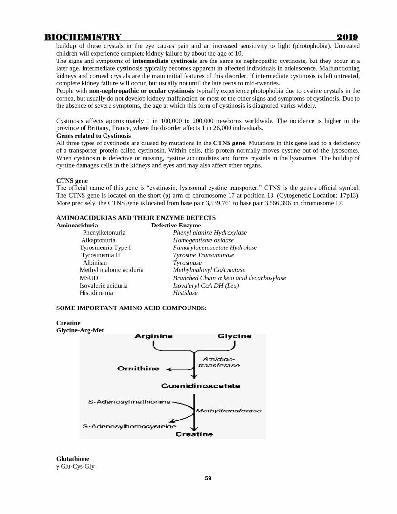
BIOCHEMISTRY 2019
59
buildup of these crystals in the eye causes pain and an increased sensitivity to light (photophobia). Untreated
children will experience complete kidney failure by about the age of 10.
The signs and symptoms of intermediate cystinosis are the same as nephropathic cystinosis, but they occur at a
later age. Intermediate cystinosis typically becomes apparent in affected individuals in adolescence. Malfunctioning
kidneys and corneal crystals are the main initial features of this disorder. If intermediate cystinosis is left untreated,
complete kidney failure will occur, but usually not until the late teens to mid-twenties.
People with non-nephropathic or ocular cystinosis typically experience photophobia due to cystine crystals in the
cornea, but usually do not develop kidney malfunction or most of the other signs and symptoms of cystinosis. Due to
the absence of severe symptoms, the age at which this form of cystinosis is diagnosed varies widely.
Cystinosis affects approximately 1 in 100,000 to 200,000 newborns worldwide. The incidence is higher in the province of Brittany, France, where the disorder affects 1 in 26,000 individuals.
Genes related to Cystinosis
All three types of cystinosis are caused by mutations in the CTNS gene. Mutations in this gene lead to a deficiency
of a transporter protein called cystinosin. Within cells, this protein normally moves cystine out of the lysosomes.
When cystinosin is defective or missing, cystine accumulates and forms crystals in the lysosomes. The buildup of
cystine damages cells in the kidneys and eyes and may also affect other organs.
CTNS gene The official name of this gene is “cystinosin, lysosomal cystine transporter.” CTNS is the gene's official symbol.
The CTNS gene is located on the short (p) arm of chromosome 17 at position 13. (Cytogenetic Location: 17p13).
More precisely, the CTNS gene is located from base pair 3,539,761 to base pair 3,566,396 on chromosome 17.
AMINOACIDURIAS AND THEIR ENZYME DEFECTS
Aminoaciduria Defective Enzyme
Phenylketonuria Phenyl alanine Hydroxylase
Alkaptonuria Homogentisate oxidase
Tyrosinemia Type I Fumarylacetoacetate Hydrolase
Tyrosinemia II Tyrosine Transaminase
Albinism Tyrosinase
Methyl malonic aciduria Methylmalonyl CoA mutase
MSUD Branched Chain keto acid decarboxylase
Isovaleric aciduria Isovaleryl CoA DH (Leu)
Histidinemia Histidase
SOME IMPORTANT AMINO ACID COMPOUNDS:
Creatine
Glycine-Arg-Met
Glutathione
γ Glu-Cys-Gly
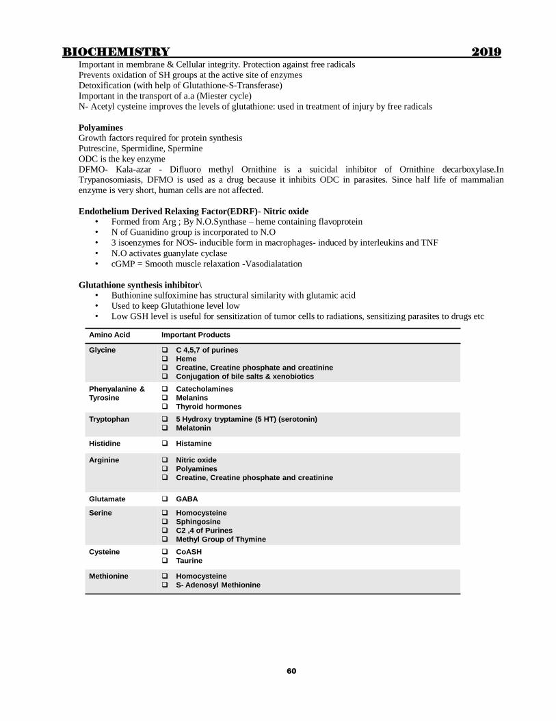
BIOCHEMISTRY 2019
60
Important in membrane & Cellular integrity. Protection against free radicals
Prevents oxidation of SH groups at the active site of enzymes
Detoxification (with help of Glutathione-S-Transferase)
Important in the transport of a.a (Miester cycle)
N- Acetyl cysteine improves the levels of glutathione: used in treatment of injury by free radicals
Polyamines
Growth factors required for protein synthesis
Putrescine, Spermidine, Spermine
ODC is the key enzyme
DFMO- Kala-azar - Difluoro methyl Ornithine is a suicidal inhibitor of Ornithine decarboxylase.In Trypanosomiasis, DFMO is used as a drug because it inhibits ODC in parasites. Since half life of mammalian
enzyme is very short, human cells are not affected.
Endothelium Derived Relaxing Factor(EDRF)- Nitric oxide
• Formed from Arg ; By N.O.Synthase – heme containing flavoprotein
• N of Guanidino group is incorporated to N.O
• 3 isoenzymes for NOS- inducible form in macrophages- induced by interleukins and TNF
• N.O activates guanylate cyclase
• cGMP = Smooth muscle relaxation -Vasodialatation
Glutathione synthesis inhibitor\
• Buthionine sulfoximine has structural similarity with glutamic acid
• Used to keep Glutathione level low
• Low GSH level is useful for sensitization of tumor cells to radiations, sensitizing parasites to drugs etc
Amino Acid Important Products
Glycine C 4,5,7 of purines
Heme
Creatine, Creatine phosphate and creatinine
Conjugation of bile salts & xenobiotics
Phenyalanine &
Tyrosine
Catecholamines
Melanins
Thyroid hormones
Tryptophan 5 Hydroxy tryptamine (5 HT) (serotonin)
Melatonin
Histidine Histamine
Arginine Nitric oxide
Polyamines
Creatine, Creatine phosphate and creatinine
Glutamate GABA
Serine Homocysteine
Sphingosine
C2 ,4 of Purines
Methyl Group of Thymine
Cysteine CoASH
Taurine
Methionine Homocysteine
S- Adenosyl Methionine

BIOCHEMISTRY 2019
61
Chapter 6
ENZYMES
Important points to remember
Enzyme classes that do not require coenzymes are Class 3 & 4 i.e. Hydrolases & Lyases
Coenzymes are Non–protein, low molecular wt., dialyzable and thermostable substances
Coenzymes act as 2nd substrates or group transfer agents Coenzymes can be classified according to the group whose transfer they facilitate
Isoenzymes are physically distinct forms of the same enzyme
Isoenzymes differ in structure, properties, electrophoretic mobility, and Tissue of Expression
Isoenzyme has one common property in that they catalyze the same reaction
CLASSIFICATION OF ENZYMES: All enzymes are classified into following 6 classes as below
1. Oxidoreductase : Oxidation of one substrate with simultaneous reduction of another substrate/coenzyme.
Includes Oxidase, Reductase, Dehydrogenase, Catalase, Peroxidase, Oxygenases, ( hydroxylases also)
2. Transferase: Transfer a group other than Hydrogen.
Includes Transaminases, Phospho transferases (Polymerases and kinase),Sulphotransferases, Methyl transferases,
Acyl transferases, Glycosyl transferase,
3. Hydrolase: Splits the substrate into two by adding water. Includes Esterases, Peptidases, Glycosidases etc
4. Lyase: splits the substrate into two without adding water: Eg Aldolase
5. Isomerase: Catalyzes intramolecular rearrangement.
Includes Epimerase, Racemase, Mutase, Cis-trans isomerase
6. Ligase: Bind together 2 substrates.
Includes Synthetases and Carboxylases
COENZYMES
Organic substances required by the enzyme for its proper action. (Cofactor: organic/inorganic)
Classified as
Coenzymes taking part in the transfer of Hydrogen NAD+, NADP+, FMN, FAD, Lipoic acid, Coenzyme Q
Coenzymes taking part in the transfer of other groups
Coenzyme A – Transfer of Acyl groups
TPP – Transfer of Hydroxyethyl groups
PLP – Transfer of Amino group
Folate coenzyme – One carbon units
Biotin - Carbon dioxide
MECHANISM OF ENZYME ACTION
i) Lowering of activation energy
Activation energy is the energy required to bring a substrate from its ground energy state to the transition state.
Enzymes reduce this energy ii) Michaelis Menten theory : States that Enzyme combines with substrate to form as complex which
dissociates into Enzyme and products.
iii) Fischer’s template theory : “Active site” of the enzyme will have a structure complementary to that of
substrate. Explains Enzyme specificity and competitive inhibition
iv) Koshland’s theory: Binding of substrate to the active site induces a conformatonal change at the active site
which facilitates further binding of substrate: explains allosteric regulation
RATE LIMITING ENZYME (KEY ENZYME)
These enzymes are easily regulated by physiological mechanisms
Rate limiting enzyme (Key enzyme) catalyses the slowest reaction in a pathway, (Km value high) so that regulatory
mechanisms can exert their effect. Key enzymes are allosterically regulated and Key enzymes are inducible enzymes

BIOCHEMISTRY 2019
62
Km VALUE (MICHAELIS CONSTANT)
Substrate concentration at which half the maximum velocity is achieved
Significance
Signature of the enzyme
Inversely related to affinity
To find out the natural substrate: Natural substrate has the lowest Km value
To identify the key enzyme: Highest Km
To find out the type of enzyme inhibition
SERINE PROTEASES:
Contain Ser at the active site. Ser, His, Asp- FORMS THE CATALYTIC TRIAD
Mostly secreted as zymogens- undergoes “limited proteolysis
Serpin: Serine protease inhibitor
ENZYME INHIBITION
Various types as follows:
i) Competitive inhibition
Inhibitor is a substrate analogue; binds to the active site
Vmax is not affected; Km is increased
Reversible on adding excess substrate
Examples
Sulphonamides as antibacterial drugs Methotrexate as anticancer drug
Ethyl alcohol in treatment of methyl alcohol poisoning
ii) Non-competitive inhibition
Inhibitor has no structural similarity with substrate
Inhibitor usually does not bind to the substrate binding site; acts by various other mechanisms
iii) Uncompetitive inhibition
Binding of substrate caused binding of inhibitor to the enzyme. Both Km and Vmax decreased
iv) Suicide inhibition
Enzyme catalyses a reaction involving the inhibitor. Product formed is a powerful inhibitor of the enzyme.
v) Allosteric inhibition
Allosterically regulated enzymes have substrate binding site and an allosteric site
Inhibitor binds to allosteric site; It induces a conformational change at the active site; So the substrate binding to
active site is altered
ENZYME REGULATION- types
BIOCHEMISTRY 2019
63
i. Induction and Repression
ii. Covalent modification
iii. Allosteric regulation
FUNCTIONAL AND NON FUNCTIONAL PLASMA ENZYMES
Functional enzymes: Those which have a specific function in plasma; Eg: Enzymes of coagulation pathway,
Lipoprotein lipase, choleinestrase
Non-functional enzymes: Their function is not in plasma: they are present in plasma due to leakage from the organ
where they are located (as a part of normal wear and tear). Their level increases in blood when there is damage to
the tissue where it is located.Eg: CK is elevated in MI and ALT in hepatitis
DIAGNOSTIC APPLICATION OF ENZYMES AND ISOENZYMES IN VARIOUS DISEASES
Enzymes in Liver diseases:
ALT: Normal value 13-40 U/L; Elevated in liver cell damage; Hepatitis, Cirrhosis, Cancer
AST: Normal value 8-20 U/L; Elevated in Liver cell damage; not specific
Alkaline phosphatase: Has 6 isoenzymes; Normal value 3-13 KAU/dl; Very high levels are seen in obstructive
liver diseases and bone diseases. High levels in children before closure of epiphyses
5’Nucleotidase: (Nucleotide triphosphatase); Elevated in obstructive liver diseases
Gamma Glutamyl Transferase: Very sensitive index of alcoholic liver disease
Enzymes in Heart disease:
Creatine kinase: (creatine phosphokinase); First enzyme to elevate after myocardial infarction; Rises within 2-3
hours. Remain elevated for 1-2 days only; For early diagnosis of MI
3 isoenzymes: CK-MB is to be measured for heart disease
Isoenzymes of CPK
CPK is a dimer – B and M subunits
CPK1 - BB – Brain : CPK2 - MB – Heart : CPK3 - MM – Muscles:
AST: Elevated in MI after 2 days; Remain elevated for 6-7 days
2 isoenzymes: one from heart; other from liver (Increased in liver diseases also)
Lactate dehydrogenase: Elevated after 2-3 days following MI; Remain elevated for 10-12 days
For late diagnosis of MI; 5 isoenzymes: LDH 1 is elevated in MI
(Normally LDH2 > LDH1- This is reversed in MI- “Flipped pattern”
Isoenzymes of LDH
LDH is a tetramer, 2 types of subunits - H and M
LDH1- HHHH – Heart; LDH2 – HHHM – RBC
LDH3 – HHMM – Brain
LDH4 – HMMM – Liver
LDH5 – MMMM – Skeletal muscles
Other proteins in heart disease
Troponin: High sensitivity & Specificity
Myoglobin- Low specificity
Ischemia-modified albumin- Low specificity
Pro-brain natriuretic peptide: Elevated in CCF
Cardiac troponins
Not enzymes: Very sensitive index for MI: Elevated early in MI, remain elevated for 10-14 days
Considered “any time marker” of MI
ISOENZYMES OF ALP
α 1- Biliary epithelium - Elevated in obstructive jaundice
α 2 HL - Hepatocytes - Elevated in heaptitis
α 2 HS - Placenta - Elevated in pregnancy, trophoblastic tumors
Pre β – Bone - Elevated in Pagets disease, Rickets. Elevated values in children
γ – Intestinal cells - Elevated in ulcerative colitis
LAP- Leukocyte ALP - Elevated in myeloid leukemia

BIOCHEMISTRY 2019
64
Chapter 7
VITAMINS
Fat soluble: A, D, E, K
Water soluble: B complex, C
Vitamin A: Active forms Retinal, retinol, retinoic acid
Provitamin – β carotene – Plants: Contains 2 β ionone rings; Cleaved by a dioxygenase: Produce 2 retinal
- β carotene has antioxidant properties
Absorbed with fat: Stored in liver as retinyl palmitate (ester of retinol and palmitic acid)
Biological role: Retinal has Visual property while retinol & retinoic acid has steriod hormone activity
Hypervitaminosis : Damages lysosomal membrane
Vitamin E
“Anti sterility” vitamin
Most potent biological antioxidant: Protects biomembranes, Prevents hemolysis, Retards ageing: Decrease
oxidation of LDL reduce the risk of atherosclerosis. Synergistic action with Se
Vitamin D
D2-Ergocalciferol- Fomed from Ergosterol in plants;
D3- Cholecalciferol – Formed from 7-dehydrocholesterol in animals
Active form of vit D – calcitriol (1, 25 DHCC); Calcitriol binds to cytoplasmic receptor
25 hydroxylation in liver- Microsomal mono oxygenase:
1 hydroxylation in kidney: Mitochondrial enz-
Other forms of Vitamin D are
D1- Molecular compound of ergocalciferol with lumisterol (1:1); D4 – Dihydrotachysterol- From D3
D5 – Sitocalciferol- from 7-dehydrocholesterol
Vitamin K
Naphthoquinone derivative
K1- Phylloquinone; K2 – Menaquinone; K3 – Menadione- Synthetic, water soluble form
Required for gamma carboxylation of glutamic acid residues (Gla) of blood clotting proteins Factors II, VII, IX
and X as well as Protein C, Protein S and Protein Z ( that is they also require vit K)
Water soluble vitamins: B complex, vitamin C
B complex vitamins act as coenzymes for various enzymes
Vitamin Active form Reactions requiring the vitamin
Thiamine TPP PDH, α KG DH, Transketolase,
Branched chain α keto acid decarboxylase
Riboflavin FMN, FAD FMN: Aminoacid oxidase, NADH dehydrogenase
FAD: PDH, α KG DH, Acyl CoA DH, Succinate
dehydrogenase, Xanthine oxidase
Niacin NAD, NADP Dehydrogenases and reductases other than riboflavin linked enzymes
Pantothenic acid ACP, CoA Acyl transferase reactions
Pyridoxine PLP Transaminases, Decarboxylation of amino acids,
Cystathionine synthase, Cystathionase, ALA synthase,
Kynureninase, Glycogen phosphorylase
Biotin Carboxylation
Folic acid THF One carbon metabolism
Cobalamin Methyl B12
Doexy adenosyl B12
HS-Met methyl transferase
Methyl malonyl CoA mutase
B COMPLEX VITAMINS WITH RING STRUCTURE
Thiamine: Pyrimidine +Thiazole

BIOCHEMISTRY 2019
65
Riboflavin: Isoalloxazine
Niacin: Pyridine
Pyridoxine: Pyridine
Biotin: Imidazole + Thiophene
Folic acid : Pteridine + PABA
Cobalamin: Corrin [Tetrapyrrole ring with Cobalt at its centre]
INDIVIDUAL VITAMINS, IMP ENZYMES REQUIRING VITAMIN & DEFICIENCY DISEASES
BIOTIN: Synthesised by intestinal bacteria; Anti egg white injury factor; Coenzyme for carboxylases
NADPH generating enzymes
G6PD
6 phosphogluconate DH
Cytoplasmic isocitrate DH
Malic enzyme (malate pyruvate)
NADPH utilising enzymes
Enzymes of reductive biosynthetic pathways
Niacin deficiency
Requirement: 20 mg/day: 2 / 3 is produced from Trp : 60 mg Trp 1 mg Niacin
Trp Niacin
Kynureninase: Require PLP as coenzyme
Pellagra: 4 D: Diarrhoea, Dementia, Dermatitis leading to Death
Maize: Niacin present as bound unavailable form known as niacytin
Sorghum: High Leucine content inhibits QPRTase PLP def: Kynureninase inhibited
Hartnup disease : Trp absorption defective
FMN and FAD – coenzyme forms of Riboflavin (B2)
FMN: Aminoacid oxidase, NADH dehydrogenase
FAD: Acyl CoA dehydrogenase, Succinate DH, X.O, PDH, α KG DH
B6 (Pyridoxine)
Enzymes which require PLP: Transaminase, Decarboxylation of amino acids, Cystathionine synthase,
Cystathionase, ALA synthase, Kynureninase (Trp Niacin)
Sulphur containing B complex vitamins: Thiamine, Biotin
Pantothenic acid does not contain sulphur, but its active forms CoA and ACP contain
Pantothenic acid (B5): Present in CoA, Acyl Carrier Protein; Contains β alanine ; Deficiency: Burning foot
syndrome
VITAMIN C
Only L-ascorbic acid and dehydroascorbic acid have vitamin activity; D-ascorbic acid –not active
Most animals-synthesise from glucose
Humans cannot: Absence of gulonolactone oxidase
Functions: Hydroxylation of Pro and Lys; Trp hydroxylase ; Dopamine β oxidase, Folate reductase Antioxidant reduce the risk of cancer
Infantile form – Barlow’s disease
Chapter 8
MINERALS
BIOCHEMISTRY 2019
66
Macrominerals (Major elements): Daily requirement >100 mg
Ca, Mg, P, Na, K, Cl and S
Microminerals (Trace elements): Daily requirement <100 mg
Fe, I, Cu, Co, Mn, Mo, Se, Zn and F
Ultratrace elements: Requirement < 1 mg/day
Metalloenzymes
Iron: Cytochrome oxidase, catalase, peroxidase, xanthine oxidase
Copper: Tyrosinase, Cytochrome oxidase, SOD, lysyl oxidase
Zinc: Carbonic anhydrase, Carboxy peptidase, Alcohol dehydrogenase, ALP, LDH, Glu DH, RNA
polymerase Magnesium: Hexokinase, Enolase, PFK
Manganese: Hexokinase, Enolase
Molybdenum: Xanthine oxidase
Calcium: Lipase, Lecithinase
Calcium: Normal level: 9-11 mg/dl; 50 % ionised; 40% protein bound; 10 % complexed with anions
Alkalosis favours protein binding
Calpains
Calcium dependent Cysteine proteases that modulate cellular functions
Role in DM, Cataract, Alzheimer’s disease, Muscular dystrophies
Inhibited by calpastatins
Calcium level- Reciprocal relation with phosphorus Ionic product-constant
S.Calcium x S. Phosphorus = 40 : Children, it is 50 (Ca is more)
Zinc: 60 % in bones, 30 % in skeletal muscles
Highest concentration in (a) Hippocampus (b) Prostatic fluid
Zn and Cu competitively inhibits each other’s absorption
Antioxidant: Because e.c SOD is Zn dependent
Stabilises insulin stored in pancreas
Copper:
Wilson’s disease
Copper binding ATPase deficient in cells; Excretion of copper from cells defective
Copper accumulates in liver and brain cells
Low serum Cu and Ceruloplasmin; High urinary excretion of Cu
As Zn prevents Cu absorption, it can be used to reduce Cu load
Menke’s kinky hair syndrome
Deficiency of intracellular Cu binding ATPase
Defective cross linking of connective tissue
X-linked recessive
Dissecting aneurysms and cardiac rupture cause death

BIOCHEMISTRY 2019
67
Mineral Deficency Toxicity
Boron Developmental defects, male sterility,
testicular atrophy
Calcium Osteoporosis, tetany Renal insufficiency (milk-alkalai
syndrome), nephrolithiasis, impaired
iron absorption
Copper Anemia, growth retardation, defective
keratinization and pigmentation of hair,
hypothermia, degenerative changes in aortic
elastin, osteopenia, mental deterioration
Nausea, vomiting, diarrhea, hepatic
failure, tremor, mental deterioration,
hemolytic anemia, renal dysfunction
Chromium Impaired glucose tolerance Occupational: renal failure, dermatitis,
pulmonary cancer
Fluoride Dental caries Dental and skeletal fluorosis,
osteosclerosis
Iodine Hypothyroidism Acne-like eruptions
Iron Anemia, tiredness, koilonychias etc Gastrointestinal effects (nausea,
vomiting, diarrhea, constipation), iron
overload with organ damage
Manganese Impaired growth and skeletal development, reproduction, lipid and carbohydrate metabolism;
upper body rash
Neurotoxicity, Encephalitis-like syndrome, Parkinson-like syndrome,
psychosis, pneumoconiosis
Molybdenum Neurologic abnormalities Reproductive & fetal abnormalities
Selenium Cardiomyopathy, heart failure, striated muscle
degeneration
Alopecia, peripheral neuropathy, garlic
odor to breath, dermatitis
Lung and nasal carcinomas, liver
necrosis, pulmonary inflammation
Phoshorus Rickets (osteomalacia), proximal muscle
weakness, rhabdomyolysis, paresthesia, ataxia,
seizure, confusion, heart failure, hemolysis,
acidosis
Hyperphosphatemia
Zinc Growth retardation, decresed taste sense, alopecia,
dermatitis, immune dysfunction,gonadal atrophy
Reduced copper absorption, gastritis,
Respiratory distress, pulmonary fibrosis

BIOCHEMISTRY 2019
68
Chapter 9
ACID BASE BALANCE & pH
pH of various cellular compartments: Plasma 7.4 ± 0.02 Compatible with life 6.8-7.8
- Intracellular pH : RBC 7.2 Other cells 7.0 Skeletal muscle – as low as 6.0
Buffer = Solutions which can resist changes in pH when an acid/alkali is added
They are mixtures of weak acids and its salt with a strong base OR Mixtures of weak bases and its salt with strong
acid
Henderson-Hasselbalch equation:
pK (Acid dissociation constant)
Bicarbonate 6.1
Phosphate 6.7-7.2
Organic phosphate esters 6.5-7.6
Protein buffers
– His side chains 6.5-7.4
– N-terminal amino grp 7.6-8.4
–
Buffers of body fluids
Plasma
Intracellular Fluid
RBC
Bicarbonate
Phosphate
Albumin
Phosphate
Protein buffer
Bicarbonate
Hb buffer
Phosphate
Bicarbonate
Factors affecting pH of a buffer
a. pK value : The lower the pK, lower the pH
b. RATIO of salt/acid (Not the exact concentration)
Factors affecting Buffering capacity: Ability to resist changes in pH: affected by actual conc. of salt or acid
A buffer is most effective when pH=pK (salt: acid = 1:1)
Effective range of buffer is pK-1 pK +1
Bicarbonate buffer system is the most important in plasma. Plasma [HCO3] = 24 mmol/L [H2CO3] = 1.2 mmol/L PaCO2 = 40 mmHg
[HCO3]/ [H2CO3] = 20 “Alkali reserve”
Mechanisms of regulation of pH
Buffers : First line of defense
Respiratory regulation: By increasing/decreasing the rate of respiration
Renal regulation:
a. Reabsorption of bicarbonate: Occurs mainly in PCT
b. Excretion of titratable acid; Occurs in DCT . Major titratable acid present in urine is sodium acid phosphate
Phosphate buffer is the most important urinary buffer
c. Excretion of ammonium ions: in DCT. Large quantity of acid can be excreted with minimum change in pH of
urine. Ammonia is derived by the action of glutaminase on glutamine. Most important in acidosis because- large amounts of acids can be excreted without much change in urinary pH; Ammonia is readily
availablefrom amino acids; Does not require sodium/potassium for its excretion
Normal pH of urine can vary from 4.4-6.0

BIOCHEMISTRY 2019
69
ANION GAP
In plasma, total amount of cations and anions should be equal.
Measured cations are Na+ and K+ (95 %) ; Measured anions are HCO3- and Cl- (86 %)
Unmeasured anions constitute anion gap
Anion gap = (Na+ + K+) – (Cl- + HCO3-) ; Normal value= 12 -16 mmol/L
Due to Protein anions, SO4, PO4, Organic acids: Increased in some types of metabolic acidosis
For clinical purposes, anion gap is calculated as Na+ – (Cl- + HCO3-); Normal 8-12 mmol/L
Unmeasured cations- Ca, Mg, K
High anion gap
Usually due to increase in unmeasured anions (lactic acid, keto acid, non- chloride containing acids etc)
Increase in anionic albumin (increased albumin, alkalosis (charge of albumin increases)
Low anion gap
Increase in unmeasured cation
Abnormal cations added to blood (Lithium, Cationic immunoglobulins-plasma cell dyscrasia)
Hypoalbuminemia
Acidosis which decreases charge of albumin
Hyperviscosity and severe hyperlipidemia (lead to under estimation of Na and Cl levels)
Norma plasma pH: 7.4 ± 0.02
Acidosis: decrease in plasma pH Alkalosis: increase in plasma pH
“Respiratory” = Primary decrease/increase in pCO2
“Metabolic” = Primary decrease/increase in hco3-
Metabolic acidosis
High anion gap: Renal failure,Ketoacidosis
Lactic acidosis, Ingestion of acids
Salicylate poisoning, Methanol
High fever, severe exercise
Haemorrhage, Shock, ischemia
INH, iron toxicity
Normal anion gap (Both anions and cations lost)
Diarrhoea
Small intestinal fistula
Ureterosigmoidostomy
These are associated with hypokalemia Renal Tubular Acidosis
C.A Inhibitors
Metabolic alkalosis
Chloride responsive: (Urine Cl- < 10 mmol/L)
Intake of alkali: Sodium bicarbonate, Sodium lactate
Loss of acid: Prolonged vomiting, gastric lavage
Diuresis
Chloride resistant (Urine Cl- > 10 mmol/L)
Hyperaldosteronism, Cushing’s syndrome
Respiratory acidosis
Any condition which produces hypoventillation / perfusion
CNS diseases Drugs: Morphine, Barbiturates, Anaesthetics
Bulbar poliomyelitis
Pulmonary / Mediastinal tumors
COPD, Emphysema, Pulmonary oedema, Pleurisy
Rebreathing from a closed space
Respiratory alkalosis
Any condition leading to hyperventillation
Anxiety, Hysteria
CNS diseases involving respiratory centre, Salicylate poisoning

BIOCHEMISTRY 2019
70
Chapter 10
HEME SYNTHESIS AND DEGRADATION
Hemoproteins They are proteins containing heme as the prosthetic group. Heme is a tetra-pyrrole with iron in centre
Examples of Hemoproteins are:
1. Hemoglobin & Myoglobin
2. Cytochromes
• Respiratory chain: cyt b, cyt c1, cyt c, cyt aa3
• cytochrome P450 (involved in xenobiotic metabolism)
• cytochrome b5 (a component of Methemoglobin reductase)
• cyt b558 (a component of NADPH oxidase in phagocytes)
3. Enzymes like Catalase and Tryptophan Pyrrolase etc

BIOCHEMISTRY 2019
71
Differential diagnosis of jaundice
Test Hemolytic (Prehepatic) Hepatic Obstructive (Post hepatic)
Blood Direct Bilirubin N N/↑ ↑
Blood indirect Bilirubin ↑ ↑/N N
Urine Bile salts (-) (-) / (+) (+)
Urine bile pigments (-) (-) / (+) (+)
Urine UBG ↑ N/↓ (-)
Blood ALT Normal ↑↑ Normal
Blood AST Normal ↑↑ Normal
Blood ALP Normal ↑ ↑↑↑↑
Hb derivatives
Oxy Hb – orange red (dark red) Deoxy Hb – Purple
Met Hb – Dark brown
COHb – Cherry red
Sulph Hb – Green (Bluish green)
Carboxy Hb Affinity of CO to Hb is 200 times higher than Oxygen
Occupational hazard in mines: Breathing automobile exhaust in closed space
Normal level <0.16 % Smokers-High level
CO also binds to Mb and Cyt p450
C/F when COHb > 20 %: Irritability, headache, vomiting, chest pain At 40-60 % saturation, confusion, coma and death can occur
Treatment: Hyperbaric oxygen
Met Hb: Fe in Ferric state
Met Hb emia
Congenital: Cytochrome b5 reductase deficiency, Hemoglobin M
Acquired: Drugs: Phenacetin, Sulpha,nitrites, sodium nitroprusside
Sulph Hb Drugs : sulphonamides, phenacetin, dapsone
Irreversible: Seen as Basophilic stippling of RBC
HbS-Sickle cell anemia
First molecular disease which supported one gene-one protein theory Hydrophilic Glu Hydrophobic Val: leads to stickiness
Abnormality leads to polymerization of Hb during deoxygenation Distortion of RBC
Protection against P.Falciparum
Diagnosis
Sickling test: Blood smear kept at low oxygen tension by adding sodium dithionite: Smear shows sickled RBCs
after a few hours
Solubility tests: Deoxy Hb is less soluble
Electrophoresis: slower moving than HbA
(Due to loss of Glu which is negatively charged at pH 8.6)
Antisickling agents: Urea, cyanates, aspirin
Transport of CO2 [ Isohydric transport-as bicarbonate-78%; Carbamino Hb-13%; Dissolved- 9%]
CO2 binds to N-terminal amino acid of both chains; In beta chain it has to compete with 2,3 BPG
Hb A – α2 β2 HbA2 - α2 δ2 Hb F - α2 γ2
α chain – 141 amino acids 1-Val 141-Arg
β chain – 146 amino acids 1-Val 146-His
γ chain - 146 amino acids 1-Gly 146-His
δ chain - 146 amino acids 1-Val 146-His
BIOCHEMISTRY 2019
72
HbF (α 2 γ 2) has got decreased interaction with 2,3 BPG
γ chain – decreased binding with 2,3 BPG
HbF1 – 15- 20 % of HbF is acetylated at the N terminal: does not bind 2,3 BPG
Hb H= 4 beta chains (Can be considered as alpha thalassemia)
Embryonic Hb
Produced from 3rd to 8th week of gestation
Hb Gover-1 : ε2 ζ 2 (Embryonic chains only)
Hb Gover – 2 : α2 ε2
Hb Portland : γ2 δ2 By 7th week of gestation, site of erythropoiesis shifts from liver spleen, and synthesis of HbF starts
Hb M = Proximal or Distal His Tyr
“Proximal and Distal Histidine” (The term is In relation to the proximity of His residues of both chains to Iron atom
of heme
In Alpha, Distal 58 and Proximal 87; In others Distal 63 and proximal 92
Hb M Boston: α 58 His Tyr
HbM Iwate : α 87 His Tyr
Hb M Saskatoon: β 63 His Tyr
Hb M Hyde Park: β 92 His Tyr
Hb variants with high oxygen affinity
Hb Olympia (β 20 Val Met)
Hb Yakima (β 99 Asp His)
Hb Kempsey (β 99 Asp Asn)
Hb Hiroshima (β 143 HisAsp)
Hb Rainier (β 145 Tyr His)
Hb Chesapeake (α 92 Arg Leu)
Hb F Chesapeake (α 92 Arg Leu, γ2)
Hb variants with low oxygen affinity
Hb Kansas : β 102 Asn Thr Hb Rothschild : β 37 Trp Arg
Hb Hope : β 136 Gly Asp
One Litre of plasma can carry 1.34 ml of oxygen at 380 C . One litre of blood (150 g Hb) can carry about 200 ml of
oxygen . [87 times more efficient than plasma]
2, 3 BPG - Intermediate of glycolysis in RBC: Synthesis with the help of mutase enzyme (BPG shunt) : Normal
level: 15 ±1.5 μg / g of Hb
Child>Adult Female>Male
Preferentially binds to deoxyHb: Stabilizes T state: Lowers intracellular pH
Shifts ODC to right
↑ed in anemia, cardiopulmonary insufficiency, high altitude. Decreased binding with HbF
BPG level increases after birth and reaches a peak by 3 months
Shift to right of ODC is caused by
(a) Increase in pCO2 (b) Increase in [H+] (c) Increase in 2,3 BPG (d) Increase in temperature
Haldane effect: Deoxygenation of the blood increases its ability to carry carbon dioxide
Bohr effect: Increase in CO2 or acidity causes less affinity of Hb to Oxygen
Hill coefficient: Indicates cooperative binding
Value 1 indicates independent binding
> 1 indicates positive co operativity (Hb – 2.8 – 3)

BIOCHEMISTRY 2019
73
Chapter 11
BIOENERGETICS - ELECTRON TRANSPORT CHAIN
The study of this energy transduction in our body is called Bioenergetics. It is also called Biochemical
thermodynamics
Energy – capacity to do work.
1. Free energy (G) – Energy available to do work. (Useful energy or chemical potential).
∆ G = Change in free energy
2. Entropy (S) = Randomness/ Disorderliness ∆ S= Change in entropy
3. E = Total internal energy ∆ E = Change in total internal energy
In biological systems
Standard free energy change (∆G0)
The free energy change (∆G) when all reactants are at a concentration of 1 moles / L including H+
{pH = – log [H+] = – log [1] = 0}
However in biologic system pH is 7
Therefore ∆G in biological system is denoted by ∆G/
i.e. All reactants at 1 mole/L except H+ which is 10 -7 mole/ L i.e. pH = 7
R = Gas constant
T = Absolute temp in 0K
Keq = equilibrium constant
According to 2nd law of thermodynamics, for any change to occur spontaneously entropy must increase
Based on the pattern of energy transductions, biochemical reactions can be categorized
∆G 0/ – ve ∆S + ve Spontaneous exergonic Oxidative breakdown catabolic
∆G 0/ + ve ∆S – ve Not spontaneous endergonic Reductive synthesis anabolic
∆G = 0 T∆S = ∆E Equilibrium No energy change
Metabolism: A study of all the processes which take place in our body, whether oxidative breakdown (catabolism)
or reductive synthesis (anabolism).
Energy currency
Endergonic reactions cannot proceed unless they are coupled with exergonic reactions. Similarly the energy
liberated from exergonic reactions has to be trapped for future use. Such coupling is provided by ATP (the energy
currency of the cell).
Advantages of ATP
1. If all high energy positive compounds are arranged in increasing or decreasing order of their ∆G 0/
of hydrolysis ATP occupies a middle position.
2. It can easily take up form compounds with higher ∆G 0/ and easily give to compounds with lower ∆G 0/.
Tyrosinase

BIOCHEMISTRY 2019
74
ATP ADP + Pi (∆G 0/ = – 30.5 KJ/ mole or – 7.3 kcal / mole)
ATP AMP + PPi (∆G 0/ = – 32.2 KJ/ mole or – 7.7 kcal / mole)
ADP AMP + Pi (∆G 0/ = – 27.6 KJ/ mole or – 6.6 kcal / mole)
PPi Pi + Pi (∆G 0/ = – 19.2 KJ/ mole or – 4.6 kcal / mole)
Creatine phosphate creatine + Pi (∆G 0/ = – 43.1/KJ/ mole or – 10.3 Kcal/ mole)
Note: Other functions of ATP/ ADP:
1. ADP binding results in platelet activation
2. ADP – Ribosylation of proteins important in their functioning
3. Poly ADP ribosylation of nucleoproteins is important in DNA repair mechanism
Major sources of generations of ATP
1. Respiratory Chain (oxydative phosphorylation)
Greatest quantitative source of ATP
Requires molecular O2
2. Glycolysis : 2 steps generate ATP (Substrate level phosphorylation)
i. Phosphoglycerate kinase step
ii. Pyruvate Kinase step
3. TCA cycle : only one step generates ATP (substrate level phosphorylation)
Succinate Thiokinase or succinyl CoA synthetase step
Respiratory Chain (Electron Transport Chain)
It is a series of catalysts present in the inner mitochondrial membrane in the form of four Lipid protein complexes
arranged in the order of increasing redox potential.
Redox span of respiratory chain
NAD+/NADH O2/ H2O is –1.1 V
Four lipid protein complexes are:
Complex I: NADH Dehydrogenase (NADH – Q oxidoreductase)
Complex II: Succinate Dehydrogenase (Succinate – Q reductase) Complex III: Q cytochrome c oxidoreductase
Complex IV: cytochrome c oxidase
All cytochromes except cytochrome oxidase (cyt a-a3) are anaerobic dehydrognases
Important components of respiratory chain:
1. Iron sulphur protein (FeS)
a. Non heme iron
b. Associated with metalloflavoproteins & cyt b (complex I, II, III)
c. Involves single electron change
d. Transfers electrons from flavins to Coenzyme Q and from Coenzyme Q to cyt b

BIOCHEMISTRY 2019
75
2. Ubiquinone – also called coenzyme Q
a. Mobile component (other mobile component being cyt c)
b. Transfers electron from flavoproteins to cytochromes.
c. Quinone (oxidized form) in aerobic condition
d. Quinol (Reduced form) in anaerobic condition
e. Isoprenoid (polyprenoid) resembling vitamin E & vitamin K.
f. In large stoichiometric excess as compared to other components
g. Constituent of mitochondrial membrane lipids (other lipids being phospholipid cardiolipin)
3. Cytochromes
a. Hemoproteins (other Hemoproteins Hb, Mb, Catalase, Tryptophan Pyrrolase) b. Iron oscillates from Fe 3+ to Fe 2+
Cytochromes of respiratory chain
Cytochrome b: Only cytochrome containing FeS protein.
Cytochrome c 1
Cytochrome c: Only soluble cytochrome.
Cytochrome oxidase (aa 3):
- Terminal component of respiratory chain
- Only irreversible reactions of respiratory chain
- Main driving force of respiratory chain which gives direction to the
flow of reducing equivalents. Functions maximally till virtual depletion
of O2. - Directs ATP production coupled with flow of reducing equivalents.
- Has very high affinity for O2.
- Only cytochrome with 2 subunits i.e. a & a3.
- Each subunit contains heme with iron & Cu.
- Only cytochrome containing Cu in addition to iron
Oxidative Phosphorylation
Oxidation (i.e. movement of reducing equivalents or electrons through respiratory chain) is coupled with
phosphorylation of ADP to form ATP.
How the 2 processes are coupled is explained by chemiosmotic theory proposed by Peter Mitchell in 1961.
Passage of reducing equivalents (electrons) across respiratory chain leads to translocation of H+ (protons)
from inside (matrix) to outside (inter membranous space). This generates
- Chemical gradient
- Osmotic force
Inner mitochondrial membrane is impermeable to H+, which comes inside through ATP synthase complex having two subunits:
F0 subunit
– Trans-membranous
– Forms a passage
– Connected to F1 subunit through stalk
F1 subunit
– Extends into matrix.
– Has ATP synthase enzyme activity
ADP + Pi ATP
P/O ratio – No. of inorganic phosphates incorporated into ATP for every atom (1/2 mole) if O2 consumed.
4 H+ are equivalent to one ATP. Only complex I (4 H+), III (4 H+) & IV (2 H+) act as proton pump (Not complex II).
1 mole NADH produces 2.5 moles ATP / ½ mole O2 consumed (P/ O ratio = 2.5)
1 mole FADH2 produces 1.5 moles ATP / ½ moles O2 consumed (P/ O ratio = 1.5)

BIOCHEMISTRY 2019
76
In skeletal muscle, cytoplasmic NADH are transported to mitochondria as FADH2 through glycerol-3-PO4
shuttle. So only 1.5 ATPs produced
In other tissues, it is by malate shuttle – generates 2.5 ATP
Inhibitors of respiratory chain There are 2 types of inhibitors of respiratory chain
I. Inhibitors of respiratory chain proper
1. complex I (inhibit electron transfer from FeS to Q): Piericidin, Amobarbital & Rotenone
2. Complex II (inhibit electron transfer from FeS to Q): Carboxin, TTFA
Malonate is inhibitor of succinate dehydrogenase
3. Complex III (inhibit electron transfer from cytochrome b to c):BAL &
antimycin
4. cytochrome oxidase: H2S, CO and Cyanide
II. Inhibitor of oxidative phosphorylation: oligomycin (Block Fo)
III. Adenine nucleotide transport inhibitor: atractyloside
IV. Uncouplers:
- Amphipathic molecules.
- Form channels
- Allow H+ to pass through - Oxidation takes place without ATP production (uncoupled)
- Heat is produced instead of ATP

BIOCHEMISTRY 2019
77
Examples:
- 2, 4 dinitrophenol
- Dinitrocresol
- Pentachlorophenol
- CCCP (chloro carbonyl cyanide phenylhydrazone)
Thermogenin – physiological Uncoupler present in brown adipose tissue and functions to generate heat in
newborns
I
2
3
4
5

BIOCHEMISTRY 2019
78
Chapter 12
NUCLEIC ACID – CHEMISTRY AND METABOLISM
Nucleic acids contain apart from A, T, G, C and U Certain unusual or minor bases like 5 Methyl cytosine, N6 methyladenine, N6N6 dimethyl adenine
1, 3-dimethyl derivative of xanthine is called Theophylline present in tea) and 1,3,7 trimethyl derivative of xanthine
is Caffeine)
The nitrogen atom of purine or pyrimidine to which the sugar is attached to form a nucleoside are N9 and N1
respectively. The carbon atom of sugar is always C1.
In a Nucleotide the phosphate group is generally attached to carbon at 3’ or 5’ of the sugar
Nucleotide or NA (DNA or RNA) at physiological pH has Negative charge (due to phosphate group)
Nitrogenous bases
Purine Bases Adenine- 6 amino purine
Guanine – 2 amino 6 oxo purine
Hypoxanthine – 6 Oxo purine
Xanthine -2,6 dioxo purine
Uric acid – 2,6,8 tri oxo purine
Pyrimidine Bases
Uracil - 2,4 Dioxo Pyrimidine :
Cytosine – 2 Oxo 4 amino Pyrimidine
Thymine – 5 Methyl Uracil
Nitrogen base Its corresponding nucleoside Corresponding Nucleotide
Purines
Adenine
Guanine
Hypoxanthine
Adenosine
Guanosine
Inosine
Adenosine monophosphate or adenylate
Guanosine mono phosphate or guanylate
Inosine mono phosphate IMP
Pyrimidines
Cytosine
Thymine
Uracil
Cytidine
Thymidine
Uridine
Cytidine monophosphate or cytidylate
Thymidine monophosphate or thymidylate
Uridine monophosphate or uridylate
Functions of Nucleotides

BIOCHEMISTRY 2019
79
I. Adenosine Derivatives
1. ATP: - Energy currency of the cell
2. Cyclic AMP (Adenosine 3′ 5′ mono phosphate)
a. Formed by the action of adenylate cyclase
b. 2nd messenger to many hormones
i. Adr & Noradrenalin (α2 & β receptors)
ii. CRH, ADH
iii. ACTH, FSH, LH, TSH
iv. Calcitonin, PTH
v. Glucagon
vi. hCG, Lipotrophin H (LPH), MSH, Angiotensin II, Somatostatin.
c. Acts through cAMP dependent protein kinases.(Protein kinase
A)
d. Hydrolyzed to 5′ AMP by phosphodiesterase.(PDE)
3. Adenosine 3′ phosphate 5′ phosphosulfate (PAPS) or active sulphate.
a. Sulfate donor for sulphated proteoglycans.
b. Sulfate donor for conjugation of drugs.
4. S- Adenosyl Methionine (Active Methionine):
a. Donor of methyl groups in methylation reactions.
b. Source of propylamine in synthesis of polyamines.
5. Coenzyme A - Carrier of Acyl radicals e.g. Acyl CoA, Acetyl CoA, Propionyl CoA etc.
6. NAD/ NADP: coenzymes in redox reactions
7. FAD (FMN is a nucleotide but does not contain adenine): both are coenzymes
in redox reactions
8. Amino Acid Adenylate: COOH group of Amino Acid is attached to
Adenosine of CCA of tRNA
II. Guanosine Derivative
1. GTP:
a. Provides energy in various reactions.
b. Energy source for protein synthesis
c. Required for activation of adenylate cyclase through G
proteins d. Allosteric regulator
2. cGMP (Guanosine 3′ 5′ Mono phosphate):
a. Formed by the action of Guanylate cyclase.
b. 2nd messenger to
i. ANF (Atrial Natriuretic Factor)
c. NO (Nitric Oxide)
d. Acts via cGMP dependent protein kinase
e. Hydrolyzed by phosphodiesterase to 5′ GMP
3. GDP Mannose: for glycoprotein synthesis
4. GDP Fucose: for glycoprotein synthesis
III. Uridine Derivatives 1. UDP- glucose
a. Glycogen synthesis
b. Galactose metabolism
c. Lactose synthesis
d. Glycoprotein, Glycolipid & proteoglycan synthesis
2. UDP- Galactose
a. Galactose metabolism
b. Lactose synthesis
c. Glycoprotein, Glycolipid & proteoglycans synthesis.
3. UDP- glucuronic acid
a. Proteoglycan synthesis
b. Billirubin conjugation

BIOCHEMISTRY 2019
80
c. Conjugation of steroids & xenobiotics
4. UDP – Xylose for Glycoprotein synthesis
5. UDP – N acetyl Glucosamine for Glycoprotein synthesis
6. UDP – N acetyl Galactosamine for Glycoprotein synthesis
IV. Cytidine derivatives 1. CMP – neuraminic acid for glycoprotein synthesis
2. CDP-diacylglycerol: in synthesis of cardiolipin & phosphatidyl inositol.
3. CDP-choline: in synthesis of PAF, Lecithin, Phosphatidyl ethanolamine,
Phosphatidyl serine.
4. CDP-ethanolamine- in synthesis of plasmalogens.
Coenzymes as Nucleotide derivatives 1. S- Adenosyl Methionine (Active methionine).
2. Adenosine 3′phosphate 5′phosphosulphate (3′–phosphoadenosyl – 5′–
phosphosulfate) (PAPS).
3. NAD+ & NADP+
4. FAD & FMN
5. Coenzyme A (CoASH)
“Central dogma” of genetics • Genetic information in a DNA is copied and transferred to daughter cells (replication)
• The informations are selectively transferred to mRNA (Transcription)
• Protein is synthesised as per the information provided by the mRNA (Translation)
DNA in different organism
Eukaryotic DNA (e.g. Human) Prokaryotic DNA (e.g. Bacterial)
Chromosomal DNA:
- 23 pairs of long stretches of DNA
- Present in nucleus
- Stabilized by histones
Chromosomal DNA
- Single circular DNA
- Nucleoid
- Stabilized by Histone like proteins
& RNA
Extra chromosomal DNA:
- Human mitochondrial (similar to
prokaryotic DNA)
- plants Chloroplasts
Extra chromosomal DNA:
Plasmids;
- Circular DNA different from
bacterial chromosomal DNA
- Found in gm – ve bacteria
- Replicated independently
- Carries genes for antibiotic
resistance & conjugation
STRUCTURE OF DNA DNA is a polymer of deoxyribonucleoside monophosphates covalently linked by 3’5’ phosphodiester bonds.
Most are double stranded (ds): some viruses are (ss)
Two strands wind around each other, forming a double helix. Phosphodiester bonds join the 3’ OH group of the
deoxypentose of one nucleotide to the 5’ OH of the deoxypentose of an adjacent nucleotide through a phosphate
group. The resulting long chain has a polarity with a 5’ end (the end with free phosphate) and a 3’ end (the end with
free OH group)
Watson and Crick Model
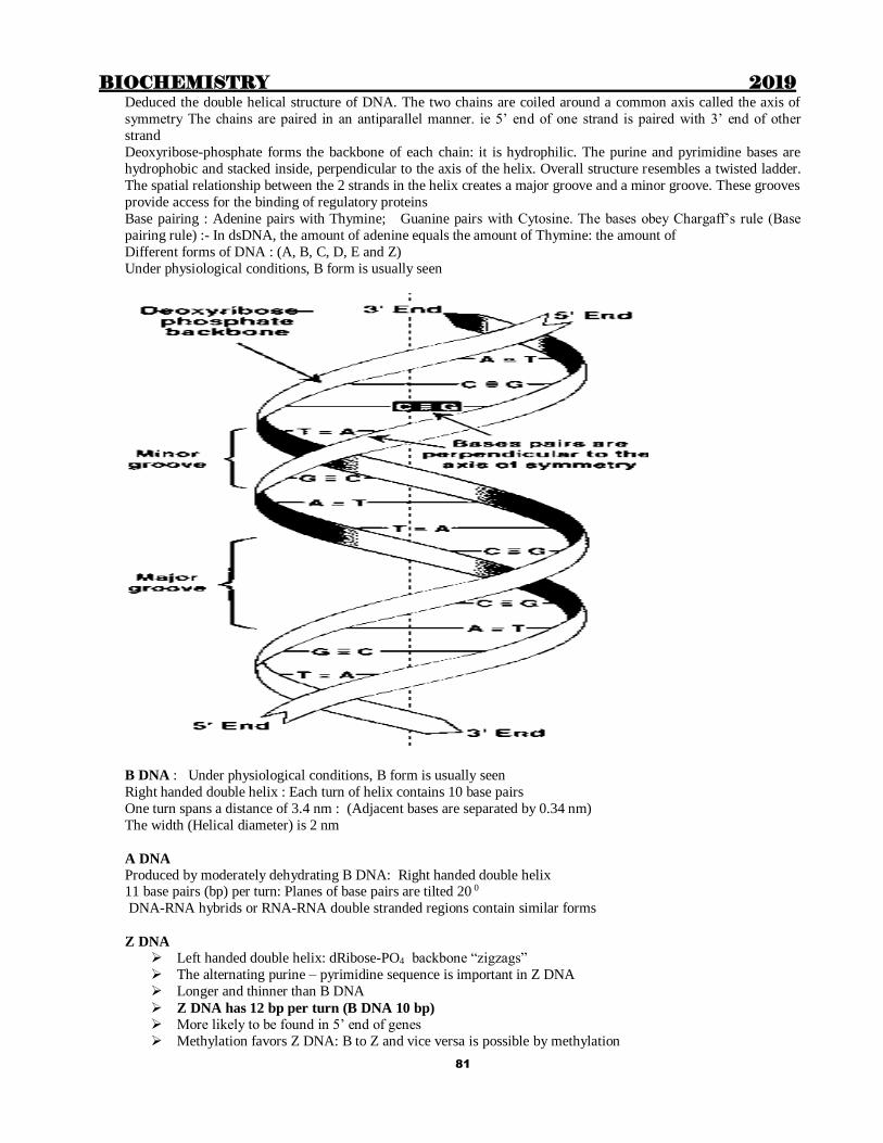
BIOCHEMISTRY 2019
81
Deduced the double helical structure of DNA. The two chains are coiled around a common axis called the axis of
symmetry The chains are paired in an antiparallel manner. ie 5’ end of one strand is paired with 3’ end of other
strand
Deoxyribose-phosphate forms the backbone of each chain: it is hydrophilic. The purine and pyrimidine bases are
hydrophobic and stacked inside, perpendicular to the axis of the helix. Overall structure resembles a twisted ladder.
The spatial relationship between the 2 strands in the helix creates a major groove and a minor groove. These grooves
provide access for the binding of regulatory proteins
Base pairing : Adenine pairs with Thymine; Guanine pairs with Cytosine. The bases obey Chargaff’s rule (Base
pairing rule) :- In dsDNA, the amount of adenine equals the amount of Thymine: the amount of
Different forms of DNA : (A, B, C, D, E and Z)
Under physiological conditions, B form is usually seen
B DNA : Under physiological conditions, B form is usually seen
Right handed double helix : Each turn of helix contains 10 base pairs
One turn spans a distance of 3.4 nm : (Adjacent bases are separated by 0.34 nm)
The width (Helical diameter) is 2 nm
A DNA
Produced by moderately dehydrating B DNA: Right handed double helix 11 base pairs (bp) per turn: Planes of base pairs are tilted 20 0
DNA-RNA hybrids or RNA-RNA double stranded regions contain similar forms
Z DNA
Left handed double helix: dRibose-PO4 backbone “zigzags”
The alternating purine – pyrimidine sequence is important in Z DNA
Longer and thinner than B DNA
Z DNA has 12 bp per turn (B DNA 10 bp) More likely to be found in 5’ end of genes
Methylation favors Z DNA: B to Z and vice versa is possible by methylation

BIOCHEMISTRY 2019
82
Circular DNA Mitochondria of eukaryotes
Chloroplasts
Prokaryotes contain one double-stranded, supercoiled, circular chromosome
Many bacteria contain circular, extrachromosmal DNA called plasmids
Denaturation of DNA
Denaturation of DNA is called melting of DNA. The strands of a given molecule of DNA is separated .The
temperature at which half of DNA strands is separated is known as melting temperature(Tm)
Tm is greater for GC pairs than for A=T pairs
Primary structure is intact on denaturation. Breaks the hydrogen bonds and becomes single stranded. Causes
stacking of the bases and becomes a rigid rod like structure and looses viscosity. This causes an increase in optical
absorbance (260 nm)which is known as hyperchromicity of denaturation Used to analyze DNA structure. Can be done by increasing the temperature or decreasing the salt concentration. In
recombinant DNA tech, Formamide is used to lower Tm, as it destabilizes H bonds
Organization of Eukaryotic DNA DNA is associated with tightly bound basic proteins called HISTONES
DNA + Histones form nucleosomes
Histones Histones are a small family of closely related basic proteins
Most abundant of chromatin proteins
They are positively charged at physiologcal pH due to the high content of Arginine and Lysine.
H2A and H2B are Lys rich: H3 and H4 are Arg rich.
The aminoterminal region is rich in basic amino acids
There are 5 classes of Histones - H1, H2A, H2B, H3 and H4
Because of the +ve charge, Histones form ionic bonds with –vely charged DNA. Along with Mg2+ Histones help
neutralize the –vely charged DNA phosphate groups
2 molecules of H2A, H2B, H3 and H4 form an octamer. They are known as “core” histones as DNA is wound
around this octamer.
A segment of DNA is wound nearly twice (1.75 turns) around a Histone octamer, forming a negatively supertwisted helix
Neighbouring nucleosomes are joined by “linker” DNA (approx 30 - 50 bp)
H1 binds to the linker DNA between the nucleosomes. H1 is the most tissue specific and species specific of the
Histones It facilitates packing of nucleosomes into the more compact structures
Core histones are subject to covalent modifications
H1 is bound less tightly and can be separated by salt solution
SUMO= Small Ubiquitin-related Modifier
Modified Histones
Modification Role

BIOCHEMISTRY 2019
83
Acetylation (H3 and 4) Regulation of transcription
Acetylation Chromosomal assembly
Phosphorylation (H1) Chromosomal condensation
ADP ribosylation DNA repair
Methylation Regulation of transcription
Mono ubiquitylation Gene activation, repression
Sumoylation Transcription repression
RNA mRNA transfer genetic information from DNA to protein synthesizing machinery
tRNA – for translation of RNA information into specific amino acids
rRNA – role in protein synthesizing machinery by synthesizing ribosomes
snRNA- role in RNA processing
Transfer RNA
tRNA are the smallest (4S) of the three major species of RNA molecules:
Also known as “soluble RNA (sRNA)” Transfer amino acids from cytoplasm to ribosomal assembly for protein synthesis
Eukaryotic RNA’s
RNA Types Abundance Stability
Ribosomal (rRNA) 28 S, 18 S, 5.8 S, 5 S 80% of total High (very stable)
Messenger (mRNA) About 105 different species 4-5% of total Unstable to stable (vary)
Transfer (tRNA) About 60 different species 15% of total High
Small nuclear (snRNA) About 30 different species 1% of total High
METABOLISM OF NUCLEOTIDES: Nucleotides are synthesized by 2 pathways; Salvage and De-novo
Salvage is single step and uses preformed adenine, guanine and hypoxanthine nitrogenous base.
The enzymes are APRTase and HGPRTase
Brain and RBC are dependent on salvage pathway and do not have De-novo pathway.
DeNovo pathway of pyrimidines and purines:

BIOCHEMISTRY 2019
84
There are six enzymes involved in synthesis of Pyrimidines, activities of which are located in three gene products.
CPS II, Aspatate transcarbamoylase, Dihydro orotase : in cytoplasm
Dihydro orotate dehydrogenase : in mitochondria.
OPRTase, Orotate decarboxylase : in cytoplasm
Source of atoms in pyrimidine ring
1
5
6
3
2
4Glutamine
CO2
Aspartate
Pyrimidine
Source of atoms in purine ring
N5N10Methenyl
H4Folate
N
N
N
N
H
GlycineCO2
Amide group of Glutamine
N10Formyl H4 Folate
Aspartate
1
2
3
4
5
6
7
8
9
Catabolism of purines and pyrimidines Purine catabolism
End product of purine catabolism – Uric acid (2,6,8 trioxopurine)
Rate limiting enzyme – Xanthine oxidase
The solubility of uric acid is less. At physiological pH of 7.4 uric acid is Mono Na urate, which is present in plasma.
In urinary tract:
a. Up to DCT & CD (pH 7.4) – Mono Na urate (less risk of stones)
b. After DCT & CD (pH falls to 6.0) – Uric acid (more risk of stones)
c. Mono Na urate is much more soluble than uric acid i. Uric acid stones are far more common than mono sodium Urate
ii. Uric acid stones are far more likely in distal nephron than proximal nephron.
iii. Alkalization of urine reduces stone formation.
iv. Lactic acidosis (alcoholism) increases Uric Acid form
Pyrimidine catabolism
Uracil and Cytosine Beta alanine , NH3 and CO2

BIOCHEMISTRY 2019
85
Thymine Beta amino isobutyric acidMethyl malonate semialdehyde Succinate, NH3 and
CO2
Note: Pyrimidine catabolism: forms highly water soluble β – Alanine & β – Amino isobutyrate. Therefore no
problem like uric acid
Disorders of Pyrimidine Metabolism:
1. Orotic Aciduria
2. Reye′s Syndrome- Severely damaged mitochondria → ↑ Carbamoyl phosphate (Underutilized) →
↑Orotate production → Orotic Aciduria
3. Deficiency of Ornithine transcarbamoylase (OTC) of urea cycle also has same effects. 4. Allopurinol –Competitive inhibitor of orotidylate decrboxylase → Orotic Aciduria & Orotidinuria
Disorders of purine catabolism:
1. ADA deficiency:
a. Deficiency leads to accumulation adenosine, AMP, ADP, ATP and their deoxy forms
b. dATP inhibits Ribonucleotide Reductase
c. This leads to deficiency of dGDP, dGTP, dCTP, TMP, TDP and TTP
d. Deficiency of deoxy ribonucleotides affects DNA synthesis, replication & cell division.
e. ADA deficiency leads to deficient Lymphocyte proliferation (both T cells & B cells are
affected) This prevents proliferation especially of T and B lymphocytes
f. This leads to deficient humoral as well as cellular immunity ultimately leading to severe combined immuno deficiency (SCID)
2. Hyperuricemia
a. There is increased deposition of uric acid crystals & stones in urinary tract mainly as uric acid
after DCT & CD.
b. Deposition of Na urate crystals in soft tissues & joints leading to their inflammation called
gout
c. Na Urate crystals are negatively Birefringent i.e. in polarizing microscope (plane polarized
light) they appear yellow when their long axis is parallel (y e l l o w) & blue when
perpendicular to the plane of light (D/D pseudo gout in which calcium pyrrophosphate is
deposited)
d. Na Urate crystals are needle shaped (Rhomboid in pseudogout)
Inhbitors of purine synthesis
Azaserine Formyl glycinamide R5P synthetase
Diazanorleucine PRPP-Gln amidotransferase
6-mercaptopurine Adenylosuccinase
IMP DH
Mycophenolic acid IMP DH
Azathioprine
Suppress CMI. Used to prevent graft rejection
Converted to 6-mercaptopurine in the body
Azathioprine and 6-mercaptopurine are metabolized by Xathine oxidase Therefore increase uric acid level
Inhibited by allopurinol

BIOCHEMISTRY 2019
86
CELL CYCLE Events occurring during the period between 2 mitotic divisions
Takes about 20-22 hours in mammalian cells
Divided into M, G1, S and G2 phases
M phase (Mitosis) Shortest phase (1 hr). Cell division takes place in M phase: After cell division, the cells can enter a Go phase-
dormant: Cells can also re-enter the cycle for growth and repair
G0 phase: Dormant phase; In a normal cell population, most of the cells are in Go phase
G1 phase (gap-1): 12 hours. Protein and RNA contents increase
S (Synthetic phase); 6-8 hrs; DNA replication occurs during this phase
G2 (Gap-2) phase: 4-5 hrs. Cytoplasmic enlargement; DNA repair; Production of Histones
G1, S and G2 phases are together called interphase (the phase between 2 mitosis)
Regulation of Cell Cycle
Cell cycle is controlled by
4 types of cyclins (A, B, D, E) and 5 cyclin dependent kinases (CDK 1,2,4,5,6) Cyclins activate CDK; CDK phosphorylate regulatory proteins
The most important check points occur in G1 and G2 phase entry points
Growth factors trigger the cells from Go G1
CDK2-cyclin E complex - G1S
CDK2-cyclin A completes S phase - (SG2)
CDK2- cyclins A&B completes G2 - (G2M)
MPF (M phase promoting factor) Promotes mitosis
Cyclins are synthesised throughout cell cycle, and are destroyed during mitosis
BIOCHEMISTRY 2019
87
Chapter 13
GENETICS
REPLICATION OF DNA New DNA synthesis during cell division
Semiconservative : demonstrated by messelson-Stahl experiment
The strands separate
Each strand acts as a template for the synthesis of a new strand
Bases in the new strand will be complementary to those in the template strand, obeying Chargaff’s rule
The synthesis of new strand is with the help of DNA polymerase, occurs in 5’ 3’ direction (or: the
template is read in 3’5’ direction)
New strand is joined to the template strand by H bonds between base pairs
Thus 2 double stranded (duplex) DNA are produced: Each goes to one daughter cell
Initiation of replication
DNA polymerase can use only ssDNA as a template: So, DNA strands should be separated first
In prokaryotes, replication begins at a single site called the ORIGIN OF REPLICATION. : AT rich sequence (called
consensus sequence)
In eukaryotes, replication begins at multiple sites- AUTONOMUS REPLICATNG SEQUENCES
Formation of the replication fork
As the 2 strands separate, they form a “V” shaped area: REPLICATION FORK
It moves along the DNA molecule as synthesis occurs
Replication of dsDNA is bidirectional, ie, replication forks move in both directions from the origin (But the synthesis in each new strand occurs in 5’3’ direction only)
Proteins required for strand separation:
ori binding proteins (dna A proteins in prokaryotes). It recognizes the origin site, rich in AT base pairs and separate
the strands forming local single stranded DNA. Progressive unwinding is brought by DNA helicases (known as
dnaB protein in prokaryotes). SSB proteins stabalise the complex by keeping two starand separated. SSB (single
strand binding proteins) prevent premature reannealing
Thus dnaA protein, DNA helicases cause strand separation: Both utilize ATP.
Positive supercoiling during replication
When 2 strands are separated, positive supercoils (“Supertwists”) appear in the region of DNA ahead of the
replication fork. These interfere with further unwinding of DNA: solved by enzymes called topoisomerases
Topoisomerases : Produce nicks in DNA and reseal it There are 2 types of topoisomerases
Topoisomerase I Topoisomerase II
Cuts single strand Cuts both strands
Does not insert super coiling Inserts super coiling
ATP not required ATP is required
Note: DNA Gyrase is a type of Topoisomerase II in E. coli which produces negative super coils using ATP
before replication
RNA primer: First an RNA primer is synthesized by RNA primase (dna G in EColi is primase). Once the RNA
primer is formed, DNA polymerase starts adding nuleotides to the 3’ end of primer. The added nucleotides will have bases complementary to the DNA strand
Synthesis can occur continuously in 5’3 direction: (Leading strand). The other strand is synthesized as small
fragments (okazaki fragments: (Lagging strand)

BIOCHEMISTRY 2019
88
Primosome: Complex of dnaA, SSB proteins, Helicase (dnaB) & Primase (dnaG), formed in the lagging
strand. It moves backwards i.e. 3′ to 5′ (opposite to the direction of synthesis). Forms RNA primers at several
steps in the lagging strand
Primosome is followed by DNA Polymerase III which synthesizes both leading and lagging strands.
After replication, DNAP III proof reads it
RNA primers are removed by DNAP I, gaps filled by DNAP I, and joined by DNA ligases
Telomere: 5’ end of the new strand is not synthesized by this ssDNA area is called telomere: Corrected by
telomerase which acts like reverse transcriptase and adds (TTAGGG)n to telomere.
This prevents shortening of chromosome with each cell division
Inhibitors of DNA replication Affects Bacterial DNA Gyrase - (antibacterials): Ciprofloxacin, Norfloxacin, Naidixic acid, Novobiocin
Affects Human topoisomerase – (Anticancer): Etoposide, Doxorubicin, Daunorubicin
Prokaryotic DNA polymerase
Pol I – removal of RNA primers and Gap filling (also called Kornberg’s enzyme)
Pol II – DNA proof reading and repair
Pol III – Synthesis of leading strand and okazaki fragments, Proof reading
Eukaryotic DNAP
Alpha- RNA primase activity (+) Initiates DNA synthesis, Gap filling
Beta- Repair
Gamma- Mitochondrila DNA synthesis
Delta- Synthesis of leading strand and Okazaki fragments Epsilon- Repair
End replication problem in eukaryotes:
Since the eukaryotic DNA is in the form of long segments, a small portion at the end of lagging strand after
removal of RNA primer remains unreplicated due to lack of free 3′ OH group to initiate replication. Primer
gaps are formed at the ends of both new DNA strands. This phenomenon occurs at the ends of chromosomes
i.e. telomeres.
Telomeres:
- Several thousand tandem repeats of a 5–8 bp sequence e.g. AGGGTTAGGGTTAGGGTT
- Few hundred nucleotide long segment of ssDNA folds back on itself and stabilized by proteins
- In a normal cell, telomeres get shortened in each replication cycle. This is inevitable.

BIOCHEMISTRY 2019
89
After the telomeres get shortened by a critical length, the cell stops dividing and enters GO phase (senescence).
However germ cells, cancer cells & stem cells have telomerase activity which prevents telomere shortening
A specific enzyme Telomerase fills these gaps
Telomerase:
- A Ribonucleoprotein
- Has reverse transcriptase activity (RNA dependent DNA polymerase)
- 9 – 30 nucleotide long region of RNA with
5′ – AACCCUAACCCU – 3′ sequence
- It adds 3′ – TTGGGATTGGGA – 5′ sequence to 3′ end of parent strand
- Now fresh DNA can be synthesized on this template

BIOCHEMISTRY 2019
90
TRANSCRIPTION
Synthesis of RNA from DNA template is called transcription.
Cellular Structure & Function
Main feature of Transcription is that it is highly selective i.e. RNA polymerase recognizes the signals on DNA
where to start and where to end the transcription.
Main enzyme for transcription is RNA polymerase.
Bacterial RNA polymerase is a multi-subunit enzyme.
Prokaryotic RNA polymerase
In prokaryotes all types of RNA except RNA primers for replication are synthesized by the same type of
RNA polymerase: Multisubunit enzyme: 2 identical α subunits, similar but non-identical β and β’ subunits
and 1 ω subunit constitute the core complex (E)
The core enzyme (E) associates with a protein factor called sigma (σ) factor to form the holoenzyme (E σ): σ factor helps RNAP to recognize the “promoter” on the DNA
2α, 1β, and 1β', are responsible for the 5′→3′ RNA polymerase activity
Function of ω subunit is not clear
Inhibited by the Rifampicin, which binds to the β subunit
Eukaryotic RNA Polymerase
3 different types of RNAP: They have 2 large subunits and many small subunits- the number of this varies in
different types
Type Inhibition by amanitin Products
I Insensitive 5.8, 18, 28SrRNA
II Highly sensitive mRNA, miRNA, snRNA
III Intermediate sensitivity tRNA, 5S rRNA
Mitochondrial RNA polymerase is similar to prokaryotic RNA polymerase.
Differences between DNA polymerase & RNA polymerase:
1. RNA Polymerase doesn′t require primer.
2. RNA polymerase does not have proof reading ability.
3. RNA Polymerase does not require Helicase. It has its own helices activity. Similarities between DNA & RNA polymerases:
1. Both are 5′ → 3′ polymerases
2. Both require Nucleoside Tri phosphates (NTPs) as precursors
Transcription in prokaryotes
Fumarate+ Acetoacetate
Transaminase
∆ G = ∆E – T∆S

BIOCHEMISTRY 2019
91
I. Initiation σ Protein recognizes & binds to promoter region in prokaryotic DNA. After initiation, the factor dissociates and
RNA polymerase starts forming RNA.
II. Elongation-
Core RNA polymerase synthesizes RNA similar to DNA polymerase. It has its own helicase activity and super
coils are taken care of by Topoisomerases.
Main features of Elongation:
- RNA synthesis always takes place from 5′→ 3′ direction.
- Ribonucleoside tri phosphates are used to form phosphodiester linkage.
- PPi released is broken to 2Pi pulling the reaction to completion.
- Sequence of RNA transcript is same as coding strand (sense strand) except U in place of A.
- The strand complimentary to coding strand (anti-coding or anti-sense strand) serves as a template.
- Transcription is a continuous process.
- No Endo or Exonuclease activity.
- No proof reading.
- No repair of mistakes.
III. Termination

BIOCHEMISTRY 2019
92
There are two mechanisms for termination of transcription.
1) ρ Dependent termination
- ρ factor binds C rich ρ recognition site near 3′ end of RNA transcript
- Gradually it moves to RNA polymerase paused at the termination site
- ρ factor has RNA dependent ATPase activity
- It also has intrinsic ATP dependent RNA DNA helicase activity which helps release the
primary RNA transcript.
2) ρ independent termination
- Formation of a stable hairpin of RNA transcript due to DNA Palindromes.
- Formation of poly Us after hairpin. - A–U bonding is weak, which helps RNA to detach from DNA.
Note: Palindromes –A sequence of double stranded DNA, where two strands have the same sequence from
5′→ 3′.
Transcription in Eukaryotes
Formation of pre-initiation complex (PIC): at least 50 different factors are involved in formation of PIC
ensuring the fidelity of transcription
Elongation: same as prokaryotes
III. Termination: poorly understood
Inhibitors of RNA synthesis Actinomycin D Inserts phenoxazone ring between 2 G-C bp of DNA
Rifampicin Binds to β subunit of RNA polymerase and inactivates it
Alpha amanitin Inactivates RNA polymerase II
3’-deoxy adenosine incorrect entry into chain causing chain termination
Usually transcription is by DNA dependent RNA polymerase
Reverse transcriptase is RNA dependent DNA polymerase Present in some RNA viruses-retroviruses
Produce a dsDNA copy of their RNA genome
PROMOTERS OF TRANSCRIPTION ◦ Sequences in DNA which directs RNAP to start transcription: they are on coding strand
Eukaryotes:
◦ 25 bp upstream – HG box (TATAAA)
◦ 70bp upstream - CAAT box GGCCAATCT
Prokaryotes :
◦ 10 bp upstream - Pribnow box/ TATA box (TATAAT)
35 bp upstream -5’TGTTGACA3
Sigma factor recognizes promoter site
Rho factor is involved in termination of transcription
Post-transcriptional processing:
mRNA is transcribed as a primary transcript, which is long and contains exons & introns. Undergoes processing to
get mRNA. Includes endonuclease cleavage, Poly-A tailing, 5’ capping, Methylation, Removal of introns, Splicing
of exons.
5’ capping: 7 methyl GTP cap. Helps in recognition of mRNA by translation machinery
Prevents attack by 5’ exonuclease
Poly A tail: 20-250 adenylate residues. Prevents attack by 3’ exonuclease

BIOCHEMISTRY 2019
93
A few mRNAs esp those for some histones do not have poly A tail
Areas that are transcriptionally active are less densly stained and are called euchromatin.
Areas that are transcriptionally inactive are densly packed at interphase and appear as densly stained areas under
electron microscope- called heterochromatin.
Some areas are always condensed and transcriptionally inactive- Constitutive heterochromatin
Certain areas of heterochromatin decondenses at times and become transcriptionally active Facultative
heterochromatin
Features of genetic code
Triplet codons : Each codon consist of 3 bases Non-overlapping; Non punctuated
Degeneracy- One amino acid has more than one codon
Unambiguous – one codon stands only for 1 amino acid
Universal (“from E.ColiElephant): In all organisms, the one codon codes for the same amino acid
Wobbling- reduced stringency between 3rd base of codon and complementary base in anticodon
Initiator-AUG
Terminator- UGA,UAG, UAA
Tota 64 codons. 3 are stop codons. Remaining 61 code for 20 amino acid. Some amino acids have more than one
codon
Single Codon: AUG - Met, UGG - Trp
2 Codons: Asp, Glu, Asn, Gln, Phe, Tyr, Lys, Cys, His.
3 Codons: Ile. 4 Codons: Gly, Ala, Val, Pro, Thr.
6 Codons: Ser, Leu, Arg.
Stop Codons -3
Mitochondrial DNA (mtDNA)- Closed circular dsDNA Majority of proteins in mitochondria are coded by nuclear genes. Others are by mitochondrial DNA
Encodes 13 protein subunits of respiratory chain 7 subunits of NADH dehydrogenase
Cyt b
3 subunits of cyt oxidase
2 subunits of ATP synthase
Genetic code differs slightly from that of mtDNA
UGA Trp
AGA and AGG stop codons
Special features of mitochondrial DNA
Mitochondrial DNA (mtDNA) is somewhat similar to prokaryotic DNA:-
i) It is a circular double stranded helically coiled DNA.
ii) There are 2 – 10 copies of circular DNA per mitochondrion. Certain cells in embryo which are
undergoing differentiation may contain up to 100 copies of circular duplex DNA
iii) In embryo all the mitochondria are derived from ovum and hence in an individual mDNA is maternally
acquired
iv) mDNA codes for mitochondrial tRNAs, rRNAs and a few mitochondrial proteins. More than 95% mitochondrial proteins are encoded by nuclear DNA
v) Mitochondria also divide along with cell division. The mDNA is replicated before and during cell
division
vi) mtDNA genes have a much higher mutation rate than nuclear DNA genes.
vii) Mitochondria undergo replicative segregation at cell division.
viii) Somatic mDNA mutations accumulate in post-mitotic tissues with age, reducing the ATP generating
capacity.
SnRNAs: (Snurps or (short nuclear ribonucleoproteins):
BIOCHEMISTRY 2019
94
Small nuclear RNAs. SnRNAs combine with proteins to form small nuclear Ribonucleoproteins (snurps)
Involved in splicing of exons
Size: 90 -300 nucleotides.About 1 million copies of Sn RNAs/cell.
6 different SnRNAs- uracil rich: U1,U2, U4, U5,U6 and U7 .
U1- Cuts the 5’end: U5 - attacks the 3’ end of the intron
U2 – is the main splicing enzyme.
U7- Specific for Histones
All of them are located in nucleus.
TRANSLATION It has following steps:
Activation of amino acids
Initiation
Elongation
Termination
For many proteins, this is followed by post translational modifications
Activation of amino acid With the help of aminoacyl tRNA synthetases
Amino acid + tRNA + ATP Aminoacyl tRNA + AMP
DHU arm of tRNA is important in the recognition of tRNA by this enzyme
Initiation of translation
A mRNA is selected by ribosome for translation
This involves the action of many eukaryotic initiation factors (eIF)
Can be divided into 4 steps
Ribosomal dissociation
Formation of Pre-initiation complex
Formation of initiation complex Ribosoml assembly
Ribosomal dissociation
80 s ribosome splits into 40s and 60s subunits
eIF-3 and eIF-1A bind to the 40s subunit
This delays its reassociation with 60s subunits
It allows other initiation factors to associate with 40s
Formation of 43s pre-initiation complex
GTP binds with eIF-2
This binds to Met-tRNA
This binds to 40s to form 43s pre initiation complex
eIF-3 and 1A stabilizes it
Formation of 48s initiation complex mRNA cap facilitates binding of mRNA to 43s pre initiation complex
A protein complex called eIF-4F binds to the cap. eIF-4A and 4B reduces the complex
secondary structure of 5’ end of mRNA through ATP dependent helicase activities
Now this complex scans mRNA for a suitable initiator codon
First AUG after the marker sequence is taken as initiator codon
In prokaryotes, the marker sequence is a purine rich sequence (eg:- UAAGGAGG)
known as Shine-Dalgarno sequence, located 6-10 bases upstream of the initiator codon. It helps in binding of
30 S ribosomal subunit to mRNA
In eukaryotes, the marker is known as “Kozak” sequence

BIOCHEMISTRY 2019
95
Formation of 80s initiation complex (Ribosomal assembly)
60 s ribosomal subunit binds to 48s initiation complex, using GTP
Initiation factors bound to 48s are released
40s and 60s assembles to form 80s ribosomal assembly
The initiating AUG is recognized by a Met-tRNA. This is facilitated by eIF2 and GTP
(in prokaryotes and in mitochondria this tRNA carries N-formylated Met)
The charged Met-tRNA enters the P site of ribosomal assembly
This Met or fMet is cleaved even before completion of termination
Elongation

BIOCHEMISTRY 2019
96
Involves
Binding of new aminoacyl tRNA
Peptide bond formation
Translocation
Binding of new amino acyl – tRNA to ‘A’ site
The codon after the initiator codon decides which aminoacyl-tRNA is to be added next.
The new aminoacyl-tRNA comes to the ‘A’ site: This requires elongation factor EF-1 and GTP
GTP hydrolysed; EF-1 released
Peptide bond formation
The amino group of the new aminoacid forms a peptide bond with the COOH of the previous amino acid,
catalysed by “peptidyl transferase” (a ribozyme) As the amino acid is already activated this does not require energy
Now the previous tRNA, which is on the ‘P’ site does not carry amino acid
Translocation
Ribosome moves in 5’ 3’ direction of the mRNA, to te next codon
Now the empty tRNA reaches the ‘E’ site and is expelled from ribosome
tRNA which carries the growing peptide reaches the ‘P’ site
New incoming aminoacyl tRNA as decided by the next codon comes to the ‘A’ site
This requires EF-2 and GTP
Energy requirements
For each peptide bond formed, a total of 4 high energy bonds are used
2 for activation of amino acid + 1 for EF-1 step (binding of tRNA to A site) + 1 for EF-2 step (translocation)
Step of formation of peptide bond does not require energy
In addition, Energy is required for initiation complex formation and termination
TERMINATION
When the A site of ribosomal assembly reaches a terminator codon sequence in mRNA, translation stops.
There is no tRNA with anticodon for these
eRF recognises terminator codon (in prokaruotes, it is by RF-1 and RF-2)
It induces peptidyltransferase to hydrolyze the polypeptide from tRNA
eRF-3 releases the releasing factors, utilizing GTP (in prokaryotes, RF-3)
80S ribosomal assembly dissocates into 60S and 40S subunits
Example of chain terminating codons are UAA (Ochre), UGA (opal) and UAG (amber)
INHIBITORS
Ciprofloxacin, Nalidixic acid and Novobiocin inhibit Bacterial DNA gyrase
Etoposide, Adriamycin and doxorubicin inhibit Human topoisomerase
6-mercaptopurine inhibits DNA polymerase
5-Fluoro uracil inhibits Thymidylate synthase
PROTEIN TARGETING Most proteins are synthesized in cytoplasm; But many of them functions in specific cellular organelles.
So, after translation, these should reach the correct destination
This is with the help of specific amno acid sequences signals in the amino terminal region
In addition to this the carboxy terminal region also contains sequences which carry an “address” for its destination
Co-translational glycosylation also has a role in proein targetting
Defective protein targeting can lead to diseases like Zellweger’s syndrome, Primary hyperoxaluria etc

BIOCHEMISTRY 2019
97
Zellweger’s syndrome: Due to defective protein targetting, the enzymes required for oxidation of very long chain
fatty acids do not reach peroxisomes. Accumulation of VLCFA in nervous tissue leads to neurological impairment
and early death
Primary hyperoxaluria: Defective targeting of alanine glyoxalate aminotrasferase
Normally it is present in peroxisomes ; Due to targeting defect it is seen in mitochondria
Leads to increased pool size of glyoxalate excess production of oxalate
REGULATION OF GENE EXPRESSION “Gene expression” means the production of a functional gene product (Protein or RNA) from the information in a
gene. This is mainly regulated at the level of transcription. In eukaryotes, regulation also occurs at the level of post-
transcriptional and post-translational modifications.
All genes are not regulated. Constitutive genes (House keeping genes) encode products required for basic cellular
functions. They are continuously expressed. Regulated genes are expressed only under certain conditions
Eg: Insulin gene is expressed only in β cells of pancreas
Some proteins are produced only when needed (Lactose metabolising enzymes in E.Coli)
Regulatory sequences in DNA (cis-acting elements) interact with regulatory proteins (trans-acting elements) to
regulate transcription. Binding of regulatory proteins to DNA is through structural motifs like zinc finger, leucine
zipper or helix-turn-helix
Examples of DNA binding regulatory proteins with binding motifs
Regulatory proteins binding to DNA Motif
Lac repressor and CAP of E coli Helix turn helix
Homeo box proteins in mammals Helix turn helix
Gene 32 protein of E coli Zinc finger
Steroid receptor family in mammals Zinc finger
CRE binding protein in mammals Leucine zipper
Induction and repression
Induction and repression acts as important mechanisms of regulation Induction is the increased synthesis of a protein/enzyme in response to a signal. (the gene is “on”)
Repression is decreased synthesis (the gene “off”)
OPERON CONCEPT OF GENE REGULATION
Put forward by Jacob and Monod
This was based on studies in E.Coli . E.Coli grown in glucose medium do not produce lactose metabolizing
enzymes. But when they are grown in lactose,(and no glucose) lactase enzyme(beta galactosidase) is
produced . The mechanism suggested is as follows:
In bacteria, the structural genes that code for proteins in a particular pathway are sequentially grouped on
the DNA along with cis-acting elements. The product is a polycistronic mRNA. The genes for this thus act
as a unit which can be cocordinately controlled. This entire package is called “OPERON”
Operon is a unit of gene expression; It includes
Structural genes: control elements: regulator/inhibitor gene: Promoter and operator areas

BIOCHEMISTRY 2019
98
LAC OPERON This is the operon related to lactose metabolism
Includes 3 structural genes (Z,Yand A) which code for 3 proteins, a regulator(inhibitor) gene, a
promoter area and an operator
Z gene codes for β galactosidase, Y gene codes for permease (which transports lactose and galactose into
cell) and A gene codes for thiogalactoside transacetylase
Transcription of these 3 genes start from a common promoter located close to Z gene. RNAP binds to the promoter
and transcribes these 3 genes as a single mRNA.
When glucose is available and lactose is absent, transcription is repressed
Regulator (Inhibitor) gene produces a repressor molecue (150kD)
Repressor has strong affinity to the operator site and binds tightly
RNAP finds the promoter and moves forwards towards the structural genes, but its movement beyond
operator is prevented by repressor protein bound to the operator
So transcription of structural genes does not occur; lactose metabolising enzymes not produced
When lactose is present and glucose is absent, transcription is induced or “derepressed”
Lactose combines with repressor protein
This Lactose-repressor complex cannot bind to the operator site
RNAP can move forward and transcribe the structural genes
The mRNA produced will be translated to the enzymes for lactose metabolism
This shows that the synthesis of these proteins are normally repressed
Lactose is an inducer; When lactose is present, the genes are de-repressed or induced
BIOCHEMISTRY 2019
99
Role of Catabolite Activator Protein
If both glucose & lactose present, E.Coli utilize Glucose. Lactose metabolizing enzymes are produced only
if glucose is absent
This is because RNAP can attach only when CAP –cAMP complex is available
cAMP accumulates only when the bacteria starves (when no glucose) ; then CAP binds with cAMP to
produce the complex. This helps binding of RNAP to promoter site.
MUTATIONS “Spontaneous abrupt origin of a new character”
Due to an alteration in the genetic material
One or a few nucleotides may be altered, or a large area may be altered
Mutagenicity of a substance is detected by Ame’s test
Mutation can occur due to Point mutations (base substitution) OR Frameshift mutations (base addition or deletion)
Point mutations
Transitions (Pyrimidine Pyrimidine or Purine Purine)
Transversions (Purine Pyrimidine / Pyrimidine Purine)
Results in: Silent mutations
Missense mutation (Acceptable, Partially acceptable, Unacceptable)
Nonsense mutation
DNA REPAIR MECHANISMS
Repair mechanism Defect Rectification
Mismatch repair Single base or 2-5 base unpaired loops Methyl directed strand cutting,
Exonuclease digestion and replacement
Base excision repair Damage to a single base (Spontaneous,
chemical or radiation induced)
Base removal by N-glycosylase, abasic
sugar removal, replacement
Nucleotide excision
repair
Damage to a DNA segment Removal of approx 30 nucleotide
oligomer, and replacement
Double-strand break repair
Damage to strands (radiation, chemotherapy etc)
Ku protein, DNA-PK acts Unwinding, alignment and ligation
Xeroderma pigmentosum
Autosomal recessive disorder
Exposure to UV light causes dimerisation of Thymine bases
Not corrected because Nucleotide excision repair is defective
This requires excision endonuclease which is defective in Xeroderma Pigmentosa.
Disease characterized by marked sensitivity to UV light leading to cancer.
Diseases associated with DNA repair mechanisms Disease Defect Features
Xeroderma Pigmentosum Nucleotide excision repair defective Sensitivity to UV light, Skin malignancy
Fanconi’s anemia DNA cross link repair AR; Anemia, increased incidence of
cancer
Ataxia telangectasia ATM gene Sensitivity to UV light, Lymphoreticular
neoplasms
Bloom’ syndrome DNA ligase / helicase Lymphoreticular neoplasms
Hereditary polyposis colon
cancer (Lynch syndrome)
Mismatch repair Ca Colon
Cockayne syndrome NER defective A.R. Progeria like features
No increased tendency for cancer
ONCOGENIC VIRUSES Infection by some viruses can produce cancer
This is because the viral DNA is incorporated into the human DNA
Viral DNA multiplication will not be controlled by human mechanisms

BIOCHEMISTRY 2019
100
Leads to uncontrolled multiplication of the cells
Virus Cancer produced
Epstein-Barr virus (EBV)
Burkitt’s lymphoma Nasopharyngeal carcinoma
Human papilloma virus (HPV) Cancer of uterine cervix
Hepatitis B(HBV) Hepatoma
Antimutagens
Substances which interefere with development of cancer
Eg; Vitamin A, Carotenoids, Vitamin E, Vitamin C, Dietary fibre
ONCOGENES
They are certain genes, normally present in cells, which are capable of causing cancer. Also called proto-oncogenes
; First demonstrated in Rous sarcoma virus
This virus can produce sarcoma in birds; But, if the virus is deficient in a particular gene, it cannot do; This gene is
named sarcoma gene (src)
Same sequences are present in normal avian cells- they are called C-src (cellular). Viral gene is called V-src Proto oncogenes are regulatory genes, and they are expressed only when required
Oncogene Location in
human chromosome
Virus carrying the gene Product of the gene
abl 9 Abelson leukemia virus in mouse
Tyrosine kinase
erb-A 17 Erythroblastosis virus in chicken
Receptor for epidermal growth factor
myc 8 Myelocytoma virus in chicken DNA-binding protein
sis 22 Simian sarcoma virus in monkey
Platelet Derived Growth Factor (PDGF)
src 20 Rous sarcoma in chicken Tyrosine kinase
ras 12 Rat sarcoma GTPase
Activation of oncogenes by carcinogens or due to point mutations may result in malignancy
Oncosuppressor genes (Anti oncogenes)
These are genes which protect the cell from developing cancer
When these are deleted or mutated, it results in cancer
Eg:- Retinoblastoma oncosuppressor gene
P53 oncosuppressor gene
P53 Oncosuppressor gene
Present in short arm of chromosome 17
Producese a phosphoprotein with m:wt 53 kD This prevents development of cancer by 3 means
If DNA of a cell is damaged, it blocks cell division untill the damage is repaired. (It downregulates
PCNA gene transcription and causes arrest in G1 phase)
If the damage is irreparable, it directs the cell for apoptosis
It can also prevent the activity of oncogenic viruses
Many cancer cells show an absence of p53 gene or a mutated non functional p53
Retinoblastoma gene
Located in chromosome 13; Encodes a protein called p105 (m:wt 105 kD)
Called so because, firs isolated from retinoblastoma patients
It inactivates transcription factor E2F
Inhibits cell cycle at G1 phase
In normal cells, this arrest is prevented by the action of cyclin D (Separates Rb from E2F) If the genome is deleted of this gene, retinoblastoma results
Certain tumor antigens from oncogenic viruses combine with Rb then cannot inhibit cell cycle cancer

BIOCHEMISTRY 2019
101
ONCOFETAL ANTIGENS
These are products of certain genes which are normally expressed only in fetal life after which the gene is repressed.
But when malignant transformation occurs in some cells, the gene is de repressed and these proteins are produced.
They are clinically used as tumor markers
Eg:-
Alpha fetoprotein – in hepatoma
Carcino embryonic antigen – Colon cancer
TUMOR MARKERS Enzymes
ALP: Bone
Regan isoenzyme: Ca of Liver, Lung and Gut
Nagao isoenzyme: Pleural malignancy
ACP, PSA : Ca prostate
Neuron specific enolase: tumors involving nervous system
Hormones Beta hCG- Choriocarcinoma
Calcitonin- Medullary Ca thyroid
Vasoactive Intestinal polypeptide- Apudomas
VMA – Phaeochromocytoma and neuroblastoma
5 HIAA- Carcinoid syndrome
Oncofetal products
Alpha fetoprotein: Hepatoma, germ cell tumors
CEA: Colorectal, GI and Lung cancers
Carbohydrate antigens
CA – 125: Ovarian cancer
Tissue catabolic product Hydroxy proline- Bone metastasis
Other proteins
Immunoglobulins- Multiple myeloma, macroglobulinemia
Bence-Jones proteins- Multiple myeloma
RECOMBINANT DNA TECHNONLOGY:
Following are the Steps: Preparation of specific human gene
Preparation of chimeric DNA
Transfection / Transformation of vector into the host
Selection of colony having the required gene
Preparation of specific human gene
The specific mRNA is first selected from the tissue
A cDNA (complementary copy DNA) is prepared using reverse transcriptase: (This is done because
isolation of a specific gene from human DNA is a difficult process)
Preparation of chimeric DNA
A vector carrying a foreign DNA is called Chimeric DNA (Hybrid DNA) (Recombinant DNA)
A vector DNA (Eg: Plasmid) is cut with a specific R.E. This may produce either sticky ends or blunt ends
Human DNA is also cut with the same R.E: Same sequences are generated at the ends of the cut pieces
Now the sticky ends of both vector and human DNA have complementary sequences.When these are
incubated together, annealing takes place. The fragments are joined together by DNA ligase - a circular
chmeric DNA is formed

BIOCHEMISTRY 2019
102
Following are the different vectors with the size
Vector DNA insert size
Plasmid pBR322 0.01-10 kb
Lambda phage 10-20 kb
Cosmids 35-50 kb
BAC 50-250 kb
YAC 500-3000 kb
Transfection/Transformation
The process of introducing vector into a cell is called transformation for bacteria and transfection for
eukaryotes
The host E.Coli cells and vectors are incubated in hypertonic medium with calcium. Opens up calcium
channels and vector enters the cell
The bacteria are cultured
As the host cell multiplies, it forms a clone in which every bacterium carries copies of the same inserted
DNA fragment, hence the name “cloning.”
Selection of colonies having the required gene The colonies which contain the desired foreign DNA are selected using properties like antibiotic resistance.
The cloned DNA is released from its vector by cleavage (using the appropriate restriction endonuclease) and is isolated. By this mechanism, many identical copies of the DNA of interest can be produced.
pBR322 has Apr, Tcr and Cmr genes
EcoRI cuts in the middle of Cmr gene
PstI cuts wthin Apr gene
Expression vectors: If a vector carrying foreign gene is translated to the foreign protein, it is called ‘expression
vector”.
Homopolymer tailing
Ends of sticky ends may get sponatneously reconneted to produce tandem hetrogenous inserts
To prevent this, blunt ends produced first and then homopolymer tailing is given using terminal transferase
GENE THERAPY Therapeutic delivery of genes into a cell: Applied to patient with a defective gene which produces disease.
The normal, cloned DNA for a gene is introduced into the somatic cells of the patient. The DNA become
permanently integrated into the patient's chromosomes expressed to produce the correct protein Vectors used for gene delivery are
Retroviruses
Adenoviruses
Plasmid-liposome complex: (can carry human genome of big size
Gene gun method: Tungsten particles coated with plasmid are accelerated to the tissues by helium pressure
discharge
Diseases treated with gene therapy
SCID (Severe Combined Immuno Deficiency): ADA deficiency: First successful gene therapy
Cystic fibrosis; Hemophilia; Familial hypercholesterolemia ; Duchenne muscular dystrophy
TRANSGENESIS A form of germ cell gene therapy: The hybrid DNA is inroduced into the fertilized ova
Embryos develop in the uterus of another animal; produce transgenic animal
incresed milk producton in cows
to correct genetically determined hypogonadism in mice
FUSION PROTEINS: Product of a fusion gene
Fusion gene: produced by removing a stop codon from a gene and appending DNA sequence coding for
another protein
Useful in tumor cell targeted immunotherapy
Important Terms /Tools In Genetics

BIOCHEMISTRY 2019
103
Antisense Therapy mRNA contains a “sense” or message that is to be translated
If an olignucleotide with a complementary sequence is made, it is called “antisense”
If antisense RNA is introduced into a cell, it can trap the normal mRNA and can inhibit the protein
synthesis
This is being tried on AIDS and Cancer
Antisense molecules can be introduced into cells by liposome encapsulation
Balanced Polymorphism When a gene is mutated, the abnormal gene will be eliminated from the next generation in population . So the ratio
of normal & abnormal gene is maintained in all generation (Hardy-Weinberg law). But an abnormal gene need not
always be eliminated from population, especially if it has got an advantage.
Eg: Sickle cell trait and malaria
The lethal nature of the gene in homozygous state is counterbalanced by the advantage in heterozygous state. This is
called balanced polymorphism
Apoptosis: Programmed Cell Death Removal of aged or partially damaged cells is done by apoptosis
The changes include Chromatin condensation, Nuclear shrinkage, membrane blebbing and removal of these
bodies by macrophages: In electrphoresis- Step ladder pattern of DNA
Apoptosis mediating genes- suicidal genes
Apoptosis is finally effected by activation of caspases : ( Cysteinyl aspartate specific proteases)
Restriction Endnucleases Presence in a given bacterium restrict the growth of bacteriophages
Cut DNA in a sequence-specific manner
Protects the host bacterial DNA from DNA of foreign organisms
Always exist in pair with site-specific DNA methylases
Site - specific DNA methylases protect host DNA from digestion by Restriction endonucleases
Regulation by repression in humans ALA synthase (rate limiting enzyme in heme synthesis) is repressed by heme
An aporepressor is produced by a regulatory gene. The aporepressor binds with heme (co-repressor) to
form the holo-repressor
Holorepressor binds to the operator site and prevents the movement of RNAP
Thus transcription of ALA synthase gene is repressed
When heme is not available, holo repressor is not formed; So de-repression occurs and ALA synthase is
produced
Hormone Response Elements (HRE) in regulation of gene expression Hormones or their second messengers act as inducers in higher organisms
Steroid hormones bind to cytoplasmic receptors and the complex goes to nucleus. Exert their effect by
attaching to HRE in DNA
The receptor binds at the enhancer region activates promoter accelerates transcription
Gene Switching Sometimes one gene is switched off while a closely related gene takes up its function
Eg:- In fetal life, genes for alpha & gamma chains of Hb are actve, After birth, it gradually switch over to
genes of alpha and beta chains
Gene Amplification • Amplification of a few genes by repeated initiation of DNA synthesis
• Eg: drug resistance to methotrexate can be produced by amplification of Folate reductase gene by cancer
cells
Gene Silencing:
It is switching OFF a gene by mechanisms other than genetic modifications
Transcriptional gene silencing- by histone modifications

BIOCHEMISTRY 2019
104
Post-transcriptional gene silencing – by mRNA of a particular gene being destroyed or blocked Eg:
RNAinterference (by miRNA and siRNA)
mi RNA si RNA
Form imperfect duplexes with 3’ untranslated
regions of mRNA and arrest translation
Form perfect hybrids anywhere with mRNA.
These hybrids are degraded in P bodies
Leads to arrest of translation
DNA Finger Printing • Tandem repeats are short sequences of DNA at scattered locations in the genome, repeated in tandem (one
after another)
• Probes are used to hybridize to these tandem repeats
• The number of these units varies from person to person, but is unique for any given individual
Therefore, it serves as a molecular fingerprint.
Applications
• Disputed paternity / parenthood
• To identify the culprit of a crime
– DNA could be isolated form blood /seminal stains even after several years
RFLP (Restriction Fragment Length Polymorphism) Genome variations are differences in the sequence of DNA among individuals; include both polymorphisms and
mutations
A polymorphism is a clinically harmless DNA variation that does not affect the phenotype; occur in the intervening
sequences that do not code for proteins.When variation occurs in more than 1% of population, it is called
polymorphism
Two types of DNA variation commonly result in RFLPs:
single-base changes
tandem repeats
single-base changes at restriction sites or a change at other sites may result in fragments of lengths differing from the normal
Tandem repeats: short sequences of DNA at scattered locations in the genome, repeated in tandem (one
after another)
RFLP can be found out by cleaving the DNA into fragments (restriction fragments) with a restriction endonuclease .
The existence of 2 or more types of restriction fragment patterns in population is called RFLP
Nucleic Acid Hybridization Techniques: In Situ Hybridization DNA / mRNA can be viewed in the context of tissue morphology
When the probe is tagged with fluorescin, it is called FISH
CISH is chromogenic in situ hybridization
DNA Microarray DNA probes of about 500-5000 genes are prepared and added to small wells in polystyrene plates
cDNA from patient’s cell is added to the wells: Hybridized and fluorescence assessed

BIOCHEMISTRY 2019
105
Used to identify expression of various genes
Blot techniques: Sothern,Northern,Western,South-western
CHROMOSOME JUMPING A technique for physical mapping of genome
Used to bypass regions difficult to clone eg: repetitive DNA sequences
DNA of interest is identified, cut into fragments with REand circularised From a known sequence a primer is designed to sequence across the circularised junction
This primer is used to jump 100 kb-300 kb intervals: a sequence 100 kb away would have come near the
known sequence on circularisation
Thus, sequences not reachable by chromosome walking can be sequenced
JUMPING GENES- “TRANSPOSONS”
Discovered by Barabara McClintock
Sequences of DNA that can move or transpose themselves to new positions within the genome of a
single cell [either "copy and paste" or "cut and paste“]
Cause mutations and can alter genome size
Transposons are considered “junk” DNA
Used in biotechnology to alter DNA in a living organism
POLYMERASE CHAIN REACTION In vitro amplification of a segment of DNA, whose flanking sequences are used as primers.
Enzyme used is Taq polymerase
Reverse transcriptase PCR
Polymerase from Thermus thermophilus ; Has RTase and Polymerase activity
Produce cDNA from mRNA present in the cell, it is amplified ie , it detects mRNA; Means it is from a live organism
Nested PCR
Use 2 different sets of primers in 2 successive cycles
This is to avoid contamination in products due to unexpected primer binding sites
RACE PCR: Rapid Amplification of cDNA Ends
Unknown sequence at the ends of mRNA is amplified by a RT PCR, using known information from the
centre of the transcript
Can be used to generate 5’ or 3’ end of mRNA
Also called one sided PCR or anchored PCR
TRIPLEX DNA (TRIPLE STRANDED DNA) 3 oligonucleotides wind around each other and form a triple helix. In this structure, one strand binds to a B-form
DNA double helix through Hoogsteen or reversed Hoogsteen hydrogen bonds
A Hoogsteen base pair is a variation of base-pairing in nucleic acids such as the A•T pair. In this manner,
two bases on each strand can be held together by hydrogen bonds in the major groove. A Hoogsteen base pair
applies the N7 position of the purine (as a hydrogen bond acceptor) and C6 amino group (as a donor), which bind the Watson-Crick (N3–N4) face of the pyrimidine base.

BIOCHEMISTRY 2019
106
Fluorescence In Situ Hybridization (FISH)
FISH (Fluorescent in situ hybridization) is a cytogenetic technique which can be used to detect and localize the
presence or absence of specific DNA sequences on chromosomes. It uses fluorescent probes which bind only to
those parts of the chromosome with which they show a high degree of sequence similarity. Fluorescence microscopy
can be used to find out where the fluorescent probe bound to the chromosome. FISH is often used for finding
specific features in DNA. These features can be used in genetic counseling, medicine, and species identification.
FISH uses fluorescent molecules to vividly paint genes or chromosomes. This technique is particularly useful for
gene mapping and for identifying chromosomal abnormalities.
How does FISH work?
FISH involves the preparation of short sequences of single-stranded DNA, called probes, which are complementary
to the DNA sequences the researchers wish to paint and examine. These probes hybridize, or bind, to the
complementary DNA and, because they are labeled with fluorescent tags, allow researchers to see the location of
those sequences of DNA. Unlike most other techniques used to study chromosomes, which require that the cells be
actively dividing, FISH can also be performed on nondividing cells, making it a highly versatile procedure.
What is FISH used for?
Scientists use three different types of FISH probes, each of which has a different application:
Locus specific probes hybridize to a particular region of a chromosome. This type of probe is useful when scientists
have isolated a small portion of a gene and wish to determine which chromosome that gene is located on. They
prepare a probe from the piece of the gene and observe which chromosome the probe hybridizes to.
Alphoid or centromeric repeat probes are generated from repetitive sequences found at the centromeres of
chromosomes. Because each chromosome can be painted in a different color, researchers use this technique to
determine whether an individual has the correct number of chromosomes or, for example, whether a person has an
extra copy of a chromosome.
Whole chromosome probes are actually collections of smaller probes, each of which hybridizes to a different
sequence along the length of the same chromosome. Using these libraries of probes, scientists are able to paint an
entire chromosome and generate a spectral karyotype. This full color image of the chromosomes allows scientists to
distinguish between the chromosomes based on their colors, rather than based on their dark and light banding
patterns, viewed in black and white through traditional karyotyping. Whole chromosome probes are particularly
useful for examining chromosomal abnormalities, for example, when a piece of one chromosome is attached to the
end of another chromosome.
.
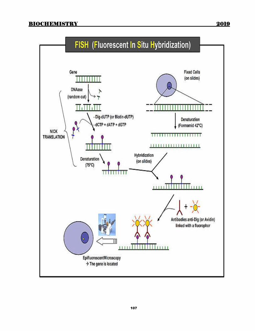
BIOCHEMISTRY 2019
107
BIOCHEMISTRY 2019
108
SITE-DIRECTED MUTAGENESIS
Site-directed mutagenesis is a molecular biology technique in which a mutation is created at a defined site in a DNA
molecule, usually a circular molecule known as a plasmid. In general, site-directed mutagenesis requires that the
wild type gene sequence be known.
This technique is also known as site-specific mutagenesis or oligonucleotide-directed mutagenesis. For our
purposes, we will define site-directed mutagenesis as any technique for introducing specific base-pair changes at
specific locations within a target DNA sequence such as a gene. In general, changes are limited in extent - most
often just one or two base pairs - with the intention of changing, for example, a restriction site, a codon, or the
sequence of some regulatory element. Occasionally, more extensive changes can be made such as, for example,
introducing six histidine codons so that a His-tagged protein my be expressed and isolated.
Site-directed mutagenesis can also be achieved by using PCR
This approach to mutagenesis was pioneered by the late Michael Smith at the Univerity of British Columbia. He
shared the 1993 Nobel Prize in Chemistry for his fundamental contributions to the establishment of oligonucleotide-
based, site-directed mutagenesis and its development for protein studies. Kary Mullis was the co-recipient. He
received the prize for his invention of the polymerase chain reaction (PCR) method
In this strategy, a piece of DNA is first cloned in a suitable vector. A ssDNA copy of the recombinant plasmid is
then prepared to which is annealed an oligonucleotide containing the desired sequenced changes. (Sometimes
additional oligonucleotides are used as suggested in this figure.) The annealed DNA now serves as a template for
DNA polymerase and the resulting heteroduplex DNA is then transformed into E. coli where the mutated clones will
be identified.
In theory, if all goes well then 50% of the clones will contain the original sequence and 50% will contain the
mutated sequence. In practice, the bias lies greatly in favour of the original sequence. This is due to inefficiencies in
synthesising the complementary DNA strand, primer annealing improperly to other sequences in template DNA, and
to the host cell repair systems which recognise the heteroduplex and remove the mutation that you are attempting to
introduce.
Site-directed mutagenesis has been widely used in the study of protein functions. There are many approaches. An
oligonucleotide-based method is illustrated in Figure below
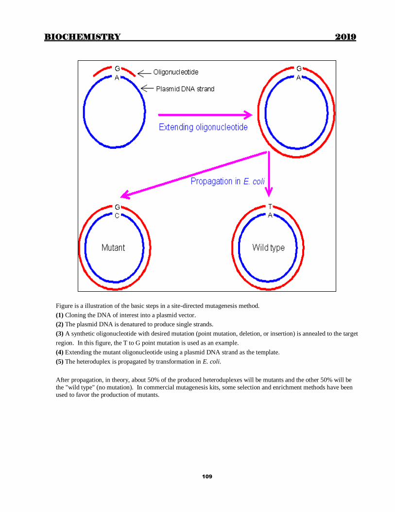
BIOCHEMISTRY 2019
109
Figure is a illustration of the basic steps in a site-directed mutagenesis method.
(1) Cloning the DNA of interest into a plasmid vector.
(2) The plasmid DNA is denatured to produce single strands.
(3) A synthetic oligonucleotide with desired mutation (point mutation, deletion, or insertion) is annealed to the target
region. In this figure, the T to G point mutation is used as an example.
(4) Extending the mutant oligonucleotide using a plasmid DNA strand as the template.
(5) The heteroduplex is propagated by transformation in E. coli.
After propagation, in theory, about 50% of the produced heteroduplexes will be mutants and the other 50% will be
the "wild type" (no mutation). In commercial mutagenesis kits, some selection and enrichment methods have been
used to favor the production of mutants.

BIOCHEMISTRY 2019
110
Same site directed mutagenesis can be demonstrated by figure below

BIOCHEMISTRY 2019
111
STUDYING GENE FUNCTION: CREATING KNOCKOUT MICE
Construction a knockout mouse
For decades researchers have tried to create tools that allowed for precise control over a specific gene in order to study its function. In the early 1980′s a breakthrough technology known as transgenics or gene transfer was
developed. This new technology involved the process of pronuclear microinjection, a method involving the
injection of genetic material into the nuclei of fertilized eggs (Genetic material is injected directly into the fertilized
egg which is then implanted back into a mouse and allowed to come to term). Following injection, DNA would
incorporate into the genome of the cell. The transformed fertilized eggs were then injected back into pregnant
females and brought to term. A major downfall of this technique is that researchers could neither predict nor
control where in the genome the foreign genetic material would be inserted. Since a gene’s location in the
genome is important for its expression pattern, mouse lines carrying the same transgene could display wildly varying
phenotypes.
An innovative solution that resolved this conflict was originated by a team of scientists led by Martin Evans, Oliver
Smithies and Mario Capecchi who created what is known as a genetic “knockout”. By creating a knockout they
proved that it was possible to aim the inserted gene at a precise location in the mouse genome. This gave scientists
the ability to replace, or knock out, a specific gene with an inactive or mutated allele. Therefore, knockout animals
are considered an investigative technique that allows for a particular gene of interest to be removed in an attempt to
define what effect that gene has in the life of the organism. The gene knockout is created by selectively disabling a specific target gene in a particular type of cells called embryonic stem cells. The three scientists received the Lasker
Award in 2001 for this achievement. The technique has been used to make several thousand different knockout
mice. Knockout mice have become one of the most useful scientific tools in helping to understand the human
genome and its roles in disease.
The process of gene targeting involves two processes. First a vector must be constructed. Once this has been done,
the vector is put into embryonic stem cells and a new mouse is made from these cells. The vector consists of two
regions of homology at either end, the disrupted gene of interest, and two additional genes to allow for quality
control in the process. The regions of homology are located at either end of the vector and are complementary to
specific sites on the genome surrounding the gene of interest. The vector binds to the genome at these two points of
homology. This technology then takes advantage of the process of recombination to swap the disrupted gene for the
real one. Here the two additional genes are used. In order to make sure that the vector has been inserted into the
genome, the neomycin resistance gene is included. If the vector has not been inserted then all cells will die when the
cells are grown in neomycin. It is also possible that the vector has been inserted into the genome in the wrong place.
To make sure this has not happened, the thymidine kinase (tk) gene is also added into the construct outside the
region of homology. If recombination has taken place then the tk gene will not be in the genome and so cells will be insensitive to gancyclovir. However if the vector has been inserted into the genome completely then the cell will die
when grown in gancyclovir. Thus through positive and negative selection, cells with the correct insert can be
isolated.
Embryonic stem cells are used as the target cells for the insertion of these vectors because they are pluripotent, meaning that they can give rise to every different type of cell in the adult body. Scientists interested in examining a
specific gene will remove or “knock out” the gene in an embryonic stem cell as described above, then put the cell
into a recently fertilized embryo. The manipulated cell then divides and eventually develops into many different
tissues. The result is a mouse in which some of the cells have developed from the knocked-out embryonic stem cells.
Often, embryonic stem cells which give rise to mice with a brown coat colour are injected into blastocysts which
give rise to mice with a white coat colour. This allows the researcher to see which mice have contributions from
both the injected cells and the blastocyst: if a mouse is completely white, then no injected cells grafted with the
blastocyst. On the other hand if the mouse has both brown and white markings on its coat, then the injection of
knockout embryonic stem cells was successful. After finding mice which have contributions from both cell types,
the mice are bred to propagate the knockout gene to a certain percentage of the progeny, a process known as germ-
line transmission. Once germ-line transmission is achieved, it is possible to breed mice where some are missing the knocked-out gene while others are not. In this case the latter group can act as a control group to see if the experiment
has worked.

BIOCHEMISTRY 2019
112
Uses of knockout mice
Knockout mice are used in a variety of ways. They allow to test the specific functions of particular genes and to
observe the processes that these particular genes could regulate. What actions does this gene turn off and on? By
examining what is happening in an in vivo (inside the body) model, we are able to determine the effects a particular gene may have. These effects would be impossible to observe in a culture dish. However, to completely establish
and assign an action to a particular gene is very challenging and requires an incredible amount of work.
One of the most exciting applications of knockout technology is in biomedical research. Scientists are using these
models to study the progress of thousands of genetically based diseases at the molecular level. The hope is that by
better understanding how a certain gene contributes to a particular disease, researchers can then take the knowledge
a step further and look for drugs that act on that gene. Another essential application of knockout mice is in drug
development. The knockout technology may lead to the discovery of the next generation of blockbuster therapies for
curing numerous diseases based on novel targets from the human genome.
Although knockout technology is highly advantageous for both biomedical research and drug development, it also
contains a number of limitations. For example, because of developmental defects, many knockout mice die while
they are still embryos before the researcher has a chance to use the model for experimentation. Even if a mouse
survives, several mouse models have somewhat different physical and physiological (or phenotypic) traits than their
human counterparts. An example of this phenomenon is the p53 knockout. Gene p53 has been implicated in as many
as half of all human cancers. However p53 knockout mice develop a completely different range of tumors than do
humans. In particular, mice develop lymphomas and sarcomas, whereas humans tend to develop epithelial cell-derived cancers [2]. Because such differences exist it cannot be assumed that a particular gene will exhibit identical
function in both mouse and human, and thus limits the utility of knockout mice as models of human disease.
Disadvantages of knockout technology
Although knockout mice are extremely useful in studying gene function, producing custom knockout mice is very
expensive costing between $3,000 to $30,000. There are hundreds of knockout mice commercially available and can
be purchased from a variety of companies. It is also important to consider the cost of care for genetically altered
animals, as they require special transgenic core facilities where staff can assist researchers with housing and taking
proper measures to keep these knockout animals at optimal conditions for research purposes. The cost of equipping
and maintaining such a facility is usually very high and may be a limiting factor.
Thus knockout mice are a wonderful tool used to study the function of specific genes in a system. Despite numerous
concerns and limitations applying this technique, it provides tremendous insight into understanding the disease process. The development of a gene knockout mouse has been a massive advance to the biomedical and
pharmaceutical field presenting researchers with a very powerful tool for analyzing gene function during
development, as well as in disease. In short, gene knockout technologies have become invaluable experimental tools
for modeling genetic disorders, assigning functions to genes, evaluating drugs and toxins, and for helping to answer
fundamental questions in basic and applied research.
BIOCHEMISTRY 2019
113
MICROSATELLITES AND DISEASE
Microsatellite sequences are repeating DNA sequences. For example, in one part of chromosome number 4, CAG
nucleotides are repeated many times over. They look like this: CAGCAGCAGCAG...... If the trinucleotides are
repeated too many times this would cause the person to get Huntington's disease in adult life. Other diseases that
involve repeats of three nucleotides are also known to cause neurological diseases (see below). At this time 14
neurological disor ders have been shown to result from the expansion of trinucleotide repeats, establishing an
expanding class of diseases. Trinucleotide repeat diseases can be categorised into two subclasses based on the
location of the trinucleotide repeats: diseases involving noncoding repeats (untranslated sequences) and diseases
involving coding sequences (exonic). In general trinucleotide repeat disorders are either dominantly inherited or X-
linked, the one exception being Friedrich's ataxia, which is autosomal recessive (Goldstein and Schlotterer, 1999; Cummings and Zoghbi, 2000).
Not all diseases are caused by a mistake in one gene. Sometimes many genes may be involved in a disease, for
example, in schizophrenia. For these diseases microsatellite sequences have been used as a marker for locating the
diseased region of the chromosome. This method is called positional cloning. Microsatellite markers close to the
disease gene correlate with the heredity of the disease, and by analysis of these markers within families scientists
can predict how the disease will be inherited (Risch, 2000).
EXAMPLES OF DISEASES INVOLVING TRINUCLEOTIDE REPEATS-
Huntington's disease
Symptoms: Late onset dementia and loss of motor control, resulting in full-blown chorea after 10-20 years. Motor disorder is often preceded or accompanied by memory deficits, cognitive decline or changes in personality. Juvenile
onset is rare and patients show rigidity, bradykinesia, epilepsy, severe dementia and an accelerated disease course.
Involvement of microsatellites: CAG coding repeat in the first exon of the HD gene. Normal gene contains between
6 and 35 repeats and the affected gene from 36 to 121 repeats. Adult onset typically occurs when the repeat contains
40 - 50 units, whereas alleles containing more than 70 repeats typically result in the more severe juvenile form. The
microsatellite adds a string of glutamine amino acids to the huntingtin protein
Chromosome location: #4
Fragile X
Symptoms: Mental retardation, long and prominent ears and jaws, high-pitched speech, hyperactivity, poor eye
contact, and stereotypic hand movements (e.g. hand-flapping and hand-biting). 1 in 4000 males are affected, and fewer females are affected, depending on the ratio of cells with normal X chromosome active to abnormal X active.
Involvement of microsatellites: The repeat is CGG in a non-coding region of the FMR2 gene, and normal is 6 - 53
repeats. The disease occurs if the repeat is between 60 - 200.
Chromosome location: X
Myotonic dystrophy
Symptoms: Congenital DM is the most severe form of this disease involving hypotonia, respiratory distress at birth
and developmental abnormalities. Adult onset includes variable loss of mental function, myotonia, muscle
weakness and progressive muscle wasting. Other features may include facial dysmorphology, presenile cataracts,
testicular atrophy, premature balding in males, kidney failure, hyperinsulin secretion and cardiac conduction
abnormalities.
Involvement of microsatellites: CTG repeats in a non-coding region of the DMPK gene. Normal is between 5 and 37, the disease may involve from 50 - 1000s of repeats.
Chromosome location: #19
Spinalbulbar muscular atrophy
Symptoms: Neurological degeneration leading to difficulties in speech, articulation and swallowing, muscle
weakness and atrophy. Signs of mild androgen insensitivity are typically seen at adolescence.
Involvement of microsatellites: CAG repeat in first coding exon of the androgen receptor (AR) gene. Between 9 and
36 repeats is normal, and people with 38-62 repeats develop the disease.
Chromosome location: X, recessive.

BIOCHEMISTRY 2019
114
Friedrich's ataxia
Symptoms: Ataxia, diminished tendon reflexes, loss of position and vibratory senses, dysarthria (slurred speech),
cardiomyopathy, diabetes mellitus, optical atrophy, scoliosis and skeletal abnormalities. Age of onset is typically
early childhood.
Involvement of microsatellites: GAA repeat in non-coding region of gene X25. Normal gene contains between 7
and 34 repeats. The disease gene has 34 to 80 repeats.
Chromosome location: #9, recessive
EXAMPLES OF DISEASES FOUND BY POSITIONAL CLONING
Schizophrenia and Bipolar Disorder Symptoms: Schizophrenia is characterized by auditory and visual hallucinations and delusions; symptoms of
bipolar disorder involve severe mood swings, between mania and depression.
Involvement of microsatellites: Using 388 microsatellite markers within 8 families the researchers (Bailer et al.,
2002) found a susceptibility locus for both schizophrenia and bipolar disorder on chromosome 3.
Chromosome location: #3
Congenital generalized hypertrichosis
Symptoms: Rare hair growth disorder, characterized by excessive hair growth on the face and upper body;
sometimes called "werewolf" syndrome.
Involvement of microsatellites: Using hypervariable microsatellite markers the researchers (Figurea et al., 1995)
localized the gene to a part of the X chromosome.
Chromosome location: X, dominant.
Asthma and Bronchial Hyperresponsiveness
Symptoms: Common respiratory disorder characterized by recurrent episodes of coughing, wheezing and
breathlessness.
Involvement of microsatellites: A putative asthma susceptibility gene was identified on the ADAM33 region of
chromosome 20 (Van Eerdewgh et al., 2002).
Chromosome location: #20
Detecting Cancer
The rate of microsatellite expansion (that is, increase in the number of repeats) or contraction (decrease in number of repeats) in cells is increased in some types of cancers, due to defects in enzymes that correct copying mistakes in
DNA. Early clinical detection of some types of colon and bladder cancers using changes in microsatellite repeats
have been successful (Yonekura et al., 2002; Moxon and Willis, 1999).
BIOCHEMISTRY 2019
115
Chapter 14
BIOTECHNOLOGY
RESTRICTION ENDONUCLEASES Cleave double-stranded (ds) DNA into smaller fragments. Each enzyme cleaves DNA at a specific nucleotide
sequence-used experimentally to obtain precisely defined DNA segments called restriction fragments.
-Named after the bacterium from which they are isolated
EcoRI From E Coli
BamHI from Bacillus amyloliquifaciens
RE recognize short stretches of DNA (4-6 base pairs) that contain specific palindrome nucleotide sequences; this
site is called restriction site
Produce “sticky or blunt” ends
Using the enzyme DNA ligase sticky ends of a DNA fragment can be covalently joined with other DNA fragments
that have sticky ends produced by cleavage with the same RE. Another ligase, encoded by bacteriophage T4, can
covalently join blunt-ended fragments.
DNA CLONING
Introduction of a foreign DNA molecule into a replicating cell permits the amplification (production of many
copies) of the DNA.
To clone a nucleotide sequence of interest, the total cellular DNA is first cleaved with a specific restriction
enzyme, creating millions of fragments.
Each of the resulting DNA fragments is joined to a DNA vector molecule to form a recombinant molecule.
This molecule conveys its inserted DNA fragment into a single host cell, (eg: a bacterium), where it is replicated
(or “amplified”).
The process of introducing foreign DNA into a cell is called transformation for bacteria and transfection for
eukaryotes.
As the host cell multiplies, it forms a clone in which every bacterium carries copies of the same inserted DNA fragment, hence the name “cloning.”
The cloned DNA is released from its vector by cleavage (using the appropriate restriction endonuclease) and is
isolated.
By this mechanism, many identical copies of the DNA of interest can be produced.
VECTORS
A vector is a molecule of DNA to which the fragment of DNA to be cloned is joined
Prokaryotic plasmids: Plasmids may carry genes that convey antibiotic resistance to the host bacterium, and
may facilitate the transfer of genetic information from one bacterium to another. They can be readily isolated
from bacterial cells, their circular DNA cleaved at specific sites by RE and foreign DNA inserted. The hybrid
plasmid can be reintroduced into a bacterium, and large numbers of copies of the plasmid containing the foreign DNA produced
Other vectors:
o viruses that infect bacteria (bacteriophage λ) or mammalian cells (eg: retroviruses)
o artificial constructs such as cosmids and bacterial or yeast artificial chromosomes (BACs or YACs,
respectively)
Recombinant DNA techbology
DNA LIBRARIES
A DNA library is a collection of cloned restriction fragments of the DNA of an organism.
Genomic libraries: contain a copy of every dsDNA nucleotide sequence in the genome. cDNA libraries contain those DNA sequences that appear as mRNA molecules, and these differ from one
cell type to another.
Cloned cDNA lack introns and the control regions of the genes, whereas these are present in genomic
libraries.

BIOCHEMISTRY 2019
116
PROBES
Cleavage of large DNA molecules by restriction endonucleases produces an array of fragments. To pick a specific
gene or DNA sequence of interest from the mixture of millions of DNA fragments, probes are used. Probe is a short,
single-stranded piece of DNA, labeled with a radioisotope, or with a nonradioactive probe, such as biotin. The
nucleotide sequence of a probe is complementary to the target DNA
BLOT TECHNIQUES
Southern blot detects DNA
Northern blot – RNA
Western blot - Protein
Southern Blotting
Southern blotting is a technique that can detect mutations in DNA. It combines the use of restriction enzymes,
electrophoresis, and DNA probes.
Invented by Edward M Southern
DNA is extracted from cells
DNA is cleaved into many fragments using a restriction enzyme
The fragments are separated by electrophoresis.
The DNA fragments in the gel are denatured and blotted to a nitrocellulose membrane for analysis.
A probe is used to identify the DNA fragments of interest.
The patterns observed on Southern blot analysis depend both on the specific restriction endonuclease and on
the probe used to visualize the restriction fragments
RESTRICTION FRAGMENT LENGTH POLYMORPHISM
Genome variations are differences in the sequence of DNA among individuals. It has been estimated that the
genomes of nonrelated people differ at about 0.1% of the genome.
These genome variations include both polymorphisms and mutations. A polymorphism is a clinically harmless
DNA variation that does not affect the phenotype. In contrast, mutation refers to an infrequent, but potentially
harmful, genome variation that is associated with a specific human disease.. Polymorphisms often occur in the
intervening sequences that do not code for proteins.
A restriction fragment length polymorphism (RFLP) is a genetic variant that can be examined by cleaving the
DNA into fragments (restriction fragments) with a restriction enzyme. The length of the restriction fragments is
altered if the genetic variant alters the DNA so as to create or abolish a restriction site.
RFLP can be used to detect human genetic variations
Two types of DNA variation commonly result in RFLPs:
Single-base changes in the nucleotide sequence
Tandem repeats of DNA sequences.
POLYMERASE CHAIN REACTION
Invented by Karry Mullis 1989 (Nobel prize 1993)
In vitro DNA amplification: Millions of copies of a sequence of DNA produced: no cloning needed
DNAP is Taq polymerase
Advantages of PCR
The major advantages of PCR over cloning are sensitivity and speed. Applications
Comparison of a normal cloned gene with an uncloned mutant form of the gene:
Detection of low-abundance nucleic acid sequences: eg: viruses during window period
Forensic analysis of DNA samples: DNA fingerprinting by means of PCR has revolutionized the analysis
of evidence from crime scenes.
Prenatal diagnosis and carrier detection of diseases like cystic fibrosis:
Detection of malignancy
Fossil studies
ANALYSIS OF GENE EXPRESSION
Can be done by
Determination of mRNA levels

BIOCHEMISTRY 2019
117
Analysis of proteins
Determination of mRNA levels can be by
Northern blot
DNA microarray
NORTHERN BLOTS: Very similar to Southern blots except that the original sample contains a mixture of mRNA molecules that are
separated by electrophoresis, then transferred to a membrane and hybridized to a radioactive probe. The bands
obtained by autoradiography give a measure of the amount and size of particular mRNA molecules in the sample
Microarrays: DNA microarrays contain thousands of immobilized DNA sequences organized in an area no larger
than a microscope slide.
used to analyze a sample for the presence of gene variations or mutations (genotyping)
used to determine the patterns of mRNA production (gene expression analysis), analyzing thousands of genes at
the same time.
For genotyping analysis, the cellular sample is genomic DNA.
For expression analysis, mRNA molecules from a particular cell type is converted to cDNA and labeled with a
fluorescent tag
This mixture is then exposed to a gene chip, which is a glass slide or membrane containing thousands of tiny
spots of DNA, each corresponding to a different gene.
The amount of fluorescence bound to each spot indicates the amount of that particular mRNA in the sample.
DNA microarrays are often used to determine the differing patterns of gene expression in two different types of
cell—for example, normal and cancer cells
ANALYSIS OF PROTEINS
Can be done by
ELISA
Western blot
GENE THERAPY
The goal of gene therapy is to insert the normal, cloned DNA for a gene into the somatic cells of a patient
who is defective in that gene.
The DNA must become permanently integrated into the patient's chromosomes in such a way as to be
properly expressed to produce the correct protein.
TRANSGENIC ANIMALS
Transgenic animals can be produced by injecting a cloned gene into the fertilized egg.
If the gene becomes successfully integrated into a chromosome, it will be present in the germline of the
resulting animal, and can be passed along from generation to generation.
Transgenic goats and cows can be designed that produce human proteins, such as blood clotting factors, in
their milk.
BIOCHEMISTRY 2019
118
Chapter 15
SEPARATION TECHNIQUES
CHROMATOGRAPHY Chromatography is a technique by which the components in a mixture of similar substances are separated by a
continuous redistribution between 2 phases, one stationary and the other mobile. The differences in the attractive
forces between the stationary phase and the substances to be separated leads to selective retardation of these
substances, relative to the mobile phase.
The technique was first applied by Tswett to a mixture of plant pigments. (Hence the name).
Depending on the type of stationary and mobile phases, chromatography can be of different types.
Liquid-solid chromatography mobile phase liquid; stationary phase solid
Liquid-liquid chromatography mobile phase liquid; stationary phase also liquid
Gas-solid chromatography mobile phase gas; stationary phase solid
Gas-liquid chromatography mobile phase gas; stationary phase liquid
When the stationary phase is liquid, it is supported on a solid like paper, silica etc.
Reverse phase chromatography
In liquid-liquid chromatography, the stationary phase is polar and mobile phase is less polar. But in some cases, we
make the stationary phase non-polar and mobile phase polar. This is called reverse phase chromatography.
Depending on the mechanism of selective retardation of the particles, chromatography can be of different types like;
Partition chromatography
Adsorption chromatography
Ion exchange chromatography
Affinity chromatography
Gel filtration chromatography (Molecular sieve chromatography)
Partition chromatography Most commonly done method. Various components of the mixture are separated depending on their partition
coefficient.
There are mainly 3 types of partition chromatography.
1. Paper chromatography
2. Thin layer chromatography
3. Gas-liquid chromatography
Paper chromatography This is a liquid-liquid partition chromatography. The stationary phase is water, held on a solid support of filter
paper. Mobile phase is a mixture of solvents like butanol and acetic acid or phenol and ammonia.
If the mobile phase is applied from the bottom, it is called ascending chromatography and if it is applied from the
top, descending chromatography.
The mixture to be separated is applied as a small spot near a corner of the paper and this paper is mounted on the
solvent in a tank. The solvent ascends in the filter paper by capillary action and the substances are separated by the
difference in partition coefficient.
Thin layer chromatography (TLC) Silica gel is spread as a thin layer on a glass slide. The sample is applied on one corner. The plate is then placed in a
tank containing the solvent. Water is held as the stationary phase and the non-polar solvent acts as the mobile phase (so it is also a liquid-liquid chromatography). It is less time consuming than paper chromatography (takes only 3-4
hours whereas paper chromatography takes 12-16 hours).
In paper as well as thin layer chromatography, once the procedure is completed it is necessary to visualize the spots
formed by various substances. For this, substances called location reagents are sprayed to the paper or glass slide,
which will react with the substances to give coloured spots.
Examples of location reagents:
Ninhydrin for amino acids
Diphenylamine for sugars
Identification of the spots
BIOCHEMISTRY 2019
119
After locating the spots, the next aim is to identify the substances. For this we have to calculate the Rf values (Ratio
of fronts) of the spots and compare them with Rf values of known standards.
Rf value of a particular substance is a constant for a particular solvent system at a given temperature.
In paper chromatography, the solvent used is butanol-acetic acid-water mixture. Water trapped in between cellulose fibres act as the stationary phase. So hydrophilic amino acids move less than hydrophobic amino acids In a paper
chromatography, Rf value (distance travelled by the solute/ distance travelled by the solvent) is calculated. The more
the Rf. The more it moves
Highest Rf is for Leucine.
Leucine > Isoleucine > Phenyl alaine > valine > Tyrosine ……. …….
…… …… Arg > His > Asparagine > Lysine > Cystine
2 dimensional chromatography: When the Rf values of the substances are very close to one another, the separation
may not be satisfactory. Then we do a second chromatography in a direction perpendicular to the first one, using a
different solvent system.
Gas liquid chromatography A type of partition chromatography.
Stationary phase is liquid supported by inert materials like silica kept as a long narrow column. This method is
suitable for those substances which can resist high temperature. The mixture is made volatile at one end of the
column, mixed with an inert gas and passed through the column. A detecting device is used for detecting the various
fractions which emerge at the other end of the column.
Adsorption chromatography Based on the differences in adsorption of the substances to a solid medium. Silica gel, alumina etc are made into
columns and used as the stationary media. The mixture is applied in a solvent on the top of the column. Due to the
difference in adsorption of each substance, they move down with different speeds and get separated. Each fraction
can be separated using suitable solvents.
Ion-exchange chromatography In this, charged biological molecules are separated based on their electrostatic attraction with substances called ion-
exchange resins. Ion-exchange resins are cross-linked polymers with ionic groups as part of their structure. These are 2 types: anion
exchange resins (contain a quarternary N+) and cation exchange resins (contain sulphonic or carboxylic groups).
Column chromatography is done using the resin as stationary medium. The substances bind to the resins depending
on their charges. Then these are eluted by using buffers of increasing pH gradient.
Affinity chromatography Based on the high affinity of some proteins to specific substances. These specific substances are used in columns to
isolate or purify the proteins.
Eg:-
Coenzymes are used for isolating enzymes.
Amino phenyl boronic acid is used for isolating glycated Hb
Antibodies are used for purification of antigens.
Gel filtration chromatography: also known as (Molecular sieve chromatography)
Here the mixture to be separated is passed through a column of porous hydrophilic gel (eg:Agarose, Acryl amide).
This column acts as a molecular sieve, which allows small molecules
to enter into the gel particles, and prevent large molecules from entering. So large molecules bypass the gel particles and move down at a faster rate than the smaller particles. This technique is mainly used for separation, purification
or molecular weight determination of proteins.
HPLC (High performance liquid chromatography)
It is a type of column chromatography in which the basic mechanisms are the same as adsorption, partition or ion-
exchange chromatography. But the mobile phase is passed through the column under high pressure. This leads to a
high resolution, and it requires only a few minutes.

BIOCHEMISTRY 2019
120
ELECTROPHORESIS Electrophoresis is the movement of charged particles through an electrolyte when subjected to an electric field.
Factors affecting electrophoresis
The rate of movement of the particles during electrophoresis depends on:
1. Net charge on the particles
2. Strength of the electric field
3. Mass and shape of the particles 4. pH of the medium
5. Properties of the supporting medium
6. Temperature
Electrophoresis apparatus
The electrophoresis apparatus consists of a power pack for
a stable continuous supply of electricity, a tank to hold the
buffer, electrodes and the support medium for
electrophoresis, which is connected to the buffer by
wicks.
Common supporting media in use are:
1. Filter paper- requires 16-18 hours 2. Cellulose acetate membrane strips- takes only <1
hour; expensive
3. Agar gel- takes 1½ -2 hours; less expensive;
reasonably good separation.
4. Poly acryl amide gel-it has high molecular
sieving effect; so ensures good separation.
Different types of buffer can be used depending on the
substances to be separated.
After the electrophoretic run is completed, the substances are fixed to the supporting medium using a fixative. The
mixture will be separated into different bands. Amount of each component in the mixture can be calculated by 2 methods:
Elution
Scanning using densitometer.
Serum protein separation using agar gel electrophoresis
The buffer used is barbitone buffer at the pH of 8.6. At this pH all the serum proteins have a net negative charge and
will move towards the anode.
Agar gel is prepared in barbitone buffer. This gel is liquid when hot and is spread on a glass slide. This solidifies on
cooling. A few microlitres of sample is applied by cutting into the gel using a sharp object. Then it is subjected to
electrophoresis for about 90 minutes.
The slide is taken out, dried. The various fractions are fixed using methanol. Then the bands are stained by using a
dye (Amidoschwarz). Excess stain is removed by dil. acetic acid. Albumin moves most rapidly, so is far away from the point of application. This is followed by α 1 , α 2, β and γ
globulins. Gamma globulins have the smallest negative charge, so move only slightly- or it may slightly move to the
opposite side due to electroendosmosis. If plasma is used, fibrinogen is found as a narrow band between β and γ
globulins.
Variations in the patterns can occur depending on the blood level of various fractions. In multiple myeloma we get a
sharp spike of γ globulin band which is called myeloma band (M band). Similar bands may be seen in
Waldenstorm’s macroglobulinemia and heavy chain disease.
PAGE (Poly Acrylamide Gel Electrophoresis)
Poly acryl amide has a high molecular sieving effect and so gives a better separation. Serum proteins are separated
into more than 20 different bands. In SDS-PAGE, proteins are boiled for 1-2 minutes with a denaturing agent, sodium dodecyl sulphate (SDS). The
negative charge of SDS will cover the protein molecules, making them strongly electronegative: so the separation is
then based on their molecular size. This method is mainly used for molecular weight determination of proteins.

BIOCHEMISTRY 2019
121
Isoelectric focusing
A pH gradient is generated from anode to cathode. Each protein gets concentrated at the point where the local pH is
the same as the isoelectric point. This method is useful for separation of isoenzymes, Hb variants etc.
Immunoelectrophoresis
Agarose gel electrophoresis is done first with the serum sample applied in wells cut into the gel. To visualize the
different fractions, a specific antibody is applied to a trough cut into the gel and incubated. Precipitation arcs are
formed when the antigen and antibody molecules are at 1:1 ratio. It is a highly sensitive method to study proteins.
High voltage electrophoresis
400 – 2000 V is used; Takes < half an hour; Used for separation of nucleotides
Capillary electrophoresis
Very low quantity of sample is required
Gel taken in capillary tube
High voltage 25,000 V
Complete in a few minutes
Pulsed field gel electrophoresis
Direction of current is altered at regular intervals (electrical field is cycled between electrodes)
Used to separate DNA fragments of > 50 kBp length
2 dimensional electrophoresis
First electrophoresis based on charge in one direction; Second, based on m:wt
Identify mutant DNA, abnormal proteins in genetic disorders

BIOCHEMISTRY 2019
122