Embed Size (px)
Citation preview

Hindawi Publishing CorporationJournal of NanomaterialsVolume 2012, Article ID 397206, 9 pagesdoi:10.1155/2012/397206
Research Article
Changes in Cardiac Autonomic Regulation afterAcute Lung Exposure to Carbon Nanotubes: Implications forOccupational Exposure
Jacopo M. Legramante,1, 2, 3, 4 Sergio Sacco,1, 3, 4 Patrizio Crobeddu,1, 2, 3, 4 Andrea Magrini,5
Federica Valentini,6 Giuseppe Palleschi,6 Marco Pallante,1, 4 Rita Balocchi,7 Ivo Iavicoli,8
Antonio Bergamaschi,8 Alberto Galante,1, 2, 3 Luisa Campagnolo,9 and Antonio Pietroiusti5
1 Dipartimento di Medicina Interna, Universita “Tor Vergata”, Via Montpellier 1, 00133 Roma, Italy2 A.F.O. Medicina d’Urgenza-Pronto Soccorso, Dipartimento delle Emergenze, Policlinico Tor Vergata, Viale Oxford 81,00133 Roma, Italy
3 Divisione di Riabilitazione Cardiologica, IRCCS S. Raffaele, Via dei Laghi Km 19.600, 00049 Velletri, Italy4 Stazione per la Tecnologia Animale, Universita “Tor Vergata”, Via Montpellier 1, 00133 Roma, Italy5 Sezione di Medicina del Lavoro, Dipartimento di Biopatologia e Diagnostica per Immagini, Universita di Roma “Tor Vergata”,Via Montpellier 1, 00133 Rome, Italy
6 Dipartimento di Scienze e Tecnologie Chimiche, Universita di Roma “Tor Vergata”, Via della Ricerca Scientifica 1,00133 Roma, Italy
7 CNR-Centro Nazionale delle Ricerche, Istituto di Fisiologia Clinica, Via Giuseppe Moruzzi 1, 56124 Pisa, Italy8 Istituto di Medicina del Lavoro, Universita Cattolica del Sacro Cuore, Largo F. Vito 1, 00168 Roma, Italy9 Dipartimento di Salute Pubblica e Biologia Cellulare, Universita “Tor Vergata”, Via Montpellier 1, 00133 Roma, Italy
Correspondence should be addressed to Antonio Pietroiusti, [email protected]
Received 14 October 2011; Accepted 20 February 2012
Academic Editor: Sergio Iavicoli
Copyright © 2012 Jacopo M. Legramante et al. This is an open access article distributed under the Creative Commons AttributionLicense, which permits unrestricted use, distribution, and reproduction in any medium, provided the original work is properlycited.
Carbon nanotubes (CNTs) are among the most relevant engineered nanomaterials (ENMs). Given the expected rise of exposure toENMs, there is concern that they may adversely affect health of exposed people. Aim of the study was to test the hypothesis thatsingle wall carbon nanotubes (SWCNTs) pulmonary exposure acutely affect the autonomic cardiovascular regulation in consciousrats. We studied Wistar-Kyoto rats in which a telemetry transmitter for continuous arterial pressure (AP) and heart rate (HR)recordings was surgically implanted. SWCNTs dispersed in phosphate buffer saline (PBS) or PBS alone were randomly adminis-tered intratracheally. Immediately before, and 24 hours after each instillation a 30 min AP recording was performed. The sequenceanalysis was performed to evaluate the baroreflex function. In the control group, PBS instillation did not induce any significantchanges. At variance the SWCNT exposure induced a significant reduction of baroreflex system (BRS) (3.5 ± 0.6 versus 2.6 ±0.40 msec/mmHg) without significant changes in the occurrence of baroreflex sequences (7.5 ± 0.47% versus 7.4 ± 0.38%). Ourresults show that SWCNT pulmonary exposure might affect the cardiovascular autonomic regulation thus contributing to cardiacand arrhythmic events.
1. Introduction
There is strong evidence that episodic high levels of airborneparticulate matter (PM) are associated with stroke, heartattacks, heart arrhythmias, and sudden death [1]; theseevents may be precipitated, at least in part, by alterations in
the autonomic input to the heart [2–5], which can be dis-played by a partial or total loss in spontaneous heart ratevariability (HRV). Indeed, decreased HRV is highly pre-dictive for increased risk of arrythmias and sudden cardiacdeath [6], especially in subjects with ischemic heart disease[7].

2 Journal of Nanomaterials
There are also reports showing that higher levels of ambi-ent air pollutants increase the risk of emergency department(ED) visits for cardiac arrhythmia [8]. Moreover it has beensuggested that alterations in the autonomic tone eventuallydue to increased environment air pollution might contributeto the instability of a vascular plaque or initiate cardiac ar-rhythmias thus representing a plausible explanation for theoccurrence of acute cardiovascular events, such as increasedmyocardial infarctions [9].
However, previous studies on PM have shown thatultrafine (UF) particles are more cytotoxic, inflammatory,and fibrogenic on an equivalent mass basis than fine-sizedparticles of the same composition. It is therefore not surpris-ing that a recent large European study showed that the ultra-fine (UF) component of PM is the major factor contributingto the alterations of cardiovascular autonomic control [10].The responsible underlying mechanisms remain unclear butmay involve activation of pulmonary neural reflex arcs,direct effects of pollutants on cerebral areas responsible forautonomic control or on cardiac ion channels, or may followthe heightened systemic inflammatory state.
Synthetic nanomaterials, developed in recent years toengineer new structures, materials, and devices, generallyoccur in size ranges similar to UF particles. An importantclass of synthetic nanomaterials is represented by carbonnanotubes. These cylindrical carbon molecules have novelproperties, making them potentially useful in many applica-tions in nanotechnology, electronics, optics, and other fieldsof materials science, as well as potential uses in architecturalfields. Therefore, their wide utilization raises concern aboutpossible adverse effects on cardiovascular system. Indeed,some preliminary animal data suggest that carbon nanotubesmay promote atherosclerosis [11].
No definitive data are currently available on their possibleeffects on the cardiovascular autonomic control.
The introduction of techniques able to monitor arterialpressure and heart rate in conscious, freely moving animalsallows to evaluate possible modifications of autonomic car-diovascular regulation over time in response to various expe-rimental conditions [12]. In particular, the possibility tomonitor the baroreflex control of sinus node by studying thebaroreflex sensitivity (BRS), in a noninvasive and nonobtru-sive way [13], is really intriguing due to its prognostic valuein cardiovascular diseases [14, 15].
Therefore aim of the present study was to test the hypo-thesis that single wall carbon nanotubes (SWCNTs) pul-monary exposure in a physiological model of conscious ratacutely affect the autonomic cardiovascular regulation andin particular the arterial baroreflex function which has beenwidely considered as a predictive index of cardiac mortalityin patients affected by ischemic heart disease and myocardialinfarction [14, 15].
2. Material and Methods
2.1. SWCNT Preparation. SWCNTs (CarboLex, Aldrich,Steinheim, Germany) produced by the arc-discharge process,employing CO in a continuous-flow gas phase as the carbon
Figure 1: Scanning electron microscopy imaging of SWCNTsadministered to rats. A high degree of agglomeration is evidenced.
feedstock and Fe(CO)5 as the iron-containing catalyst pre-cursor and purified by the HNO3 acid treatment at roomtemperature, were used in this study. For the SWCNTs mor-phological and structural characterization, Raman spec-troscopy and scanning electron microscopy (SEM) were rou-tinely used to characterize the as-received nanotube samplesand the samples submitted to the purification processingsteps. The Raman spectroscopy revealed a typical spectrumassociated with the radial breathing mode (RBM) of carbonatoms, representing the fingerprint of single-walled carbonnanotubes. The analysis of the Raman signals in the tangen-tial modes of the graphite lattice allowed information aboutthe phase purity of the purified carbon nanotubes. This lastshowed a Raman spectrum unaffected by the acidic treat-ments.
2.2. Physicochemical Characterization. Purified suspendedSWCNTs were used in the study at the final concentration of1 mg/mL. The range of diameter and length and surface areaof SWCNTs were 1.2–1.6 nm, 2–5 µm, and 300 m2/g, respec-tively. Surface area was determined by Brunauer, Emmett,and Teller analysis (BET method). Carbon nanotubes weresuspended in phosphate-buffered saline (0,1 M PBS, pH 7.4);immediately before the pulmonary exposure, they were soni-cated for 10 minutes with a Branson Sonifier B-12 (Cell Dis-ruptor, Sonic Power Company Danbury Connecticut at100 W) in order to obtain a better dispersion of the nano-material. The choice to disperse SWCNTs in PBS was takenin order to reproduce the same experimental conditions ofa previous relevant study showing a proatherosclerotic effectafter intratracheal instillation of SWCNTs [11]; the 10 minsonication procedure was chosen because in preliminaryexperiments we found that after 10 min sonication, the dis-persion remained stable up to 1 min. However, as shown inFigure 1, a high degree of agglomeration was detectable atscanning electron microscopy. During all the instillationexperiments, the purified SWCNT dispersion was subjected

Journal of Nanomaterials 3
260
R-R
160
60
(mse
c)
150
100
50
0
(mm
Hg)
AP
++
Baroreflex sequences 260
R-R
160
60
(mse
c)
150
100
50
0
(mm
Hg)
AP
−−
Figure 2: Examples of the arterial pressure, ECG signals and of the corresponding pulse intervals. The signals refer to sequences during whichSAP and PI of the fifth heart beat (i.e., lag 5, used for the high heart rate of rats, see Section 2 for details) changed in the same direction,either increasing, hypertension/bradycardia, or decreasing, hypotension/tachycardia (baroreflex sequences).
to the ultrasonic bath (LBS2, 135 W) for 10 minutes, andinstilled intratracheally within 1 minute after sonication, toensure stability of the homogeneous SWCNT dispersion. Theamount of SWCNTs given during each session was 1 µg/grbody weight. This concentration is in the lower range ofthe dosage given in previous experimental studies aimed atassessing lung and cardiovascular toxicology of SWCNTs [11,16, 17].
2.2.1. General Procedures. The study was performed on 16adult Wistar-Kyoto (WKY) rats of both sexes (350 g body wt)and the experimental procedures were carried out accord-ing to the Association for Assessment and Accreditation ofLaboratory Care International and approved by the animalcare facility (Stazione per la Tecnologia Animale) of theUniversity “Tor Vergata” and by the Italian Health Minister.Rats were used and housed individually in the animal carefacility, allowed to normal rat chow and drinking water adlibitum, and kept on a 12 h light-12 h dark cycle.
2.2.2. Surgical Procedures. As previously described [13], afterhaving induced the anesthesia by Ketamine (Ketavet 5060 mg/kg i.p.) and Medetomidine (Domitor 0.3 mg/kg i.p.) atelemetry transmitter (TA11PA-C40, Data Sciences, St. Paul,MN, USA) was implanted for recordings of AP signals ac-cording to manufacturer specifications. The tip of the arterialcatheter was inserted into the abdominal aorta previouslyexposed by a midline incision via a hole made by a 21-gaugeneedle below the bifurcation of the renal arteries just proxi-mal to the iliac bifurcation and secured in place with tissueglue (Vetbond, 3 M). The transmitter body was attached tothe abdominal wall along the incision line with sutures asthe incision was closed. After surgery, the rats were givenantibiotics (ceftriaxone) and housed individually in cages for5–7 days of recovery before any experimental protocol began.
2.2.3. Measurement of Arterial Pressure Using Radiotelemetry.As previously reported [13] the system used to record arterialpressure consists of three basic elements: (1) a transmitter for
monitoring BP (TA11PAC40); (2) a receiver (RPC-1); (3) anadapter (R11CPA) with an ambient pressure monitor (APR-1) that produces analog output signals of pulsatile AP. Thetelemetered AP signal was digitized using an analog I/O PCcard (National Instrument 6024E, Austin, TX, USA) at a rateof 2000 Hz, displayed on the computer screen and processedby an algorithm based on feature extraction to detect andmeasure the characteristics of AP cycles developed in our lab-oratory based on a Lab view platform software. Pulse inter-val (PI) was measured from the pressure pulses and used tocalculate heart rate (HR).
2.2.4. Experimental Protocol. Rats were randomly dividedinto two groups: control rats (n = 7) and SWCNT instilledrats (n = 9). SWCNTs or PBS were given to SWCNT instilledand control group, respectively. Briefly, after having inducedthe anesthesia as previously described, the trachea was intu-bated with a polyethylene cannula of the same diameter andthe instillation was performed. Rats in both groups under-went 30 min duration AP recordings performed in baselineconditions and 24 hours after the instillation.
2.3. Baroreflex Analysis by “Sequence Technique”. The sequ-ence analysis was performed as previously reported [13, 18].Briefly, the beat-by-beat time series of systolic arterial pres-sure (SAP) and PI were analyzed by a computer to identifyspontaneously occurring sequences of three or more con-secutive beats in which SAP and PI of the fifth heart beat(i.e., lag 5, used for the high heart rate of rats) [19] changedin the same direction, for example, SAP increasing andPI increasing (i.e., hypertension and bradycardia) or SAPdecreasing and PI decreasing (i.e., hypotension and tachy-cardia). These sequences were identified as “baroreflex”sequences (Figure 2). A linear regression was applied to eachindividual sequence, similarly to the Oxford techniqueemploying bolus injections of vasoactive drugs. Only thosesequences in which r2 was >0.85 were accepted and thenumber of baroreflex sequences was calculated.

4 Journal of Nanomaterials
The mean individual slope of the baroreflex sequences,obtained by averaging all slopes computed within a givenexperimental period, was calculated and taken as a measureof the baroreflex sensitivity (BRS) for that period [18].
The engagement time was also calculated as the fractionaloccurrence of the sequences independently on recordingtime and/or HR as previously reported [13]. Briefly, thisindex has been obtained by dividing the sum of the R-Rorganized in sequences, according to the criteria reportedabove, by the total recording duration and multiplying it by100.
2.4. Power Spectral Analysis. The heart rate variability (HRV)was assessed in the frequency domain by computing thepower spectral density (PSD) in selected frequency bands.
As expected, dealing with conscious and freely movinganimals, the 30 min RR recordings showed periods of non-stationarities both in mean and standard deviations. To com-ply with the requirements of the analysis we first processed allthe recordings to extract stationary epochs (in weak sense).To this aim a mixed, automatic and visual, procedure was ap-plied. First, mean, standard deviation (SD) and slope of thelinear trend were computed over nonoverlapping intervalsof 1 min spanning the whole recording. The selection ofstationary intervals was then performed by visual inspectionof the time course of mean, SD, and slope values. Stationaryintervals of different lengths were detected inside the record-ings: for uniformity of analysis a 5 min stationary intervalwas selected for each animal.
Spectral estimation was performed using Welch’s aver-aged modified periodogram method with a Hamming win-dow and a minimum of 50% epochs overlap. The variableoverlap comes from the choice of having, for each animal, atleast a preset minimum number of epochs to average. PSDwas assessed in the frequency range of 0.2–6 Hz involvingvery low frequency (VLF: from 0 to 0.2 Hz), low frequency(LF: from 0.2 to 0.6 Hz), and high frequency (HF: from 0.6to 3.0 Hz) spectral bands [20]. According to the literature, thePSD values VLFn, LFn, and HFn, expressed in normalizedunits, are obtained as the ratio between the VLF, LF, andHF powers and the cumulative power LF + HF, respectively,[21, 22].
In addition, the ratio of low-frequency (LF) to high-fre-quency modulation (HF) of the RR intervals which is consid-ered to be an indirect marker of the sympathovagal balance(LF/HF) was also assessed [7]. All the software code waswritten in Matlab 7.10.
2.5. Data Analysis and Statistics. AP data were stored andanalyzed by a computerized on line system for biological dataelaboration developed in our laboratory based on a Lab viewplatform software.
Within groups changes in the reported variables wereevaluated by one-way ANOVA for repeated measures for nor-mally distributed variables and by Kruskal-Wallis one wayANOVA on Ranks for nonnormally distributed variables.The significance of differences of baseline values between thetwo experimental groups was evaluated by t-test. All data are
Table 1: Comparison of cardiovascular and autonomic valuesin baseline (i.e., before intratracheal instillation of either PBS orSWCNTs) condition between control and SWCNT instilled rats.
Control (n = 7) Instilled (n = 9)
SAP (mmHg) 130.7± 5.1 127.2± 5.0 NS
DAP (mmHg) 99.9± 4.4 95.8± 3.2 NS
HR (b/min) 333.1± 17.0 351.6± 12.0 NS
BARO% 8.3± 0.7 7.5± 0.5 NS
BRS (msec/mmHg) 4.1± 0.9 3.5± 0.6 NS
NBARO% 4.1± 0.5 3.9± 0.2 NS
NBRS (msec/mmHg) 4.1± 0.9 3.4± 0.5 NS
Mean values ± SEM. SAP: systolic arterial pressure; DAP: diastolicarterial pressure; HR: heart rate; BARO%: engagement time for baroreflexsequences; BRS: baroreflex sensitivity; NBARO%: engagement time fornonbaroreflex sequences; NBRS: gain of the nonbaroreflex sequences.Statiscal analysis has been performed by one-way repeated measures analysisof variance or Friedman repeated measures analysis of variance on rankstests.
presented as means ± SEM. A value of P < 0.05 was con-sidered statistically significant.
3. Results
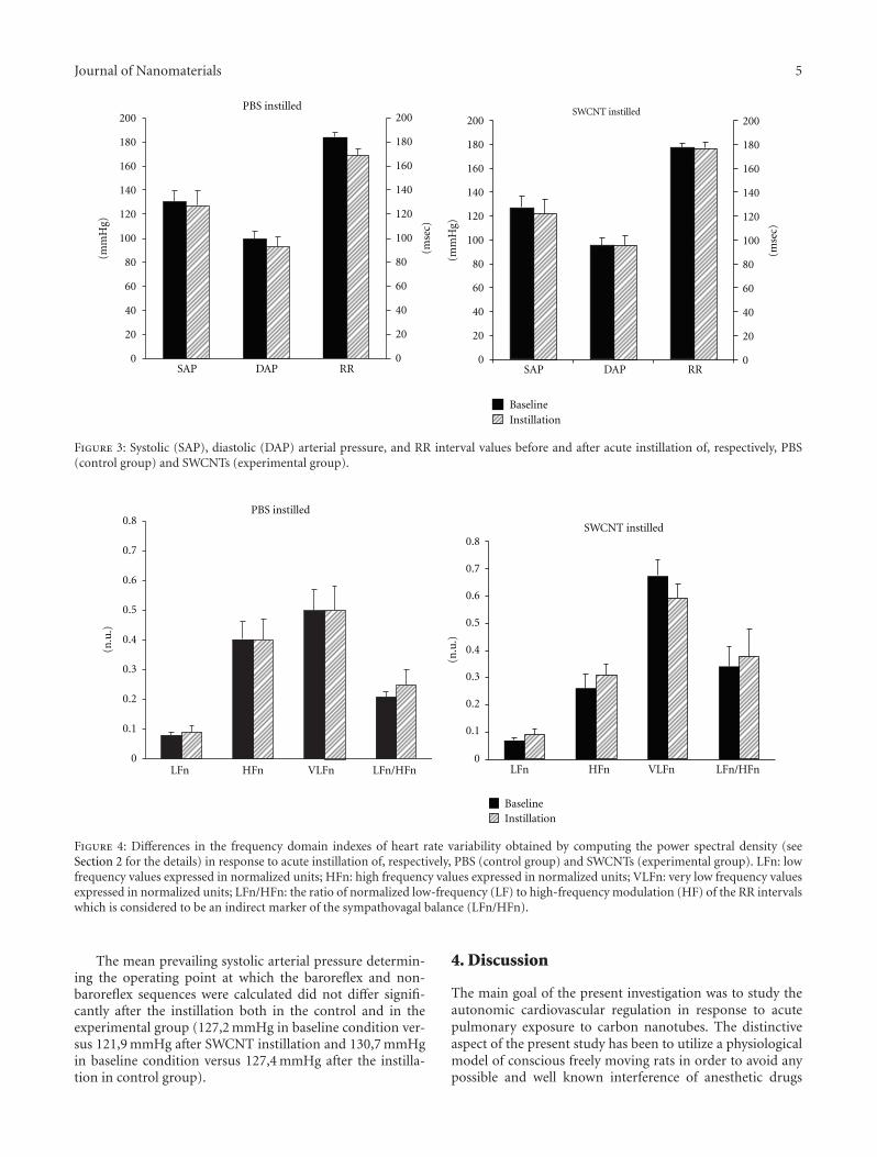
In baseline conditions (i.e., before intratracheal instillationof either PBS or SWCNTs) cardiovascular parameters did notdiffer between control and SWCNT instilled rats (Table 1)and no significant cardiovascular changes were evident after24 hours (Figure 3) in both experimental groups.
Similarly the occurrence of baroreflex sequences and BRSin baseline conditions did not show significant changes bet-ween control and SWCNT instilled rats (Table 1). Also theindexes evaluating the spectral analysis of the heart ratevariability were similar in baseline condition between the twoexperimental groups and in particular LFn was 0, 079± 0, 01for control group and 0, 066 ± 0, 009 for SWCNT instilledrats, HFn 0, 402 ± 0, 06 versus 0, 261 ± 0, 05, VLFn 0, 519 ±0, 07 versus 0, 673± 0, 05, and LFn/HFn 0, 209± 0, 02 versus0, 342± 0, 07.
The power spectral analysis of the heart rate variabilitydid not appear to be affected by the SWCNT experimentalacute exposure. In fact LFn, HFn, VLFn, LFn/HFn did notshow significant changes between basal condition and after24 hour from the instillation both in the control and in theSWCNT instilled rats (Figure 4).
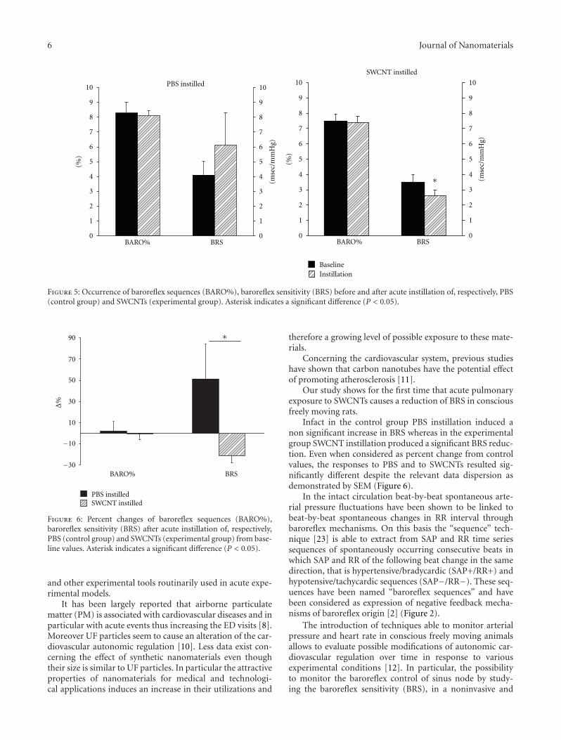
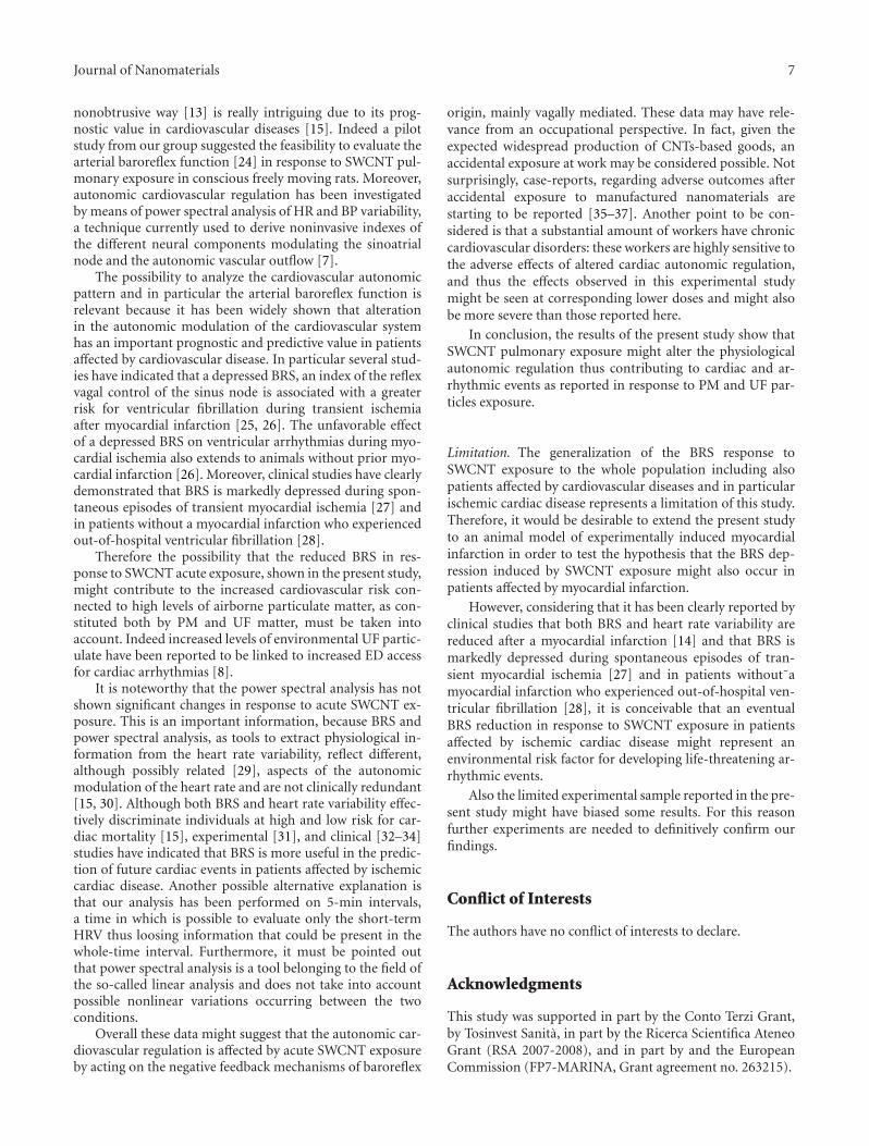
In the control group the PBS instillation did not induceany significant changes in the occurrence and in the sensi-tivity of baroreflex sequences (Figure 5). At variance theSWCNT experimental acute exposure induced a significantreduction of BRS without significant changes in the occur-rence of baroreflex sequences (Figure 5). These differencesare not trivial because whereas in SWCNT instilled rats thesignificant reduction accounts about 21% for BRS, in thecontrol group BRS shows an increase of about 51%. Indeedthe BRS percent changes from baseline versus acute exposureare significantly different between PBS and SWCNT instilledrats (Figure 6).
Journal of Nanomaterials 5
PBS instilled200
180
160
140
120
100
80
60
40
20
0
(mse
c)
(mm
Hg)
SAP DAP RR
200
180
160
140
120
100
80
60
40
20
0
SWCNT instilled200
180
160
140
120
100
80
60
40
20
0
(mse
c)
(mm
Hg)
BaselineInstillation
SAP DAP RR
200
180
160
140
120
100
80
60
40
20
0
Figure 3: Systolic (SAP), diastolic (DAP) arterial pressure, and RR interval values before and after acute instillation of, respectively, PBS(control group) and SWCNTs (experimental group).
PBS instilled0.8
0.7
0.6
0.5
0.4
0.3
0.2
0.1
0
(n.u
.)
LFn HFn VLFn LFn/HFn
0.5
0.3
SWCNT instilled0.8
0.7
0.6
0.4
0.2
0.1
0
(n.u
.)
LFn HFn VLFn
BaselineInstillation
LFn/HFn
Figure 4: Differences in the frequency domain indexes of heart rate variability obtained by computing the power spectral density (seeSection 2 for the details) in response to acute instillation of, respectively, PBS (control group) and SWCNTs (experimental group). LFn: lowfrequency values expressed in normalized units; HFn: high frequency values expressed in normalized units; VLFn: very low frequency valuesexpressed in normalized units; LFn/HFn: the ratio of normalized low-frequency (LF) to high-frequency modulation (HF) of the RR intervalswhich is considered to be an indirect marker of the sympathovagal balance (LFn/HFn).
The mean prevailing systolic arterial pressure determin-ing the operating point at which the baroreflex and non-baroreflex sequences were calculated did not differ signifi-cantly after the instillation both in the control and in theexperimental group (127,2 mmHg in baseline condition ver-sus 121,9 mmHg after SWCNT instillation and 130,7 mmHgin baseline condition versus 127,4 mmHg after the instilla-tion in control group).
4. Discussion
The main goal of the present investigation was to study theautonomic cardiovascular regulation in response to acutepulmonary exposure to carbon nanotubes. The distinctiveaspect of the present study has been to utilize a physiologicalmodel of conscious freely moving rats in order to avoid anypossible and well known interference of anesthetic drugs
6 Journal of Nanomaterials
PBS instilled10
9
8
7
6
5
4
3
2
1
0
(%)
10
9
8
7
6
5
4
3
2
1
0
(mse
c/m
mH
g)
BARO% BRS
SWCNT instilled
10
9
8
7
6
5
4
3
2
1
0
(mse
c/m
mH
g)
10
9
8
7
6
5
4
3
2
1
0
(%)
BARO% BRS
BaselineInstillation
∗
Figure 5: Occurrence of baroreflex sequences (BARO%), baroreflex sensitivity (BRS) before and after acute instillation of, respectively, PBS(control group) and SWCNTs (experimental group). Asterisk indicates a significant difference (P < 0.05).
90
70
50
30
10
−10
−30
Δ%
BARO% BRS
PBS instilledSWCNT instilled
∗
Figure 6: Percent changes of baroreflex sequences (BARO%),baroreflex sensitivity (BRS) after acute instillation of, respectively,PBS (control group) and SWCNTs (experimental group) from base-line values. Asterisk indicates a significant difference (P < 0.05).
and other experimental tools routinarily used in acute expe-rimental models.
It has been largely reported that airborne particulatematter (PM) is associated with cardiovascular diseases and inparticular with acute events thus increasing the ED visits [8].Moreover UF particles seem to cause an alteration of the car-diovascular autonomic regulation [10]. Less data exist con-cerning the effect of synthetic nanomaterials even thoughtheir size is similar to UF particles. In particular the attractiveproperties of nanomaterials for medical and technologi-cal applications induces an increase in their utilizations and
therefore a growing level of possible exposure to these mate-rials.
Concerning the cardiovascular system, previous studieshave shown that carbon nanotubes have the potential effectof promoting atherosclerosis [11].
Our study shows for the first time that acute pulmonaryexposure to SWCNTs causes a reduction of BRS in consciousfreely moving rats.
Infact in the control group PBS instillation induced anon significant increase in BRS whereas in the experimentalgroup SWCNT instillation produced a significant BRS reduc-tion. Even when considered as percent change from controlvalues, the responses to PBS and to SWCNTs resulted sig-nificantly different despite the relevant data dispersion asdemonstrated by SEM (Figure 6).
In the intact circulation beat-by-beat spontaneous arte-rial pressure fluctuations have been shown to be linked tobeat-by-beat spontaneous changes in RR interval throughbaroreflex mechanisms. On this basis the “sequence” tech-nique [23] is able to extract from SAP and RR time seriessequences of spontaneously occurring consecutive beats inwhich SAP and RR of the following beat change in the samedirection, that is hypertensive/bradycardic (SAP+/RR+) andhypotensive/tachycardic sequences (SAP−/RR−). These seq-uences have been named “baroreflex sequences” and havebeen considered as expression of negative feedback mecha-nisms of baroreflex origin [2] (Figure 2).
The introduction of techniques able to monitor arterialpressure and heart rate in conscious freely moving animalsallows to evaluate possible modifications of autonomic car-diovascular regulation over time in response to variousexperimental conditions [12]. In particular, the possibilityto monitor the baroreflex control of sinus node by study-ing the baroreflex sensitivity (BRS), in a noninvasive and
Journal of Nanomaterials 7
nonobtrusive way [13] is really intriguing due to its prog-nostic value in cardiovascular diseases [15]. Indeed a pilotstudy from our group suggested the feasibility to evaluate thearterial baroreflex function [24] in response to SWCNT pul-monary exposure in conscious freely moving rats. Moreover,autonomic cardiovascular regulation has been investigatedby means of power spectral analysis of HR and BP variability,a technique currently used to derive noninvasive indexes ofthe different neural components modulating the sinoatrialnode and the autonomic vascular outflow [7].
The possibility to analyze the cardiovascular autonomicpattern and in particular the arterial baroreflex function isrelevant because it has been widely shown that alterationin the autonomic modulation of the cardiovascular systemhas an important prognostic and predictive value in patientsaffected by cardiovascular disease. In particular several stud-ies have indicated that a depressed BRS, an index of the reflexvagal control of the sinus node is associated with a greaterrisk for ventricular fibrillation during transient ischemiaafter myocardial infarction [25, 26]. The unfavorable effectof a depressed BRS on ventricular arrhythmias during myo-cardial ischemia also extends to animals without prior myo-cardial infarction [26]. Moreover, clinical studies have clearlydemonstrated that BRS is markedly depressed during spon-taneous episodes of transient myocardial ischemia [27] andin patients without a myocardial infarction who experiencedout-of-hospital ventricular fibrillation [28].
Therefore the possibility that the reduced BRS in res-ponse to SWCNT acute exposure, shown in the present study,might contribute to the increased cardiovascular risk con-nected to high levels of airborne particulate matter, as con-stituted both by PM and UF matter, must be taken intoaccount. Indeed increased levels of environmental UF partic-ulate have been reported to be linked to increased ED accessfor cardiac arrhythmias [8].
It is noteworthy that the power spectral analysis has notshown significant changes in response to acute SWCNT ex-posure. This is an important information, because BRS andpower spectral analysis, as tools to extract physiological in-formation from the heart rate variability, reflect different,although possibly related [29], aspects of the autonomicmodulation of the heart rate and are not clinically redundant[15, 30]. Although both BRS and heart rate variability effec-tively discriminate individuals at high and low risk for car-diac mortality [15], experimental [31], and clinical [32–34]studies have indicated that BRS is more useful in the predic-tion of future cardiac events in patients affected by ischemiccardiac disease. Another possible alternative explanation isthat our analysis has been performed on 5-min intervals,a time in which is possible to evaluate only the short-termHRV thus loosing information that could be present in thewhole-time interval. Furthermore, it must be pointed outthat power spectral analysis is a tool belonging to the field ofthe so-called linear analysis and does not take into accountpossible nonlinear variations occurring between the twoconditions.
Overall these data might suggest that the autonomic car-diovascular regulation is affected by acute SWCNT exposureby acting on the negative feedback mechanisms of baroreflex
origin, mainly vagally mediated. These data may have rele-vance from an occupational perspective. In fact, given theexpected widespread production of CNTs-based goods, anaccidental exposure at work may be considered possible. Notsurprisingly, case-reports, regarding adverse outcomes afteraccidental exposure to manufactured nanomaterials arestarting to be reported [35–37]. Another point to be con-sidered is that a substantial amount of workers have chroniccardiovascular disorders: these workers are highly sensitive tothe adverse effects of altered cardiac autonomic regulation,and thus the effects observed in this experimental studymight be seen at corresponding lower doses and might alsobe more severe than those reported here.
In conclusion, the results of the present study show thatSWCNT pulmonary exposure might alter the physiologicalautonomic regulation thus contributing to cardiac and ar-rhythmic events as reported in response to PM and UF par-ticles exposure.
Limitation. The generalization of the BRS response toSWCNT exposure to the whole population including alsopatients affected by cardiovascular diseases and in particularischemic cardiac disease represents a limitation of this study.Therefore, it would be desirable to extend the present studyto an animal model of experimentally induced myocardialinfarction in order to test the hypothesis that the BRS dep-ression induced by SWCNT exposure might also occur inpatients affected by myocardial infarction.
However, considering that it has been clearly reported byclinical studies that both BRS and heart rate variability arereduced after a myocardial infarction [14] and that BRS ismarkedly depressed during spontaneous episodes of tran-sient myocardial ischemia [27] and in patients without˜amyocardial infarction who experienced out-of-hospital ven-tricular fibrillation [28], it is conceivable that an eventualBRS reduction in response to SWCNT exposure in patientsaffected by ischemic cardiac disease might represent anenvironmental risk factor for developing life-threatening ar-rhythmic events.
Also the limited experimental sample reported in the pre-sent study might have biased some results. For this reasonfurther experiments are needed to definitively confirm ourfindings.
Conflict of Interests
The authors have no conflict of interests to declare.
Acknowledgments
This study was supported in part by the Conto Terzi Grant,by Tosinvest Sanita, in part by the Ricerca Scientifica AteneoGrant (RSA 2007-2008), and in part by and the EuropeanCommission (FP7-MARINA, Grant agreement no. 263215).

8 Journal of Nanomaterials
References
[1] B. T. Mossman, P. J. Borm, V. Castranova, D. L. Costa, K. Don-aldson, and S. R. Kleeberger, “Mechanisms of action of inhaledfibers, particles and nanoparticles in lung and cardiovasculardiseases,” Particle and Fibre Toxicology, vol. 4, p. 4, 2007.
[2] D. Liao, J. Creason, C. Shy, R. Williams, R. Watts, and R.Zweidinger, “Daily variation of particulate air pollution andpoor cardiac autonomic control in the elderly,” EnvironmentalHealth Perspectives, vol. 107, no. 7, pp. 521–525, 1999.
[3] C. A. Pope III, R. L. Verrier, E. G. Lovett et al., “Heart ratevariability associated with particulate air pollution,” AmericanHeart Journal I, vol. 138, no. 5, pp. 890–899, 1999.
[4] D. R. Gold, A. Litonjua, J. Schwartz et al., “Ambient pollutionand heart rate variability,” Circulation, vol. 101, no. 11, pp.1267–1273, 2000.
[5] S. R. Magari, R. Hauser, J. Schwartz, P. L. Williams, T. J. Smith,and D. C. Christiani, “Association of heart rate variability withoccupational and environmental exposure to particulate airpollution,” Circulation, vol. 104, no. 9, pp. 986–991, 2001.
[6] A. Algra, J. G. P. Tijssen, J. R. T. C. Roelandt, J. Pool, and J.Lubsen, “Heart rate variability from 24-hour electrocardiog-raphy and the 2-year risk for sudden death,” Circulation, vol.88, no. 1, pp. 180–185, 1993.
[7] Task Force of the European Society of Cardiology and theNorth American Society of Pacing and Electrophysiology,“Heart rate variability: standards of measurement, physiolog-ical interpretation, and clinical use,” Circulation, vol. 93, pp.1043–1065, 1996.
[8] S. S. Tsai, H. F. Chiu, T. N. Wu, and C. Y. Yang, “Air pollutionand emergency room visits for cardiac arrhythmia in a sub-tropical city: Taipei, Taiwan,” Inhalation Toxicology, vol. 21, no.13, pp. 1113–1118, 2009.
[9] A. Peters, D. W. Dockery, J. E. Muller, and M. A. Mittleman,“Increased particulate air pollution and the triggering ofmyocardial infarction,” Circulation, vol. 103, no. 23, pp. 2810–2815, 2001.
[10] K. L. Timonen, E. Vanninen, J. de Hartog et al., “Effects ofultrafine and fine particulate and gaseous air pollution on car-diac autonomic control in subjects with coronary artery dise-ase: the ULTRA study,” Journal of Exposure Science and Envi-ronmental Epidemiology, vol. 16, no. 4, pp. 332–341, 2006.
[11] Z. Li, T. Hulderman, R. Salmen et al., “Cardiovascular effectsof pulmonary exposure to single-wall carbon nanotubes,”Environmental Health Perspectives, vol. 115, no. 3, pp. 377–382,2007.
[12] H. Waki, K. Katahira, J. W. Polson, S. Kasparov, D. Murphy,and J. F. R. Paton, “Automation of analysis of cardiovascularautonomic function from chronic measurements of arterialpressure in conscious rats,” Experimental Physiology, vol. 91,no. 1, pp. 201–213, 2006.
[13] J. M. Legramante, S. Sacco, G. Raimondi et al., “Investigatingfeedforward neural regulation of circulation from analysis ofspontaneous arterial pressare and herat rate fluctuations inconscious rats,” American Journal of Physiology, vol. 296, no.1, pp. H202–H210, 2009.
[14] M. T. la Rovere, G. Specchia, A. Mortara, and P. J. Schwartz,“Baroreflex sensitivity, clinical correlates, and cardiovascularmortality among patients with a first myocardial infarction: aprospective study,” Circulation I, vol. 78, no. 4, pp. 816–824,1988.
[15] M. T. la Rovere, J. T. Bigger Jr., F. I. Marcus, A. Mortara, and P.J. Schwartz, “for ATRAMI (Autonomic Tone and Reflexes AfterMyocardial Infarction) Investigators. Baroreflex sensitivity
and heart rate variability in prediction of total cardiac mor-tality after myocardial infarction,” The Lancet, vol. 351, no.9101, pp. 478–484, 1998.
[16] A. A. Shvedova, E. R. Kisin, R. Mercer et al., “Unusual inflam-matory and fibrogenic pulmonary responses to single-walledcarbon nanotubes in mice,” American Journal of Physiology,vol. 289, no. 5, pp. L698–L708, 2005.
[17] A. A. Shvedova, E. R. Kisin, R. Mercer et al., “Vitamin E defi-ciency enhances pulmonary inflammatory response and oxi-dative stress induced by single walled carbon nanotubes inC57BL/6 mice,” Toxicology and Applied Pharmacology, vol.221, no. 3, pp. 339–348, 2007.
[18] J. M. Legramante, G. Raimondi, M. Massaro, S. Cassarino,G. Peruzzi, and F. Iellamo, “Investigating feed-forward neuralregulation of circulation from analysis of spontaneous arterialpressure and heart rate fluctuations,” Circulation, vol. 99, no.13, pp. 1760–1766, 1999.
[19] J. Oosting, H. A. J. Struijker-Boudier, and B. J. A. Janssen,“Validation of a continuous baroreceptor reflex sensitivityindex calculated from spontaneous fluctuations of bloodpressure and pulse interval in rats,” Journal of Hypertension,vol. 15, no. 4, pp. 391–399, 1997.
[20] C. Mostarda, B. Rodrigues, M. Vane et al., “Autonomic impair-ment after myocardial infarction: role in cardiac remodellingand mortality,” Clinical and Experimental Pharmacology andPhysiology, vol. 37, no. 4, pp. 447–452, 2010.
[21] A. Malliani and M. Pagani, “Spectral analysis of cardiovascularvariabilities in the assessment of sympathetic cardiac regula-tion in heart failure,” Pharmacological Research, vol. 24, no. 1,pp. 43–53, 1991.
[22] E. Rondon, M. S. Brasileiro-Santos, E. D. Moreira et al., “Exer-cise training improves aortic depressor nerve sensitivity inrats with ischemia-induced heart failure,” American Journal ofPhysiology, vol. 291, no. 6, pp. H2801–H2806, 2006.
[23] G. Bertineri, M. di Rienzo, A. Cavallazzi, A. U. Ferrari, A.Pedotti, and G. Mancia, “Evaluation of baroreceptor reflex byblood pressure monitoring in unanesthetized cats,” AmericanJournal of Physiology, vol. 254, no. 2, pp. H377–H383, 1988.
[24] J. M. Legramante, F. Valentini, A. Magrini et al., “Cardiac auto-nomic regulation after lung exposure to carbon nanotubes,”Human and Experimental Toxicology, vol. 28, no. 6-7, pp. 369–375, 2009.
[25] G. E. Billman, P. J. Schwartz, and H. L. Stone, “Baroreceptorreflex control of heart rate: a predictor of sudden cardiacdeath,” Circulation, vol. 66, no. 4, pp. 874–880, 1982.
[26] P. J. Schwartz, E. Vanoli, M. Stramba-Badiale, G. M. de Ferrari,G. E. Billman, and R. D. Foreman, “Autonomic mechanismsand sudden death: new insights from analysis of baroreceptorreflexes in conscious dogs with and without a myocardial in-farction,” Circulation I, vol. 78, no. 4, pp. 969–979, 1988.
[27] G. Pomidossi, A. Saino, R. Perondi et al., “Impairment of thearterial baroreflex durino sympathetic and silent myocardialischemia in humans,” Journal of the American College ofCardiology, vol. 22, pp. 1866–1872, 1993.
[28] P. J. Schwartz, A. Zaza, M. Pala, E. Locati, G. Beria, and A.Zanchetti, “Baroreflex sensitivity and its evolution during thefirst year after myocardial infarction,” Journal of the AmericanCollege of Cardiology, vol. 12, no. 3, pp. 629–636, 1988.
[29] M. Piepoli, P. Sleight, S. Leuzzi et al., “Origin of respiratorysinus arrhythmia in conscious humans: an important role forarterial carotid baroreceptors,” Circulation, vol. 95, no. 7, pp.1813–1821, 1997.
Journal of Nanomaterials 9
[30] J. T. Bigger Jr., M. T. la Rovere, R. C. Steinman et al., “Com-parison of baroreflex sensitivity and heart period variabilityafter myocardial infarction,” Journal of the American College ofCardiology, vol. 14, no. 6, pp. 1511–1518, 1989.
[31] S. S. Hull Jr., A. R. Evans, E. Vanoli et al., “Heart rate variabilitybefore and after myocardial infarction in conscious dogs athigh and low risk of sudden death,” Journal of the AmericanCollege of Cardiology, vol. 16, no. 4, pp. 978–985, 1990.
[32] G. M. de Ferrari, M. Landolina, M. Mantica, R. Manfredini, P.J. Schwartz, and A. Lotto, “Baroreflex sensitivity, but not heartrate variability, is reduced in patients with life-threateningventricular arrhythmias long after myocardial infarction,”American Heart Journal I, vol. 130, no. 3, pp. 473–480, 1995.
[33] S. H. Hohnloser, T. Klingenheben, A. van de Loo, E.Hablawetz, H. Just, and P. J. Schwartz, “Reflex versus tonicvagal activity as a prognostic parameter in patients with sus-tained ventricular tachycardia or ventricular fibrillation,” Cir-culation, vol. 89, no. 3, pp. 1068–1073, 1994.
[34] R. E. Kleiger, J. P. Miller, J. T. Bigger Jr., A. J. Moss, and TheMulticenter Post-Infarction Research Group, “Decreased heartrate variability and its association with increased mortalityafter acute myocardial infarction,” Journal of the AmericanCollege of Cardiology, vol. 59, no. 4, pp. 256–262, 1987.
[35] Y. Song, X. Li, and X. Du, “Exposure to nanoparticles is relatedto pleural effusion, pulmonary fibrosis and granuloma,” Euro-pean Respiratory Journal, vol. 34, no. 3, pp. 559–567, 2009.
[36] J. I. Phillips, F. Y. Green, J. C. A. Davies, and J. Murray, “Pul-monary and systemic toxicity following exposure to nickelnanoparticles,” American Journal of Industrial Medicine, vol.53, no. 8, pp. 763–767, 2010.
[37] T. Toyama, H. Matsuda, I. Ishida et al., “A case of toxic epi-dermal necrolysis-like dermatitis evolving from contact der-matitis of the hands associated with exposure to dendrimers,”Contact Dermatitis, vol. 59, no. 2, pp. 122–123, 2008.


![非接触による心拍検出方法 - JST · 800 900 1000 1100 Time [sec] R-R Interval [msec] 心電計 補間前提案法 RMSE = 39.4 msec • K. Yamamoto, K. Toyoda, and T. Ohtsuki,](https://img.dokumen.tips/doc/110x75/6061e169d3a1f91bed4abbda/eefoe-jst-800-900-1000-1100-time-sec-r-r-interval.jpg)