Embed Size (px)
Citation preview
Arq Neuropsiquiatr 2008;66(1):15-21
15
CERVICAL DYSTONIA
Clinical and therapeutic features in 85 patients
Carlos Henrique F. Camargo1, Hélio A.G. Teive2, Nilson Becker1, Maria Helena Herdoíza Baran1, Rosana Hermínia Scola2, Lineu César Werneck3
Abstract – We studied patients with cervical dystonia (CD) to determine clinical features and response to botulinum toxin A (BoNT/A). Patients were submitted to clinical, laboratory and neuroimaging evaluation. BoNT/A was injected locally in 81 patients using electromyographic guidance. Four patients who had had previous treatment were considered to be in remission. The average ages at onset of focal dystonia and segmental dystonia were greater than for generalized dystonia (p<0.0003). The severity of the abnormal head-neck movements were more severe among the patients with generalized dystonia (p<0.001). Pain in the cervical area was noted in 59 patients. It was not possible to determine the etiology of the disease in 62.3% of patients. Tardive dystonia was the most common secondary etiology. A major improvement in the motor symptoms of CD and pain was observed in patients following treatment with BoNT/A. The tardive dystonia subgroup did not respond to the treatment. Dysphagia was observed in 2.35% of the patients.
Key WorDs: dystonia, cervical dystonia, botulinum toxin, dysphagia.
Distonia cervical: aspectos clínicos e terapêuticos de 85 pacientes
Resumo – Para identificar os aspectos clínicos e a resposta a toxina botulínica A (TxBA), pacientes com distonia cervical (DC) foram submetidos a avaliação clínica, laboratorial e neuroimagem. o tratamento com TxBA foi aplicado a 81 pacientes guiado por eletroneuromiografia. Quatro pacientes, com tratamento prévio, foram considerados em remissão. A média de idade de início dos sintomas de pacientes com distonia focal e segmentar foi maior que a encontrada em pacientes com distonia generalizada (p<0,0003). A gravidade das alterações motoras cervicais foi maior entre os pacientes com distonia generalizada que nos pacientes com distonia focal (p<0,001). Graus diferentes de dor na região cervical foram relatados por 59 dos pacientes. Não foi possível determinar a etiologia da doença em 62,3% dos pacientes sendo distonia tardia a mais comum. Houve acentuada melhora dos sintomas motores e da dor da DC com a aplicação de TxBA. o subgrupo de pacientes com distonia tardia não respondeu ao tratamento. Disfagia ocorreu em 2,35% dos pacientes.
PAlAvrAs-CHAve: distonia, distonia cervical, toxina botulínica, disfagia.
Movement Disorders Unit, Neurology service, Department of Clinical Medicine, Hospital de Clínicas, Federal University of Paraná, Curitiba Pr, Brazil: 1Neurologist; 2Associate Professor; 3Full Professor.
received 13 August 2007, received in final form 21 November 2007. Accepted 22 December 2007.
Dr. Hélio A.G. Teive – Rua General Carneiro 181 / 12º andar - 80060-900 Curitiba PR - Brasil. E-mail: [email protected]
Dystonia is defined as a syndrome characterized by prolonged muscle contraction causing twisting, repetitive movements or abnormal posture1. Most voluntary muscles can be affected and, in the case of the neck muscles, the condition is referred to as cervical dystonia. The term spas-modic torticollis was previously used for this syndrome, but it does not stress the dystonic nature of the disease2.
A wide range of therapies are available for cervical dystonia, from clinical treatment to brain surgery (palli-dotomy and deep brain stimulation) or even peripher-al surgery. However, BoNT/A is currently considered the treatment of choice3.
The objectives of this study were to identify the clini-cal profile of patients with cervical dystonia who attend-ed the Hospital das Clínicas, UFPr, and to analyze their re-sponse to treatment with botulinum toxin A (BoNT/A) in terms of the severity of the motor alterations and pain.
METHODPatient selectionPatients with cervical dystonia who attended the Botulinum
Toxin and Movement Disorders outpatient Unit in the Neurol-ogy service, Hospital de Clínicas, Federal University of Paraná, were selected for the study.
Arq Neuropsiquiatr 2008;66(1)
16
Cervical dystoniaCamargo et al.
The inclusion criteria were: (1) the presence of cervical or segmental dystonia; (2) the presence of generalized dystonia, hemidystonia or multifocal dystonia, with referral for botuli-num-toxin-A treatment for cervical dystonia.
The exclusion criteria were: (1) refusal to submit to diag-nostic investigation; (2) failure to agree to the chosen therapy; (3) the presence of hemidystonia, multifocal dystonia or gener-alized dystonia with referral for surgery or stable with clinical treatment; (4) inability to attend for reassessment; and (5) fail-ure to sign the informed consent form. The exclusion criteria for treatment with BoNT/A were: (1) the presence of myasthenia gravis or other diseases of the neuromuscular junction; (2) the use of aminoglycoside antibiotics; and (3) pregnancy and lacta-tion. All patients signed the informed consent form.
Clinical assessmentAll the patients were diagnosed with cervical dystonia by
one or more neurologists, and this was confirmed by the co-ordinator for the movement disorders unit (HT). The patients were then assessed by the author by means of a detailed clini-cal history and a physical and neurological examination to iden-tify clinical characteristics, an association with other movement disorders and neurological diseases, epidemiological data, the time during which the disease had evolved, a history of trauma, the use of medicines, signs and symptoms that might indicate a secondary cause and a family history of dystonia or other move-ment disorders. Head tremor was classified as dystonic tremor or essential tremor4.
All the patients were submitted to brain computed tomog-raphy and cervical-spine radiography. Additional tests included hemogram, TsH, vDrl, blood glucose test, esr, electrolyte lev-els and liver and kidney function in all the patients. Computed tomography of the cervical spine, magnetic resonance imaging of the brain and other laboratory tests were requested accord-ing to the clinical assessment of each patient.
The patients were classified according to the clinical pre-sentation of the cervical dystonia (torticollis, laterocollis, ret-rocollis, anterocollis or combined forms) and whether this was focal dystonia, multifocal dystonia, hemidystonia or general-ized dystonia.
For primary dystonia to be diagnosed, the following con-ditions had to be met: (1) normal perinatal and developmental history; (2) no history of diseases or medication that could have precipitated the appearance of dystonia; (3) no evidence of py-ramidal or cerebellar signs, alterations in sensitivity, or cognitive dysfunction on examination; (4) exclusion of secondary causes by specific tests.
The criteria published by Burke et al.5 were used for tar-dive dystonia, and those published by Cardoso and Jankovic6 for posttraumatic cervical dystonia. secondary causes of cervi-cal dystonia were related to craniocerebral trauma, stroke, en-cephalitis and brain tumor. Dystonic cerebral palsy was associat-ed with a history of birth trauma and perinatal anoxia.
The presence of dystonia concomitantly with a heredode-
generative disease or neurochemical disorder was considered to be dystonia in a heredodegenerative disease and dystonia-plus7.
TreatmentThe clinical treatment patients had been receiving prior to
inclusion in the study was continued, and none of the patients were submitted to surgery during the study. Patients received botulinum-toxin-A therapy (Botox®, Allergan, Irvine, CA, UsA).
The lyophilized preparation of the toxin (1 flask=100 U=5 ng) was stored at –20ºC and reconstituted with 1 ml of 0.9% saline solution at the time of injection. The 10 U per 0.1 ml solution was administered in a 1cc (1 ml) tuberculin syringe.
Patients who had already used BoNT/A and had reported a subjective improvement continued to receive the same dose. For new patients the initial dose varied from 100 to 280 U. The dose and number of points where the toxin was injected were adjusted for each muscle (Table 1).
The choice of muscle, location and amount of BoNT/A for each muscle were determined based on clinical evaluation with the aid of electromyography (Table 2). To inject BoNT/A, the tuberculin syringe was coupled to a Teflon-coated monopolar electrode needle (37 mm x 27 G) (oxford Instruments®) connect-ed to a Nihon-Koden multichannel appliance.
Follow-upThe patients were assessed on admission and approximate-
ly 14, 30 and 120 days after the treatment had started to com-pare severity, disability and pain using the Toronto Western spas-modic torticollis rating scale (TWsTrs – severity); Fahn-Marsden dystonia scale (FMs) (for the cervical segment); Jankovic disabil-ity scale (JDs); and visual analog pain scale (0=absence of pain, 1–3=mild pain, 4–6=moderate pain, 7–9 = strong pain, 10=dis-abling pain).
Table 1. Dose and number of injection points per muscle.
Muscle Dose Number of points
sternocleidomastoid 15-75 2-4
Trapezium 30-100 5-10
splenius capitis 15-50 2-4
levator scapulae 15-50 2-4
Paraspinalis 15-50 2-4
Table 2. Muscles chosen for injection of botulinum toxin accord-ing to clinical presentation.
subtype Muscles in which applied
Torticollis Contralateral sternocleidomastoid and ipsilateral trapezium and splenius
laterocollis splenius, trapezium and ipsilateral sternocleidomastoid
Anterocollis Bilateral sternocleidomastoid
retrocollis Trapezium and bilateral paraspinalis
Arq Neuropsiquiatr 2008;66(1)
17
Cervical dystoniaCamargo et al.
Adverse effects, latency to start, peak and duration of ac-tion of the medication and interference in daily life were mea-sured with a questionnaire.
Statistical analysisThe distribution pattern for all the data was tested (normal
or non-normal). The statistical differences between the means of the groups were measured using the one-tailed Student t-test and ANovA for normal distributions and the Mann-Whitney and Kruskal-Wallis tests for non-normal distributions. The correla-tions were measured using the Pearson and Spearman correla-tion coefficients. For the differences between the expected val-ues and the values actually found, the chi-square test with Yates correction and the Fisher exact test were used. The results are given as mean ±sD (standard deviation). The differences were considered significant if p<0.05.
RESULTSClinical and epidemiological characteristicsA total of 85 patients with cervical dystonia were in-
cluded in the study: 51 females and 34 males, giving a ra-tio of 1.5:1. Forty-five patients (52.9%) only had cervical dystonia while twenty patients (23.5%) also had segmen-tal dystonia, 13 of whom had cranial dystonia, 6 dystonia in the upper limbs, and 1 oromandibular dystonia and dys-tonia in an upper limb. In the 2 patients (2.4%) with multi-focal dystonia, a lower left limb was involved. Hemidys-tonia was observed in 5 patients (5.9%), and 13 (15.3%) had generalized dystonia.
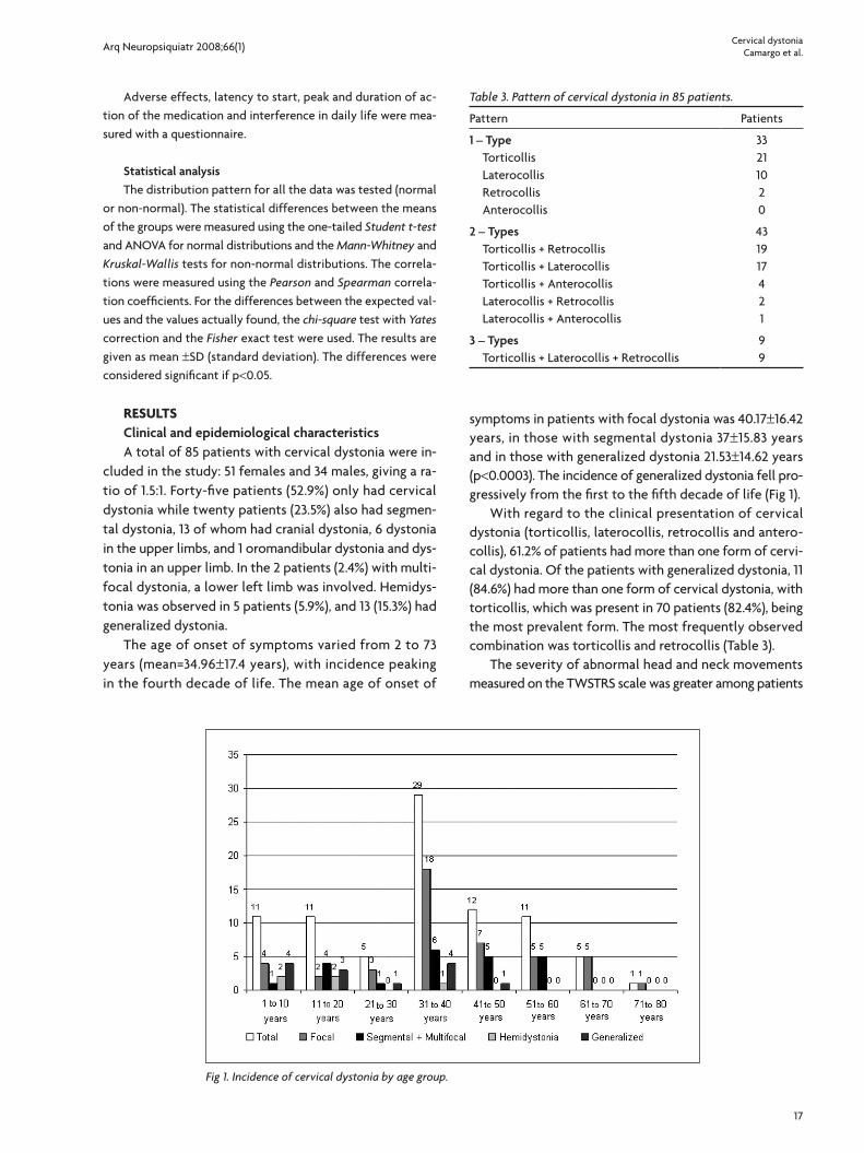
The age of onset of symptoms varied from 2 to 73 years (mean=34.96±17.4 years), with incidence peaking in the fourth decade of life. The mean age of onset of
symptoms in patients with focal dystonia was 40.17±16.42 years, in those with segmental dystonia 37±15.83 years and in those with generalized dystonia 21.53±14.62 years (p<0.0003). The incidence of generalized dystonia fell pro-gressively from the first to the fifth decade of life (Fig 1).
With regard to the clinical presentation of cervical dystonia (torticollis, laterocollis, retrocollis and antero-collis), 61.2% of patients had more than one form of cervi-cal dystonia. of the patients with generalized dystonia, 11 (84.6%) had more than one form of cervical dystonia, with torticollis, which was present in 70 patients (82.4%), being the most prevalent form. The most frequently observed combination was torticollis and retrocollis (Table 3).
The severity of abnormal head and neck movements measured on the TWsTrs scale was greater among patients
Table 3. Pattern of cervical dystonia in 85 patients.
Pattern Patients
1 – Type Torticollis laterocollis retrocollis Anterocollis
33211020
2 – Types Torticollis + retrocollis Torticollis + laterocollis Torticollis + Anterocollis laterocollis + retrocollis laterocollis + Anterocollis
431917421
3 – Types Torticollis + laterocollis + retrocollis
99
Fig 1. Incidence of cervical dystonia by age group.

Arq Neuropsiquiatr 2008;66(1)
18
Cervical dystoniaCamargo et al.
with generalized dystonia than among those with focal dystonia (p<0.001) or segmental dystonia (p<0.001). Using the same scale, severity was found to be higher in patients with two or three types of cervical dystonia than in those with only one type (p<0.001) and p<0.0009, respectively.
Different degrees of pain in the cervical region were reported by 59 (69.4%) of the patients. of these, 43 (72.9%) reported moderate pain. The presence of spasms and jerks, reported by 60 patients (70.6%), was an aggravat-ing factor (p<0.0001).
Comparison of patients with combinations of cervical dystonic movements revealed more complaints of pain in the groups with two and three movements (p<0.009 and p<0.039, respectively). There was no difference between the number of patients who complained of pain and the level of pain among patients with focal, segmental, mul-tifocal or generalized dystonia or hemidystonia.
“Gestes antagonistes” were reported by only one pa-tient; however, during clinical examination, “gestes antag-onistes” such as holding the chin and touching the face were observed in 15 patients (14.6%). These movements rarely lasted for more than a minute.
some form of tremor was observed in 20 patients (23.5%). Head tremor (‘no-no’ type) was present in 9 pa-tients (45%) and essential-type tremor of the upper limbs in 3 (15%). A combination of the two types of tremor was observed in 8 patients (40%), and three of these reported a family history of tremor.
Two patients (one with focal dystonia and one with segmental dystonia) had associated Parkinsonism. The lat-ter of these two patients also had a family history of Par-kinsonism. Another patient with segmental dystonia had Tourette’s syndrome, and his brother and father had a his-tory of tics. The only patient with Behçet’s syndrome and hemidystonia also had chorea and hemiballism.
The etiology of the disease could not be determined
in 53 patients (62.3%), and the most prevalent etiology found was tardive dystonia (8.2%). In this group of pa-tients, the severity measured on the TWsTrs scale was greater than in patients with other etiologies (p<0.002). All the patients with dystonia secondary to neuroleptic treatment had either the isolated form of retrocollis or retrocollis combined with one or two other types of dys-tonia (five cases and one case, respectively). Five patients (5.9%) without any defined etiology and a mean age at on-set of symptoms of 43.5±21.68 years reported cases of cer-vical dystonia in their families; three of these patients had focal dystonia and two segmental dystonia (Table 4).
TreatmentBefore treatment with BoNT/A, 48 of the patients
(56.5%) had undergone clinical treatment. Anticholinergic agents, particularly trihexyphenidyl (in doses of 6 to 30 mg daily), were the main medication used (42.35%). eighty-one patients (92.3%) received BoNT/A injections. Four pa-tients (4.7%), who had both been diagnosed and had re-ceived BoNT/A previously, with an average disease dura-tion of 3.5±1 years, were considered to be in remission as they did not show any symptoms during the evaluation period (14 months) and therefore did not have any more BoNT/A injections. All the patients who received BoNT/A presented for evaluation at the end of the evaluation period, and 53 (65.4%) presented during the course of the evaluation period.
The mean BoNT/A dose used was 151.05±52.55 U. The greater the severity of the motor symptoms, the more BoNT/A used (r=0.5 and p<0.05). However, no correlation was found between an increase in BoNT/A dosage and an improvement in symptoms measured on the TWsTrs scale (r=0.15 and p<0.05). Nevertheless, the group of pa-tients with three types of cervical dystonia benefited from larger dosages of BoNT/A (r=0.798 and p<0.05).
Table 4. Etiology by distribution of cervical dystonia.
etiology Focal segmental Generalized Hemidystonia Multifocal Total
Indeterminate 31 9 9 2 2 53
Neuroleptic treatment 2 4 1 7
Perinatal anoxia 1 2 2 1 6
Craniocerebral trauma 5 1 6
Indeterminate with family history 2 3 5
Cervical trauma 3 3
Brain infarct 1 1 2
Meningitis 1 1
Behçet’s syndrome 1 1
Wilson’s disease 1 1
Total 45 20 13 5 2 85
Arq Neuropsiquiatr 2008;66(1)
19
Cervical dystoniaCamargo et al.
Thirty-one (36.5%) had not received BoNT/A previous-ly. of the 54 patients previously treated, 10 (18.5%) had had one treatment session, 25 (46.3%) had had between 2 and 4 sessions and 19 (35.2%) had had more than 5. The previous treatment sessions did not interfere with the as-sessment of the severity of the disease at the start of the study according to the results using the TWsTrs scale (p<0.53), JDs (p<0.16) and FMs (p<0.16).
There was a similar and highly positive correlation be-tween the results for the response to BoNT/A in relation to the severity of the disease obtained with the three different scales. The correlations observed were as fol-lows: correlation between TWsTrs and JDs: r=0.82 and p<0.05; between TWsTrs and FMs: r=0.9 and p<0.05; and between JDs and FMs: r=0.82 and p<0.05. There was a ma-jor improvement in the symptoms of cervical dystonia on the TWsTrs (Fig 2), FMs and JDs scales following treat-ment sessions with BoNT/A, and these returned to their initial levels at the end of the evaluation. The patients re-ported a mean time until the medication started to have an effect of 10.07±5.84 days and a mean duration of effect before symptoms returned of 89.15±21.79 days.
BoNT/A also had a beneficial effect in controlling cer-vical pain. There was a significant improvement in relation to complaints of pain and pain intensity (Fig 3). An im-provement in the degree of pain was also observed when the groups with one (p<0.045), two (p<0.0001) or three (p<0.026) types of dystonic movement were analyzed separately. There was no recurrence of previous levels of cervical pain as the analgesic effect of BoNT/A in the group with one dystonic movement was partially main-tained at the end of the evaluation (p<0.094).
The subgroup of patients with tardive dystonia, whose clinical characteristics and degree of disability differed from those of the other patients in the group, did not re-spond to treatment with BoNT/A on the TWsTrs scale (p<0.13). Patients with posttraumatic cervical dystonia, for whom the duration of trauma before onset of symp-toms was 6.66±8.08 years, responded positively to BoNT/A on the TWsTrs scale (p<0.004) and reported pain relief (p<0.002). In this subgroup there was no change in the an-algesic effect of BoNT/A on the 120th day compared with the 30th day (p<0.3).
Transitory dysphagia was reported in 2 patients (2.35%). There were no other reports of adverse effects associat-ed with the medication.
DISCUSSION
Although cultural and economic issues made it dif-ficult for some patients to present for evaluation during follow-up in this longitudinal study, we were able to as-semble a sample that yielded representative data about
the clinical characteristics and response to proposed therapy of patients with cervical dystonia. In agreement with previously published studies, we found that cervi-cal dystonia was predominant in females8,9. The onset of symptoms occurred mainly between the fourth and sixth decades of life (61.2%)8,9. Generalized forms had earlier onset, and focal and segmental forms later onset. Genetic studies have proved helpful in understanding this observa-tion. Dystonias related to the DyT1 and DyT5 genes start as focal dystonias and become predominantly generalized in childhood. Dystonias 7 and 13, which are associated with the DyT7 and DyT13 genes, have later onset and tend to remain focal. When dystonia 13 occurs in childhood, it has a much greater tendency to become generalized10,11.
Technical and economic limitations prevented us from carrying out genetic tests on the patients in this study.
Fig 2. Response to treatment with botulinum toxin on the TWSTRS scale.
Fig 3. Response to cervical pain following treatment with botulinum oxin.

Arq Neuropsiquiatr 2008;66(1)
20
Cervical dystoniaCamargo et al.
some of the patients who were classified as having “in-determinate” etiology may have genetic alterations that were not investigated. of particular note are the five pa-tients with a family history of focal and segmental dys-tonia and a mean age of onset of more than 40 years and the two patients with a family history of other movement disorders (parkinsonism and tics).
Tardive dystonia is the main form of secondary dys-tonia, with the cervical form being the most common12. In agreement with published figures, 8.2% of the patients studied were found to have dystonia secondary to the use of neuroleptic agents9. The clinical behavior of patients in this group was different from that of the remainder of the sample. All the patients had retrocollis, and 85.71% had more than one type of dystonic movement, two char-acteristics that could be considered aggravating factors. our finding that retrocollis was predominant agrees with those of other authors who consider its presence to be highly suggestive of tardive dystonia13. Unlike the patients in the study by Molho et al.13, our patients with tardive dystonia did not respond to BoNT/A treatment, and the size of the sample of patients with tardive dystonia had to be increased to confirm these results.
The natural history of cervical dystonia is not fully understood, and it is important to take great care to sepa-rate those patients who are in remission from those who still have the disease. In our study we found that 4.7% of the patients had remission from dystonia during the fol-low-up period and that all of these had had the disease for less than five years. other studies have reported that from 12% to 21% of patients with dystonia may have remis-sion14-16. The variables that are usually present in sustained remission from cervical dystonia, such as early onset of the disease, predominantly tonic dystonic movements and single-direction head and neck movement were not observed. In addition, there was only one female patient in this group14,16. Therefore, longer follow-up of these pa-tients is required to observe the extent to which remission from dystonia is sustained.
essential tremor is probably the best definition for tremor of the upper members seen in patients with cer-vical dystonia4. The definitions used for tremors varied between studies, making comparisons difficult. We found some type of tremor in 23.5% of the patients, less than the 33% to 71% found in other studies of cervical dystonia pa-tients9,15. We found postural tremor in the upper limbs in 3.5% of our patients while Ferraz et al.17 observed the same type of tremor in 22.2% of their study population. our finding of dystonic head tremor in 20% of the patients was similar to the 28% found by Chan et al.8. The reported incidence of family history of tremor (3.53%) was also dif-ferent from the 39% observed by Jankovic et al.9.
Another finding that differed from the literature was the small percentage of patients with “gestes antago-nistes” (14.6%), which is as high as 88.9%18 in the literature. The limited educational background of the patients in our study population may explain this distortion, as these pa-tients are not normally able to relate the onset of dysto-nia to “gestes antagonistes”, and 92% of them were un-able to describe when they first realized they were making these gestures19.
The total number of patients with a family history of movement disorders (11.77%) was also less than the 44% found in the literature9, probably because of the difficulty most of the patients have in contacting their relatives, their poor socioeconomic background and the absence in the study population (which consists of patients from public health care institutions in Paraná) of any ethnic groups, such as Jews, associated with genetic transmis-sion of dystonias.
The most frequently observed presentation of cervical dystonia was torticollis, followed by laterocollis, retrocol-lis and anterocollis, as observed in previous studies8,9. In addition, as previously described, cases of anterocollis are rare, and a combination of abnormal head and neck pos-tures was more prevalent than only one abnormal posture9.
Patients with two or three types of dystonic move-ments had higher severity and pain indexes than those with a single presentation. Patients with generalized dys-tonia with more than one type of abnormal head move-ments had higher degrees of severity. Therefore, a larger number of dystonic movements of the neck and larger number of extracervical dystonia sites appear to be ag-gravating factors in cervical dystonia. In spite of this, pa-tients with generalized dystonia showed a good response to BoNT/A without the need for higher doses.
BoNT/A was very effective at controlling the severity or intensity of pain irrespective of the clinical presenta-tion of the dystonia in the group of patients assessed. The 10-day latency period for the BoNT/A to start having an effect was greater than that reported by Barbosa et al.20. The 12-week maximum duration of improvement was simi-lar to the 11 weeks observed by various authors20,21.
The success of BoNT/A therapy depends on correctly selecting the muscles where it is to be applied and the dose of BoNT/A injected22. Although some studies have yielded good results without the use of electromyogra-phy, we believe that our use of this technique as a guide to help us choose the muscles in which to inject BoNT/A was important in achieving the excellent clinical response observed in the patients in this study20. When used as a guide, electromyography helps locate and choose the muscle, optimizes the BoNT/A dose and reduces the in-cidence of adverse effects23.
Arq Neuropsiquiatr 2008;66(1)
21
Cervical dystoniaCamargo et al.
The BoNT/A dose, which averaged 151 U, could be op-timized by choosing the correct muscle group, with larger doses only being of benefit for patients with three com-bined forms of dystonic movements. Jankovic et al.24, us-ing an average dosage of 209 U and without the aid of electromyography, obtained very good results, which we managed to reproduce with lower doses of BoNT/A. In our study, a larger proportion of patients had an improve-ment in their dystonia and fewer complications than in studies by other authors who used electromyography21.
The choice of the correct muscle groups with the aid of electromyography and the use of low dosages of BoNT/A may account for the few adverse effects report-ed, as only two patients reported dysphagia. In a study by Jankovic et al.24, in which the authors used higher average doses of BoNT/A and did not use electromyography guid-ance, 24% of the patients had adverse effects, and 23% of these had dysphagia. In a study by Barbosa et al.20, in which the average dose was 191 U and electromyography was not used, 47% of the patients developed dysphagia. other factors may also have contributed to the low dys-phagia indexes in our study population. Firstly, a larger number of application points in each muscle and simul-taneous application in both sternocleidomastoids in only 16% of patients (thus reducing diffusion of the medicine to the pharynx) can reduce the incidence of dysphagia22. sec-ondly, it is not easy for patients with cervical dystonia to notice dysphagia, so that it is very often underdiagnosed25.
The high incidence of pain distinguishes cervical dys-tonia from other focal dystonias and contributes signifi-cantly to patient disability8. Different degrees of pain in the cervical region were reported by 69.4% of the patients, a similar figure to those reported previously in the litera-ture15,26 . BoNT/A was highly effective in controlling pain, and its analgesic effect was sustained for a long time in patients with fewer disability factors.
We conclude that BoNT/A is an effective and safe treatment for cervical dystonia despite the heterogeneous and complex presentation of this disease. our findings agree with reports in the literature that BoNT/A improves the quality of life of patients with dystonia27,28.
REFERENCES 1. FahnS.Thevariedclinicalexpressionsofdystonia.NeurolClin1984;
2:541-554.
2. TsuiJK.Cervicaldystonia.InTsuiJK,CalneD(Eds).Handbookofdis-tonia.NewYork:MarcelDekker,Inc,1995:115-127.
3. AlbaneseA,BarnesMP,BhatiaKP,etal.Asystematicreviewonthedi-agnosisandtreatmentofprimary(idiopathic)dystoniaanddystoniaplussyndromes:reportofanEFNS/MDS-ESTaskForce.EurJNeurol2006;13:433-444.
4. DeuschlG,BainP,BrinM.ConsensusstatementoftheMovementDis-order Society on Tremor. Ad Hoc Scientific Committee. Mov Disord 1998;13(Suppl3):S2-S23.
5. BurkeRE,FahnS,JankovicJ,etal.Tardivedystonia:late-onsetandpersistentdystoniacausedbyantipsychoticdrugs.Neurology1982;32:1335-1346.
6. CardosoF,JankovicJ.Peripherallyinducedtremorandparkinsonism.ArchNeurol1995;52:263-270.
7. FahnS,BressmanSB,MarsdenCD. Classification of dystonia. Adv Neu-rol1998;78:1-10.
8. ChanJ,BrinMF,FahnS.Idiopathiccervicaldystonia:clinicalcharac-teristics.MovDisord1991;6:119-126.
9. JankovicJ,LederS,WarnerD,etal.Cervical dystonia: clinical findings andassociatedmovementdisorders.Neurology1991;41:1088-1091.
10. NémethAH.Thegeneticsofprimarydystoniasandrelateddisorders.Brain2002;125:695-721.
11. O’RiordanS,RaymondD,LynchT,etal.Ageatonsetasafactorinde-terminingthephenotypeofprimarytorsiondystonia.Neurology2004;63:1423-1426.
12. FerrazHB,AndradeLA. Symptomatic dystonia: clinical profile of 46 Brazilianpatients.CanJNeurolSci1992;19:504-507.
13. MolhoES,FeustelPJ,FactorSA.Clinicalcomparisonoftardiveandid-iopathiccervicaldystonia.MovDisord1998;13:486-489.
14. FriedmanA,FahnS.Spontaneousremissionsinspasmodictorticollis.Neurology1986;36:398-400.
15. LowensteinDH,AminoffMJ.Theclinicalcourseofspasmodictorticol-lis.Neurology1988;38:530-532.
16. JahanshahiM,MarionMH,MarsdenCD.Naturalhistoryofadult-on-setidiopathictorticollis.ArchNeurol1990;47:548-552.
17. FerrazHB,AndradeLA,SilvaSM,etal.Tremorposturaledistonia:as-pectos clínicos e considerações fisiopatológicas. ArqNeuropsiquiatr.1994;52:466-470.
18. JahanshahiM.Factorsthatameliorateoraggravatespasmodictorticol-lis.JNeurolNeurosurgPsychiatry2000;68:227-229.
19. MüllerJ,WisselJ,MasuhrF,etal.Clinicalcharacteristicsofthegesteantagonisteincervicaldystonia.JNeurol2001;248:478-482.
20. BarbosaER,SilvaHC,BittarMS,etal.Tratamentodasdistoniascervi-caiscomtoxinabotulínica:análisede19casos.ArqBrasNeurocirurg1995;14:135-138.
21. JankovicJ,SchwartzK.Botulinumtoxininjectionsforcervicaldysto-nia.Neurology1990;40:277-280.
22. BerardelliA,AbbruzzeseG,BertolasiL,etal.Guidelinesforthether-apeuticuseofbotulinumtoxininmovementdisorders.ItalianStudyGroupforMovementDisorders,ItalianSocietyofNeurology.ItalJNeurolSci1997;18:261-269.
23. DresslerD.Electromyographicevaluationofcervicaldystoniaforplan-ningofbotulinumtoxintherapy.EurJNeurol2000;7:713-718.
24. JankovicJ,SchwartzK,DonovanDT.Botulinumtoxintreatmentofcra-nial-cervicaldystonia,spasmodicdysphonia,otherfocaldystoniasandhemifacialspasm.JNeurolNeurosurgPsychiatry1990;53:633-639.
25. ErtekinC,AydogduI,SecilY,etal.Oropharyngealswallowingincra-niocervicaldystonia.JNeurolNeurosurgPsychiatry2002;73:406-411.
26. KutvonenO,DastidarP,NurmikkoT.Paininspasmodictorticollis.Pain1997;69:279-286.
27. HilkerR,SchischniaschviliM,GhaemiM,etal.HealthrelatedqualityoflifeisimprovedbybotulinumneurotoxintypeAinlongtermtreat-edpatientswithfocaldystonia.JNeurolNeurosurgPsychiatry2001;71:193-199.
28. MüllerJ,KiechlS,WenningGK,etal.Theprevalenceofprimarydys-toniainthegeneralcommunity.Neurology2002;59:941-943.