Embed Size (px)
Citation preview
ORIGINALRESEARCH
Cervical and Intracranial Arterial Anomalies in 70Patients with PHACE Syndrome
C.P. HessH.J. Fullerton
D.W. MetryB.A. DroletD.H. Siegel
K.I. AugusteN. Gupta
A.N. HaggstromC.F. Dowd
I.J. FriedenA.J. Barkovich
BACKGROUND AND PURPOSE: Cerebral and cervical arterial abnormalities are the most commonnon-cutaneous anomaly in PHACE syndrome, but the location and type of arterial lesions that occurhave not been systematically assessed in a large cohort. Our aim was to characterize the phenotypicspectrum of arteriopathy, assess the frequency with which different arteries are involved, and evaluatespatial relationships between arteriopathy, brain structural lesions, and hemangiomas in PHACEsyndrome.
MATERIALS AND METHODS: Intracranial MRA and/or CTA images from 70 children and accompanyingbrain MR images in 59 patients with arteriopathy and PHACE syndrome were reviewed to identify thetype and location of arterial lesions and brain abnormalities. Five categories of arteriopathy wereidentified and used for classification: dysgenesis, narrowing, nonvisualization, primitive embryoniccarotid-vertebrobasilar connections, and anomalous arterial course or origin. Univariate logistic regres-sion analyses were performed to test for associations between arteriopathy location, hemangiomas,and brain abnormalities.
RESULTS: By study design, all patients had arterial abnormalities, and 57% had �1 form of arteriopa-thy. Dysgenesis was the most common abnormality (56%), followed by anomalous course and/ororigin (47%), narrowing (39%), and nonvisualization (20%). Primitive embryonic carotid-vertebrobasilarconnections were present in 20% of children. Hemangiomas were ipsilateral to arteriopathy in all but1 case. The frontotemporal and/or mandibular facial segments were involved in 97% of cases, but noother specific associations between arteriopathy location and hemangioma sites were detected. Allcases with posterior fossa anomalies had either ICA anomalies or persistent embryonic carotid-basilarconnections.
CONCLUSIONS: The arteriopathy of PHACE syndrome commonly involves the ICA and its embryonicbranches, ipsilateral to the cutaneous hemangioma, with dysgenesis and abnormal arterial course themost commonly noted abnormalities. Brain abnormalities are also typically ipsilateral.
ABBREVIATIONS: ACA � anterior cerebral artery; AcomA � anterior communicating artery; AP �anteroposterior; BA � basilar artery; CTA � CT angiography; ICA � internal cerebral artery; MCA �middle cerebral artery; MRA � MR angiography; OMIM � Online Mendelian Inheritance in Man;PCA � posterior cerebral artery; PcomA � posterior communicating artery; PHA � persistenthypoglossal artery; PSA � persistent stapedial artery; PTA � persistent trigeminal artery; VA �vertebral artery
The acronym PHACE (OMIM #606519) summarizes thefeatures of an uncommon neurocutaneous syndrome in
which infantile Hemangiomas of the head and neck arefound in association with Posterior fossa malformations,
Arterial anomalies, Cardiac defects, and/or abnormalitiesof the Eye.1 Cervical and intracranial arteriopathy has beenreported as the most common extracutaneous abnormalityin this disorder, occurring with an estimated prevalence of84% in a national registry of patients.2 The range of arterialanomalies includes agenesis, luminal narrowing, dolicho-ectasia, persistence of primitive embryonic arteries, aneu-rysms,3-7 and, in a small number of subjects, progressivepostnatal narrowing, Moyamoya-like collaterals, andthrombosis.6,8,9 We retrospectively reviewed abnormal an-giographic cross-sectional imaging studies in 70 childrenmeeting consensus criteria for PHACE syndrome to achievethe following objectives: 1) characterize the phenotypicspectrum of arteriopathy in the head and neck, 2) assess thefrequency with which different arteries are involved, and 3)explore relationships between arteriopathy, segmentalhemangiomas, and structural brain lesions in the context ofembryologic hypotheses that have been proposed for thedevelopment of the disorder.
Materials and MethodsThis study was performed under a research protocol approved by the
Committee on Human Research at the University of California, San
Received February 11, 2010; accepted after revision May 12.
From the Departments of Radiology (C.P.H., C.F.D., A.J.B.), Neurology (H.J.F.), Pediatrics(H.J.F., A.J.B.), Neurological Surgery (K.I.A., N.G., C.F.D., A.J.B.), and Dermatology (I.J.F.),University of California, San Francisco, San Francisco, California; Department of Derma-tology (D.W.M.), Texas Children’s Hospital, Houston, Texas; Department of Dermatology(B.A.D.), Medical College of Wisconsin, Milwaukee, Wisconsin; Department of Dermatol-ogy (D.H.S.), Oregon Health & Science University, Portland, Oregon; and Department ofDermatology (A.N.H.), Indiana University, Bloomington, Indiana.
This work was supported by the American Society of Pediatric Neuroradiology Award inPediatric Neuroradiology Research. Dawn Siegel is partially funded through the OregonClinical Translational Research Institute UL1 RR024140 01.
I.J.F. and A.J.B. contributed equally.
Paper previously presented in part at: 47th Annual Meeting of the American Society ofNeuroradiology, May 16 –21, 2009; Vancouver, British Columbia, Canada; and 18th Inter-national Workshop on Vascular Anomalies, April 21–24, 2010; Brussels, Belgium.
Please address correspondence to Christopher P. Hess, MD, PhD, Neuroradiology Section,Department of Radiology and Biomedical Imaging, Box 0628, University of California, SanFrancisco, 505 Parnassus Ave, L-358, San Francisco, CA 94143-0628; e-mail:[email protected]
DOI 10.3174/ajnr.A2206
1980 Hess � AJNR 31 � Nov-Dec 2010 � www.ajnr.org
Francisco and by institutional review at participating sites. Criteria for
inclusion were the following: 1) a clinical diagnosis of PHACE syn-
drome according to recently proposed diagnostic criteria,10 and 2)
abnormal brain and/or neck arterial imaging findings according to
consensus interpretation by 2 neuroradiologists.
PatientsThe population considered for inclusion in this study consisted of a
historic cohort of 91 individuals with large segmental hemangiomas
of the head and neck (�5 cm in diameter) and clinically diagnosed
PHACE syndrome. Subjects were evaluated in pediatric dermatology
clinics at 4 participating academic institutions during a 10-year pe-
riod from 1999 to 2008. Age and sex were recorded at each site, along
with the results of a dermatologic examination that included an as-
sessment of the hemangioma locations according to the segment map
of Haggstrom et al.11 Although all study subjects underwent cross-
sectional angiographic imaging to exclude arterial abnormalities, only
70 patients had arterial lesions in the brain, head, and/or neck that
met inclusion criteria (see below). Twenty-two of these subjects were
concurrently enrolled in a separate study assessing the risk of PHACE
syndrome in high-risk infants with facial hemangiomas of �24 cm2.12
The clinical aspects of some cases included in this study have also been
previously reported.9,13
Radiology ReviewImaging was performed according to various protocols at 17 different
institutions and imaging facilities. Because many subjects initially un-
derwent imaging locally before being referred to 1 of the 4 primary
treating institutions, image quality varied depending on specific ac-
quisition parameters, magnetic field strengths, and the presence of
artifacts. All examinations included either time-of-flight MRA or bo-
lus CTA images suitable for evaluation of the upper cervical and in-
tracranial arterial system around the skull base and circle of Willis.
Among the 91 imaging studies initially reviewed, 21 were excluded for
the following reasons: common normal variations in the circle of
Willis without other arterial anomalies (15 subjects), insufficient spa-
tial coverage (2 subjects), or poor technical scan quality (4 subjects).
Detailed analysis of arteriopathy type and location was then per-
formed on the remaining 70 patients by 2 subspecialty-certified neu-
roradiologists by using MRA (59 subjects), CTA (3 subjects), or both
(8 subjects). All 59 patients with MRA also had conventional brain
MR imaging that included at least T1-, T2-, and gadolinium-en-
hanced T1-weighted sequences. These images of the brain were as-
sessed for the presence or absence of infarcts and/or other supra- and
infratentorial structural abnormalities that have been previously as-
sociated with PHACE sydrome.2-4,7,10,13-15
Classification and Analysis of Arterial AbnormalitiesOn the basis of prior studies of arterial anomalies in PHACE sy-
drome,3-9 we used 5 categories to classify arteriopathy on cross-sec-
tional imaging:
● “Dysgenesis” was used to describe bizarre looping, elongation, ec-
tasia, kinking, and focal or fusiform aneurysmal enlargement.
● “Narrowing” was characterized as either focal or long-segment (�1
cm). Arterial stenosis was not distinguished from developmental
hypoplasia because time-of-flight MRA did not reliably allow this
distinction.
● “Nonvisualization” was defined as the absence of a normal artery.
Similar to the case of narrowing, arterial agenesis, occlusion, or
severe stenosis was considered as a single category because time-of-
flight MRA did not consistently allow this distinction.
● “Persistent embryonic carotid-vertebrobasilar arterial connec-
tions” included PTA, PSA, and PHA. No cases of persistent otic or
proatlantal segmental arteries were observed.
● “Abnormalities in arterial course and/or origin” were assigned
when there were significant differences from conventional arterial
branching, including persistent embryonic arteries other than ca-
rotid-vertebrobasilar connections.
Only abnormalities in the large and medium cerebral arteries (de-
fined as the ICA, ACA, MCA, PCA, AcomA, PcomA, VA, or BA) were
assessed because smaller more peripheral intracranial arteries were
not reliably depicted on all studies. In addition to the type of arteri-
opathy, we also assessed the arteries involved and the laterality of
arterial abnormalities with respect to the hemangioma (ipsilateral
and/or contralateral). ICA arteriopathy was further categorized as
segmental or pleurisegmental by using the following embryologic seg-
ments16: cervical, ascending and/or horizontal petrous, ascending
and/or horizontal cavernous, or clinoid and/or terminal carotid.
The relative frequency of cases with abnormalities assigned to
each category of arteriopathy and the different arteries involved was
determined, and univariate logistic regression analyses were per-
formed to assess the following: 1) whether unilateral arteriopathy was
associated with the presence of an ipsilateral hemangioma, 2) whether
bilateral arteriopathy was associated with bilateral hemangiomas, 3)
whether the artery involved was associated with the segmental loca-
tion of the observed hemangioma, and 4) whether the artery involved
was associated with the presence of a brain structural abnormality. All
statistical analyses were performed by using Stata, Version 9 (Stata-
Corp, College Station, Texas).
Results
Patient DemographicsSimilar to previous findings in PHACE syndrome,17 a signifi-cant difference in sex was noted, with 60 female and 10 maleindividuals showing imaging evidence for arteriopathy. Age atthe time of imaging varied from 1 day to 11.4 years (mean, 7.2months; interquartile range, 3.5 months to 1.7 years). Approx-imately 85% of subjects were younger than 2 years of age.The older ages reflected the fact that imaging performed atthe time of initial diagnosis was not available for somepatients.
Segmental Facial HemangiomasSegmental hemangiomas were more commonly unilateral(42/70 cases) than bilateral (28/70 cases). When unilateral,hemangiomas were present in nearly equal proportions on theright (22 cases) and the left (20 cases). In 48/70 cases, heman-giomas involved �1 facial segment. The S1 (frontotemporal)and/or S3 (mandibular) segments were involved in all but 2cases; S1 segment hemangiomas, seen in 59/70 of subjects,were more common than S3 hemangiomas, which werepresent in 42/70 subjects. The S4 (frontonasal) segment wasleast frequently involved, present in 23/70 of cases. A singlecase involved the S2 (maxillary) segment alone. All except 1 S4lesion were associated with an S1 lesion.
PEDIA
TRICSORIGIN
ALRESEARCH
AJNR Am J Neuroradiol 31:1980 – 86 � Nov-Dec 2010 � www.ajnr.org 1981

ArteriopathyAmong the different categories used for classification of arte-riopathy, dysgenesis was the most common anomaly, presentin 39/70 cases. Abnormalities in this category were also themost variable in appearance (Fig 1). Also included in this cat-egory were 10 aneurysms, all located within segments of arter-ies that also met the criteria for dysplasia. These were charac-terized morphologically as eccentric outpouchings in 2 cases,smooth fusiform enlargement in 4 cases, and irregular fusi-form enlargement in 4 cases. Aneurysms involved the petrousor supraclinoid ICA (5 cases), the right precommunicatingsegment of the PCA (3 cases), the ACA (1 case), and the C2segment of the vertebral artery (1 case).
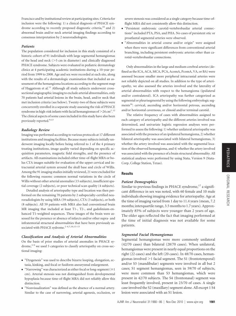
Abnormal arterial course or origin or both (Fig 2) were the
second most common type of arteriopathy, observed in 33/70cases. Some of these cases represented persistent embryonicarteries; others did not correspond to known embryonic arte-rial segments. For example, we encountered 7 cases with aber-rant origin of the ophthalmic artery, which arose from themiddle meningeal artery (3 cases), the ACA (2 cases), the cav-ernous segment of the ICA (1 case), or the BA (1 case), corre-sponding to persistence of primitive embryonic variants ofthis artery.18,19 In many cases, enlarged anomalous branchesprovided arterial supply to a large facial or scalp hemangioma.
Embryonic carotid-basilar connections were present in14/70 cases. PTA was most common (11 cases); PHA and PSAwere observed in 1 and 2 cases, respectively.
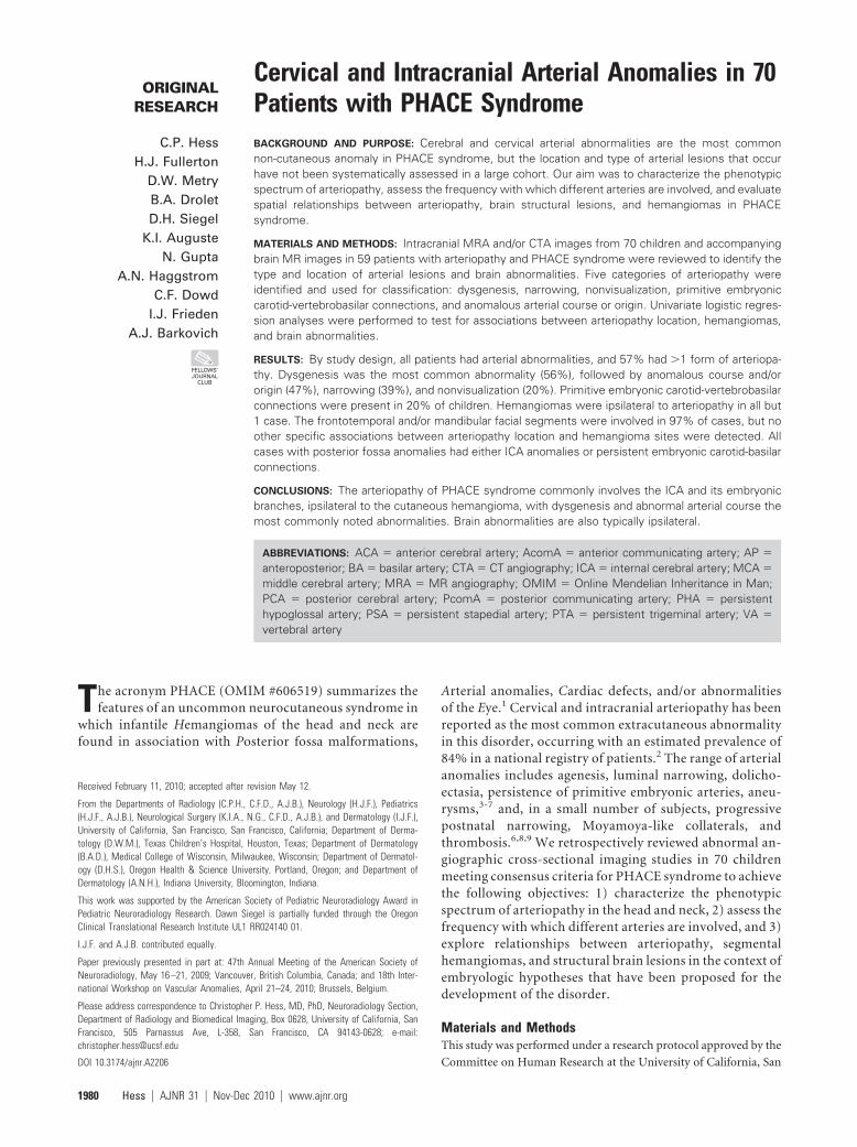
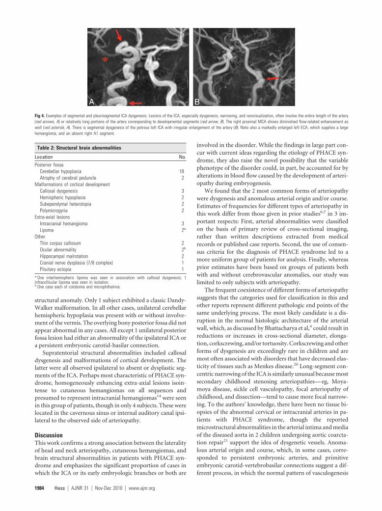
Narrowing (Fig 3) was seen in 27/70 cases, involving a seg-
Fig 1. Representative arterial dysgenesis images are shown as 3D volume renderings from MRA source data, generated by using OsiriX (http://www.osirix-viewer.com/Downloads.html).32
A, Enlargement of the right ICA, MCA, and ACA (white asterisks). There is also an accessory left MCA (red arrow), and the left PCA is duplicated with an anomalous branching pattern(red asterisk). B, Proximal irregularity of a markedly enlarged left ICA (red arrows) and tortuosity of the right PCA (white asterisk). The origin of the right MCA (white arrow) is also anomalous,arising from the cavernous ICA. C, Looping course of the left M1 segment (white arrow) and enlarged left A1/A2 junction (red arrow). D, Marked dysgenesis of the cavernous and supraclinoidright ICA (red arrow) and contralateral dysgenesis of the cavernous and supraclinoid left ICA (white arrows). Note also that the right ICA is not visualized proximal to the cavernous sinus,the right PcomA supplies the right MCA, and the right MCA is markedly narrowed (white asterisks).
Fig 2. Anomalous course and/or origin. A, 3D rendering from MRA shows elongation of the right PcomA, an anomalous connection between the right P1 segment and the right MCA (redarrow). The right ICA and left PCA are also diffusely narrow (white arrow), and the right A1 segment is hypoplastic (white asterisk). B, 3D rendering from CTA shows the tortuous loopedcourse of the supraclinoid right ICA (white arrow). Note also the presence of 2 ACAs, a separate artery of the corpus callosum, and aneurysms at the right carotid terminus and distalright A1 segment (white asterisks). Volume renderings were generated by using OsiriX.32
1982 Hess � AJNR 31 � Nov-Dec 2010 � www.ajnr.org

ment of �1 cm in length in all except 2 subjects. There were nocases of MCA or supraclinoid ICA narrowing associated withclassic Moyamoya collaterals, though small collaterals wereobserved around segments of the ICA and MCA, together withfocal dysgenesis and narrowing in 3 cases.
Nonvisualization (Fig 3) was a feature in 14/70 cases. Al-though agenesis was not distinguished from occlusion in mostcases with MRA, both were observed in the few patients whounderwent CTA. A single case showed segmental agenesis ofthe supraclinoid ICA.5
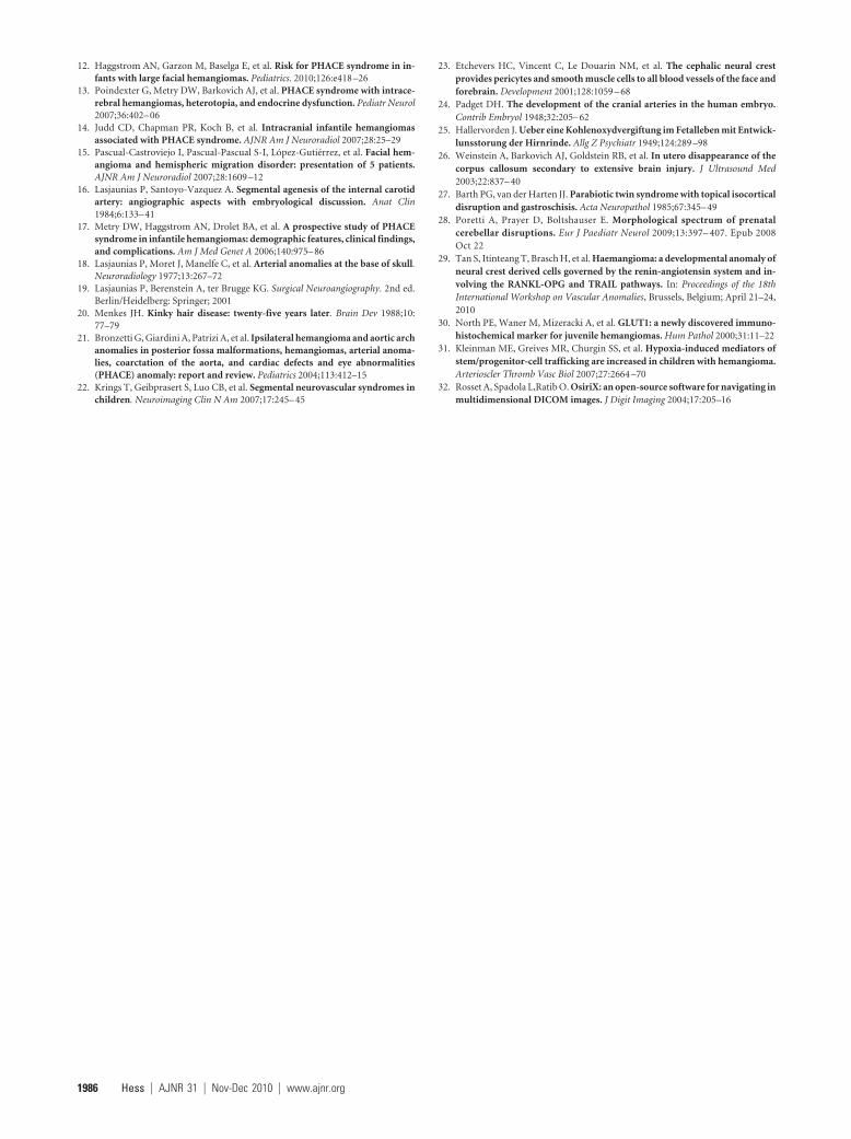
Arteriopathy was more common in the anterior circulationthan in the posterior circulation (Table 1). The ICA was themost commonly involved artery, followed by the MCA andACA, the PCA, and, least commonly, the BA and VA. Abnor-malities were most frequent in the cervical segment of the ICA,though other embryologic segments were involved in similarproportions. A majority of cases with ICA abnormalities(60%) showed pleurisegmental lesions (Fig 4). Univariatelogistic regression analysis revealed no associations between
the artery involved and the segmental location of thehemangioma.
Arteriopathy was also more commonly unilateral (54/70cases) than bilateral (16/70 cases) and, when unilateral, oc-curred with similar frequency on the right (25/70 cases) andon the left (29/70 cases). Among the 54 cases of unilateralarteriopathy, 53 (98%) had ipsilateral hemangiomas. Bilateralarteriopathy was not associated with bilateral hemangiomas.
InfarctsThree patients had remote infarcts on imaging. One had evi-dence of right MCA in utero ischemic injury, right frontalpolymicrogyria, a right globe coloboma, and multiple arterialanomalies, including an absent left ICA, a large AcomA, and apersistent right hypoglossal artery. One had evidence of a peri-natal stroke and hypoplasia of the left ACA and narrowing ofthe left MCA; an acute left MCA infarction had been previ-ously diagnosed in this patient. The third had a remote leftMCA and lateral lenticulostriate territory infarction on an MRimaging performed at 3.6 years of age; MRA in this patientrevealed a thin left M1 segment of the MCA with corkscrewelongation.
Brain Structural AbnormalitiesIn the 59/70 patients with conventional MR imaging availablefor review, structural abnormalities were present in 41% (24/59). Both supra- and infratentorial lesions were observed (Ta-ble 2). All unilateral lesions were associated with both arteri-opathy and hemangioma ipsilateral to the side of the brainabnormality on MR imaging.
Posterior fossa abnormalities were the most common
Fig 3. Arterial narrowing and nonvisualization. All images are shown as maximum-intensity-projection reconstructions from MRA source data. A, Submentovertex projection showingnonvisualization of the left ICA. B, AP projection shows long-segment narrowing of the entire visualized course of the right ICA (red arrow). C, AP projection shows nonvisualization ofthe left ICA from the distal cervical to the cavernous segments. The intradural left VA is also absent. D, AP projection shows absence of the entire left ICA and long-segment taperednarrowing and luminal irregularity of the distal cervical right ICA (red arrow).
Table 1: Location of arterial abnormalities
Location No. (%)ICA 53/70 (76%)
Cervical 35Petrous 33Cavernous 27Supraclinoid 32
MCA 13/70 (20%)ACA 11/70 (16%)PCA 10/70 (14%)BA or VA 5/70 (7%)
AJNR Am J Neuroradiol 31:1980 – 86 � Nov-Dec 2010 � www.ajnr.org 1983
structural anomaly. Only 1 subject exhibited a classic Dandy-Walker malformation. In all other cases, unilateral cerebellarhemispheric hypoplasia was present with or without involve-ment of the vermis. The overlying bony posterior fossa did notappear abnormal in any cases. All except 1 unilateral posteriorfossa lesion had either an abnormality of the ipsilateral ICA ora persistent embryonic carotid-basilar connection.
Supratentorial structural abnormalities included callosaldysgenesis and malformations of cortical development. Thelatter were all observed ipsilateral to absent or dysplastic seg-ments of the ICA. Perhaps most characteristic of PHACE syn-drome, homogeneously enhancing extra-axial lesions isoin-tense to cutaneous hemangiomas on all sequences andpresumed to represent intracranial hemangiomas14 were seenin this group of patients, though in only 4 subjects. These werelocated in the cavernous sinus or internal auditory canal ipsi-lateral to the observed side of arteriopathy.
DiscussionThis work confirms a strong association between the lateralityof head and neck arteriopathy, cutaneous hemangiomas, andbrain structural abnormalities in patients with PHACE syn-drome and emphasizes the significant proportion of cases inwhich the ICA or its early embryologic branches or both are
involved in the disorder. While the findings in large part con-cur with current ideas regarding the etiology of PHACE syn-drome, they also raise the novel possibility that the variablephenotype of the disorder could, in part, be accounted for byalterations in blood flow caused by the development of arteri-opathy during embryogenesis.
We found that the 2 most common forms of arteriopathywere dysgenesis and anomalous arterial origin and/or course.Estimates of frequencies for different types of arteriopathy inthis work differ from those given in prior studies6,7 in 3 im-portant respects: First, arterial abnormalities were classifiedon the basis of primary review of cross-sectional imaging,rather than written descriptions extracted from medicalrecords or published case reports. Second, the use of consen-sus criteria for the diagnosis of PHACE syndrome led to amore uniform group of patients for analysis. Finally, whereasprior estimates have been based on groups of patients bothwith and without cerebrovascular anomalies, our study waslimited to only subjects with arteriopathy.
The frequent coexistence of different forms of arteriopathysuggests that the categories used for classification in this andother reports represent different pathologic end points of thesame underlying process. The most likely candidate is a dis-ruption in the normal histologic architecture of the arterialwall, which, as discussed by Bhattacharya et al,4 could result inreductions or increases in cross-sectional diameter, elonga-tion, corkscrewing, and/or tortuosity. Corkscrewing and otherforms of dysgenesis are exceedingly rare in children and aremost often associated with disorders that have decreased elas-ticity of tissues such as Menkes disease.20 Long-segment con-centric narrowing of the ICA is similarly unusual because mostsecondary childhood stenosing arteriopathies— eg, Moya-moya disease, sickle cell vasculopathy, focal arteriopathy ofchildhood, and dissection—tend to cause more focal narrow-ing. To the authors’ knowledge, there have been no tissue bi-opsies of the abnormal cervical or intracranial arteries in pa-tients with PHACE syndrome, though the reportedmicrostructural abnormalities in the arterial intima and mediaof the diseased aorta in 2 children undergoing aortic coarcta-tion repair21 support the idea of dysgenetic vessels. Anoma-lous arterial origin and course, which, in some cases, corre-sponded to persistent embryonic arteries, and primitiveembryonic carotid-vertebrobasilar connections suggest a dif-ferent process, in which the normal pattern of vasculogenesis
Fig 4. Examples of segmental and pleurisegmental ICA dysgenesis. Lesions of the ICA, especially dysgenesis, narrowing, and nonvisualization, often involve the entire length of the artery(red arrows, A) or relatively long portions of the artery corresponding to developmental segments (red arrow, B). The right proximal MCA shows diminished flow-related enhancement aswell (red asterisk, A). There is segmental dysgenesis of the petrous left ICA with irregular enlargement of the artery (B). Note also a markedly enlarged left ECA, which supplies a largehemangioma, and an absent right A1 segment.
Table 2: Structural brain abnormalities
Location No.Posterior fossa
Cerebellar hypoplasia 18Atrophy of cerebral peduncle 2
Malformations of cortical developmentCallosal dysgenesis 3Hemispheric hypoplasia 2Subependymal heterotopia 2Polymicrogyria 2
Extra-axial lesionsIntracranial hemangioma 3Lipoma 2a
OtherThin corpus callosum 2Ocular abnormality 2b
Hippocampal malrotation 2Cranial nerve dysplasia (7/8 complex) 1Pituitary ectopia 1
a One interhemispheric lipoma was seen in association with callosal dysgenesis; 1infracollicular lipoma was seen in isolation.b One case each of coloboma and microphthalmia.
1984 Hess � AJNR 31 � Nov-Dec 2010 � www.ajnr.org

is altered during development of the cervical and intracranialarterial system.
As elegantly summarized by Krings et al,22 current hypoth-eses regarding the developmental errors that link the abnor-malities in PHACE syndrome have focused on the role of theneural crest, the adjacent cephalic mesoderm, and even theneural plate in cases of brain structural lesions. Chimera stud-ies in the avian embryo provide evidence that the muscularand connective tissue wall of the ICA and its branches arederived from cephalic neural crest cells, in contrast to the ar-terial wall of the basilar and vertebral arteries, which derivefrom the cephalic mesoderm.23 The disproportionate fre-quency with which the anterior circulation is involved com-pared with the posterior circulation follows the embryologicdivision between neural crest and mesodermal-derived arter-ies. However, for reasons outlined by other authors,metameric disruption in the neural crest and adjacent cephalicmesoderm does not fully account for the spectrum of abnor-malities in PHACE syndrome.4,19,22
The primitive ICA is vital to the normal development of thebrain and surrounding structures because it is the embryologicprecursor to the MCA, the ACA, the PcomA arteries, thePCAs, the superior cerebellar arteries, and the upper BA and itis the primary source of blood to the developing vertebrobasi-lar system. According to Padget,24 this artery is first seen as thecranial extension of the dorsal aorta at the 3-mm embryonicstage (22–23 days) and gives rise to its primary branches by the16-to 18-mm stage (48 –51 days). During this roughly 4-weekperiod, the primitive ICA also supplies the posterior circula-tion through a series of transient intersegmental arteries thateach normally develop and regress for 7–10 days until the ver-tebrobasilar system is fully developed. Blood supply to thecerebellum in particular is critically dependent on the ICAduring this phase of embryogenesis.
While different brain structural anomalies observed in thisstudy and previously associated with PHACE syndrome mayindividually be caused by a variety of factors, both the com-mon posterior fossa abnormalities and the less frequent supra-tentorial abnormalities share in utero ischemia as a commonpotential cause. Before fusing and becoming the BA between29 and 33 days’ gestation, the paired dorsal longitudinal arter-ies are supplied mainly by the primitive ICA by means of thetrigeminal arteries. It is not until nearly 6 weeks’ gestation thatthe vertebral arteries become the major contributor to hind-brain blood supply. Compromised vascular blood flow within1 longitudinal artery before this point could impair formationof the ipsilateral cerebellar hemisphere and vermis. Abruptdisruption of an otherwise normally developing cerebellum issuggested by the normal bony posterior fossa,4 consistent withan ischemic etiology for cerebellar anomalies. Similarly, tran-sient or permanent vascular insult and in utero ischemia arewell-established causes for polymicrogyria,25 callosal dysgen-esis,26 heterotopia,27 cerebellar hypoplasia,28 and other mal-formations of cortical development.
Although almost all cases with intracranial arteriopathyhave S1 or S3 segment hemangiomas, the lack of a significantassociation between the arteries involved and the segmentallocation of the facial hemangioma militates against a commonmetameric origin for the simultaneous development of bothabnormalities. Recent work provides evidence that the endo-
thelial progenitor cells that give rise to hemangiomas may ul-timately derive from the neural crest,29 though the cellular andmolecular events that would induce the neural crest to differ-entiate along these lines have not yet been elaborated. At birth,most hemangiomas represent “precursor lesions” that subse-quently proliferate. Alterations in blood flow or hypoxia mayserve as the trigger that leads to the development of latenthemangioma precursors. Hemangiomas at all stages of evolu-tion express glucose transporter 1,30 an intrinsic cellularmarker of tissue hypoxia, and the combination of hypoxia andestrogen demonstrates a synergistic effect on hemangioma en-dothelial cell proliferation.31
There are 2 important limitations of this study. First, be-cause most patients underwent only MRA to study their arter-ies, it was not possible to distinguish between agenesis andocclusion or between stenosis and hypoplasia, which representfundamentally different processes. This limitation hinders ourability to divide these anomalies from an embryologic stand-point. Second, the average age of children included in thiswork was relatively old with respect the diagnosis of PHACEsyndrome. To the extent that arteriopathy may change withtime, this ascertainment bias could lead to inaccuracies in theestimated frequencies of different types of arteriopathy.
ConclusionsPleurisegmental dysgenesis, especially involving the ICA, andanomalous course and origin of the cervical and intracranialarteries, often representing persistence of embryonic arteries,are the 2 most frequent arterial anomalies observed on cross-sectional angiographic imaging in patients with PHACE syn-drome. The phenotypic variability of brain structural lesionsand hemangiomas could conceivably be explained by alter-ations in blood flow and, in some cases, hypoxia, as a result ofarteriopathy during embryogenesis.
References1. Frieden IJ, Reese V, Cohen D. PHACE syndrome: the association of posterior
fossa brain malformations, hemangiomas, arterial anomalies, coarctation ofthe aorta and cardiac defects, and eye abnormalities. Arch Dermatol1996;132:302–11
2. Metry DW, Garzon MC, Drolet BA, et al. PHACE syndrome: current knowl-edge, future directions. Pediatr Dermatol 2009;26:381–98
3. Pascual-Castroviejo I. Vascular and nonvascular intracranial malformationassociated with external capillary hemangioma. Neuroradiology 1978;16:82– 84
4. Bhattacharya JJ, Luo CB, Alavarez H, et al. PHACES syndrome: a review of eightpreviously unreported cases with late arterial occlusions. Neuroradiology2004;46:227–33
5. Baccin CE, Krings T, Alvarez H, et al. A report of two cases with dolichoseg-mental intracranial arteries as a new feature of PHACES syndrome. ChildsNerv Syst 2007;23:559 – 67. Epub 2006 Oct 13
6. Heyer GL, Dowling MM, Licht DJ, et al. The cerebral vasculopathy of PHACESsyndrome. Stroke 2008;39:308 –16. Epub 2008 Jan 3
7. Oza VS, Wang E, Berenstein A, et al. PHACES association: a neuroradiologicreview of 17 patients. AJNR Am J Neuroradiol 2008;29:807–13
8. Burrows PE, Robertson RL, Mulliken JB, et al. Cerebral vasculopathy and neu-rologic sequelae in infants with cervicofacial hemangioma: report of eightpatients. Radiology 1998;207:601– 07
9. Drolet BA, Dohil M, Golomb MR, et al. Early stroke and cerebral vasculopathyin children with facial hemangiomas and PHACE association. Pediatrics2006;117:959 – 64
10. Metry D, Heyer G, Hess C, et al. Consensus statement on diagnostic criteria forPHACE syndrome. Pediatrics 2009;124:1447–56
11. Haggstrom AN, Lammer EJ, Schneider RA, et al. Patterns of infantilehemangiomas: new clues to hemangioma pathogenesis and embryonic facialdevelopment. Pediatrics 2006;117:698 –703
AJNR Am J Neuroradiol 31:1980 – 86 � Nov-Dec 2010 � www.ajnr.org 1985
12. Haggstrom AN, Garzon M, Baselga E, et al. Risk for PHACE syndrome in in-fants with large facial hemangiomas. Pediatrics. 2010;126:e418 –26
13. Poindexter G, Metry DW, Barkovich AJ, et al. PHACE syndrome with intrace-rebral hemangiomas, heterotopia, and endocrine dysfunction. Pediatr Neurol2007;36:402– 06
14. Judd CD, Chapman PR, Koch B, et al. Intracranial infantile hemangiomasassociated with PHACE syndrome. AJNR Am J Neuroradiol 2007;28:25–29
15. Pascual-Castroviejo I, Pascual-Pascual S-I, Lopez-Gutierrez, et al. Facial hem-angioma and hemispheric migration disorder: presentation of 5 patients.AJNR Am J Neuroradiol 2007;28:1609 –12
16. Lasjaunias P, Santoyo-Vazquez A. Segmental agenesis of the internal carotidartery: angiographic aspects with embryological discussion. Anat Clin1984;6:133– 41
17. Metry DW, Haggstrom AN, Drolet BA, et al. A prospective study of PHACEsyndrome in infantile hemangiomas: demographic features, clinical findings,and complications. Am J Med Genet A 2006;140:975– 86
18. Lasjaunias P, Moret J, Manelfe C, et al. Arterial anomalies at the base of skull.Neuroradiology 1977;13:267–72
19. Lasjaunias P, Berenstein A, ter Brugge KG. Surgical Neuroangiography. 2nd ed.Berlin/Heidelberg: Springer; 2001
20. Menkes JH. Kinky hair disease: twenty-five years later. Brain Dev 1988;10:77–79
21. Bronzetti G, Giardini A, Patrizi A, et al. Ipsilateral hemangioma and aortic archanomalies in posterior fossa malformations, hemangiomas, arterial anoma-lies, coarctation of the aorta, and cardiac defects and eye abnormalities(PHACE) anomaly: report and review. Pediatrics 2004;113:412–15
22. Krings T, Geibprasert S, Luo CB, et al. Segmental neurovascular syndromes inchildren. Neuroimaging Clin N Am 2007;17:245– 45
23. Etchevers HC, Vincent C, Le Douarin NM, et al. The cephalic neural crestprovides pericytes and smooth muscle cells to all blood vessels of the face andforebrain. Development 2001;128:1059 – 68
24. Padget DH. The development of the cranial arteries in the human embryo.Contrib Embryol 1948;32:205– 62
25. Hallervorden J. Ueber eine Kohlenoxydvergiftung im Fetalleben mit Entwick-lunsstorung der Hirnrinde. Allg Z Psychiatr 1949;124:289 –98
26. Weinstein A, Barkovich AJ, Goldstein RB, et al. In utero disappearance of thecorpus callosum secondary to extensive brain injury. J Ultrasound Med2003;22:837– 40
27. Barth PG, van der Harten JJ. Parabiotic twin syndrome with topical isocorticaldisruption and gastroschisis. Acta Neuropathol 1985;67:345– 49
28. Poretti A, Prayer D, Boltshauser E. Morphological spectrum of prenatalcerebellar disruptions. Eur J Paediatr Neurol 2009;13:397– 407. Epub 2008Oct 22
29. Tan S, Itinteang T, Brasch H, et al. Haemangioma: a developmental anomaly ofneural crest derived cells governed by the renin-angiotensin system and in-volving the RANKL-OPG and TRAIL pathways. In: Proceedings of the 18thInternational Workshop on Vascular Anomalies, Brussels, Belgium; April 21–24,2010
30. North PE, Waner M, Mizeracki A, et al. GLUT1: a newly discovered immuno-histochemical marker for juvenile hemangiomas. Hum Pathol 2000;31:11–22
31. Kleinman ME, Greives MR, Churgin SS, et al. Hypoxia-induced mediators ofstem/progenitor-cell trafficking are increased in children with hemangioma.Arterioscler Thromb Vasc Biol 2007;27:2664 –70
32. Rosset A, Spadola L,Ratib O. OsiriX: an open-source software for navigating inmultidimensional DICOM images. J Digit Imaging 2004;17:205–16
1986 Hess � AJNR 31 � Nov-Dec 2010 � www.ajnr.org


![Atypical Intracranial Epidermoid Cysts: Rare Anomalies with … · 2017-10-31 · the parasellar region [1]. Conversely, atypical epidermoid cysts are rare, with intra-axial epidermoid](https://img.dokumen.tips/doc/110x75/5f7ff1f90cbb51524d18b285/atypical-intracranial-epidermoid-cysts-rare-anomalies-with-2017-10-31-the-parasellar.jpg)