Embed Size (px)
Citation preview

Cerebral Palsy: MR Findings in 40 Patients
Charles L. Truwit, 1'2 A . James Barkovich,2
'3 Thomas K . Koch,3 and Donna M. Ferriero3
.4
Purpose: , We used MR to retrospectively analyze the brains of patients suffering from cerebral palsy, our aim being to determine MR's role in the assessment of brain damage and the relationship of pre-, peri-, and post-natal events to cerebral palsy. Methods: Forty patients (aged 1 month to 41 years) underwent MR scanning and findings were correlated with clinical histories in all cases. Results: Review of MR scans of 11 patients who had been born prematurely revealed findings of periventricular white matter damage, indicative of hypoxic-ischemic brain injury (82%), the
chronology of which was difficult to determine. Among 29 patiel")ts who had been born at term, three major patterns emerged: (1), gyral anomalies, suggestive of polymicrogyria, consistent with mid-second trim'ester injury; (2), isolated periventricular leukomalacia reflecting late second- or
early third-trimester injury; and (3), watershed cortical or deep gray nuclear damage, consistent with late third-trimester, perinatal or postnatal injury. In 16 (55%) of 29 patients born at term, MR findings of intrauterine brain damage were observed; in over half of these cases MR revealed developmental anomalies, which is nearly twice the rate reported in prior studies employing CT. Conclusion: Our results support a growing consensus that cerebral palsy in term infants is often
the result of prenatal factors, and less commonly related to the perinatal period.
Index term: Cerebral palsy
AJNR 13:67-78, January /February 1992
Cerebral palsy (CP) has been the subject of numerous clinical, neuropathologic, and neuroradiologic studies to assess the relationship of pre-, peri-, and postnatal events to CP (1-9). Clinical studies have been limited by the availability of historical information regarding prenatal care and possible prenatal insults. Neuropathologic studies have been limited by the small
Received February 20, 1991 ; accepted and revision requested April
15, revision received May 22; final acceptance May 29. The views expressed in this article are those of the authors and do
not reflect the official policy or position of the Department of the Army,
the Department of Defense, or the United States Government. 1 U.S. Army Medical Department, Academy of Health Sciences, HSHA
GPS Fort Sam Houston , TX 78234-6100. Address reprint requests to C.L. Truwit, Department of Radiology, Fitzsimons Army Medical Center, Au
rora , CO 80045-5000. 2 Department of Radiology, Neuroradiology Section, University of
California, San Francisco, 505 Parnassus Ave., Box 0628, San Francisco,
CA 94143-0628. 3 Department of Pediatric Neurology, University of California, San
Francisco, San Francisco, CA 94143. 4 Department of Pediatric Neurology, San Francisco General Hospital,
San Francisco, CA 94110.
AJNR 13:67-78, Jan/ Feb 1992 0195-6108/ 92/ 1301-0067 © American Society of Neuroradiology
67
numbers of cases that have gone to autopsy. Neuroradiologic studies have been limited in their usefulness, primarily because of the limited capabilities of neuroimaging in the era before computed tomography (CT) and magnetic resonance (MR) imaging. With the development of CT, radiographic correlation with clinical data regarding prenatal, perinatal, and postnatal asphyxia became possible, particularly with respect to morphologic changes of the cerebral white matter (1, 7-1 0). The sensitivity of CT, however, is limited in many cases of cerebral palsy. In particular, while state-of-the-art CT is sensitive to dramatic anomalies of brain development, such as schizencephaly, MR is more sensitive than CT in the detection of both subtle brain malformations, such as callosal hypogenesis, polymicrogyria, and mild degrees of white matter damage. Therefore, we studied MR images to retrospectively analyze the brains of patients with cerebral palsy.
Materials and Methods
Forty patients, aged 1 month to 41 years, underwent
MR scanning at either mid (0.35 T) or high (1.5 T) field

68
strength. Three patients were initially scanned at less than 1 year of age. They are now 4 years, 6 months (case 15), 3 years, 4 months (case 24) and 2 years , 6 months (case 27) and have spastic , hypotonic and spastic CP, respectively . As the study was undertaken at a major referral center, the study population was limited by selecting only those patients with the referral diagnosis of CP, and only those for whom MR studies were included. The MR studies were retrospectively reviewed by two of the authors with knowledge of the preceding clinical diagnosis of CP, but without knowledge of the specific clinical manifestations of each case. Thus, the MR review was only partially blinded, since the diagnosis of CP was known. Nevertheless, because we set out to assess what abnormalities appeared on MR scans of patients with CP, not the efficacy of MR in diagnosing CP, we believe this methodology was appropriate.
All MR studies included sagittal and axial T1-weighted images, 400-800/ 20-35/ 1-2 (TR/ TE/ excitations), and axial T2-weighted images, 2000-3000/ 30-120/ 1-2. The MR studies were evaluated for developmental or acquired abnormalities of the corpus callosum, deep and peripheral white matter, cerebral cortex, basal ganglia/ thalami , and brainstem, and for the presence of parenchymal cysts, hydrocephalus, and ventricular contour defects. The corpus callosum, brainstem , cerebral cortex , and basal ganglia/ thalami were subjectively assessed as thinned or normal. As the radiologic manifestations of preterm and term injuries differ, patients born prematurely were grouped separately.
Classification of cerebral anomalies was based on typical MR findings. Polymicrogyria was diagnosed by the presence of abnormally thick cortex, diminished and absent arborization of subjacent white matter, and mild ventriculomegaly beneath the anomalous parenchyma ( 11 , 12). Hypoplasia of the ipsilateral thalamus, cerebral peduncle, and pons was commonly associated. The cortical mantle typically demonstrated multiple sulci , in contradistinction to the broad, shallow gyri of pachygyria , and in contradist inct ion to the mushroom-shaped, small gyri of ulegyria. We recognize that this definition is broader than the strict pathologic definition, which requires an abnormal fourlayered cortex. Nevertheless, although it is possible that the MR findings reflect another, unspecified migration anomaly , in our experience, this MR pattern has repeatedly correlated with pathologic specimens of polymicrogyria .
The diagnosis of schizencephaly required MR definition of an abnormal gray m atter cleft extending all the way to t he lateral ventricle. A cleft that reached toward, but that was clearly separated from, the ventricle was defined as a cleft of polymicrogyria (13) . Although others have considered this to be type I schizencephaly (14), we consider the presence of white matter between the gray matter cleft and the lateral ventricle to distinguish the two entities, since a pial-ependymal seam , the hallmark of schinzencephaly, is impossible in this situation.
MR findings were correlated with cl inical histories, which were available for all patients. In one patient (case 4), no birth history was available other than delivery at 28 weeks
AJNR: 13, January / February 1992
gestation. Specifically, pre-, peri- and postnatal histories were assessed. Eleven patients (27 %) were born prematurely , by the criterion of a gestational age (by dates) of 36 weeks or less at the time of birth. Twenty-nine patients (73 %) were delivered at or near term. Although included in Table 1, we did not segregate the patients by type of CP (e.g. , diplegic, quadriplegic), as we commonly encountered clinical discrepancies, such as one clinician labeling the patient diplegic only to find the next clinician labeling the same patient quadriplegic or hemiplegic.
For purposes of this study, we defined the perinatal period as that time starting 2 weeks before birth and continuing through the first 2 weeks of life. Perinatal asphyxia was defined to include fetal distress and respiratory distress (endotracheal oxygenation, meconium aspiration, arrest, or near-arrest). We did not try to separate the types and degrees of perinatal asphyxia. Periventricular leukomalacia (PVL), isolated periventricular white matter damage without cortical , subcortical, or deep gray matter lesions, was considered to be representative of brain damage that occurred prior to the 36th week of gestation. This concept is supported by recent reports on the MR assessment of perinatal asphyxia (15, 16). As a result , isolated PVL in the term infant was considered to be consequent to intrauterine brain injury, even in the face of perinatal insults.
Results
As seen in Table 1, the MR scans revealed readily apparent abnormalities in every patient except three patients born at term. One of these patients (case 16) had choreoathetoid CP; her mother had experienced third-trimester pre eclampsia. Another patient (case 22) had spasticity and was cortically blind. The third patient (case 29) underwent meconium aspiration and had spasticity.
MR Findings in Patients Born Prematurely
Among the 11 patients born prematurely, several radiographic features were apparent. MR scans in all 11 revealed deep white matter loss, especially in the peritrigonal regions. In nine of the 11, scans demonstrated thinning of the corpus callosum, especially in the posterior body (Fig. 1 ). In case 8, the corpus callosum was severely hypogenetic. The one patient with a normal corpus callosum (case 5) had hypotonic CP and had diminished deep, and to a lesser extent peripheral , white matter. The ventricular trigones were slightly rounded. All had abnormal ventricles, usually manifested as irregular contour of the trigones, consistent with prior incorporation of peri ventricular cysts. One patient (case 9) had discrete periventricular cysts. In three patients (cases 5 , 6, and 8) , born at 30-32 weeks'
TA
BL
E
1:
Clin
ical
and
MR
Fin
din
gs
in P
atie
nts
wit
h C
ereb
ral
Pal
sy
Gro
up
/ G
esta
tio
nal
Ag
e
Cas
e A
ge
at
MR
S
ex
at
Bir
th:
Per
i-,
Do
min
an
t C
linic
al
Co
rpu
s C
allo
sum
D
eep
Wh
ite
Ma
tte
r P
erip
hera
l C
ysts
L
ate
ral
Ven
tric
les
Co
rte
x B
asal
B
rain
ste
m
Oth
er
Fin
din
gs
Po
stn
ata
l F
ind
ing
s W
hit
e M
att
er
Gan
glia
N
o.
Hi s
tory
Bo
rn p
rem
atu
rely
I 9
yr,
8 m
o
M
27
wk:
Gra
de 4
S
pa
stic
dip
legi
a D
iffu
sely
th
inn
ed,
Dim
inis
hed
, es
peci
ally
N
orm
al
No
Ir
reg
ula
r co
nto
ur,
esp
ecia
lly
No
rma
l N
orm
al
No
rma
l
intr
aven
tric
ula
r es
peci
ally
po
s-p
eri t
rig
on
al
mid
bo
dy
and
trig
on
es
h em
orr
hag
e,
teri
orl
y
shu
nt,
RD
S
2 I
yr,
6 m
o
F
28
wk:
Gra
de 3
S
pa
stic
dip
leg
ia
Th
inn
ed
po
ste
rio
r D
imin
ishe
d p
eri
trig
onal
&
No
rma
l N
o
Dila
ted
trig
one
s N
orm
al
No
rma
l N
orm
al
intr
aven
tric
ula
r b
od
y lo
ng
T2
he
mo
rrh
ag
e,
RD
S
3 I
yr,
2 m
o
M
28
wk:
RD
S,
Spa
stic
ity
Dif
fuse
ly t
hin
ned
, D
imin
ish
ed
pe
ritr
igo
nal
No
rma
l N
o
Min
ima
lly i
rre
gu
lar
con
tou
r N
orm
al
No
rma
l S
ma
ll C
ereb
ella
r h
em
isp
he
ric
intr
av
en
tric
ula
r p
ost
eri
or >
an
-h
ypo
pla
sia
vs
atr
o-
he
mo
rrh
ag
e
teri
or
phy
4 41
yr
F
28
wk
Sp
ast
icit
y, b
lindn
ess
Th
inn
ed
bo
dy
Dim
inis
he
d &
lo
ng
T2
No
rma
l N
o
Ab
no
rma
l co
nto
ur
No
rmal
N
orm
al
No
rma
l
5 2
yr,
6 m
o
F
30
wk:
Su
rviv
ing
H
ypo
ton
ic,
ath
eto
id,
No
rmal
D
imin
ish
ed
pe
ritr
igo
na
l &
S
ligh
tly
di-
No
S
ligh
tly
rou
nd
ed
tri
go
nes
No
rma
l N
orm
al
No
rma
l
twin
; en
do
trac
hea
l p
arap
ares
is
occ
ipit
al
min
ishe
d
intu
ba
tio
n fo
r 2
wk
6 7
yr,
5 m
o
M
30
wk:
Sev
ere
RD
S,
Sp
ast
icit
y, L
E
Th
inn
ed
po
ste
rio
r D
imin
ish
ed
pe
ritr
igo
na
l S
catt
ere
d fo
ci
No
Ir
reg
ula
r co
nto
ur
post
e-N
orm
al
No
rma
l N
orm
al
intr
aven
tric
ula
r b
od
y &
sp
len
-o
f p
ro-
rio
rly
he
mo
rrh
ag
e, s
hu
nt
ium
lo
ng
ed
T2
7 2
yr,
8 m
o
F
30
-31
wk:
5 d
ays
' S
pa
stic
dip
leg
ia
Th
inn
ed p
ost
eri
or
Dim
inis
he
d p
eri
trig
on
al,
No
rma
l N
o
Dila
ted
trig
on
es
& o
ccip
ita
l N
orm
al
No
rma
l N
orm
al
en
do
trac
heal
in
tu-
bo
dy
& s
ple
n-
lon
g T
2 h
orn
s w
ith
irr
egu
lar
con
-
ba
tion
, du
ctu
s lig
a-
ium
to
ur
tio
n
8 10
yr
M
32
wk:
NS
VD
, h
y-S
pa
stic
dip
leg
ia,
Sev
ere
hyp
o-
Ma
rke
dly
dim
inis
he
d
Mild
y d
imin
-N
o
Ma
rke
dly
irr
eg
ula
r co
nto
ur
No
rma
l N
orm
al
No
rma
l C
hia
sma
tic h
ypo
pla
sia
dro
cep
ha
lus
note
d
me
nta
l re
tard
a-
gene
sis
ishe
d p
os-
thic
k-
at
bir
th;
shu
nt
at
3 ti
on
te
rio
rly
ness
, w
k di
s-
tort
ed
20
shu
nt
9 2
yr
F
32
wk:
Fe
tal d
istr
ess
S
pa
stic
ity
Th
inne
d p
ost
eri
or
Dif
fuse
ly d
imin
ishe
d &
N
orm
al
Yes
, p
eri
-M
ild c
on
tou
r a
bno
rma
lity
No
rma
l N
orm
al
No
rma
l
pre
cip
ita
tin
g e
me
r-b
od
y lo
ng
T2
tr
igo
na
l
gen
cy c
esar
ean
de
live
ry;
Ap
ga
rs,
7/9
10
3
yr,
8 m
o
F
35
wk
: P
ree
cla
mp
sia
, H
ypo
ton
ia,
blin
d-
Se
vere
ly t
hin
ned
Dim
inis
hed;
glio
sis
No
rma
l N
o
En
larg
ed
ex
vacu
o
No
rma
l N
orm
al
Sm
all
feta
l di
stre
ss,
ce-
nes
s sa
rean
de
live
ry
II
4 yr
, 6
mo
M
3
6 w
k: ?
In
ute
ro
Sp
ast
ic d
iple
gia
, D
iffu
sely
th
inn
ed
D
iffu
sely
th
inn
ed
& l
on
g
No
rma
l N
o
Min
ima
lly i
rre
gu
lar
con
tou
r,
No
rma
l N
orm
al
Sm
all
toxo
pla
smo
sis
, L
> R
; d
eve
lop
-b
od
y T
2
trig
on
es
pre
ecl
am
ps
ia,
feta
l m
en
tal
dela
y
dis
tres
s,
cesa
rean
de
live
ry, h
ypo
gly
-
cem
ia a
t b
irth

TA
BL
E
1:
Clin
ical
and
MR
Fin
din
gs
in P
atie
nts
wit
h C
ereb
ral
Pa
lsy-C
onti
nued
Gro
up
/ G
esta
tiona
l A
ge
a
t B
irth
: P
eri-
, D
om
ina
nt
Clin
ica
l P
erip
hera
l B
asal
C
ase
Ag
e a
t M
R
Sex
C
orp
us
Ca
llosu
m
Dee
p W
hit
e M
att
er
Cys
ts
Late
ral
Ve
ntr
icle
s C
ort
ex
B
rain
stem
O
the
r F
indi
ngs
Po
stn
ata
l F
ind
ing
s W
hit
e M
att
er
Gan
glia
N
o.
His
tory
Bo
rn a
t te
rm
12
I y
r, I
m
o
F
37
wk:
Cya
no
sis
at
Hyp
oto
nia
, se
vere
N
orm
al
Del
ayed
mye
lina
tio
n
De
laye
d m
ye-
No
No
rma
l N
orm
al
No
rma
l N
orm
al
bir
th, o
xyg
en
for
de
velo
pm
en
tal
lin
atio
n
24
hr
de
lay
13
2 y
r M
3
6 w
k S
pa
stic
ity,
R m
on
o-
Th
inn
ed
po
ste
rio
r D
imin
ish
ed
pe
ritr
igo
na
l N
orm
al
No
A
bn
orm
al
con
tou
r N
orm
al
No
rma
l N
orm
al
pare
sis
bo
dy
, sp
len
ium
&
slig
ht
fro
nta
l
14
I y
r, I
I m
o M
3
6 w
k S
pa
stic
ity
, L
E >
U
E
Mild
ly t
hin
ne
d
Pe
ritr
igo
nal
th
inn
ing
N
orm
al
No
M
ildly
irr
eg
ula
r co
nto
ur,
N
orm
al
No
rma
l N
orm
al
post
eri
or
bod
y tr
igo
ne
& s
ple
niu
m
15
II m
o
M
42
wk
: D
ecre
ased
S
pa
stic
ity, R
UE
M
ildly
th
inn
ed
D
imin
ishe
d p
eri
trig
ona
l, N
orm
al
Yes
Ir
reg
ula
r co
nto
ur,
L t
rig
on
e
No
rma
l In
farc
ts,
L A
tro
ph
y, L
feta
l m
ove
me
nts
b
od
y L
side
in
tern
al
cere
bral
for
24
hr
at
30
wk
; ca
psul
e &
p
ed
un
-
Ap
ga
rs:
7/6
th
alam
us
cle
16
4
yr,
11
mo
F
Th
ird
-tri
me
ste
r p
re-
Cho
reo
ath
eto
id
No
rma
l N
orm
al
No
rma
l N
o
No
rma
l N
orm
al
No
rma
l N
orm
al
ecl
am
psi
a:
Ap
ga
rs,
mo
vem
ents
7/9
17
4 y
r M
4
3 w
k: M
eco
niu
m
Sp
ast
ic q
ua
dri
ple
-T
hin
ne
d b
ody
&
Dim
inis
he
d p
eri
trig
on
al
Slig
htl
y di
-N
o
Slig
htl
y b
ulb
ou
s tr
igo
ne
s N
orm
al
Th
ala
mi
No
rma
l
sta
inin
g;
Ap
ga
rs,
gia
, se
izur
es
sple
niu
im
min
ish
ed
slig
htl
y
4/7
sm
all
16
3 yr
, 3
mo
F
T
erm
: M
icro
cep
ha
lic,
Sp
ast
ic q
ua
dri
ple
-T
hin
ned
bo
dy
&
Dim
inis
he
d d
iffu
sely
wit
h
Dim
inis
hed
No
E
nla
rged
wit
h a
bn
orm
al
Th
inn
ed
L
on
g T
2,
L N
orm
al
de
laye
d d
eliv
ery
gi
a,
R >
L
; d
eve
l-sp
len
ium
lo
ng
T2
d
iffu
sely
co
nto
ur
po
ste
-th
ala
mu
s
afte
r tw
in r
equ
irin
g
op
me
nt
de
lay
wit
h l
on
g ri
or
pa
-&
L p
uta
-
Pit
oc
in,
me
co
niu
m
T2
riet
al
me
n
stai
nin
g
19
6 yr
, 11
mo
M
T
erm
: A
no
xia
, m
eco
-S
past
ic h
ype
rto
nic
D
iffu
sely
th
inn
ed
Dif
fuse
ly t
hin
ne
d,
Dif
fuse
ly
Mu
ltip
le
Por
ence
ph
aly
, L
>
R,
Dif
fuse
ly
No
rmal
N
orm
al
Mu
ltic
ysti
c e
nce
ph
alo
-
niu
m a
spir
atio
n,
mo
no
ple
gia
, se
i-L
>R
th
inn
ed,
occ
ipit
al
>
fro
nta
l th
inn
ed
m
alac
ia,
L >
R
mic
roce
pha
ly
zure
s,
dys
ton
ia,
L>
R
seve
re m
en
tal
re-
tard
ati
on
20
6
yr,
9 m
o
F
Te
rm
Sp
ast
ic q
ua
dri
ple
gia
S
eve
rely
th
inn
ed
V
ery
th
inn
ed
dif
fuse
ly
Ve
ry t
hin
ned
N
o
En
larg
ed
Dif
fuse
ly
Sho
rt T
2
No
rma
l S
hort
T2
sub
cort
ica
lly,
dif
fuse
ly
diff
use
ly
thin
ne
d
diff
use
atr
op
hy
21
II y
r M
T
erm
S
eizu
res,
L h
em
ipa
-T
hin
ned
bo
dy
Sch
ize
nce
ph
aly
in
to
Sch
izen
-N
o A
bn
orm
al c
on
tou
r, L
sid
e S
chiz
-N
orm
al
No
rma
l
resi
s p
ost
eri
or
limb
ce
phal
y en
ce-
into
po
ste
-ph
aly
rio
rlim
b
22
4
yr
F
Te
rm
Sp
ast
icit
y, c
ort
ica
l N
orm
al
No
rma
l N
orm
al
No
N
orm
al
No
rmal
N
orm
al
No
rma
l
blin
dnes
s
23
1
6 y
r F
T
erm
: N
SV
D
Sp
ast
ic q
ua
dri
ple
-N
orm
al
No
rma
l F
oca
l T2
pro
-N
o
Dis
tort
ed
bo
dy
Par
ieta
l N
orm
al
No
rma
l
gia,
de
velo
p-
lon
ga
tion
p
oly
-
me
nta
l d
ela
y m
icro
-
gy
ri a
wit
h
cle
ft
24
I
mo
F
T
erm
: M
ild m
eco
-H
ypo
ton
ic q
ua
dri
-N
orm
al
Del
ayed
my
elin
atio
n N
one
No
D
iffu
sely
en
larg
ed,
L si
de
No
rma
l C
lum
pe
d
Th
in m
id-
niu
m s
tain
ing
, n
ar-
ple
gia
fo
ci o
f T
l b
rain
row
ly a
void
ed
& T
2
SID
S,
arr
est
on
sh
ort
en
-
day
6 in
g
TA
BL
E
1:
Cli
nica
l an
d M
R F
indi
ngs
in P
atie
nts
wit
h C
ereb
ral
Pal
sy-C
on
tin
ued
Gro
up
/ G
est
atio
na
l A
ge
Cas
e A
ge
at
MR
S
ex
at
Bir
th:
Per
i-,
Do
min
an
t C
linic
al
Co
rpu
s C
allo
sum
D
eep
Wh
ite M
att
er
Per
iphe
ral
Cys
ts
Late
ral
Ve
ntr
icle
s C
ort
ex
Bas
al
Bra
ins
tem
O
the
r F
ind
ings
N
o.
Pos
tna
tal
Fin
din
gs
Wh
ite
Ma
tte
r G
ang
lia
His
tory
25
3
yr,
6 m
o
M
Ter
m:
No
rma
l u
nti
l S
pa
stic
ity,
me
nta
l T
hin
ne
d p
ost
eri
or
Ve
ry d
ela
yed
mye
lina
tion
Ve
ry d
elay
ed
No
Bu
lbou
s fr
on
tal
tips
&
No
rma
l N
orm
al
Sm
all
6 m
o re
tard
atio
n,
bod
y &
spl
en-
mye
l ina-
trig
on
es
de
velo
pm
en
tal
ium
ti
on
de
lay
26
2
yr,
1 m
o
F
Te
rm:
Ap
ga
rs,
9/9
S
pa
stic
dip
legi
a,
Slig
htl
y th
inn
ed
D
elay
ed m
yelin
ati
on
D
elay
ed m
ye-
No
N
orm
al
No
rma
l N
orm
al
No
rma
l
tru
nca
lata
xia
, ve
r-d
iffu
sely
, b
ut
lin
atio
n;
tica
l n
ysta
gm
us,
no
rma
l fo
r d
e-
slig
htl
y
L si
de
laye
d m
yelin
a-
pro
lon
ge
d
tio
n
T2,
pe
ritr
i-
go
nal
27
10
mo
M
Ter
m:
Sp
ott
ing
sin
ce
Spa
stic
dip
legi
a, L
T
hin
ned
sp
len-
Lo
ng T
1 &
T2
, tr
igo
nal,
D
imin
ishe
d N
o
En
larg
ed t
em
po
ral
& o
ccip
-D
iffu
se
Sm
all
R
Dif
fuse
ly
firs
t m
on
th,
pre
-h
em
ipa
res
is
ium
te
mp
ora
l, o
ccip
ital
on
R,
ita!
ho
rns,
tri
go
nes
p
oly
-th
ala
mu
s sm
all,
R
ma
ture
ru
ptu
re o
f b
ilate
ral
mic
ro-
side
me
mb
ran
es
>3
6
sub
co
rtic
al
gy
ria,
R
hr,
nu
cha
l X
2,
ence
pha
la-
fro
nta
l,
ho
me
bir
th
mal
acia
, pa
rie
tal
tem
po
ral
tip
s
28
1
yr,
7 m
o M
T
erm
: N
uch
al c
ord
; S
past
ic d
iple
gia
T
hin
ned
bo
dy
&
Dim
inis
hed
per
iven
tric
u-
No
rma
l N
o
Pro
ba
bly
no
rma
l N
orm
al
No
rma
l N
orm
al
Ap
ga
rs, 9
/9
sp
len
ium
Ja
r &
lo
ng
T2
29
2
yr
M
Ter
m:
Me
con
ium
as-
Spa
stic
ity
No
rma
l N
orm
al
No
rma
l N
o
No
rma
l N
orm
al
No
rma
l N
orm
al
pir
ati
on
30
6
yr,
6 m
o
F
Te
rm:
NS
VD
, m
icro
-D
eve
lop
me
nta
l de
-S
ligh
tly
sho
rt,
al-
Dim
inis
hed
, R s
ide
Dim
inis
he
d
No
E
nla
rged
, R
sid
e D
iffu
se
Sm
all
R
Dim
inis
h-
L te
mp
ora
l ar
achn
oid
cep
haly
Ja
y, L
he
mip
are
sis
tho
ug
h n
orm
al
on
R s
ide;
p
oly
-th
ala
mu
s ed
R
cyst
, br
ach
yce
phal
y
thic
kne
ss
L p
ari
eta
l m
icro
-ce
rebr
al
foc
us
of
gyr
ia;
R
pe
du
n-
lon
g T
2 fr
on
tal,
d
e &
p
ari
eta
l po
ns
31
3 yr
, 4
mo
F
T
erm
: D
eh
ydra
tio
n a
t M
oto
r d
ela
y N
orm
al
Pe
ritr
igo
na
l dim
inis
he
d &
N
orm
al
No
En
larg
ed
tri
go
nes
& b
odie
s N
orm
al
No
rmal
S
ma
ll La
rge
sylv
ian
fis
sure
,
3 w
k lo
ng
T2
bra
chyc
ep
ha
ly
32
1
yr,
4 m
o
F
Te
rm:
Ap
ga
rs,
8/9
S
eizu
res,
L h
em
ipa-
No
rma
l D
imin
ish
ed o
n R
N
orm
al
No
N
orm
al
R p
oly
-N
orm
al
No
rma
l
resi
s m
icro
-
gyr
ia
33
1
yr,
8 m
o
F
Te
rm:
NS
VD
S
pa
stic
ity,
R L
E
No
rmal
D
imin
ishe
d D
imin
ish
ed
M
ult
iple
A
bn
orm
al c
on
tou
r, L
sid
e L
fro
nto
-N
orm
al
No
rma
l
sub
co
r-p
ari
eta
l
tica
l p
ol y
mi-
cro
gy
ria
34
2
yr,
5 m
o
M
Ter
m:
NS
VD
H
ypo
ton
ic
No
rma
l D
imin
ish
ed
fro
nta
lly;
few
N
orm
al
No
M
ildly
ab
no
rma
l co
nto
ur,
N
orm
al
No
rma
l N
orm
al
Bila
tera
l e
pe
nd
yma
l
foci
of
lon
g T
2
L >
R
, tr
igo
ne
s g
ray
ma
tte
r h
ete
ro-
topi
as
35
3
2 y
r F
T
erm
S
pa
stic
qu
ad
rip
le-
No
rma
l D
imin
ish
ed
fro
nta
lly
Dim
inis
he
d
No
N
orm
al
Bif
ron
tal
No
rma
l N
orm
al
gia
, L
E/U
E;
new
fr
on
tally
p
oly
mi-
seiz
ure
s c
rog
yri
a
36
1
yr, 6
mo
M
T
erm
: A
pg
ars
, 9
/10
; S
past
ic d
iple
gia
D
iffu
sely
th
inn
ed
D
imin
ishe
d, p
eri
trig
on
al
No
rma
l N
o
No
rma
l N
orm
al
No
rma
l N
orm
al
mat
ern
al h
em
or-
>
fro
nta
l; lo
ng
T2
rhag
e 6
9 d
ays
be-
fore
de
live
ry
-o c:
"' -.; u :s 0
U.J ...J a:l <: I-
"' Ol c '6 c [i:
"' .<:;
0
E .!!l "' c '§ til
~~ "' c til "' (J
)(
.!!l 0 u
"' "' u ~ "' > "iii :;; .§
E iil .2 e "' :J a. 0 u
)(
"' (fJ
" "' Ol <
"' § "' > "iii E
(fJ
"iii E 0 z:
"iii E 0 z:
:J .9 c 8 "iii E 0
..5 <
0 z:
"iii E 0 z:
E
~ ~ 0 ·" c ,~
"' a. c "' :_§lD 1-
0 E
"' "iii .<:; a. "' u c
"' c "' -u "' :r:
0 E
"iii E 0 z:
.: 8 "iii E 0 c .0
"' ~L-~ :J
"' 0 c ~ w
0 z:
0 E
..: "' "'
"iii E 0 z:
c 8 "iii E 0 c .0
"' 1 -~ :J "' 0 c ~
w
0 z:
1JN .<: 1-
:~ g> E .2 i5
"iii E 0 z:
0 > (fJ z:
0 E
__J
:c Ol ·;:
"' I . . "' .!!l E
0 0 z: ~
" c ~
gestation, peripheral white matter damage was seen on MR. This is not typically observed prior to 34 weeks of gestation (16). Two of these three patients (cases 6 and 8) had been previously shunted for hydrocephalus; it is possible that the peripheral white matter damage was related to the hydrocephalus and/or shunting (17). The other patient was a twin , and it is possible that the gestational age was underestimated.
No abnormalities were noted in the basal ganglia or thalami. Three patients (27 %) (cases 10 and 11) had diminished caliber of the brainstem. Three patients (27 %) (cases 1, 6 , and 8) had been shunted for hydrocephalus. Developmental anomalies were noted in one patient with severe hypogenesis of the corpus callosum and optic chiasmatic hypoplasia (case 8). There were no apparent significant morphologic differences between the spastic and hypotonic CP cases.
Clinical Findings in Patients Born Prematurely
Of the 11, nine (82%) had varying degrees of spastic CP (cases 1-4, 6-9, and 11) and two (18%) had hypotonic CP (cases 5 and 10). Nine patients (82%) had perinatal asphyxia (cases 1-3 and 5-1 0). One patient (case 11) had elevated serum titers of toxoplasmosis antibodies, as did the mother, which suggested intrauterine insult.
MR Findings in Patients Born at Term
The MR results in the 29 patients born at term were not as uniform as in those in the subgroup of patients born prematurely. Nineteen patients born at term (66%) had diminished deep white matter (cases 13-15, 17-21 , 28, 30-37, 39, and 40) (Figs. 2 and 3), seven of whom also had prolonged T2 relaxation of the white matter (cases 18, 28, 31, 34, 36, 39, and 40) and six of whom had overlying anomalies of neuronal migration (cases 21 , 30, 32, 33, 35, and 40) (Fig. 4) . Three patients had abnormal signal of the white matter but normal white matter quantity: two had delayed myelination (cases 25 and 26) and one had foci of T2 prolongation (case 27).
Only six of the 22 patients with abnormal white matter had perinatal asphyxia (cases 18-19, 24, 27, 37, and 39), although one of the six (case 27) had clinical and MR findings of both intrauterine damage (neuronal migration anomaly) and perinatal damage (premature rupture of membranes and nuchal cord, abnormal peripheral white matter of the contralateral hemisphere) (Fig. 4). Five of the 22 had findings typical of PVL, manifested by involvement of the corpus callosum, deep
AJNR: 13 , January/ February 1992 73
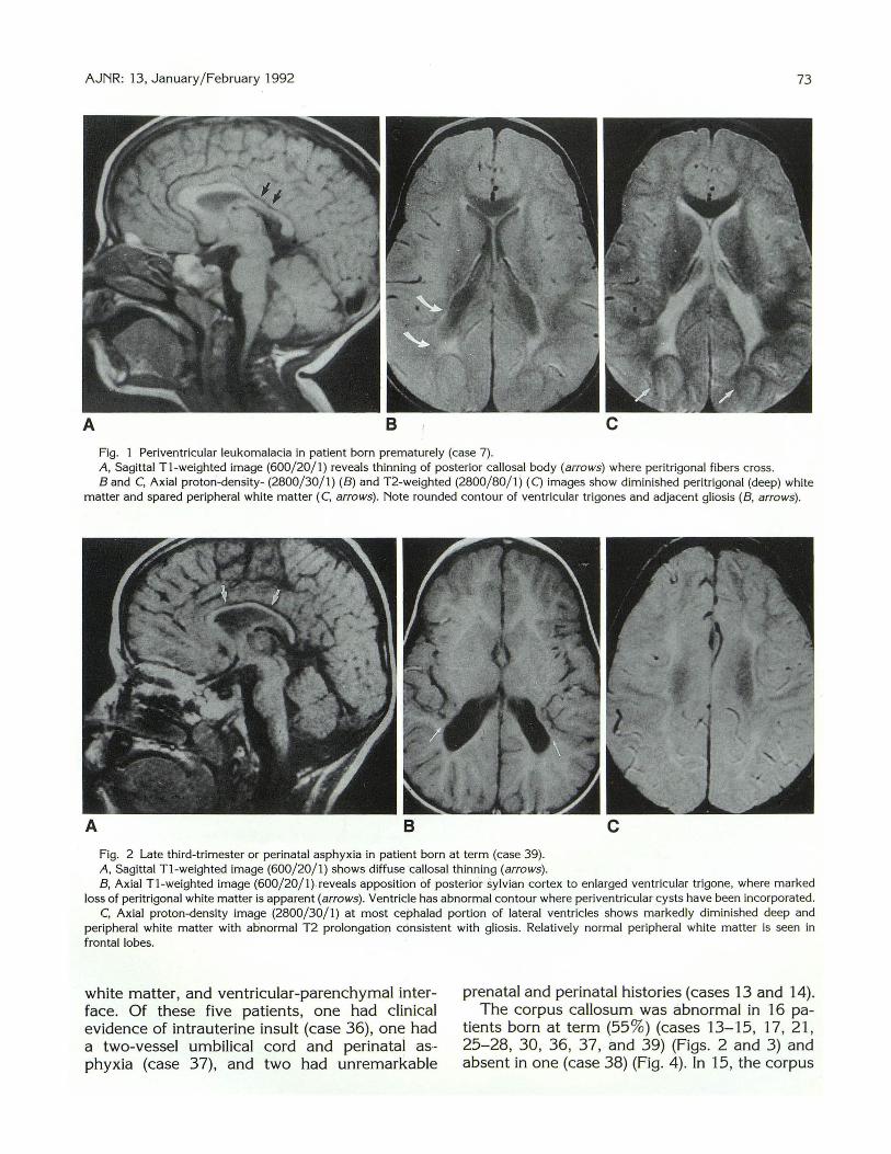
A B
Fig. Periventricular leukomalacia in patient born prematurely (case 7). A , Sagittal T1-weighted image (600/ 20/ 1) reveals thinning of posterior callosal body (arro ws) where peritrigonal fibers cross. Band C, Axial proton-density- (2800/ 30/ 1) (B) and T2-weighted (2800/ 80/ 1) (C) images show diminished peritrigonal (deep) white
matter and spared peripheral white matter (C, arrows). Note rounded contour of ventricular trigones and adjacent gliosis (B, arrows).
A B c Fig. 2 Late third-trimester or perinatal asphyxia in patient born at term (case 39) . A, Sagittal T1-weighted image (600/ 20/ 1) shows diffuse callosal thinning (arrows). B, Axial T1-weighted image (600/ 20/ 1).reveals apposition of posterior sylvian cortex to enlarged ventricular trigone, where marked
loss of peritrigonal white matter is apparent (arrows). Ventricle has abnormal contour where periventricular cysts have been incorporated. C, Axial proton-density image (2800/ 30/ 1) at most cephalad portion of lateral ventricles shows markedly diminished deep and
peripheral white matter with abnormal T2 prolongation consistent with gliosis. Relatively normal peripheral white matter is seen in frontal lobes.
white matter, and ventricular-parenchymal interface. Of these five patients, one had clinical evidence of intrauterine insult (case 36), one had a two-vessel umbilical cord and perinatal as·phyxia (case 37), and two had unremarkable
prenatal and perinatal histories (cases 13 and 14). The corpus callosum was abnormal in 16 pa
tients born at term (55%) (cases 13-15, 17, 21 , 25-28, 30, 36, 37, and 39) (Figs. 2 and 3) and absent in one (case 38) (Fig. 4). In 15, the corpus
74 AJNR: 13, January/February 1992
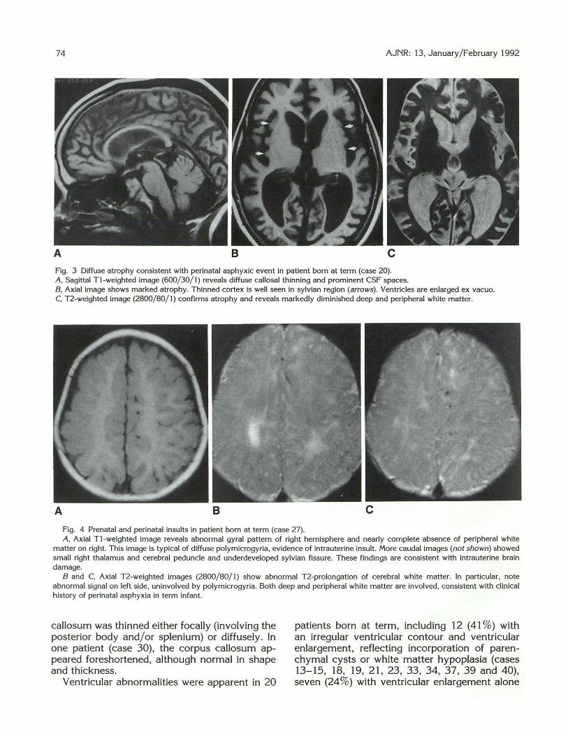
A 8 c Fig. 3 Diffuse atrophy consistent with perinatal asphyxic event in patient born at term (case 20). A, Sagittal T1-weighted image (600/30/1) reveals diffuse callosal thinning and prominent CSF spaces. B, Axial image shows marked atrophy. Thinned cortex is well seen in sylvian region (arrows) . Ventricles are enlarged ex vacuo. C, T2-weighted image (2800/80/1) confirms atrophy and reveals markedly diminished deep and peripheral white matter.
A 8
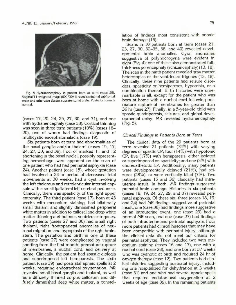
Fig. 4 Prenatal and perinatal insults in patient born at term (case 27). A, Axial T1-weighted image reveals abnormal gyral pattern of right hemisphere and nearly complete absence of peripheral white
matter on right. This image is typical of diffuse polymicrogyria, evidence of intrauterine insult. More caudal images (not shown) showed small right thalamus and cerebral peduncle and underdeveloped sylvian fissure. These findings are consistent with intrauterine brain damage.
B and C, Axial T2-weighted images (2800/80/1) show abnormal T2-prolongation of cerebral white matter. In particular, note abnormal signal on left side, uninvolved by polymicrogyria. Both deep and peripheral white matter are involved, consistent with clinical history of perinatal asphyxia in term infant.
callosum was thinned either focally (involving the posterior body and/or splenium) or diffusely. In one patient (case 30), the corpus callosum appeared foreshortened, although normal in shape and thickness.
Ventricular abnormalities were apparent in 20
patients born at term, including 12 (41 %) with an irregular ventricular contour and ventricular enlargement, reflecting incorporation of parenchymal cysts or white matter hypoplasia (cases 13-15, 18, 19, 21, 23, 33, 34, 37, 39 and 40), seven (24%) with ventricular enlargement alone
AJNR: 13, January/February 1992
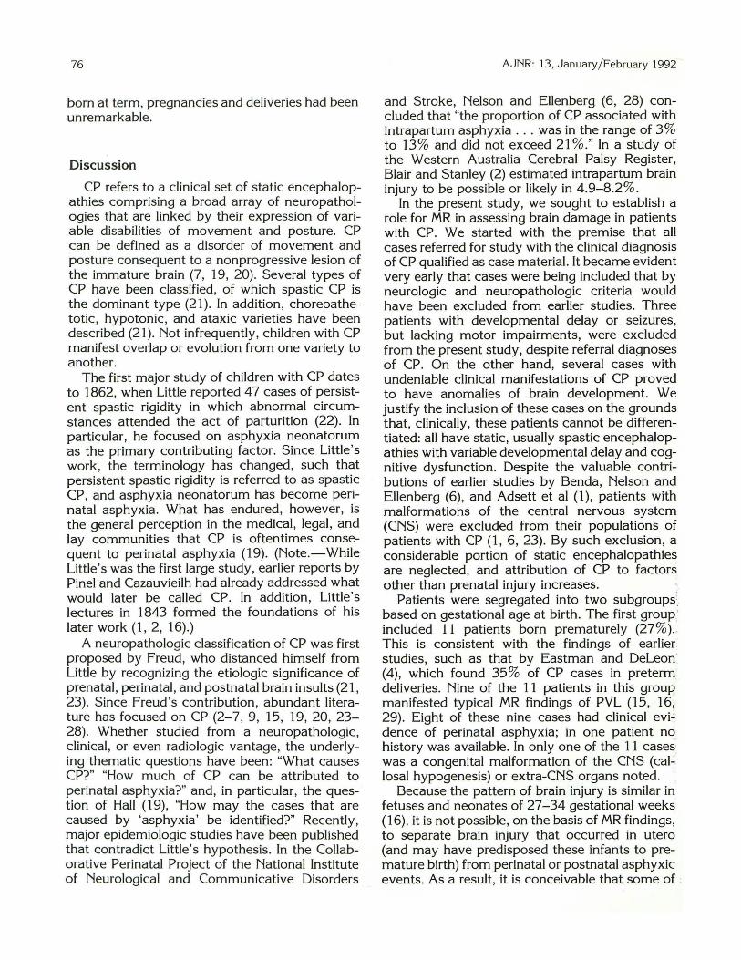
Fig. 5 Hydranencephaly in patient born at term (case 38). Sagittal Tl-weighted image (600/30/1) reveals minimal subfrontal brain and otherwise absent supratentorial brain. Posterior fossa is normal.
(cases 17, 20, 24, 25, 27, 30, and 31), and one with hydranencephaly (case 38). Cortical thinning was seen in three term patients (10%) (cases 18-20), one of whom had findings diagnostic of multicystic encephalomalacia (case 19).
Six patients born at term had abnormalities of the basal ganglia and/or thalami (cases 15, 17, 24, 27, 30, and 39). Foci of marked T1 and T2 shortening in the basal nuclei, possibly representing hemorrhage, were apparent on the scan of one patient who had had perinatal asphyxia (case 24). Another patient (case 15), whose gestation had involved a 24-hr period of decreased fetal movements at 30 weeks, had a cyst involving the left thalamus and retrolenticular internal capsule with a small ipsilateral left cerebral peduncle. Clinically, there was spasticity of the right upper extremity. The third patient (case 17), born at 43 weeks with meconium staining, had bilaterally small thalami and slightly diminished peripheral white matter in addition to callosal and deep white matter thinning and bulbous ventricular trigones. Two patients (cases 27 and 30) had small right thalami, right frontoparietal anomalies of neuronal migration, and hypoplasia of the right brainstem. The gestation and birth in one of these patients (case 27) were complicated by vaginal spotting from the first month, premature rupture of membranes, a nuchal cord, and delivery at home. Clinically, the patient had spastic diplegia and superimposed left hemiparesis. The sixth patient (case 39) had postnatal apneic spells at 2 weeks, requiring endotracheal oxygenation. MR revealed srriall basal ganglia and thalami, as well as a diffusely thinned corpus callosum and diffusely diminished deep white matter, a constel-
75
lation of findings most consistent with anoxic brain damage (16).
Scans in 10 patients born at term (cases 21, 23, 27, 30, 32-35, 38, and 40) revealed developmental brain anomalies. Gyral anomalies suggestive of polymicrogyria were evident in eight (Fig. 4); one of these also demonstrated fullthickness porencephaly (schizencephaly) (13, 18). The scan in the ninth patient revealed gray matter heterotopias of the ventricular trigones (13, 18). Clinically, these nine patients had seizure disorders, spasticity or hemipareses, hypotonia, or a combination thereof. Birth histories were unremarkable in all, except for the patient who was born at home with a nuchal cord following premature rupture of membranes for greater than 36 hr (case 27). Finally, in a 5-year-old child with spastic quadriparesis, seizures, and global developmental delay, MR revealed hydranencephaly (Fig. 5).
Clinical Findings in Patients Born at Term
The clinical data of the 29 patients born at term revealed 21 patients (72%) with varying degrees of spastic CP; four (14%) with hypotonic CP, five (17 %) with hemiparesis, either isolated or superimposed on spasticity; and one (3%) with choreoathetotic CP. Additionally, many patients were developmentally delayed (21 %), had seizures (28%), or were cortically blind (7 %). Two patients (cases 15 and 36) clinically had intrauterine insult. In both, MR findings suggested prenatal brain damage. Histories in six patients (cases 18, 19, 24, 27, 29, and 37) revealed perinatal asphyxia. Of these six, three (cases 18, 19, and ~4) had MR findings suggestive of perinatal insult, one (case 38) had findings more suggestive of an intrauterine event, one (case 29) had a normal MR scan, and one (case 27) had findings of both intrauterine and perinatal asphyxia. Four more patients had clinical histories that may have been compatible with perinatal injury, although the clinical data did not meet our criteria for perinatal asphyxia. They included two with meconium staining (cases 16 and 17), one with a nuchal cord (case 28), and one born at 37 weeks who was cyanotic at birth and required 24 hr of oxygen therapy (case 12). Two patients had clinical histories suggesting postnatal injury, including one hospitalized for dehydration at 3 weeks (case 31) and one who had several apneic spells that required endotracheal oxygenation at 2 weeks of age (case 39). In the remaining patients
76
born at term, pregnancies and deliveries had been unremarkable.
Discussion
CP refers to a clinical set of static encephalopathies comprising a broad array of neuropathologies that are linked by their expression of variable disabilities of movement and posture. CP can be defined as a disorder of movement and posture consequent to a nonprogressive lesion of the immature brain (7, 19, 20). Several types of CP have been classified, of which spastic CP is the dominant type (21). In addition, choreoathetotic , hypotonic, and ataxic varieties have been described (21). Not infrequently, children with CP manifest overlap or evolution from one variety to another.
The first major study of children with CP dates to 1862, when Little reported 47 cases of persistent spastic rigidity in which abnormal circumstances attended the act of parturition (22). In particular, he focused on asphyxia neonatorum as the primary contributing factor. Since Little's work, the terminology has changed, such that persistent spastic rigidity is referred to as spastic CP, and asphyxia neonatorum has become perinatal asphyxia. What has endured, however, is the general perception in the medical, legal, and Jay communities that CP is oftentimes consequent to perinatal asphyxia (19). (Note.-While Little 's was the first large study, earlier reports by Pinel and Cazauvieilh had already addressed what would later be called CP. In addition, Little's lectures in 1843 formed the foundations of his later work ( 1, 2, 16).)
A neuropathologic classification of CP was first proposed by Freud, who distanced himself from Little by recognizing the etiologic significance of prenatal , perinatal , and postnatal brain insults (21 , 23). Since Freud 's contribution, abundant literature has focused on CP (2-7, 9, 15, 19, 20, 23-28). Whether studied from a neuropathologic, clinical, or even radiologic vantage, the underlying thematic questions have been: "What causes CP?" "How much of CP can be attributed to perinatal asphyxia?" and, in particular, the question of Hall ( 19), "How may the cases that are caused by 'asphyxia' be identified?" Recently, major epidemiologic studies have been published that contradict Little 's hypothesis. In the Collaborative Perinatal Project of the National Institute of Neurological and Communicative Disorders
AJNR: 13, January / February 1992
and Stroke, Nelson and Ellenberg (6, 28) concluded that "the proportion of CP associated with intrapartum asphyxia .. . was in the range of 3% to 13% and did not exceed 21 %." In a study of the Western Australia Cerebral Palsy Register, Blair and Stanley (2) estimated intrapartum brain injury to be possible or likely in 4.9-8.2%.
In the present study, we sought to establish a role for MR in assessing brain damage in patients with CP. We started with the premise that all cases referred for study with the clinical diagnosis of CP qualified as case material. It became evident very early that cases were being included that by neurologic and neuropathologic criteria would have been excluded from earlier studies. Three patients with developmental delay or seizures, but Jacking motor impairments, were excluded from the present study, despite referral diagnoses of CP. On the other hand, several cases with undeniable clinical manifestations of CP proved to have anomalies of brain development. We justify the inclusion of these cases on the grounds that, clinically, these patients cannot be differentiated: all have static, usually spastic encephalopathies with variable developmental delay and cognitive dysfunction. Despite the valuable contributions of earlier studies by Benda, Nelson and Ellenberg (6), and Adsett et al ( 1 ), patients with malformations of the central nervous system (CNS) were excluded from their populations of patients with CP (1 , 6, 23). By such exclusion, a considerable portion of static encephalopathies are neglected, and attribution of CP to factors other than prenatal injury increases.
Patients were segregated into two subgroups' based on gestational age at birth. The first group ' included 11 patients born prematurely (27% ) .. This is consistent with the findings of earlier: studies, such as that by Eastman and Deleon (4), which found 35% of CP cases in preterrrf deliveries. Nine of the 11 patients in this group manifested typical MR findings of PVL (15, 16, 29). Eight of these nine cases had clinical evi.: dence of perinatal asphyxia; in one patient no_ history was available. In only one of the 11 cases was a congenital malformation of the CNS (callosal hypogenesis) or extra-CNS organs noted.
Because the pattern of brain injury is similar in fetuses and neonates of 27-34 gestational weeks ( 16), it is not possible, on the basis of MR findings , to separate brain injury that occurred in utero (and may have predisposed these infants to premature birth) from perinatal or postnatal asphyxic events. As a result, it is conceivable that some of .
AJNR: 13, January/ February 1992
the nine patients born prematurely with PVL had intrauterine events that predisposed the infants to prematurity. If so, perinatal asphyxia may not have been the sole insult resulting in brain injury.
In the second subset of 29 patients born at term (73%), a wide variety of abnormalities were observed. Most striking was the finding that, in 16 (55%) of the 29 patients, the MR scans revealed changes compatible with intrauterine insult to the developing brain. This included nine patients with anomalies of neuronal migration, one with hydranencephaly, five with PVL similar to that seen in premature infants, and one with findings of PVL and small infarcts of the left retrolenticular internal capsule and thalamus. Of these 16 patients, 10 had unremarkable gestational and birth histories, two had documented intrauterine insults, and three had histories pertinent to the perinatal period, although only two of these 16 patients are likely to have suffered perinatal asphyxia.
In contradistinction to the high percentage of intrauterine injuries in patients born at term, MR findings suggested perinatal brain injury in only seven (24% ). This subset included the patients with focal deep gray matter injury, diffuse white matter diminution and parietal cortical thinning, multicystic encephalomalacia, diffuse parasagittal cortical and white matter injury, hemorrhage within the lentiform nuclei, small basal ganglia and/ or thalami with callosal thinning and white matter loss, and the one with MR features of both intrauterine damage (polymicrogyria) and perinatal asphyxia (prolonged T2 relaxation of the deep and peripheral white matter bilaterally). In five of these seven patients, a history consistent with significant perinatal injury was present.
By cross-referencing the clinical data and MR findings, in only seven (24%) of the 29 patients born at term were we able to identify CP associated with perinatal asphyxia. Moreover, two of the seven had evidence of both intrauterine and perinatal brain damage. In most cases, therefore, neither clinical evidence nor MR findings of perinatal asphyxia could be identified. In 16 (55%) of 29 patients born at term, clinical and/or MR evidence of prenatal brain damage was present. In two cases (7% ), brain damage most likely occurred in the postnatal period.
The high percentage of cases with intrauterine brain damage (55% of term patients, 9% of preterm, 43% of both term and preterm patients combined) was quite remarkable. Even excluding the cases of PVL in term infants, which we believe
77
should be classified as intrauterine insults, 10 term and one preterm cases of congenital malformations (28%) must be accounted for. Most of these patients had a gyral pattern suggestive of polymicrogyria, an anomaly of neuronal migration that occurs at approximately 20-22 weeks' gestation (13). In the postmortem study of Malamud et al. (3) , 35% of patients with CP had CNS malformations: 10% had specific anomalies and 25% had microscopic aberrations of the CNS. A more recent clinical study of diplegic CP found seven (14%) of 49 term and one (2 %) of 4 7 preterm patients to have CNS malformations, although no specific mention was made as to how these malformations were discovered (20). Several recent radiologic studies also found CP patients with developmental brain anomalies (7 , 15). In aCT study of CP patients, Kolawole et al (9) found 15.8 % of their patients had underlying prenatal (developmental) factors. Similarly, Wiklund et al (7) observed anomalies of neuronal migration on CT in 16% of their cases of congenital hemiplegia. Interestingly, they also found periventricular atrophy (roughly equivalent to our definition of PVL) in 42%. In an MR study, Koeda et al ( 15) found two (29%) of seven patients born at term with spastic diplegia had brain anomalies, one of whom had schizencephaly and contralateral polymicrogyria. The findings of our study and that of Koeda et al are not surprising because MR is known to be more sensitive than CT in the detection of subtle gyral anomalies.
In conclusion, although our study is limited by its retrospective nature and selected population, our results support an enlarging consensus that the group of patients with clinical CP consequent to intrauterine brain injury has been underemphasized in the past (7). With the increased sensitivity of MR in the detection of subtle brain injuries, a significantly higher proportion of CP patients may be determined to have suffered intrauterine brain damage. In light of these findings, CP should not be assumed to be consequent to a perinatal event; rather, consideration of prenatal, and occasionally postnatal, causes may be productive in determining the time of the brain injury. In furtherance of this search, MR imaging may be very helpful in cases of CP.
References
1. Adsett DB, Fitz CR, Hill A. Hypoxic-ischemic cerebral injury in the
term newborn: correlation of CT findings with neurologic outcome.
Dev /VIed Child Neuro/1 985;27:155- 160

78
2. Blair E, Stanley FJ. Intrapartum asphyxia: a rare cause of cerebral
palsy. J Pediatr 1988; 11 2:5 15-51 9
3. Malamud ~ . ltabashi HH, Castor J , Messinger HB. An etiologic and diagnostic study of cerebral palsy. J Pediatr 1964;65(2):270-293
4. Eastman NJ, Del eon M. The etiology of cerebral palsy. Am J Obstet
Gy neco/1 955;69:950-961
5. Hagberg B, Hagberg G. Prenatal and perinatal ri sk factors in a survey of 681 Swedish cases. Clin Dev Med 1984;87:126-134
6. Nelson KB, Ellenberg JH. Antecedents of cerebral palsy: multivariate
analysis of risk . N Eng/ J M ed 1986;315:8 1-86
7. Wiklund L-M, Uvebrant P, Flodmark 0. Morphology of cerebral
lesions in children with congenital hemiplegia. Neuroradiology
1990;32:1 79- 186
8. Kulakowski S, Larroche J-C. Cranial computerized tomography in
cerebral palsy. An attempt at anatomo-clinical and radiological correlation. Neuropediatrics 1980; 11 :339- 353
9. Kolawole TM , Patel PJ, Mahdi AH. Computed tomographic (CT)
scans in cerebral palsy (CP). Pediatr Radio/ 1989;20:23- 27
10. Ando Y, Eda I, Nakano C, et al. Cranial computerized tomography of
prematurely born children with cerebral palsy. A cta Neonatal Jpn
1985;2 1:281-287
11 . Osborn RE, Byrd SE, Naidich TP, Bohan TP, Friedman H. MR imaging
of neuronal migrational disorders. AJNR 1988;9:1101-1106
12. Smith AS, Blaser Sl, Ross JS, Weinstein MA. Magnetic resonance
imaging of disturbances in neuronal migration: illustration of an
embryologic process. RadioGraphies 1989;9(3):509-522 13. Barth PG. Disorders of neuronal migration. Can J Neural Sci
1987;14:1 -16 14. Bird CR, Gilles FH. Type I schizencephaly: CT and neuropathologic
findings. AJNR 1987;8:451-454
15. Koeda T , Suganuma I, Kohno Y, Takamatsu T , Takeshita K. MR
imaging of spastic diplegia. Neuroradiology 1990;32: 187-1 90
16. Barkovich AJ , Truwit CL. Brain damage from perinatal asphyx ia:
AJNR: 13, January/ February 1992
correlation of MR findings with gestat ional age. AJNR 1990;11 :1087-
1096 17. Weller RO, Williams BN. Cerebral biopsy and assessment of brain
damage in hydrocephalus. Arch Dis Child 1975;50:763-768
18. Barkovich AJ. Pediatric neuroimaging. New York: Raven, 1990
19. Hall DMB. Birth asphyxia and cerebral palsy. BMJ 1989;299:279-
282 20. Veelken N, Hagberg B, Hagberg G, Olow I. Diplegic cerebral palsy in
Swedish term and preterm children: differences in reduced optimality,
relations to neurology and pathogenetic factors. Neuropediatrics
1983; 14:20-28 21. Ingram TTS. A historical review of the definition and classification of
the cerebral palsies. Clin Dev Med 1984;87:1-11 22. Little WJ . On the influence of abnormal parturition, difficult labour,
premature birth and asphyxia neonatorum on mental and physical
conditions of the child, especially in relation to deformities. Trans
Obstet Soc London 1862;3:293-344 23. Christensen E, Melchior JC. Cerebral palsy: a clinical and neuropath
ological study. Clin Dev Med 1967;25:1-14
24. Lilienfeld AM, Parkhurst EA. A study of the association of factors of
pregnancy and parturition with the development of cerebral palsy. Am J Epidemio/1951 ;53:262-282
25. Jarvis SN, Holloway JS, Hey EN. Increase in cerebral palsy in normal
birthweight babies. Arch Dis Child 1985;60:111 3-1121 26. Stanley FJ, Watson LD. The methodology of a cerebral palsy register:
the Western Australian experience. Neuroepidemiology 1985;4: 146-
160 27. Stanley FJ. The changing face of cerebral palsy? Dev Med Child
Neuro/1987;29:263-265 28. Nelson KB. What proportion of cerebral palsy is related to birth
asphyxia? J Pediatr 1988;112:572-573
29. Flodmark 0 , Lupton B, Li D, et al. MR imaging of periventricular
leukomalacia in childhood. AJR 1989; 152:583-590
Note: Please see the Commentary by Volpe on page 79 in this issue.