Embed Size (px)
Citation preview

Cephalometric predictors of treatment outcome with mandibular advancement devices in adult patients with obstructive sleep apnea: a systematic review
Objective: The efficacy of mandibular advancement devices (MADs) in the treatment of obstructive sleep apnea (OSA) ranges between 42% and 65%. However, it is still unclear which predictive factors can be used to select suitable patients for MAD treatment. This study aimed to systematically review the literature on the predictive value of cephalometric analysis for MAD treatment outcomes in adult OSA patients. Methods: The MEDLINE, Google Scholar, Scopus, and Cochrane Library databases were searched through December 2014. Reference lists from the retrieved publications were also examined. English language studies published in international peer-reviewed journals concerning the predictive value of cephalometric analysis for MAD treatment outcome were considered for inclusion. Two review authors independently assessed eli-gibility, extracted data, and ascertained the quality of the studies. Results: Fifteen eligible studies were identified. Most of the skeletal, dental, and soft ti ssue cephalometric measurements examined were widely recognized as not prognostic for MAD treatment outcome; however, controversial and limited data were found on the predictive role of certain cephalometric measurements in-cluding cranial base angle, mandibular plane angle, hyoid to mandibular plane distance, posterior nasal spine to soft-palate tip distance, anterior nasal spine to epiglottis base distance, and tongue/oral cross sectional area ratio thus justifying additional studies on these parameters. Conclusions: Currently available evi dence is inadequate for identification of cephalometric parameters capable of reliably discriminating between poor and good responders to MAD treatment. To guide further research, methodological weaknesses of the currently available stu dies were highlighted and possible reasons for their discordant results were analyzed. [Korean J Orthod 2015;45(6):308-321]
Key words: Obstructive sleep apnea, Cephalometry; Mandibular advancement
Giulio Alessandri-Bonettia Daniela Rita Ippolitob
Maria Lavinia Bartoluccic
Vincenzo D’Antòc,d
Serena Incerti-Parentia
aUnit of Orthodontics, Department of Biomedical and Neuromotor Sciences, University of Bologna, Bologna, ItalybDepartment of Orthodontics, University of Brescia, Brescia, ItalycDepartment of Neuroscience, Reproductive Science and Oral Science, University of Naples “Federico II”, Naples, ItalydDepartment of Pediatric Surgery, Bambino Gesù Children’s Hospital, Rome, Italy
Received February 25, 2015; Revised April 15, 2015; Accepted May 6, 2015.
Corresponding author: Giulio Alessandri-Bonetti. Aggregate Professor, Unit of Orthodontics, Department of Biomedical and Neuromotor Sciences (DIBINEM), University of Bologna, Via San Vitale 59, Bologna 40125, Italy. Tel +39-0512088133 e-mail [email protected]
308
© 2015 The Korean Association of Orthodontists.
The authors report no commercial, proprietary, or financial interest in the products or companies described in this article.
This is an Open Access article distributed under the terms of the Creative Commons Attribution Non-Commercial License (http://creativecommons.org/licenses/by-nc/4.0) which permits unrestricted non-commercial use, distribution, and reproduction in any medium, provided the original work is properly cited.
THE KOREAN JOURNAL of ORTHODONTICSReview Article
pISSN 2234-7518 • eISSN 2005-372Xhttp://dx.doi.org/10.4041/kjod.2015.45.6.308

Alessandri-Bonetti et al • Cephalometric predictors of MADs treatment outcome
www.e-kjo.org 309http://dx.doi.org/10.4041/kjod.2015.45.6.308
INTRODUCTION
Obstructive sleep apnea (OSA) is characterized by repe-titive episodes of complete or partial closure of the upper airway during sleep that lead to sleep fragmentation and oxygen desaturation.1 This sleep-related breathing disorder is associated with daytime sleepiness, impaired quality of life, poor work performance, neurocognitive decline, increased risk of motor vehicle accidents and, in the long term, an increased risk of cardio-vascular disease and mortality.2 Nasal continuous positive airway pressure (nCPAP) maintains a positive pressure in the upper airway through a nose mask worn during sleep and is currently the most effective treatment option for OSA patients.3 Nevertheless, adherence to this therapy is low, with rates between 60% and 80%. The availability of alternative treatment options is therefore of the ut-most importance.4 Mandibular advancement devices (MADs), which hold the mandible forward with the aim of preventing collapse of the upper airway during sleep provide a less invasive, more comfortable, and less costly treatment alternative for patients with mild to moderate OSA who do not tolerate, do not respond to, or are not appropriate candidates for treatment with nCPAP, or those who fail behavioral measures such as weight loss or sleep position change. MADs can also be used in pa-tients with severe OSA who fail treatment attempts with nCPAP or who are not appropriate candidates for upper airway surgery.5,6
Patients undergo a repeat sleep study with MAD in situ to determine its effectiveness, which usually ran-ges between 42% and 65%.7 Clinical prediction of MAD treatment outcome would allow advanced selection of suitable candidates for this treatment before manu-facturing the device, thus avoiding inappropriate delays in therapy and waste of resources. Accordingly, this topic has been defined by the American Academy of Sleep Medicine as an important area for future research.5,6
Previous studies have suggested that lateral cepha-lometry can identify craniofacial characteristics that could have an impact on treatment response.5,8-10 Cepha-lometry is a low-cost, simple, and widely available radio graphic technique, and it is therefore suitable as a screen ing procedure. Nevertheless, the clinical utility of cephalometric measurements in the prediction of MAD treatment outcomes in OSA remains controversial. A recently published review by Saffer et al.11 found no clear predictors of MAD treatment success. However, cephalometric and anatomical factors were not inves-tigated because no randomized controlled trials add-ressing this issue were included in the review. The aim of this study was to fill this gap by conducting a systematic review of published studies examining the ability of cephalometric parameters to predict MAD treatment
response in adult patients with OSA.
MATERIALS AND METHODS
Search strategy An electronic literature search was carried out on the following databases: MEDLINE, Google Scholar, Scopus, and the Cochrane Library. To identify the rele-vant studies the following search terms were used: “obstructive sleep" AND (apnea OR apnoea) AND (predict* OR outcome OR effect OR efficacy) AND (“oral appliance" OR “mandibular advancement device" OR “mandibular repositioning appliance") AND (craniofacial OR skeletal OR cephalometr*). The reference lists of all relevant publications were checked for additional studies. Searches were updated to December 2014.
Screening and study selection In the first phase of selection, duplicates were removed and irrelevant articles were excluded by reviewing the titles and abstracts from the search results. In the next phase, the full texts of potentially relevant papers were evaluated to determine if they met the eligibility criteria.The inclusion criteria were:
• Type of study: Randomized or non-randomized controlled trials, cohort or case-control studies (with a minimum sample size of 10 patients in each group) addressing the research question of the predictive value of cephalometric analysis in oral appliance treatment outcomes in adult OSA patients. Studies had to be published in English in an international peer-reviewed literature
• Population: Male or female adult patients (≥ 18 years old) with a polysomnographic diagnosis of OSA (i.e., 5 or more respiratory events [apneas or hypopneas] per hour of sleep)
• Intervention: Treatment with any MAD for OSAThe exclusion criteria were:
• Lack of a clear description of inclusion/exclusion criteria
• Previous and/or current surgical or pharmacological interventions
• Limitation to severe OSA patients Two reviewers (DRI, SIP) independently screened paper titles and abstracts, with access to full texts where necessary to select studies into the review. Disagreements were resolved by discussion. Where resolution was not possible, a third reviewer (GAB) was consulted. The selected studies underwent data extraction and quality assessment.
Data extraction and quality assessment of selected studies Two review authors (DRI, SIP) independently performed

Alessandri-Bonetti et al • Cephalometric predictors of MADs treatment outcome
www.e-kjo.org310 http://dx.doi.org/10.4041/kjod.2015.45.6.308
the data extraction. Extracted data included: first author, year of publication, study design, sample size, patient demographic and clinical characteristics, MAD type, degree of protrusion and vertical opening, time interval between polysomnographic evaluations, outcome (i.e., cephalometric parameters that differed significantly between good and poor responders). Selected studies were assigned to a class of evidence according to the cla ssification of study designs by Jovell and Navarro-Rubio (Table 1).12
Data synthesisA narrative synthesis was carried out. Data were sorted according to MAD type (1-piece or 2-piece appliances). Given the lack of homogeneity in the study settings, a quantitative synthesis seemed inappropriate. Therefore no meta-analysis was performed.
RESULTS
The electronic database search and the review of the relevant publication reference lists yielded 939 po-tentially relevant titles and abstracts after duplicates were removed from a total of 1,034 records. Following the first phase of evaluation, 907 publications were re-jected based on the title and the abstract. One further study was excluded because full text was not obtained by searching paper and digital format sources, nor after attempting to contact the authors by e-mail corres-pondence.13 In the second phase, analysis of the full text of the remaining 31 studies led to the exclusion of 16 additional publications. Fifteen studies were there-fore selected for the systematic review.5,8-10,14-24 The relevant data from each study are reported in Table 2. The PRISMA flow diagram (Figure 1) shows the number of articles reviewed in each phase of this systematic review.25
Cephalometric variables that were analyzed in the
selected studies are listed in Table 3. The landmarks and reference lines necessary to define these parameters are shown in Figure 2.
Cephalometric skeletal measurements
Cranial base• 1-piece MAD: Svanholt et al.24 found that the
distance between the sella turcica and the deepest point in the posterior cranial fossa was lower in the group that responded positively to MAD therapy.
• 2-piece MAD: Both cranial base and anterior cranial base lengths were reported to be non-predictive of MAD treatment outcome.5,8,22 Two studies addressed the predictive value of cranial base angle with con-trasting results; one found an increased cranial base angulation to be predictive of MAD treatment su-ccess22 and the other did not.5
Sagittal jaw relationship• 1-piece MAD: Data concerning sagittal jaw rela-
tionship were controversial. SNA and ANB were recognized either as predictive of treatment success when decreased10,24 or as non-predictive10,14,18 of treatment outcome. SNB was found to be non-pre-dictive14,18 or predictive of treatment success when increased10 or decreased.24
• 2-piece MAD: The majority of papers indicated that sagittal jaw relationship cephalometric parameters were not suitable for predicting treatment outcome (SNA,5,8,9,19-22 SNB,5,19-22 ANB,5,17,20-22 Wits appraisal20). However, a decreased SNB value8,9 or an increased ANB value8 were occasionally reported as predictive of treatment success.
Vertical craniofacial dimensions• 1-piece MAD: Vertical craniofacial dimension para-
meters were usually recognized as non-predictive of
Table 1. Levels of scientific evidence
Level of evidence Type of study Strength of evidence
Level 1 Meta-analyses of randomized controlled trials Good
Level 2 Large-sample randomized controlled trials
Level 3 Small-sample randomized controlled trials Good to fair
Level 4 Non-randomized controlled prospective trials
Level 5 Non-randomized controlled retrospective trials
Level 6 Cohort studies Fair
Level 7 Case-control studies
Level 8 Non-controlled clinical series, descriptive studies Poor
Level 9 Anecdotes or case reports
Derived from Jovell and Navarro-Rubio.12

Alessandri-Bonetti et al • Cephalometric predictors of MADs treatment outcome
www.e-kjo.org 311http://dx.doi.org/10.4041/kjod.2015.45.6.308
Svan
hol
t, 20
1424
Shen
, 201
29
Ros
e, 2
00223
Ng,
201
222
Mos
tafi
z, 2
01121
Mila
no,
201
320
Men
n, 1
99619
Meh
ta, 2
0015
Mar
klu
nd
, 199
818
Liu
, 200
117
Lee
, 201
016
Lee
, 200
915
Kim
, 201
414
Hoe
kem
a, 2
0078
En
do,
200
310
Firs
t au
thor
, yea
r
Pro
spec
tive
case
-con
trol
stu
dy
Pro
spec
tive
case
-con
trol
stu
dy
Pro
spec
tive
case
-con
trol
stu
dy
Ret
rosp
ecti
ve
ca
se-c
ontr
ol s
tud
y
Pro
spec
tive
case
-con
trol
stu
dy
Pro
spec
tive
case
-con
trol
stu
dy
Pro
spec
tive
case
-con
trol
stu
dy
Pro
spec
tive
case
-con
trol
stu
dy
Pro
spec
tive
case
-con
trol
stu
dy
Pro
spec
tive
case
-con
trol
stu
dy
Ret
rosp
ecti
ve
ca
se-c
ontr
ol s
tud
y
Ret
rosp
ecti
ve
ca
se-c
ontr
ol s
tud
y
Ret
rosp
ecti
ve
ca
se-c
ontr
ol s
tud
y
Pro
spec
tive
case
-con
trol
stu
dy
Pro
spec
tive
case
-con
trol
stu
dy
Des
ign
of
the
stu
dy
777777777777777
Leve
l of
evid
ence
*
27 (
85.2
)[-
]
52 (
92.3
) [-
]
57 (
89.5
) [-
]
72 (
76.4
)
53 (
79.2
) [-
]
23 (
87.0
) [-
]
29 (
95.7
) [6
]
28 (
67.9
) [4
]
32 (
100)
[-
]
47 (
89.4
) [-
]
76 (
89.5
)[-
]
50 (
92)
[-]
86 (
88.4
)[-
]
51 (
-)
[2]
103
(-)
[-]
Subj
ects
, n(%
mal
e)[w
ith
draw
als]
GR
: 53.
66 ±
7.2
7P
R: 5
2.02
± 1
0.5
43.4
± 1
0.3
(18-
)
56.5
± 7
.3
(35-
73)
49.0
± 1
1
49.5
± 1
1.8
54.0
± 1
1.1
(20-
67)
53 ±
11
(37-
69)
48 ±
9
(35-
73)
Med
ian
: 57
(37-
72)
49.1
(2
5-80
)
51.7
± 0
.3
(21-
69)
50.2
± 9
.8
(21-
69)
51.5
± 9
.8
49.2
± 9
.5
GR
: 51.
2P
R: 5
1.1
Age
(yr
)
GR
: 27.
13 ±
2.3
5P
R: 3
2.16
± 5
.42
24.8
± 2
.6
26.4
± 2
.0
30.2
± 5
.6
28.7
± 4
.2
27.8
± 4
.4
29 ±
5
29.4
± 3
.1
Med
ian
: 28
29.6
1 ±
6.49
< 3
5
GR
: 25.
5 ±
2.5
PR
: 26.
9 ±
2.6
GR
:25.
98 ±
3.1
5P
R: 2
6.17
± 2
.78
32.2
± 6
.1
GR
: 24.
5 P
R: 2
4.7
BM
I (kg
/m2 )
GR
: 28.
46 ±
15.
17P
R: 4
3.52
± 2
5.64
GR
: 34.
1 ±
15.1
PR
: 38.
6 ±
19.1
22.0
± 1
2.2
GR
: 26.
8 ±
14.4
PR
: 28.
1 ±
19.3
CR
: 28.
4 ±
14.4
PaR
: 34.
8 ±
15.1
NR
: 40.
6 ±
12.7
24.5
± 1
3.9
RD
I: 3
7 ±
23
27 ±
17
Med
ian
: 23
CR
: 44.
21 ±
11.
57Pa
R: 4
2.46
± 1
9.33
NR
: 28.
69 ±
8.8
0
38.9
± 1
9.7
AI:
GR
: 37.
6 ±
28.4
AI:
PR
: 34.
6 ±
17.4
GR
: 19.
79 ±
17.
32P
R: 3
0.08
± 2
3.28
38.0
± 2
9.8
GR
: 21.
1 ±
19.5
PR
: 22.
3 ±
19.7
bAH
I (ev
ents
/h)
Den
mar
k
Taiw
an
Ger
man
y
Au
stra
lia
Au
stra
lia
Ital
y
USA
Au
stra
lia
Swed
en
USA
Sou
th K
orea
Sou
th K
orea
Sou
th K
orea
Net
her
lan
ds
Jap
an
Cou
ntr
y
AH
I > 5
Age
> 1
8 yr
s; A
HI >
10;
suit
abili
ty fo
r M
AS
trea
tmen
t
Mild
to m
oder
ate
OSA
;
suit
abili
ty fo
r M
AS
trea
tmen
t
AH
I ≥ 1
0/h
; sym
pto
ms
of O
SA ≥
2;
su
itab
ility
for
MA
S tr
eatm
ent
Age
> 1
8 yr
s ; A
HI >
10/
h,
su
itab
ility
for
MA
Stre
atm
ent
N
o ot
her
sle
ep o
r m
edic
al d
isor
der
s
Age
> 1
8 yr
s; A
HI >
5;
at
leas
t 2 O
SAS
sym
pto
ms
Age
> 1
8 yr
s; R
DI ≥
10;
suit
abili
ty fo
r M
AS
trea
tmen
t
AH
I > 1
0/h
; sym
pto
ms
of O
SA ≥
2;
su
itab
ility
for
MA
S tr
eatm
ent;
no
sed
ativ
es
AH
I of a
t lea
st 1
5 in
the
sup
ine
and
/
or th
e la
tera
l pos
itio
n
Mild
to s
ever
e O
SAS;
suit
abili
ty fo
r M
AS
trea
tmen
t
AH
I > 5
AH
I > 5
AH
I > 5
; no
slee
p-r
elat
ed m
edic
atio
n o
r
p
revi
ous
orop
har
ynge
al s
urg
ery
Age
> 2
0 ye
ars;
AH
I ≥ 1
5
or
AH
I ≥ 5
+sym
pto
ms
AH
I ≥ 1
0; n
o p
revi
ous
surg
ical
op
erat
ion
s
Incl
usi
on c
rite
ria
Tabl
e 2.
Sum
mar
y of
the
mai
n ch
arac
teris
tics
of
the
15 s
tudi
es s
elec
ted
for
the
syst
emat
ic re
view
Alessandri-Bonetti et al • Cephalometric predictors of MADs treatment outcome
www.e-kjo.org312 http://dx.doi.org/10.4041/kjod.2015.45.6.308
Svan
hol
t, 20
1424
Shen
, 201
29
Ros
e, 2
00223
Ng,
201
222
Mos
tafi
z, 2
01121
Mila
no,
201
320
Men
n, 1
99619
Meh
ta, 2
0015
Mar
klu
nd
, 199
818
Liu
, 200
117
Lee
, 201
016
Lee
, 200
915
Kim
, 201
414
Hoe
kem
a, 2
0078
En
do,
200
310
Firs
t au
thor
, yea
r
Mon
oblo
c ty
pe
MA
D (
75 %
of m
ax p
rotr
usi
on)
[-]
IST-
Ap
plia
nce
(tw
o p
iece
s) (
ind
ivid
ual
)[-
]
Kar
wet
zky’
s m
odif
ied
act
ivat
or (
two
pie
ces)
(in
div
idu
al)
[in
div
idu
al]
Som
noM
ed (t
wo
pie
ces)
(max
imu
m c
omfo
rtab
le li
mit
of m
and
ibu
lar
adva
nce
men
t)[-
]
Som
noD
ent (
two
pie
ces)
(m
axim
um
com
fort
able
lim
it o
f man
dib
ula
r p
rotr
usi
on)
[-]
Sile
nso
r (t
wo
pie
ces)
(50
/60%
for
a st
art)
[-]
MA
D (
two
pie
ces)
(m
ax p
rotr
usi
on -
3 m
m)
[5-7
mm
]
MA
D (
two
pie
ces)
(in
div
idu
al)
[3/4
mm
]
MA
D (o
ne
pie
ce)
(4–6
mm
[for
a s
tart
])[5
mm
bet
wee
n in
ciso
rs]
MA
D (
two
pie
ces)
(2/
3 m
ax p
rotr
usi
on [f
or a
sta
rt])
[min
imu
m]
Mon
oblo
c ty
pe
MA
D (
60%
of m
ax p
rotr
usi
on)
[wit
hou
t op
en b
ites
]
Mon
oblo
c ty
pe
MA
D (
60%
of m
ax p
rotr
usi
on)
[wit
hou
t op
en b
ites
]
Mon
oblo
c ty
pe
MA
D (
60 %
of m
ax p
rotr
usi
on)
[min
imu
m]
Th
orn
ton
ad
just
able
pos
itio
ner
typ
e-1
(in
div
idu
al)
[-]
Mon
oblo
c ty
pe
MA
D (
70 %
of m
ax p
rotr
usi
on)
[2 m
m]
MA
D ty
pe
(pro
tru
sion
)[v
erti
cal o
pen
ing]
4 w
eeks
6 w
eeks
6-12
wee
ks
6 w
eeks
6-8
wee
ks
2-3
mon
ths
Mea
n:
104
day
s
19.7
± 8
.8
wee
ks
Up
to
2 m
onth
s
-
At l
east
3
mon
ths
At l
east
3
mon
ths
3 m
onth
s
2/3
mon
ths
Wit
hin
3
mon
ths
Foll
ow u
p
AH
I red
> 7
5%
AH
I red
> 5
0% a
nd
AH
I < 1
0/h
Age
> 6
0: A
HI <
5/h
an
d A
I < 2
/h;
ag
e <
60: A
HI <
10/
h a
nd
AI <
5/h
.
AH
I red
≥ 5
0% (
alon
e or
in a
dd
itio
n to
AH
I < 2
0/ <
10/
< 5
/h)
GR
: AH
I red
> 5
0%, A
HI >
5/h
MR
: AH
I red
> 5
0%, A
HI >
5/h
; PR
: AH
I red
< 5
0%
AH
I < 5
/h
RD
I red
≥ 5
0% a
nd
RD
I ≤ 2
0
Res
olu
tion
of s
ymp
tom
s an
d A
HI <
5/h
AH
I < 1
5
GR
: AH
I red
> 7
5%; M
R: A
HI r
ed: 2
5% to
75%
;
PR
: AH
I red
< 2
5%
AH
I red
> 5
0% a
nd
AH
I < 2
0/h
AH
I red
> 5
0% a
nd
AH
I < 2
0/h
AH
I red
> 5
0% a
nd
AH
I < 1
0/h
AH
I < 5
/h o
r (A
HI r
ed >
50%
)
an
d A
HI <
20/
h a
nd
no
sym
pto
ms
AH
I red
> 5
0%
Succ
ess
crit
eria
Low
er S
NA
, low
er S
NB
,
low
er S
NP
g, lo
wer
S-D
Smal
ler
SNB
; sh
orte
r N
-Me;
nar
row
er M
inR
GA
Low
er S
N-M
P; l
arge
r SG
o:N
Me;
hig
her
H-M
P, lo
wer
H-M
e
Shor
ter
PN
S-P,
incr
ease
d B
a-SN
;
larg
er S
NM
P
Gre
ater
ton
gue/
oral
en
clos
ure
CSA
rat
io
Low
er M
P-H
an
d lo
wer
SN
-MP
< 2
9°
No
cep
hal
omet
ric
pre
dic
tors
of
tr
eatm
ent o
utc
ome
Larg
er R
PAS
and
larg
er S
N-M
P
Nor
mal
(or
low
) SN
-MP
an
d
lo
wer
AN
S-M
e
Larg
er r
atio
of t
he
vert
ical
air
way
len
gth
to th
e cr
oss-
sect
ion
al a
rea
of th
e so
ft p
alat
e
Low
er P
NS-
P
No
cep
hal
omet
ric
pre
dic
tors
of
tr
eatm
ent o
utc
ome
Low
er P
NS-
Go
Hig
her
AN
B, s
mal
ler
SNB
,
larg
er O
J, O
B a
nd
N-A
NS
Larg
er S
NB
, low
er A
NB
, sh
orte
r M
P-H
,
shor
ter
AN
S- H
, sh
orte
r A
NS-
Eb
Ou
tcom
e (r
esp
onde
rs)
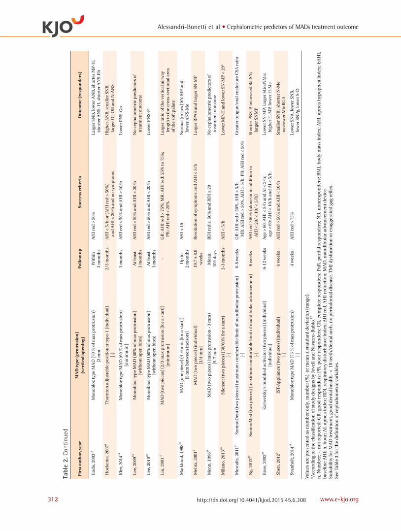
Val
ues
are
pre
sen
ted
as
nu
mb
er o
nly
, nu
mb
er (
%),
or
mea
n ±
sta
nd
ard
dev
iati
on (
ran
ge).
*Acc
ord
ing
to th
e cl
assi
fica
tion
of s
tud
y d
esig
ns
by Jo
vell
and
Nav
arro
-Ru
bio
.12
n, N
um
ber
; -, n
ot r
epor
ted
; GR
, goo
d r
esp
ond
ers;
PR
, poo
r re
spon
der
s; C
R, c
omp
lete
res
pon
der
s; P
aR, p
arti
al r
esp
ond
ers;
NR
, non
resp
ond
ers;
BM
I, b
ody
mas
s in
dex
; AH
I, a
pn
ea h
ypop
nea
ind
ex; b
AH
I,
bas
elin
e A
HI;
h, h
our;
AI,
ap
nea
ind
ex; R
DI,
res
pir
ator
y d
istu
rban
ce in
dex
; AH
I red
, AH
I red
uct
ion
; MA
D, m
and
ibu
lar
adva
nce
men
t dev
ice.
Su
itab
ility
for
MA
D tr
eatm
ent,
good
den
tal h
ealt
h,
> 1
0 te
eth
/den
tal a
rch
, no
per
iod
onta
l dis
ease
, TM
J dys
fun
ctio
n o
r ex
agge
rate
d g
ag r
efle
x.
See
Tab
le 3
for
the
def
init
ion
of c
eph
alom
etri
c va
riab
les.
Tabl
e 2.
Con
tinu
ed

Alessandri-Bonetti et al • Cephalometric predictors of MADs treatment outcome
www.e-kjo.org 313http://dx.doi.org/10.4041/kjod.2015.45.6.308
treatment outcome,14,24 with the exception of lower anterior face height,18 lower posterior face height,14 and mandibular plane angle,18 which were occasionally identified as predictive of treatment success when decreased.
• 2-piece MAD: Saddle angle, articular angle, gonial angle, palatal plane angle, posterior face height, and the ratio between upper anterior face height and lower anterior face height were not useful for predicting MAD treatment outcomes.9,20-23 The majority of the papers ascribed a non-predictive role to anterior face height, upper anterior face height, lower anterior face height, and the ratio between posterior face height and anterior face height, but a lower anterior face height value9 as well as higher values of upper anterior face hei-ght8 and of the ratio of posterior face height to anterior face height23 were also occasionally found in good responders. Data on the predictive role of mandibular plane angle were conflicting: 3 out of 7 studies found it to be non-predictive of treatment outcomes,8,9,21 2 studies indicated an increased value as a predictor of treatment success,5,22 and 2 studies reported a decreased value as a predictor of treatment success.20,23
Maxillary and mandibular lengths• 1-piece MAD: Endo et al.10 found that maxillary
and mandibular lengths were not predictive of MAD
treatment outcome.• 2-piece MAD: There was general agreement that
maxillary and mandibular lengths, ramus height, and corpus length were not suitable for prediction of MAD treatment outcome.5,17,20-22
Hard palate• 1-piece MAD: Hard palate length did not appear to
be predictive of MAD treatment outcome.10,16
• 2-piece MAD: Ng et al.22 claimed that hard palate length was not predictive of MAD treatment out-come.
Hyoid bone• 1-piece MAD: Endo et al.10 found decreased values
of MP-H, H-Me, and ANS-H to be predictive of MAD treatment success, whereas Kim et al.14 supported a non-predictive role of H-Me and ANS-H.
• 2-piece MAD: There was general agreement that H-RGN, C3ia-H, H-Go, Go-H-Me were not suitable for predicting MAD treatment outcomes.9,17,20,21 Rose et al.23 found that decreases in H-Me and ANS-H were predictive of treatment success, while Hoekema et al.8 did not. Data on MP-H were conflicting. Five out of 7 studies found it to be non-predictive,5,8,9,19,21 while the two others reported that decreased MP-H20 and increased MP-H23 were predictive of treatment success.
Figure 1. Flow chart of lite-rature search and study selec-tion.
Identification
Scre
enin
gE
ligib
ility
Inclu
ded
Records identified through databases searching
(n = 1,027)
Pubmed
(n = 46)
Scopus
(n = 96)
Google Scholar
(n = 878)
Cochrane Library
(n = 7)
Additional records identified
through other sources (n = 7)
Records after duplicates
removed (n = 939)
Records screened
(n = 939)
Full-text articles assessed
for eligibility (n = 31)
Studies included in
qualitative synthesis
(n = 15)
Records excluded (n = 907)
Full-text article not available
(n = 1)
Full-text articles excluded,
with reasons (n = 16):
Unsuitable sample (n = 7)
Inadequate methodology (n = 5)
Not published in the English
language (n = 3)
No pertinent/useful data (n = 1)

Alessandri-Bonetti et al • Cephalometric predictors of MADs treatment outcome
www.e-kjo.org314 http://dx.doi.org/10.4041/kjod.2015.45.6.308
Table 3. Summary of cephalometric variables
Variable Measurement
Cephalometric skeletal measurements
Cranial base
Cranial base length (mm) Ba-S-N
Anterior cranial base length (mm) S-N
Cranial base angle (o) BaSN
Distance between sella turcica and the deepest point in posterior cranial fossa (mm)
S-D
Sagittal jaw relationship
Anteroposterior position of the maxilla (o) SNA
Anteroposterior position of the mandible (o) SNB
SN-Pg
Anteroposterior relationship between maxilla and mandible (o, mm)
ANB
Wits appraisal (distance between A and B projections onto the occlusal plane)
Vertical craniofacial dimensions
Anterior face height (mm) N-Me
Upper anterior face height (mm) N-ANS
Lower anterior face height (mm) ANS-Me
Posterior face height (mm) S-Go
Lower posterior face height (mm) PNS-Go
Posterior face height : anterior face height (ratio) S-Go : N-Me
Upper anterior face height : lower anterior face height (ratio) N-ANS : ANS-Me
Saddle angle (o) N-S-Ar
Articular angle (o) S-Ar-Go
Gonial angle (o) Ar-Go-Me
Mandibular plane angle (o) SN-MP
FP-MP
PP-MP
Palatal plane angle (o) SN-PP
Maxillary and mandibular lengths
Maxillary length (mm) Cd-A
Mandibular length (mm) Cd-Gn
Ramus height (mm) Ar-Go
Cd-Go
Corpus length (mm) Go-Me
Go-Gn
Hard palate
Length (mm) ANS-PNS
Hyoid bone
Vertical position (mm) H-MP or Hy-MP (perpendicular)
H-Go (vertical measure)
Alessandri-Bonetti et al • Cephalometric predictors of MADs treatment outcome
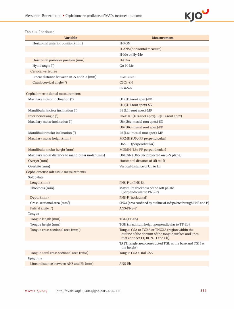
www.e-kjo.org 315http://dx.doi.org/10.4041/kjod.2015.45.6.308
Table 3. Continued
Variable Measurement
Horizontal anterior position (mm) H-RGN
H-ANS (horizontal measure)
H-Me or Hy-Me
Horizontal posterior position (mm) H-C3ia
Hyoid angle (o) Go-H-Me
Cervical vertebrae
Linear distance between RGN and C3 (mm) RGN-C3ia
Craniocervical angle (o) C2C4-SN
C2si-S-N
Cephalometric dental measurements
Maxillary incisor inclination (o) U1 (U1i-root apex)-PP
U1 (U1i-root apex)-SN
Mandibular incisor inclination (o) L1 (L1i-root apex)-MP
Interincisor angle (o) IIAA: U1 (U1i-root apex)-L1(L1i-root apex)
Maxillary molar inclination (o) U6 (U6c-mesial root apex)-SN
U6 (U6c-mesial root apex)-PP
Mandibular molar inclination (o) L6 (L6c-mesial root apex)-MP
Maxillary molar height (mm) MXMH (U6c-PP perpendicular)
U6c-FP (perpendicular)
Mandibular molar height (mm) MDMH (L6c-PP perpendicular)
Maxillary molar distance to mandibular molar (mm) U6L6SN (U6c-L6c projected on S-N plane)
Overjet (mm) Horizontal distance of Uli to Lli
Overbite (mm) Vertical distance of Uli to Lli
Cephalometric soft tissue measurements
Soft palate
Length (mm) PNS-P or PNS-Ut
Thickness (mm) Maximum thickness of the soft palate (perpendicular to PNS-P)
Depth (mm) PNS-P (horizontal)
Cross-sectional area (mm2) SPXA (area confined by outline of soft palate through PNS and P)
Palatal angle (o) ANS-PNS-P
Tongue
Tongue length (mm) TGL (TT-Eb)
Tongue height (mm) TGH (maximum height perpendicular to TT-Eb)
Tongue cross sectional area (mm2) Tongue CSA or TGXA or TNGXA (region within the outline of the dorsum of the tongue surface and lines that connect TT, RGN, H and Eb).
TA (Triangle area constructed TGL as the base and TGH as the height)
Tongue : oral cross sectional area (ratio) Tongue CSA : Oral CSA
Epiglottis
Linear distance between ANS and Eb (mm) ANS-Eb

Alessandri-Bonetti et al • Cephalometric predictors of MADs treatment outcome
www.e-kjo.org316 http://dx.doi.org/10.4041/kjod.2015.45.6.308
Cervical vertebrae• 1-piece MAD: Svanholt et al.24 found that the pre-
valence of and severity of morphological deviations of the upper spine were greater in the group of patients that failed MAD.
• 2-piece MAD: Neither the linear distance between RGN and C3ia21 nor the craniocervical angle21,22
seemed to be predictive of MAD treatment outcome.
Cephalometric dental measurements• 1-piece MAD: Studies investigating dental para-
meters agreed on the lack of predictive value of these measurements for treatment outcome.14,24
• 2-piece MAD: Overjet and overbite were widely
Table 3. Continued
Variable Measurement
Upper Airway
Width (mm)
At the level of soft palate SPAS or SP-PAS or Superior Posterior (along parallel line to Go-B line)
MAS or middle airway (along parallel line to Go-B line through P)
MinRPA or narrowest palatal airway (minimal width perpendicular to posterior pharyngeal wall)
RPAS (Phw-Spt)
At the level of tongue base PAS or IAS or IAS1 (distance between posterior pharyngeal wall and the dorsal base of tongue surface, measured on a line intersecting Go and B Point)
Posterior Inferior (distance between base of tongue and posterior pharyngeal wall)
MinRGA or narrowest lingual airway (minimal width perpendicular to posterior pharyngeal wall)
TB-PAS (at the level of the tongue base)
PPW’-BT’
Through C3 IAS2 (along parallel line to Go-B line)
Vertical length (mm) VAL (PNS-Eb)
Cross-sectional area (mm2)
Nasopharynx cross-sectional area NASOXA (area outlined by line between R and PNS, extension of palatal plane to posterior pharyngeal wall and posterior pharyngeal wall)
Oropharynx cross-sectional area OROXA (area outlined by inferior border of nasopharynx, posterior surface of soft palate, line parallel to palatal plane from point P to dorsal surface of tongue, posterior inferior surface of tongue, line parallel to palate plane through point Et, and posterior pharyngeal wall)
Cross-sectional area (mm2)
Hypopharynx cross-sectional area HYPOXA (area outlined by inferior border of oropharynx, posterior surface of epiglottis, line parallel to palatal plane through point C4, and posterior pharyngeal wall)
Pharinx cross-sectional area PHYNXA (sum of NASOXA, OROXA and HYPOXA)
Facial contours
Facial convexity (o) N’-Prn-Pog’
Prominence of the nose (mm) Prn-S (vertical)
Upper lip position (mm) Distance UL-E line
Lower lip position (mm) Distance LL-E line
See Figure 2 for definitions of landmarks and reference lines used in this table.

Alessandri-Bonetti et al • Cephalometric predictors of MADs treatment outcome
www.e-kjo.org 317http://dx.doi.org/10.4041/kjod.2015.45.6.308
re cognized as non-predict ive of treatment success.9,17,20,22,23 However, Hoekema et al.8 fo und that increased overjet and overbite were progno-stically favorable. Increased maxillary molar height also seemed to be associated with a better chance of successful treatment.17 None of the other cepha-lometric dental measurements exhibited predictive value.21,23
Cephalometric soft tissue measurements
Soft palate• 1-piece MAD: Soft palate depth and thickness and
palatal angle were identified as non-predictive of treatment outcomes,10,14 whereas data concerning soft palate length, which, when decreased,16 was recognized both as non-predictive of treatment
outcome14,15 and predictive of treatment success, were controversial.
• 2-piece MAD: Shen recognized soft palate thickness as non-predictive of treatment outcome.9 The role of soft palate length was controversial. Two out of 6 studies found that decreased soft palate length was predictive of treatment success9,22 while 4 did not.8,19-21
Tongue• 1-piece MAD: There was general agreement that
the cephalometric variables of tongue length, height, and cross-sectional area were not useful for predicting MAD treatment outcomes.10,14,15
• 2-piece MAD: Tongue-related cephalometric variables were widely recognized as non-predictive of MAD treatment outcome9,17,22,23 with the sole exception of tongue/oral enclosure cross-sectional ratio, which Mostafiz et al.21 found was increased in complete responders.
Epiglottis• 1-piece MAD: Only 1 study examined the predictive
role of the distance between ANS and Eb in treatment outcomes, finding that the greater this distance, the less effective the treatment.10
• 2-piece MAD: No study investigated the predictive role of epiglottis parameters.
Upper airway• 1-piece MAD: Upper airway parameters were unani-
mously recognized as non-predictive of MAD treat-ment outcome.10,14,16
• 2-piece MAD: Neither vertical length nor cross-sectional area were found to be predictive of out-come.9,17,20 Concerning the upper airway widths, only 1 study (Shen et al.9) found significantly decreased retroglossal width in good responders. The majority showed a non-predictive role for retropalatal width, although Mehta et al.5 and Liu et al.17 found an increased retropalatal space in good and poor res-ponders, respectively.
Facial contours• 1-piece MAD: Kim et al.14 found no differences in
profile measurements between good and poor res-ponders.
• 2-piece MAD: No profile measurements were found to be predictors for treatment outcome with 2-piece MAD.20
DISCUSSION
MADs are increasingly used for treatment of mild to
FP
PP
OP
MP
Me
Pog'
LLLLUli
B
PogPog
GnGn
RGN
H (Hy)
Eb
C3iaC3ia
C4
Iop
D
Po
SN N' SN
E line
Prn
OrR
Cd
Ba
C2
Ar
Phw
Spt
PNSPNS ANS
A
ULUL
PPW'PPW' P (Ut)P (Ut)
EtEt
GoGoBT'BT'
L6cL6c
U6cU6c
TTTT
Lli
Figure 2. Diagrammatic representation of landmarks and reference lines. A, Subspinale; ANS, anterior nasal spine; Ar, articulare; B, supramentale; Ba, basion; BT’, base of tongue; C2, tangent point on the dorsal surface of C2 vertebra to a line from C4; C3ia, C3 vertebra infe-roanterior; C4, C4 vertebra inferoposterior; Cd, condylion; D, the deepest point in posterior cranial fossa; E line, Ricketts-E line; Eb, epiglottis base; Et, tip of epiglottis; FP, Frankfurt Plane; Gn, gnathion; Go, gonion; H (Hy), hyoidale; Iop: internal occipital protuberance; L1i, lower incisor tip; LL, lower lip; Me, menton; MP, mandibular plane; N, nasion; N’, soft tissue nasion; OP, occlusal plane; Or, orbitale; P (Ut), soft-palate tip; Phw, posterior pharyngeal wall; PPW’, posterior pharyngeal wall interseption; PNS, posterior nasal spine; Po, porion; Pog, pogonion; Pog’, soft tissue pogonion; PP, palatal plane; Prn, nasal tip; R, roof of pharynx; RGN, retrognathion; S, sella; SN, S-N plane; Spt, tangent point on a line parallel to PNS-P on the dorsal surface of the soft palate at the maximum width; TT, tongue tip; U6c, maxillary first molar point; U1i, upper incisor tip; UL, upper lip.

Alessandri-Bonetti et al • Cephalometric predictors of MADs treatment outcome
www.e-kjo.org318 http://dx.doi.org/10.4041/kjod.2015.45.6.308
moderate OSA because they provide a less invasive, more comfortable, and less costly alternative to nCPAP. Nevertheless, MADs are not as efficacious as nCPAP, and the treatment success rates range from 42% to 65%.7 As certain craniofacial characteristics, including reduced posterior airway space, abnormally long soft palate, and low position of the hyoid, are commonly found in OSA patients it seems reasonable to assume that the efficacy of MAD may relate to morphological factors.26
Several modalities for assessing upper airway morphology have been recommended, including mag-netic resonance, nasopharyngoscopy (in an awake state or during drug-induced “sleep”), computed tomography, and lateral cephalometry.8,9,27-29 Cephalometery is low-cost, simple, and widely available, and these advantages may offset the disadvantages of the cephalogram being a 2-dimensional projection of a 3-dimensional structure that is performed in an awake state and in an upright position whereas the pathology of OSA arises with the patient lying down during sleep. Therefore, we aimed to provide a systematic review of cephalometric parameters predictive of MAD treatment outcome.
Summary of the evidence Within the limitations of the selected studies, some con sideration can be made with regard to cephalometric para meters predictive of MAD treatment response. Data on the skeletal cephalometric measurements were con-flicting. Among the cranial base cephalometric values, cranial base angle and the distance between the sella turcica for 2-piece MADs and the deepest point in the posterior cranial fossa for 1-piece MADs were suggested as possible prognostic factors. However, the data are still too limited to draw any conclusion. The lack of predictive value of sagittal jaw relationships (SNA, SNB, SN-Pg, ANB, Wits appraisal) as well as li-near parameters related to vertical jaw dimensions (an-terior face height, lower anterior face height, lower po sterior face height, upper anterior face height, ratio between posterior face height and anterior face height) was almost unanimously confirmed. On the other ha-nd, the mandibular plane angle was associated with widely conflicting results, with some studies reporting a non-predictive role and other studies reporting both increased and decreased predictive values for treatment success. It is therefore impossible to draw any definite conclusion on this parameter based on the available data. Hyoid bone position also showed widely conflicting results, with some studies reporting a non-predictive role for MP-H and others reporting both increased and decreased predictive value for treatment success. Available data for H-Me and ANS-H were ra-ther limited. Therefore, the prognostic value of hyoid bone parameters needs further investigation. A possible
predictive role of upper spine morphological deviations for 1-piece MAD was suggested. Further study will be required to evaluate these findings. With regard to dental cephalometric parameters, overjet and overbite, although being reported as predictive of MAD treatment outcome by Hoekema et al.8 alone, were mostly considered unlikely to carry any prognostic significance. Furthermore Liu et al.17 found that in 2-piece MADs, efficacy was decreased with greater eruption of the maxillary molars. However, being as this study was the only one examining this parameter, the recognition of a predictive role for the distance between the maxillary first molar and Frankfort plane seems premature. Therefore, further research on this parameter is also warranted. Cephalometric soft tissue measurements did not seem to be very useful in predicting MAD treatment response. Soft palate length was recognized both as predictive of treatment success when decreased and as not significantly different between good and poor responders. Therefore, prediction of treatment outcome based on this measurement is not yet unjustified and will require further investigation. Greater ratio between tongue and oral cross sectional area21 and shorter distance between Eb and ANS10 have been shown to be associated with treatment success in 2- and 1-piece MAD, respectively; however additional studies will be needed to confirm these results. Although some studies have suggested a predictive role for retropalatal and retroglossal widths in treatment with 2-piece MAD, the results were controversial. The inconsistency of predictive values of upper airway cephalometric parameters was foreseeable because cephalometry is not the preferred imaging technique for evaluation of these anatomic structures. Indeed, there appears to be a strong linear relationship between 2-dimensional cephalometric and 3-dimensional computed tomography reconstructions for tongue, soft palate, and nasopharynx, while the con-figuration of oropharynx and hypopharynx appears to be less consistent.30
Limitations
Study level The question of the predictive role of cephalometric parameters has not been addressed by randomized clinical trials because the variables of interest are not under the control of the investigators. The inclusion of observational studies therefore appeared necessary. The study design adopted in all of the studies selected for this review was case-control (either prospective and retrospective) rather than cohort: 2 or 3 groups differing in MAD treatment outcome (good and poor responders or nonresponders, partial and complete responders) were

Alessandri-Bonetti et al • Cephalometric predictors of MADs treatment outcome
www.e-kjo.org 319http://dx.doi.org/10.4041/kjod.2015.45.6.308
prospectively or retrospectively identified and compared on the basis of cephalometric parameters. As a result, the findings score low in terms of strength of evidence.A selection bias may have occurred because the sample was not randomly selected. Moreover in some of the prospective studies patients were included after they had been allowed to choose whether or not they wanted to participate or after they had decided entirely for themselves not to accept treatment with nCPAP (self-selection bias).18,19 Finally, a few studies reported that some patients dropped out (attrition bias).5,8,19
MAD treatment success has been associated with fe-male sex, a younger age, a lower body mass index (BMI), and a lower baseline apnea-hypopnea index (AHI) but, due to the design of the studies included in this review, these baseline characteristics could not be properly addressed and may actually show systematic differences between good responders and poor res-ponders (confounding bias).17,22 In the 13 studies that reported male-female ratio, males had considerably more representation, ranging from 67.9% to 100% of the sample.5,9,14-24 With the exception of the study by Mehta et al.,5 none of the others investigated the differences in sex distribution between good responders and poor responders. Six studies found no age difference bet-ween good and poor responders,5,10,14,15,19,24 while 4 studies found age to be significantly lower in good res-ponders.17,20-22 Eight studies failed to find a predictive role for BMI,5,10,14,15,19,20,22,23 while 3 studies reported lower BMI in good responders.8,21,24 Only 1 paper add-ressed the issue of weight gain or loss between the ini-tial and the final evaluations.19 Six out of 14 studies did not account for potential differences in baseline AHI,8,16,18-20,23 whereas comparison between good and poor responders in terms of AHI or apnea index led to significant differences in 2 studies, with lower valu es associated with treatment success,14,21 and no significant differences in 6 studies.5,9,10,15,17,22
With regard to observer bias, only 3 studies reported that the operator who performed the cephalometric analysis or the polysomnography was blinded to the treatment outcome.5,14,22 None of the selected studies included a power analysis or a sample size calculation. The presence of potentially underpowered studies could lead to inconsistent or misleading results, because they may find no significant difference between study sam-ples even in the presence of a real difference in the ge-neral population.
Review level The findings of the present systematic review were also limited by the heterogeneity in the methodology of the included studies. Respiration was analyzed in terms of AHI in the majority of the investigations, but some of
the earlier studies evaluated the respiratory disturbance index (RDI).19 The definition of sleep apnea was based either on AHI > 5, AHI ≥ or > 10, AHI ≥ or > 15, or RDI ≥ 10. Success criteria included RDI reduction of ≥ 50% and RDI ≤ 20, AHI < 5, AHI < 15, or AHI reduction of > 50% alone or in association with AHI < 20, < 10, or < 5. A certain degree of variability was present with respect to the type of MAD (monobloc or two pieces), as well as to the amount of protrusion (from 50 or 60% of maximum protrusion to the most advanced position without causing any discomfort) or vertical opening (ranging from “minimum” to 15 mm). The time interval between diagnostic and with-appliance poly somnography ranged from few weeks to several months. The cephalometric parameters also varied sub-stantially between different studies. Because of the lack of homogeneity among the studies, any attempt to summarize the study results seemed unjustified. This, a narrative synthesis was carried out without performing a meta-analysis. No “grey literature” or articles in non-English langu-ages were included in the present review. Therefore the review is also subject to publication bias and language bias.
CONCLUSION
This review found controversial and limited data on the predictive role of certain cephalometric parameters for MAD treatment outcome. Therefore selection criteria for suitable candidates for MAD treatment by cephalometric analysis are currently inadequate. Although no definitive clinical recommendations can be made, this systematic review highlights the methodological weaknesses of the currently available studies, analyzes the possible reasons for their discordant results, and encourages and guides further research in this field. Prospective cohort studies in large samples with cephalometric prediction made prior to the MAD construction are required to clarify the clinical utility of lateral cephalometry for adult OSA patients who are considering oral appliances as a thera-peutic option.
REFERENCES
1. Epstein LJ, Kristo D, Strollo PJ Jr, Friedman N, Mal hotra A, Patil SP, et al; Adult Obstructive Sleep Apnea Task Force of the American Academy of Sleep Medicine. Clinical guideline for the evaluation, ma-na gement and long-term care of obstructive sleep apnea in adults. J Clin Sleep Med 2009;5:263-76.
2. Skomro RP, Kryger MH. Clinical presentations of ob-structive sleep apnea syndrome. Prog Cardiovasc Dis 1999;41:331-40.

Alessandri-Bonetti et al • Cephalometric predictors of MADs treatment outcome
www.e-kjo.org320 http://dx.doi.org/10.4041/kjod.2015.45.6.308
3. Sullivan CE, Issa FG, Berthon-Jones M, Eves L. Re-versal of obstructive sleep apnoea by continuous positive airway pressure applied through the nares. Lancet 1981;1:862-5.
4. Waldhorn RE, Herrick TW, Nguyen MC, O'Donnell AE, Sodero J, Potolicchio SJ. Long-term compliance with nasal continuous positive airway pressure the-rapy of obstructive sleep apnea. Chest 1990;97:33-8.
5. Mehta A, Qian J, Petocz P, Darendeliler MA, Cistulli PA. A randomized, controlled study of a mandibular advancement splint for obstructive sleep apnea. Am J Respir Crit Care Med 2001;163:1457-61.
6. Kushida CA, Morgenthaler TI, Littner MR, Alessi CA, Bailey D, Coleman J Jr, et al; American Academy of Sleep. Practice parameters for the treatment of snoring and Obstructive Sleep Apnea with oral app-liances: an update for 2005. Sleep 2006;29:240-3.
7. Ferguson KA, Cartwright R, Rogers R, Schmidt-Nowara W. Oral appliances for snoring and obstruc-tive sleep apnea: a review. Sleep 2006;29:244-62.
8. Hoekema A, Doff MH, de Bont LG, van der Hoeven JH, Wijkstra PJ, Pasma HR, et al. Predictors of ob-structive sleep apnea-hypopnea treatment outcome. J Dent Res 2007;86:1181-6.
9. Shen HL, Wen YW, Chen NH, Liao YF. Craniofacial morphologic predictors of oral appliance outcomes in patients with obstructive sleep apnea. J Am Dent Assoc 2012;143:1209-17.
10. Endo S, Mataki S, Kurosaki N. Cephalometric evalu-ation of craniofacial and upper airway structures in Japanese patients with obstructive sleep apnea. J Med Dent Sci 2003;50:109-20.
11. Saffer F, Lubianca JF, Rösing C, Dias C, Closs L. Predictors of success in the treatment of obstruc-tive sleep apnea syndrome with mandibular reposi-tioning appliance: a systematic review. Int Arch Otorhinolaryngol 2015;19:80-5.
12. Jovell AJ, Navarro-Rubio MD. Evaluation of scientific evidence. Med Clin (Barc) 1995;105:740-3.
13. Liu Y, Lowe AA. Factors related to the efficacy of an adjustable oral appliance for the treatment of obstructive sleep apnea. Chin J Dent Res 2000;3:15-23.
14. Kim YK, Kim JW, Yoon IY, Rhee CS, Lee CH, Yun PY. Influencing factors on the effect of mandi bular advancement device in obstructive sleep apnea patients: analysis on cephalometric and polysom-nographic parameters. Sleep Breath 2014;18:305-11.
15. Lee CH, Mo JH, Choi IJ, Lee HJ, Seo BS, Kim DY, et al. The mandibular advancement device and pa-tient selection in the treatment of obstructive sleep apnea. Arch Otolaryngol Head Neck Surg 2009; 135:439-44.
16. Lee CH, Kim JW, Lee HJ, Seo BS, Yun PY, Kim DY,
et al. Determinants of treatment outcome after use of the mandibular advancement device in patients with obstructive sleep apnea. Arch Otolaryngol Head Neck Surg 2010;136:677-81.
17. Liu Y, Lowe AA, Fleetham JA, Park YC. Cephalo-metric and physiologic predictors of the efficacy of an adjustable oral appliance for treating obstructive sleep apnea. Am J Orthod Dentofacial Orthop 2001; 120:639-47.
18. Marklund M, Franklin KA, Stenlund H, Persson M. Mandibular morphology and the efficacy of a man-dibular advancement device in patients with sleep apnoea. Eur J Oral Sci 1998;106:914-21.
19. Menn SJ, Loube DI, Morgan TD, Mitler MM, Berger JS, Erman MK. The mandibular repositioning device: role in the treatment of obstructive sleep apnea. Sleep 1996;19:794-800.
20. Milano F, Billi MC, Marra F, Sorrenti G, Gracco A, Bonetti GA. Factors associated with the efficacy of mandibular advancing device treatment in adult OSA patients. Int Orthod 2013;11:278-89.
21. Mostafiz W, Dalci O, Sutherland K, Malhotra A, Srinivasan V, Darendeliler MA, et al. Influence of oral and craniofacial dimensions on mandibular ad-vancement splint treatment outcome in patients with obstructive sleep apnea. Chest 2011;139:1331-9.
22. Ng AT, Darendeliler MA, Petocz P, Cistulli PA. Ce-phalometry and prediction of oral appliance treat-ment outcome. Sleep Breath 2012;16:47-58.
23. Rose E, Lehner M, Staats R, Jonas IE. Cephalometric analysis in patients with obstructive sleep apnea. Part II: Prognostic value in treatment with a mandi-bular advancement device. J Orofac Orthop 2002; 63:315-24.
24. Svanholt P, Petri N, Wildschiødtz G, Sonnesen L. Influence of craniofacial and upper spine morpho-logy on mandibular advancement device treatment outcome in patients with obstructive sleep apnoea: a pilot study. Eur J Orthod 2015;37:391-7.
25. Liberati A, Altman DG, Tetzlaff J, Mulrow C, Gøtz-sche PC, Ioannidis JP, et al. The PRISMA statement for reporting systematic reviews and meta-analyses of studies that evaluate healthcare interventions: explanation and elaboration. BMJ 2009;339:b2700.
26. Hoekema A, Hovinga B, Stegenga B, De Bont LG. Craniofacial morphology and obstructive sleep ap-noea: a cephalometric analysis. J Oral Rehabil 2003; 30:690-6.
27. Sanner BM, Heise M, Knoben B, Machnick M, Laufer U, Kikuth R, et al. MRI of the pharynx and treatment efficacy of a mandibular advancement device in obstructive sleep apnoea syndrome. Eur Respir J 2002;20:143-50.
28. Chan AS, Lee RW, Srinivasan VK, Darendeliler MA,

Alessandri-Bonetti et al • Cephalometric predictors of MADs treatment outcome
www.e-kjo.org 321http://dx.doi.org/10.4041/kjod.2015.45.6.308
Grunstein RR, Cistulli PA. Nasopharyngoscopic evaluation of oral appliance therapy for obstructive sleep apnoea. Eur Respir J 2010;35:836-42.
29. Sam K, Lam B, Ooi CG, Cooke M, Ip MS. Effect of a non-adjustable oral appliance on upper airway mor-phology in obstructive sleep apnoea. Respir Med
2006;100:897-902.30. Lowe AA. Can we predict the success of dental app-
liance therapy for the treatment of obstructive sleep apnea based on anatomic considerations? Sleep 1993;16(8 Suppl):S93-5.