Embed Size (px)
Citation preview
CRITICAL REVIEW www.rsc.org/loc | Lab on a Chip
Centrifugal microfluidics for biomedical applications
Robert Gorkin,a Jiwoon Park,b Jonathan Siegrist,a Mary Amasia,a Beom Seok Lee,c Jong-Myeon Park,cd
Jintae Kim,c Hanshin Kim,c Marc Madouab and Yoon-Kyoung Cho*b
Received 16th November 2009, Accepted 3rd March 2010
First published as an Advance Article on the web 28th May 2010
DOI: 10.1039/b924109d
The centrifugal microfluidic platform has been a focus of academic and industrial research efforts for
almost 40 years. Primarily targeting biomedical applications, a range of assays have been adapted on the
system; however, the platform has found limited commercial success as a research or clinical tool.
Nonetheless, new developments in centrifugal microfluidic technologies have the potential to establish
wide-spread utilization of the platform. This paper presents an in-depth review of the centrifugal
microfluidic platform, while highlighting recent progress in the field and outlining the potential for
future applications. An overview of centrifugal microfluidic technologies is presented, including
descriptions of advantages of the platform as a microfluidic handling system and the principles behind
centrifugal fluidic manipulation. The paper also discusses a history of significant centrifugal microfluidic
platform developments with an explanation of the evolution of the platform as it pertains to academia
and industry. Lastly, we review the few centrifugal microfluidic-based sample-to-answer analysis
systems shown to date and examine the challenges to be tackled before the centrifugal platform can be
more broadly accepted as a new diagnostic platform. In particular, fully integrated, easy to operate,
inexpensive and accurate microfluidic tools in the area of in vitro nucleic acid diagnostics are discussed.
1.0 Introduction
In the field of microfluidic lab-on-a-chip systems, the centrifugal
microfluidic platform has emerged as an advanced technology
for biological analysis. ‘‘Lab-on-a-CD’’ systems are the focus of
intense research, where complex assays are embedded in fluidic
networks on centrifugal microfluidic systems, especially towards
the development of in vitro diagnostics (IVD). The centrifugal
microfluidic platform has the potential to become a standard tool
for mainstream diagnostics. Fluidic processing steps such as the
aUniversity of California, Irvine, Irvine, CA, 92697, USAbUlsan National Institute of Science and Technology (UNIST), Banyeon-ri100, Ulsan, 689-798, Korea. E-mail: [email protected]; Fax: +82-52-217-2509; Tel: +82-52-217-2511cSamsung Electronics Co. Ltd., 416, Maetan-3Dong, Yeongtong-Gu,Suwon-City, Gyeonggi-Do, 443-746, KoreadYonsei University, Seoul, 120-749, Korea
Jiwoon Park and Yoon-Kyoung Cho
Jiwoo
Chem
interes
nostic
Yoon-
neerin
Since
(SAIT
NBC
progra
fluidic
and m
1758 | Lab Chip, 2010, 10, 1758–1773
mixing of reagents or metering of sample fluids can be automated
simply by implementing different spinning profiles. Through the
adaption of miniaturization technology, multiple analysis steps
can be integrated on a single disc, often towards the development
of microfluidic sample-to-answer systems, or micro total analysis
systems (mTAS). Moreover, many individual assays can be run
simultaneously on the same disc. With the development of
optical systems to interface with disc-based assays, centrifugal
microfluidic systems allow for operators to not only perform the
often complex and timely sample preparation steps required in
most assays, but also to rapidly identify biological targets all on
the same platform.
In comparison to common chip-based microfluidic systems,
centrifugal microfluidic platforms offer many advantages. For
fluid propulsion, centrifugal pumping involves a minimal
amount of instrumentation, requiring only a simple and compact
motor to create the forces needed for fluid manipulation and
n Park is a masters course student of Nano-Biotechnology and
ical Engineering (NBC) at UNIST, Korea. Her current research
ts are in the development of microfluidic devices for clinical diag-
s and on-chip cell biology.
Kyoung Cho received her Ph.D. in Materials Science and Engi-
g from the University of Illinois at Urbana-Champaign in 1999.
then, she worked at Samsung Advanced Institute of Technology
) until 2008. Currently, she is an assistant professor of School of
at UNIST and the director of the World Class University (WCU)
m at the same University. Her research interests include micro-
devices for biomedical applications and the interface of cell biology
icro/nanofluidics.
This journal is ª The Royal Society of Chemistry 2010

eliminating the need for external syringe pumps. Additionally,
centrifugal pumping requires no external interconnects to induce
fluid movement. This allows the complete fluidic network (and
indeed the inclusive assay) to be contained within a single disc.
As centrifugal microfluidics can be mass-produced from inex-
pensive materials like polycarbonate, they can be manufactured
to be disposable in an economical way. Centrifugal pumping also
holds advantages over other chip-based pumping, such as elec-
trokinectic methods, because it is not strongly dependent on
physicochemical properties of the fluid, such as pH or ionic
strength,1 and does not require large high-voltage power
supplies. Additionally, a variety of fluids have been successfully
pumped using the microfluidic centrifugal microfluidic platform
including aqueous solutions, solvents, surfactants and biological
fluids (e.g., blood, mucus, urine, milk).1
Mary Amasia; Marc Madou; Robert Gorkin; and Jonathan Siegrist
M
g
t
c
d
M
E
a
a
V
P
M
R
UCI in 2010. His background includes experience developing point-of-c
and South Korea. Dr Gorkin is currently joining the Biomedical Diagno
biotechnology platforms for research and clinical use.
Jonathan Siegrist received his Ph.D. in Biomedical Engineering from U
in Dublin, Ireland as a Postdoctoral Researcher. His interests lie in th
Jintae Kim; Beom Seok Lee; Hanshin Kim; and Jong-Myeon Park
J
S
l
c
b
o
B
t
e
c
m
H
U
Korea in 2005. Currently he is group leader of HME Business team, S
system and POCT devices.
Jong-Myeon Park is a senior research engineer at Bio Lab at SAIT and
University, Korea. His current research interests center on the developm
analysis.
This journal is ª The Royal Society of Chemistry 2010
Beyond pumping of liquids, many other fluidic functions have
been successfully integrated on the centrifugal microfluidic plat-
form including valving, decanting, calibration, mixing, metering,
sample splitting, and fluid separation. Valving is essential for all
microfluidic analysis platforms, as sample fluids and reagents
must be properly retained until needed. Multiple types of valves,
including passive and active valves, have been developed for the
centrifugal microfluidic system and have been successfully
implemented. All of these centrifugal microfluidic technologies
and functions make the centrifugal microfluidic platform
powerful, and insure complete automation to reduce time and
error due to handling. In terms of complexity, centrifugal
microfluidic technology replaces complex fluidic handling
equipment and intricate interconnects. Finally, by reducing
experiment size and complexity, costs can be kept to a minimum.1
ary Amasia is a senior member of the Madou BioMEMS research
roup at the University of California, Irvine (UCI), working
owards her Ph.D. in Chemical and Biochemical Engineering. Her
urrent research interests are in the development of microfluidic
evices as medical and diagnostic systems.
arc Madou is Chancellor Professor of Mechanical and Aerospace
ngineering at UCI, as well as Professor of Biomedical Engineering
nd Materials Concentration at the same University. Dr Madou is
lso Distinguished Honorary Visiting Professor IIT Kanpur, India,
isiting Professor UNIST (WCU Scholar), Korea and Visiting
rofessor, Department of Biomedical Engineering, Kuala Lumpur,
alaysia.
obert Gorkin received his Ph.D. in Biomedical Engineering from
are CD diagnostics with collaborations in Canada, India, Malaysia
stics Institute in Dublin and is interested in advancing microfluidic
CI in 2009. He recently joined the Biomedical Diagnostics Institute
e development of microfluidic diagnostic devices and biosensors.
intae Kim received his Ph.D. in Electrical and Computer Eng. from
ungKyunKwan University, Korea in 2002. Currently he is a project
eader in the HME business team at Samsung Electronics. His
urrent research interests include bioMEMS, molecular biology,
iochemistry, electrochemistry, biosensors, actuators, microfluidics,
ptical/electronic microscopy, and spectroscopy.
eom Seok Lee received his Ph.D. in Chemical Engineering from
he POSTECH, Korea in 2006. Currently he is a senior research
ngineer in the HME Business team at Samsung Electronics. His
urrent research interests are in clinical assays, immunoassays,
icrofluidics, and electrohydrodynamics.
anshin Kim received his M.S. in Biochemistry from Yonsei
niversity, Korea in 1993, and MBA from Sogang University,
amsung Electronics. His current research interests are diagnostic
working towards his Ph.D. in the Chemistry department at Yonsei
ent of centrifugal microfluidics for sample preparation and rare cell
Lab Chip, 2010, 10, 1758–1773 | 1759
Although centrifugal microfluidics have many advantages,
the simple motor control of fluid handling on a CD has its own
inherit limitations. For example, the passive fluid valving
operation on a CD strongly depends on the balancing of spin
speed and surface tension in most of the conventional centrif-
ugal microfluidic devices utilizing either hydrophobic or capil-
lary valves. But surface tension depends on the surface energy
which in the case of a polymer CD is dynamic in nature. That
is why recently Park et al.2 explored an active type valving
mechanism utilizing laser diodes to melt wax valves avoiding
the surface energy changes associated with passive valving. This
approach constitutes a solution but it adds other complexities
to the realization of the platform (e.g., the need for wax
deposition systems and heating actuators). Another example of
a technological barrier involves the difficulty in implementing
additional forces onto the CD. Say one wants to implement
electrical fields on the CD, perhaps to separate cells by die-
lectrophoresis or to affect osmotic pumping through channels
too narrow for centripetal forces to pump. Along this line,
Martinez-Duarte et al. utilized electric fields on the spinning
disc to capture cells by dielectrophoresis.3 But as before the
implementation of this solution renders the platform consid-
erably more complex.
This paper is intended to give a review of the centrifugal
microfluidic platform with a focus on biomedical applications,
while highlighting the history, recent advances, and potential for
future applications. Section 2 gives a historical overview of
significant centrifugal microfluidic developments and covers
commercial microfluidic centrifugal microfluidic technologies
from the biomedical industry. Section 3 describes the principles
underlying centrifugal microfluidic functions, such as fluidic
transfer and valving that have been adapted for implementation
of biological assays. Section 4 discusses many current centri-
fugal microfluidic research initiatives, and section 5 focuses
on centrifugal microfluidic-based sample-to-answer systems.
Section 6 outlines some of the challenges still being addressed in
the field, particularly in regards to sample-to-answer systems for
nucleic acid diagnostics. The final section presents an outlook for
the future of centrifugal microfluidics.
2.0 A history of centrifugal microfluidic technology
The field of centrifugal microfluidics began in the late 1960s with
the development of the centrifugal analyzer. N. Anderson, from
Oak Ridge National Labs (ORNL), developed a clinical chem-
istry analyzer, which incorporated a rotating disc with a multi-
cuvette assembly, and a stationary optical detector designed for
use with a computer.4,5 The design and operation of the system
was simple: the disc was fabricated such that channels and risers
(i.e. 3D physical barriers) along the radial axis kept samples and
reagents separated during fluid loading. During spinning,
centrifugal forces drove fluids over the barriers into optical
cuvettes positioned on the periphery of the disc. As reactions
took place, absorbance changes were monitored with a light
source and a photomultiplier tube arranged above and below the
cuvettes, respectively. The system was initially used for kinetic
assay development.
Work on centrifugal analyzers continued to progress, and
miniaturized versions were developed (Fig. 1).5 Additionally,
1760 | Lab Chip, 2010, 10, 1758–1773
other optical technologies were incorporated to measure light
transmittance, fluorescence, chemiluminescence, and light-scat-
tering properties of several simultaneously initiated reaction
mixtures.5 The implementation of these analysis systems helped
to create new clinical laboratory tools for applications in chem-
istry, toxicology, immunology, and hematology.5
By the time Anderson published his landmark paper in 1969,4
multiple companies were offering prototype versions of the
centrifugal analyzer, with the first commercial system introduced
by Electro-Nucleonics, Inc. in 1970. In a little over 10 years, the
commercial field had grown to five companies that were offering
related products: Electro-Nucleonics, Inc.—GEMSAEC and
GEMINI, Centri Union Carbide—CentrifiChem, American
Instruments—Rotochem, Instrumentation Laboratories, Inc.—
Multistat, and Roche—Cobas-Bio.4
The next phase in development of centrifugal microfluidics
took place in 1989 with the formation of Abaxis, Inc. Abaxis
bought the patents from ORNL for their version of the clinical
analyzer and began to develop it as a tool for blood analyte
analysis.6 This represented a shift in the utilization of
centrifugal microfluidics from a research-oriented tool to
a diagnostic platform. In 1995, Abaxis introduced the Piccolo
rotor system which integrated sample processing steps required
for analyte analysis and incorporated self-contained
reagents for each step. The Piccolo system would become the
flagship for a range of blood panel products encompassing
several areas of medicine.6
In 1998, M. Madou and G. Kellogg from Gamera (a US
startup company based on centrifugal microfluidic technology)
introduced the next generation of centrifugal microfluidics as
described in ‘‘The LabCD: A centrifuge-based microfluidic
platform for diagnostics.’’7 The paper outlined basic centrifugal
theory of pumping fluids and introduced valving, mixing,
sample entry and metering as basic fluidic functions on
a centrifugal microfluidic device. Their work represented an
expansion of centrifugal technologies into new realms of bio-
logical and chemical analysis by introducing microfabrication
techniques to create and merge miniaturized fluid networks and
microsensors on a single disposable centrifugal microfluidic
platform. Madou, Kellogg, and the entire Gamera team were
the first to realize that the centrifugal microfluidic platform
could be used as an advanced sample-to-answer system.7 While
clinical analyzers had the advantage of being able to perform
high-throughput assays in a short time, they often required
trained technicians to carry out several operations on different
machines. The LabCD offered a unique and attractive platform
to overcome limitations of the macro-scale centrifugal systems
of that time and opened up the possibility for more advanced
tools to be created with applications for drug development
through genomics and proteomics, molecular diagnostics, and
genetic testing.7,8 Gamera, later acquired by Tecan in July 2000,
would continue to develop the LabCD mainly for assays
related to drug development such as ADMET—absorption,
distribution, metabolism, excretion and toxicity.9 Tecan
advanced Gamera’s system significantly but discontinued their
efforts in 2005.
The early 2000s saw a rapid development of academic and
commercial endeavors to incorporate new assay designs on
a centrifugal microfluidic platform. Kido et al. first introduced
This journal is ª The Royal Society of Chemistry 2010
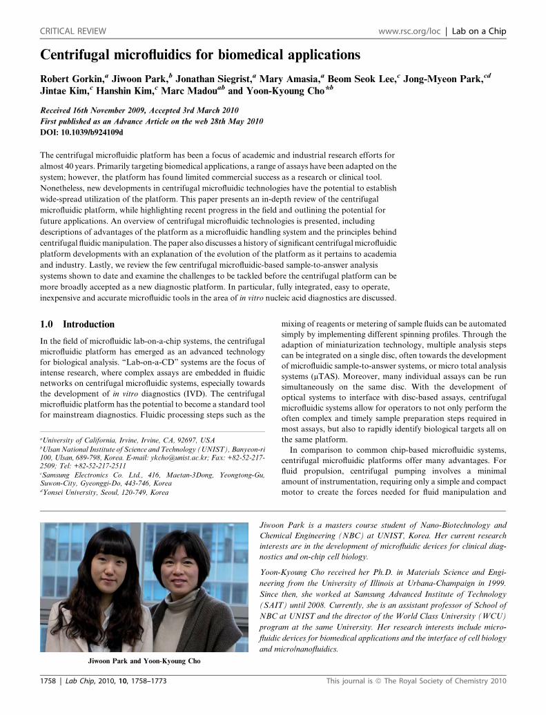
Fig. 1 A miniature fast analyzer platform developed by ORNL with
loaded analysis disc; with the disc consisted of two different sides
allowing for discrete or metered loading of samples. Figure adapted and
reproduced with permission from ref. 5.
disc-based immunoassay microarrays for agriculture and envi-
ronmental analysis in 2000.10 In their work, specialized modules
were integrated with standard CD drives to set up and analyze
protein microarrays.10 In that same year, Gyros AB was founded
and would go on to commercialize a disc-based assay system that
automated sandwich immunoassay processing. Their products
available on the market are used today as a tool for high-
throughput immunoassay analysis.11 Several other companies
would later adapt immunoassay technology on the centrifugal
microfluidic platform in various implementations: Quadraspec
developed label-free technology for identifying changes in
protein arrays,12,13 while Burstein Technologies14 and Advanced
Array Technology15 developed array methods on the disc for
analyzing nucleic acids (NA).
Although there has been extensive development of centrifugal
microfluidic technology, often with collaborations from
academia and industry, the market is relatively bare of
commercially available products today. While centrifugal
microfluidic technology has many advantages as a microfluidic
platform, the failure of companies to commercialize products
brings to the foreground several barriers to adapting micro-
fluidic platforms for widespread use. Any commercial micro-
fluidic platform needs to bring advantages to the user in terms
of cost, throughput, user-friendliness, and accuracy compared
to the current gold-standard methods. Even if a microfluidic
device with self-contained reagents guarantees a very simple
operation protocol and features significantly better accuracy,
the cost of both disposable and fixed system plays a key role in
determining commercialization success. Even if the newly
developed CD system has all of the above-mentioned advan-
tages, it takes time for it to be adapted by the major user
groups. With the commercial history of the centrifugal micro-
fluidic platform established, the sections to follow highlight
This journal is ª The Royal Society of Chemistry 2010
current research advances using centrifugal microfluidic tech-
nology.
3.0 Centrifugal microfluidic functions
This section introduces various microfluidic functions that have
been implemented on the centrifugal microfluidic systems for
biological processing. An explanation of the individual functions
serves as an introduction to give the reader insight into how the
integration of such systems enables biological processes on
a centrifugal microfluidic device. Often, the complex liquid
handling involved in biological assays requires the combination
of several of the microfluidic functions presented below.
3.1 Centrifugal pumping
In centrifugal pumping, as a disc spins, centrifugal forces induced
on sample fluids drive liquids radially outwards from the center
toward the edge of the disc. The flow of fluids in a centrifugal
platform has been well characterized; centrifugal flow rates
depend on the rotational speed, radial location of the fluid
reservoirs/channels, channel geometry, and fluidic properties
(e.g., viscosity, density, etc.) of a sample.1,16 The average velocity,
U, of centrifugally-pumped liquid in a microchannel can be
derived from centrifugal theory as:
U ¼ D2h ru2 �rDr
32mL(1)
where Dh is the hydraulic diameter of the channel (defined as 4A/
P, A is the cross-sectional area and P is the wetted perimeter of
the channel), r is the density of the liquid, u is the angular
velocity of the disc, r�is the average distance of the liquid in the
channels from the center of the disc, Dr is the radial extent of the
fluid, m the viscosity of the fluid, and L the length of the liquid in
the microchannel.1 The volumetric flow rate, Q, is then defined as
U$A, where U is from eqn (1). As shown in eqn (1), both the
channel geometries and the fluidic properties play a large role in
centrifugal pumping, in addition to spin speed.
Madou et al. and Duffy et al. demonstrated that the flow rates
predicted by simple centrifuge theory follow well with experi-
mentally measured flow rates for various kinds of samples
including water, plasma, bovine blood, urine, DMSO, and
polymerase chain reaction (PCR) products.1,8 Flow rates ranging
from 5 nL s�1 to over 0.1 mL s�1 have been achieved by various
combinations of rotational speed from 400 to 1600 RPM,
channel widths from 20 to 500 mm, and channel depths from 16
to 340 mm.8 The dynamic pumping range of the centrifugal
microfluidic platform extends far beyond these limits, as wider
channels and higher rotation speeds are easily achieved.17
Centrifugal flow rates are relatively insensitive to physico-
chemical properties such as ionic strength, pH, conductivity, and
the presence of various analytes.8 Thus, the centrifugal micro-
fluidic platform provides a unique pumping mechanism that
provides a very large dynamic range in terms of fluidic pumping
rates and volumes as well as types/properties of fluids being
pumped. This is one of the most important aspects of utilizing the
centrifugal microfluidic platform for biological applications, as it
allows successful pumping of many different fluids on the same
disc.
Lab Chip, 2010, 10, 1758–1773 | 1761
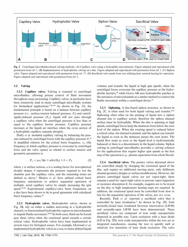
Fig. 2 Centrifugal microfluidics-based valving methods. (A) Capillary valve using a hydrophilic microchannel. Figure adapted and reproduced with
permission from ref. 1. (B) Implementation of hydrophobic valving on a disc. Figure adapted and reproduced with permission from ref. 1. (C) Siphon
valve. Figure adapted and reproduced with permission from ref. 17. (D) Sacrificial valve made from wax utilizing laser assisted heating for operation.
Figure adapted and reproduced with permission from ref. 2.
3.2. Valving
3.2.1 Capillary valves. Valving is essential to centrifugal
microfluidics, allowing precise control of fluid movement
throughout assay processing. Capillary valves, in particular have
been extensively used in many centrifugal microfluidic systems
for biomedical applications.1,16–19 As shown in Fig. 2A, the
fundamental principle is based on a balance between capillary
pressure (i.e., surface-tension induced pressure, Ps) and centrif-
ugally-induced pressure (Pu): liquid will not pass through
a capillary valve when the centrifugal pressure is less than or
equal to the capillary barrier pressure. Capillary pressure
increases at the liquid–air interface when the cross section of
a hydrophilic capillary expands abruptly.
Duffy et al. modeled capillary valving by balancing the pres-
sure induced by centrifugal forces with the capillary pressure Ps.8
A simplified relation for the critical burst frequency, uc (the
frequency at which capillary pressure is overcome by centrifugal
force and the valve opens) as related to surface tension and
geometric parameters is:
Pu ¼ ruc2r�Dr > a(4g/Dh) + b ¼ PS (2)
where g is surface tension, a is a scaling factor for non-spherical
droplet shapes, b represents the pressure required to wet the
chamber past the capillary valve, and the remaining terms are
defined as above.1 Madou et al. have utilized critical burst
frequency calculations to achieve sequential operation of
multiple, serial capillary valves by simply increasing the spin
speed.19–21 Experimental capillary-valve burst frequencies on
a disc have been shown to be in good agreement with calculated
burst frequency values.21
3.2.2 Hydrophobic valves. Hydrophobic valves, shown in
Fig. 2B, rely on either a sudden narrowing in a hydrophobic
channel, or functionalized hydrophobic regions in microchannels
to impede fluidic movement.22,23 In both cases, fluid can be forced
past these valves when the rotational speed exceeds a certain
critical value. Hydrophobic valves have been implemented in
several ways for biological assays. For example, Ekstrand et al.
implemented hydrophobic valves as a way to meter defined liquid
1762 | Lab Chip, 2010, 10, 1758–1773
volumes and transfer the liquid at high spin speeds, when the
centrifugal forces overcome the capillary pressure at the hydro-
phobic barriers,22 while Gyros AB uses hydrophobic patches at
the entrances of microchannels as a similar method to control the
fluidic movement within a centrifugal device.11
3.2.3 Siphoning. A disc-based siphon structure, as shown in
Fig. 2C, is often used for both liquid valving and transfer.6,17
Siphoning often relies on the priming of liquid into a siphon
channel due to capillary action; therefore the siphon channel
surface must be hydrophilic. When the disc is spinning at high
speeds, centrifugal forces keep the meniscus front below the crest
level of the siphon. When the rotating speed is reduced below
a critical value, the channel is primed, and the siphon can transfer
the liquid as soon as the meniscus passes the crest point. The
liquid flow stops as soon as the liquid level is hydrostatically
balanced or there is a discontinuity in the liquid column. Siphon
valving in centrifugal microfluidics provides a valving solution
for the applications that require higher spin speeds as the first
step of the operation (e.g., plasma separation from whole blood).
3.2.4 Sacrificial valves. The passive valves discussed above
are controlled simply by changing the rotational speed of the
disc, and require no additional moving parts, only deliberate
channel geometry designs or surface modifications. However, the
passive centrifugal liquid valves are not vapor-tight; there
remains a need for vapor-tight valves to prevent vapor migration
or moisture absorption if, for example, reagents are to be stored
on the disc or high temperature heating steps are required. In
addition, the rotational speed must be controlled from slow to
fast for the sequential operation of the valves, not vice versa.
Recently, Park et al. reported a sacrificial valve that is
controlled by laser irradiation.2 As shown in Fig. 2D, both
normally-closed laser irradiated ferrowax microvalves (LIFM)
and normally-open LIFM have been demonstrated. These
sacrificial valves are composed of iron oxide nanoparticles
dispersed in paraffin wax. Upon excitation with a laser diode
(LD in Fig. 2D), iron oxide nanoparticles within the wax act as
integrated nanoheaters, causing the wax to quickly melt at
relatively low intensities of laser diode excitation. The valve
This journal is ª The Royal Society of Chemistry 2010
operation is independent of the spin speed or the location of the
valves and therefore allows for more complex biological assays
integrated on the disc.24
3.3 Volume metering
Liquid volume metering is an essential function in centrifugal
fluidics to achieve proper reagent volumes for diagnostic assays
and to ensure reproducible valving processes on a disc. Volume
metering on a disc is primarily achieved through the simple use of
an overflow channel connected to a fluidic chamber.2,11 Once the
chamber has filled to the radial level of the overflow channel, any
additional fluid is routed to a waste chamber.
Steigert et al. discuss the significance of ‘‘wicking’’ that occurs
along the edges of the disc-based metering chambers, and
introduce design principles to reduce its negative effect on
metering variation. As part of an integrated colorimetric assay,
they reported the metering of 300 nL with a 5% coefficient of
variability.25
3.4 Mixing
Mixing is necessary in biomedical diagnostic applications in
order to homogenize samples and to combine various reagents
for downstream analysis. However, mixing is difficult to achieve
in microfluidic platforms due to constraints of the microscale
domain, namely low Reynolds numbers with laminar flow. This
means that there is no convective mixing; fluidic mixing is limited
to diffusive mixing which can be a very slow process.26–29
In order to overcome this challenge, a number of approaches
to achieve rapid mixing have been demonstrated on centrifugal
microfluidic platforms.17,24,30–38 One method uses rapid oscilla-
tions of the disc to achieve rapid mixing in low-Reynolds number
regimes (viz., oscillations between clockwise and counter clock-
wise rotation).32,34,35 The flow inertia and counteracting viscous
damping improves rapid mixing in larger-scale fluidic chambers.
Additionally, by introducing paramagnetic particles in the liquid
and by positioning permanent magnets aligned in non-symmet-
rical positions underneath the mixing chamber, a magnetic stir-
ring effect can further decrease mixing time.30,32,39 Ducree et al.
have shown that the Coriolis pseudo-force is efficient for
continuous-flow mixing in ‘‘macroscopic’’ fluid chambers in
centrifugal systems at high-speed flow conditions.36
3.5 Flow switching
Complex biological assays often require various analytes to be
directed to different chambers and/or channels on the disc. A
common method for flow switching in a centrifugal device is to
utilize the Coriolis force within a Y-shaped structure (a single
inlet channel splits into two symmetric outlets).17 The equation
for the Coriolis force is:Fcoriolis ¼ 2ru*y where y~ is the velocity of
flow in the rotating disc, and the remaining terms defined above.
At lower spin frequencies, u, the Coriolis force is negligible
compared to centrifugal forces and the liquid is evenly distrib-
uted between the two outlet channels. However, at higher
frequencies, the Coriolis force is large and the flow can be
directed to either of the outlet channels, depending upon the
direction of the disc rotation.
This journal is ª The Royal Society of Chemistry 2010
In addition, flow switching can also be achieved by exploiting
surface property differences of the channel structures on the disc.
In a centrifugal device developed by Gyros AB, hydrophobic
patches at the channels entrances are utilized to direct flow to
downstream chambers. Depending on the centrifugal force, the
fluid either breaks past the capillary valve, or continues past it
and flows into the second hydrophilic channel.11
4.0 Current research advances in centrifugalmicrofluidics technology
With a firm foundation in centrifugal microfluidic principles
presented, the following section provides a review of prominent
disc-based applications, including various sample preparation
techniques, and analysis and detection methods. Other applica-
tions such as cell-based assays and organism culturing are also
presented.
4.1 Sample preparation
4.1.1 Whole blood processing. The first step in many clinical
diagnostic analyses is the separation of red blood cells (RBCs)
from blood plasma, as certain cellular components of blood can
inhibit NA amplification and interfere with absorbance
measurements. Centrifugal microfluidics offer a simple approach
to this problem by automating the classical technique of RBC
sedimentation through centrifugation. By exploiting the density
differences between the RBCs and plasma, centrifugal micro-
fluidic devices are better suited for separation of plasma from
whole blood as compared to microfluidic chips driven by pres-
sure, acoustic, or electrokinetic pumping.
Plasma separation from whole blood using a centrifugal
microfluidic device has been demonstrated by several research
groups.6,25,40–47 Schembri et al., for example, demonstrated
a multiplexed centrifugal microfluidic device capable of pro-
cessing a 90 mL whole blood sample by separating and then
diluting the plasma into 12 separate testing chambers. Addi-
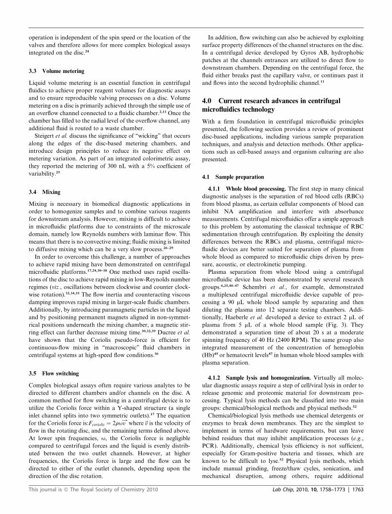
tionally, Haeberle et al. developed a device to extract 2 mL of
plasma from 5 mL of a whole blood sample (Fig. 3). They
demonstrated a separation time of about 20 s at a moderate
spinning frequency of 40 Hz (2400 RPM). The same group also
integrated measurement of the concentration of hemoglobin
(Hb)45 or hematocrit levels47 in human whole blood samples with
plasma separation.
4.1.2 Sample lysis and homogenization. Virtually all molec-
ular diagnostic assays require a step of cell/viral lysis in order to
release genomic and proteomic material for downstream pro-
cessing. Typical lysis methods can be classified into two main
groups: chemical/biological methods and physical methods.52
Chemical/biological lysis methods use chemical detergents or
enzymes to break down membranes. They are the simplest to
implement in terms of hardware requirements, but can leave
behind residues that may inhibit amplification processes (e.g.,
PCR). Additionally, chemical lysis efficiency is not sufficient,
especially for Gram-positive bacteria and tissues, which are
known to be difficult to lyse.52 Physical lysis methods, which
include manual grinding, freeze/thaw cycles, sonication, and
mechanical disruption, among others, require additional
Lab Chip, 2010, 10, 1758–1773 | 1763
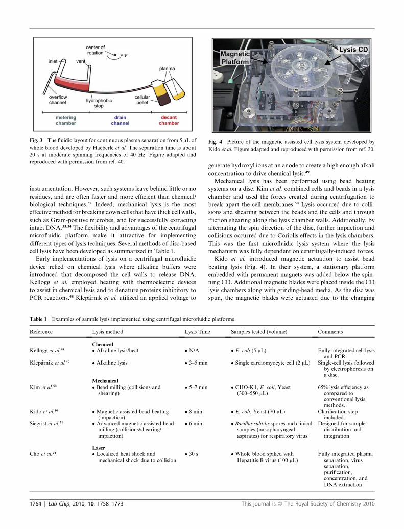
Fig. 4 Picture of the magnetic assisted cell lysis system developed by
Kido et al. Figure adapted and reproduced with permission from ref. 30.
Fig. 3 The fluidic layout for continuous plasma separation from 5 mL of
whole blood developed by Haeberle et al. The separation time is about
20 s at moderate spinning frequencies of 40 Hz. Figure adapted and
reproduced with permission from ref. 40.
instrumentation. However, such systems leave behind little or no
residues, and are often faster and more efficient than chemical/
biological techniques.52 Indeed, mechanical lysis is the most
effective method for breaking down cells that have thick cell walls,
such as Gram-positive microbes, and for successfully extracting
intact DNA.53,54 The flexibility and advantages of the centrifugal
microfluidic platform make it attractive for implementing
different types of lysis techniques. Several methods of disc-based
cell lysis have been developed as summarized in Table 1.
Early implementations of lysis on a centrifugal microfluidic
device relied on chemical lysis where alkaline buffers were
introduced that decomposed the cell walls to release DNA.
Kellogg et al. employed heating with thermoelectric devices
to assist in chemical lysis and to denature proteins inhibitory to
PCR reactions.48 Klep�arnik et al. utilized an applied voltage to
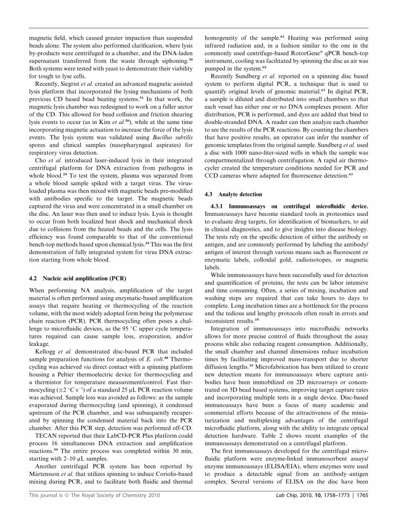
Table 1 Examples of sample lysis implemented using centrifugal microfluidi
Reference Lysis method Lysis Tim
ChemicalKellogg et al.48 � Alkaline lysis/heat � N/A
Klep�arnik et al.49 � Alkaline lysis � 3–5 min
MechanicalKim et al.50 � Bead milling (collisions and
shearing)� 5–7 min
Kido et al.30 �Magnetic assisted bead beating(impaction)
� 8 min
Siegrist et al.51 � Advanced magnetic assisted beadmilling (collisions/shearing/impaction)
� 6 min
LaserCho et al.24 � Localized heat shock and
mechanical shock due to collision� 30 s
1764 | Lab Chip, 2010, 10, 1758–1773
generate hydroxyl ions at an anode to create a high enough alkali
concentration to drive chemical lysis.49
Mechanical lysis has been performed using bead beating
systems on a disc. Kim et al. combined cells and beads in a lysis
chamber and used the forces created during centrifugation to
break apart the cell membranes.50 Lysis occurred due to colli-
sions and shearing between the beads and the cells and through
friction shearing along the lysis chamber walls. Additionally, by
alternating the spin direction of the disc, further impaction and
collisions occurred due to Coriolis effects in the lysis chambers.
This was the first microfluidic lysis system where the lysis
mechanism was fully dependent on centrifugally-induced forces.
Kido et al. introduced magnetic actuation to assist bead
beating lysis (Fig. 4). In their system, a stationary platform
embedded with permanent magnets was added below the spin-
ning CD. Additional magnetic blades were placed inside the CD
lysis chambers along with grinding-bead media. As the disc was
spun, the magnetic blades were actuated due to the changing
c platforms
e Samples tested (volume) Comments
� E. coli (5 mL) Fully integrated cell lysisand PCR.
� Single cardiomyocyte cell (2 mL) Single-cell lysis followedby electrophoresis ona disc.
� CHO-K1, E. coli, Yeast(300–550 mL)
65% lysis efficiency ascompared toconventional lysismethods.
� E. coli, Yeast (70 mL) Clarification stepincluded.
�Bacillus subtilis spores and clinicalsamples (nasopharyngealaspirates) for respiratory virus
Designed for sampledistribution andintegration
�Whole blood spiked withHepatitis B virus (100 mL)
Fully integrated plasmaseparation, virusseparation,purification,concentration, andDNA extraction
This journal is ª The Royal Society of Chemistry 2010
magnetic field, which caused greater impaction than suspended
beads alone. The system also performed clarification, where lysis
by-products were centrifuged in a chamber, and the DNA-laden
supernatant transferred from the waste through siphoning.30
Both systems were tested with yeast to demonstrate their viability
for tough to lyse cells.
Recently, Siegrist et al. created an advanced magnetic assisted
lysis platform that incorporated the lysing mechanisms of both
previous CD based bead beating systems.51 In that work, the
magnetic lysis chamber was redesigned to work on a fuller sector
of the CD. This allowed for bead collision and friction shearing
lysis events to occur (as in Kim et al.50), while at the same time
incorporating magnetic actuation to increase the force of the lysis
events. The lysis system was validated using Bacillus subtilis
spores and clinical samples (nasopharyngeal aspirates) for
respiratory virus detection.
Cho et al. introduced laser-induced lysis in their integrated
centrifugal platform for DNA extraction from pathogens in
whole blood.24 To test the system, plasma was separated from
a whole blood sample spiked with a target virus. The virus-
loaded plasma was then mixed with magnetic beads pre-modified
with antibodies specific to the target. The magnetic beads
captured the virus and were concentrated in a small chamber on
the disc. An laser was then used to induce lysis. Lysis is thought
to occur from both localized heat shock and mechanical shock
due to collisions from the heated beads and the cells. The lysis
efficiency was found comparable to that of the conventional
bench-top methods based upon chemical lysis.24 This was the first
demonstration of fully integrated system for virus DNA extrac-
tion starting from whole blood.
4.2 Nucleic acid amplification (PCR)
When performing NA analysis, amplification of the target
material is often performed using enzymatic-based amplification
assays that require heating or thermocycling of the reaction
volume, with the most widely adopted form being the polymerase
chain reaction (PCR). PCR thermocycling often poses a chal-
lenge to microfluidic devices, as the 95 �C upper cycle tempera-
tures required can cause sample loss, evaporation, and/or
leakage.
Kellogg et al. demonstrated disc-based PCR that included
sample preparation functions for analysis of E. coli.48 Thermo-
cycling was achieved via direct contact with a spinning platform
housing a Peltier thermoelectric device for thermocycling and
a thermistor for temperature measurement/control. Fast ther-
mocycling (�2 �C s�1) of a standard 25 mL PCR reaction volume
was achieved. Sample loss was avoided as follows: as the sample
evaporated during thermocycling (and spinning), it condensed
upstream of the PCR chamber, and was subsequently recuper-
ated by spinning the condensed material back into the PCR
chamber. After this PCR step, detection was performed off-CD.
TECAN reported that their LabCD-PCR Plus platform could
process 16 simultaneous DNA extraction and amplification
reactions.59 The entire process was completed within 30 min,
starting with 2–10 mL samples.
Another centrifugal PCR system has been reported by
M�artensson et al. that utilizes spinning to induce Coriolis-based
mixing during PCR, and to facilitate both fluidic and thermal
This journal is ª The Royal Society of Chemistry 2010
homogeneity of the sample.61 Heating was performed using
infrared radiation and, in a fashion similar to the one in the
commonly used centrifuge-based RotorGene� qPCR bench-top
instrument, cooling was facilitated by spinning the disc as air was
pumped in the system.62
Recently Sundberg et al. reported on a spinning disc based
system to perform digital PCR, a technique that is used to
quantify original levels of genomic material.63 In digital PCR,
a sample is diluted and distributed into small chambers so that
each vessel has either one or no DNA complexes present. After
distribution, PCR is performed, and dyes are added that bind to
double-stranded DNA. A reader can then analyze each chamber
to see the results of the PCR reactions. By counting the chambers
that have positive results, an operator can infer the number of
genomic templates from the original sample. Sundberg et al. used
a disc with 1000 nano-liter-sized wells in which the sample was
compartmentalized through centrifugation. A rapid air thermo-
cycler created the temperature conditions needed for PCR and
CCD cameras where adapted for fluorescence detection.63
4.3 Analyte detection
4.3.1 Immunoassays on centrifugal microfluidic device.
Immunoassays have become standard tools in proteomics used
to evaluate drug targets, for identification of biomarkers, to aid
in clinical diagnostics, and to give insights into disease biology.
The tests rely on the specific detection of either the antibody or
antigen, and are commonly performed by labeling the antibody/
antigen of interest through various means such as fluorescent or
enzymatic labels, colloidal gold, radioisotopes, or magnetic
labels.
While immunoassays have been successfully used for detection
and quantification of proteins, the tests can be labor intensive
and time consuming. Often, a series of mixing, incubation and
washing steps are required that can take hours to days to
complete. Long incubation times are a bottleneck for the process
and the tedious and lengthy protocols often result in errors and
inconsistent results.19
Integration of immunoassays into microfluidic networks
allows for more precise control of fluids throughout the assay
process while also reducing reagent consumption. Additionally,
the small chamber and channel dimensions reduce incubation
times by facilitating improved mass-transport due to shorter
diffusion lengths.19 Microfabrication has been utilized to create
new detection means for immunoassays where capture anti-
bodies have been immobilized on 2D microarrays or concen-
trated on 3D bead based systems, improving target capture rates
and incorporating multiple tests in a single device. Disc-based
immunoassays have been a focus of many academic and
commercial efforts because of the attractiveness of the minia-
turization and multiplexing advantages of the centrifugal
microfluidic platform, along with the ability to integrate optical
detection hardware. Table 2 shows recent examples of the
immunoassays demonstrated on a centrifugal platform.
The first immunoassays developed for the centrifugal micro-
fluidic platform were enzyme-linked immunosorbent assays/
enzyme immunoassays (ELISA/EIA), where enzymes were used
to produce a detectable signal from an antibody–antigen
complex. Several versions of ELISA on the disc have been
Lab Chip, 2010, 10, 1758–1773 | 1765
Ta
ble
2E
xam
ple
so
fim
mu
no
ass
ays
imp
lem
ente
du
sin
gce
ntr
ifu
gal
mic
rofl
uid
icp
latf
orm
s
Ref
eren
ceIn
teg
rate
dw
ith
Sa
mp
leP
rep
?T
arg
etS
am
ple
Ty
pe/
Vo
lum
eA
ssa
yT
ime
Lim
it-o
f-D
etec
tio
nD
etec
tio
nM
eth
od
Bin
din
gS
urf
ace
La
iet
al.
19
Yes
�R
at
IgG
�1
0m
L�
<1
h�
5m
gL�
1�
En
zym
ati
c2
D�
PM
MA
Zh
ao
eta
l.1
2N
o�
Ra
tIg
G�
N/A
�N
/A�
10
0n
gL�
1�
La
bel
-Fre
e�
Ta
2O
5/S
iO2
�M
ou
seIg
GM
ora
iset
al.
55
No
�C
hlo
rpy
rip
ho
s�
N/A
�6
0m
in�
0.0
2–
0.6
2m
gL�
1�
En
zym
ati
c�
Po
lyca
rbo
na
te�
Met
ola
chlo
r�
2,4
,5-T
PH
on
da
eta
l.5
6Y
es�
AF
P�
20
0n
L�
50
min
for
10
4p
ara
llel
react
ion
s�
0.1
5p
mo
lL�
1�
Flu
oro
ph
ore
3D
�B
ead
colu
mn
s:p
oly
styre
ne,
sili
ca,
sep
ha
rose
�IL
-6�
1.2
5p
mo
lL�
1
�C
EA
�1
.31
pm
ol
L�
1
Rie
gger
eta
l.5
7Y
es�
Hep
ati
tis
A�
Hu
ma
nse
rum
�N
/A�
21
5m
IUm
L�
1�
Flu
oro
ph
ore
�P
oly
sty
ren
eb
ead
s�
Tet
an
us
�2
0m
L�
15
8m
IUm
L�
1
Na
ga
iet
al.
58
No
�S
ecre
tory
IgA
�1
mL
�3
0m
in�
N/A
�E
nzy
mati
c�
Gla
ssb
ead
sL
eeet
al.
42
Yes
�H
BsA
g�
Hu
ma
nb
loo
d�
30
min
�0
.51
ng
mL�
1�
En
zym
ati
c�
Po
lyst
yre
ne
bea
ds
�A
nti
-HB
s�
15
0m
L�
8.6
mIU
mL�
1
1766 | Lab Chip, 2010, 10, 1758–1773
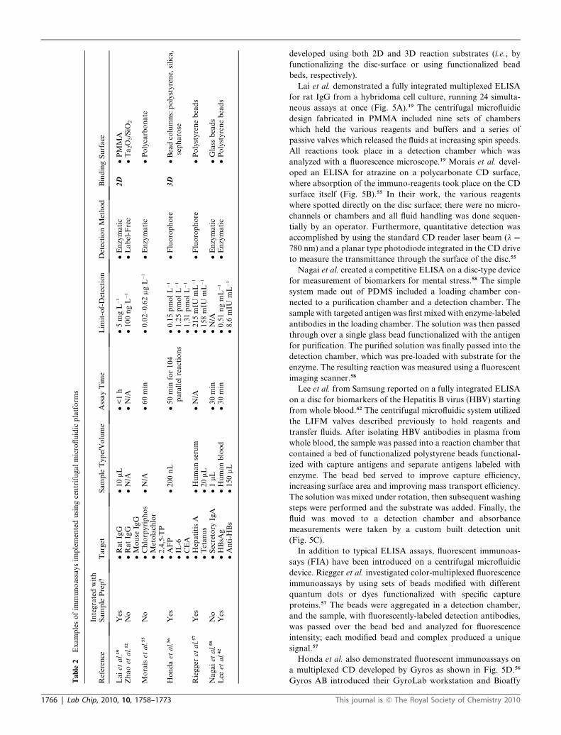
developed using both 2D and 3D reaction substrates (i.e., by
functionalizing the disc-surface or using functionalized bead
beds, respectively).
Lai et al. demonstrated a fully integrated multiplexed ELISA
for rat IgG from a hybridoma cell culture, running 24 simulta-
neous assays at once (Fig. 5A).19 The centrifugal microfluidic
design fabricated in PMMA included nine sets of chambers
which held the various reagents and buffers and a series of
passive valves which released the fluids at increasing spin speeds.
All reactions took place in a detection chamber which was
analyzed with a fluorescence microscope.19 Morais et al. devel-
oped an ELISA for atrazine on a polycarbonate CD surface,
where absorption of the immuno-reagents took place on the CD
surface itself (Fig. 5B).55 In their work, the various reagents
where spotted directly on the disc surface; there were no micro-
channels or chambers and all fluid handling was done sequen-
tially by an operator. Furthermore, quantitative detection was
accomplished by using the standard CD reader laser beam (l ¼780 nm) and a planar type photodiode integrated in the CD drive
to measure the transmittance through the surface of the disc.55
Nagai et al. created a competitive ELISA on a disc-type device
for measurement of biomarkers for mental stress.58 The simple
system made out of PDMS included a loading chamber con-
nected to a purification chamber and a detection chamber. The
sample with targeted antigen was first mixed with enzyme-labeled
antibodies in the loading chamber. The solution was then passed
through over a single glass bead functionalized with the antigen
for purification. The purified solution was finally passed into the
detection chamber, which was pre-loaded with substrate for the
enzyme. The resulting reaction was measured using a fluorescent
imaging scanner.58
Lee et al. from Samsung reported on a fully integrated ELISA
on a disc for biomarkers of the Hepatitis B virus (HBV) starting
from whole blood.42 The centrifugal microfluidic system utilized
the LIFM valves described previously to hold reagents and
transfer fluids. After isolating HBV antibodies in plasma from
whole blood, the sample was passed into a reaction chamber that
contained a bed of functionalized polystyrene beads functional-
ized with capture antigens and separate antigens labeled with
enzyme. The bead bed served to improve capture efficiency,
increasing surface area and improving mass transport efficiency.
The solution was mixed under rotation, then subsequent washing
steps were performed and the substrate was added. Finally, the
fluid was moved to a detection chamber and absorbance
measurements were taken by a custom built detection unit
(Fig. 5C).
In addition to typical ELISA assays, fluorescent immunoas-
says (FIA) have been introduced on a centrifugal microfluidic
device. Riegger et al. investigated color-multiplexed fluorescence
immunoassays by using sets of beads modified with different
quantum dots or dyes functionalized with specific capture
proteins.57 The beads were aggregated in a detection chamber,
and the sample, with fluorescently-labeled detection antibodies,
was passed over the bead bed and analyzed for fluorescence
intensity; each modified bead and complex produced a unique
signal.57
Honda et al. also demonstrated fluorescent immunoassays on
a multiplexed CD developed by Gyros as shown in Fig. 5D.56
Gyros AB introduced their GyroLab workstation and Bioaffy
This journal is ª The Royal Society of Chemistry 2010
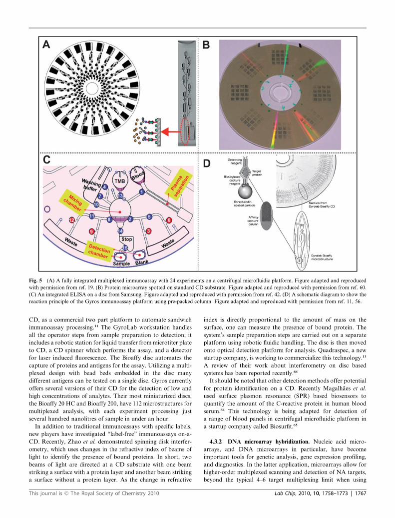
Fig. 5 (A) A fully integrated multiplexed immunoassay with 24 experiments on a centrifugal microfluidic platform. Figure adapted and reproduced
with permission from ref. 19. (B) Protein microarray spotted on standard CD substrate. Figure adapted and reproduced with permission from ref. 60.
(C) An integrated ELISA on a disc from Samsung. Figure adapted and reproduced with permission from ref. 42. (D) A schematic diagram to show the
reaction principle of the Gyros immunoassay platform using pre-packed column. Figure adapted and reproduced with permission from ref. 11, 56.
CD, as a commercial two part platform to automate sandwich
immunoassay processing.11 The GyroLab workstation handles
all the operator steps from sample preparation to detection; it
includes a robotic station for liquid transfer from microtiter plate
to CD, a CD spinner which performs the assay, and a detector
for laser induced fluorescence. The Bioaffy disc automates the
capture of proteins and antigens for the assay. Utilizing a multi-
plexed design with bead beds embedded in the disc many
different antigens can be tested on a single disc. Gyros currently
offers several versions of their CD for the detection of low and
high concentrations of analytes. Their most miniaturized discs,
the Bioaffy 20 HC and Bioaffy 200, have 112 microstructures for
multiplexed analysis, with each experiment processing just
several hundred nanolitres of sample in under an hour.
In addition to traditional immunoassays with specific labels,
new players have investigated ‘‘label-free’’ immunoassays on-a-
CD. Recently, Zhao et al. demonstrated spinning disk interfer-
ometry, which uses changes in the refractive index of beams of
light to identify the presence of bound proteins. In short, two
beams of light are directed at a CD substrate with one beam
striking a surface with a protein layer and another beam striking
a surface without a protein layer. As the change in refractive
This journal is ª The Royal Society of Chemistry 2010
index is directly proportional to the amount of mass on the
surface, one can measure the presence of bound protein. The
system’s sample preparation steps are carried out on a separate
platform using robotic fluidic handling. The disc is then moved
onto optical detection platform for analysis. Quadraspec, a new
startup company, is working to commercialize this technology.13
A review of their work about interferometry on disc based
systems has been reported recently.64
It should be noted that other detection methods offer potential
for protein identification on a CD. Recently Magalhaes et al.
used surface plasmon resonance (SPR) based biosensors to
quantify the amount of the C-reactive protein in human blood
serum.64 This technology is being adapted for detection of
a range of blood panels in centrifugal microfluidic platform in
a startup company called Biosurfit.65
4.3.2 DNA microarray hybridization. Nucleic acid micro-
arrays, and DNA microarrays in particular, have become
important tools for genetic analysis, gene expression profiling,
and diagnostics. In the latter application, microarrays allow for
higher-order multiplexed scanning and detection of NA targets,
beyond the typical 4–6 target multiplexing limit when using
Lab Chip, 2010, 10, 1758–1773 | 1767
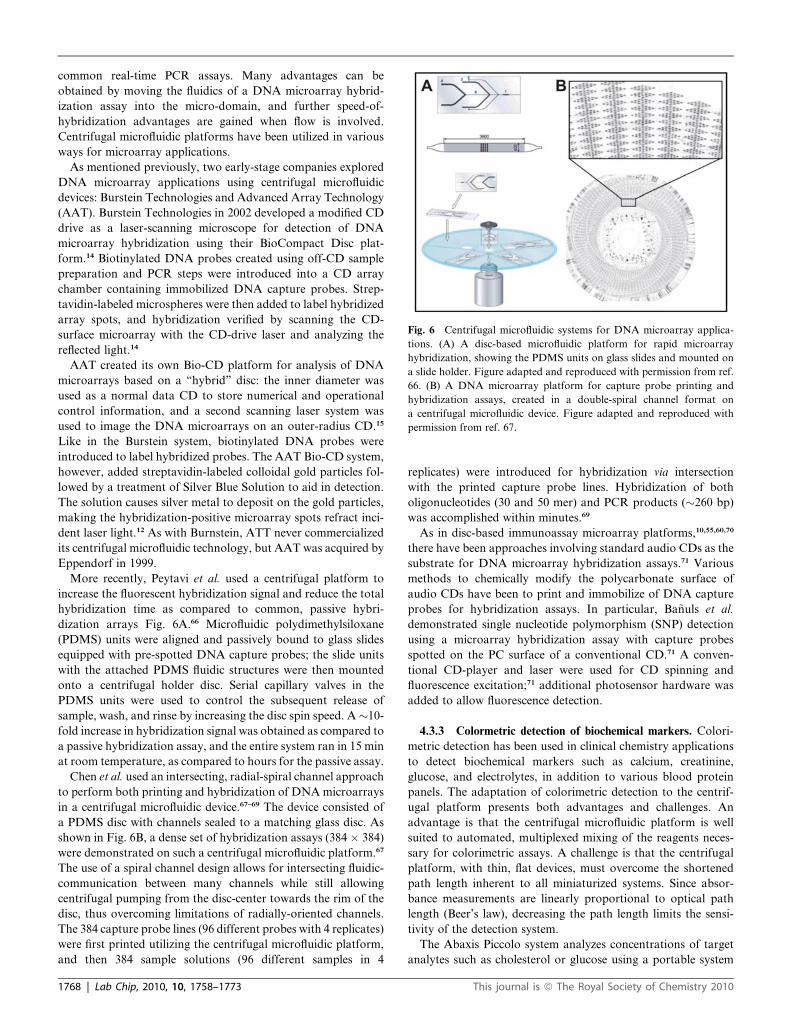
Fig. 6 Centrifugal microfluidic systems for DNA microarray applica-
tions. (A) A disc-based microfluidic platform for rapid microarray
hybridization, showing the PDMS units on glass slides and mounted on
a slide holder. Figure adapted and reproduced with permission from ref.
66. (B) A DNA microarray platform for capture probe printing and
hybridization assays, created in a double-spiral channel format on
a centrifugal microfluidic device. Figure adapted and reproduced with
permission from ref. 67.
common real-time PCR assays. Many advantages can be
obtained by moving the fluidics of a DNA microarray hybrid-
ization assay into the micro-domain, and further speed-of-
hybridization advantages are gained when flow is involved.
Centrifugal microfluidic platforms have been utilized in various
ways for microarray applications.
As mentioned previously, two early-stage companies explored
DNA microarray applications using centrifugal microfluidic
devices: Burstein Technologies and Advanced Array Technology
(AAT). Burstein Technologies in 2002 developed a modified CD
drive as a laser-scanning microscope for detection of DNA
microarray hybridization using their BioCompact Disc plat-
form.14 Biotinylated DNA probes created using off-CD sample
preparation and PCR steps were introduced into a CD array
chamber containing immobilized DNA capture probes. Strep-
tavidin-labeled microspheres were then added to label hybridized
array spots, and hybridization verified by scanning the CD-
surface microarray with the CD-drive laser and analyzing the
reflected light.14
AAT created its own Bio-CD platform for analysis of DNA
microarrays based on a ‘‘hybrid’’ disc: the inner diameter was
used as a normal data CD to store numerical and operational
control information, and a second scanning laser system was
used to image the DNA microarrays on an outer-radius CD.15
Like in the Burstein system, biotinylated DNA probes were
introduced to label hybridized probes. The AAT Bio-CD system,
however, added streptavidin-labeled colloidal gold particles fol-
lowed by a treatment of Silver Blue Solution to aid in detection.
The solution causes silver metal to deposit on the gold particles,
making the hybridization-positive microarray spots refract inci-
dent laser light.12 As with Burnstein, ATT never commercialized
its centrifugal microfluidic technology, but AAT was acquired by
Eppendorf in 1999.
More recently, Peytavi et al. used a centrifugal platform to
increase the fluorescent hybridization signal and reduce the total
hybridization time as compared to common, passive hybri-
dization arrays Fig. 6A.66 Microfluidic polydimethylsiloxane
(PDMS) units were aligned and passively bound to glass slides
equipped with pre-spotted DNA capture probes; the slide units
with the attached PDMS fluidic structures were then mounted
onto a centrifugal holder disc. Serial capillary valves in the
PDMS units were used to control the subsequent release of
sample, wash, and rinse by increasing the disc spin speed. A�10-
fold increase in hybridization signal was obtained as compared to
a passive hybridization assay, and the entire system ran in 15 min
at room temperature, as compared to hours for the passive assay.
Chen et al. used an intersecting, radial-spiral channel approach
to perform both printing and hybridization of DNA microarrays
in a centrifugal microfluidic device.67–69 The device consisted of
a PDMS disc with channels sealed to a matching glass disc. As
shown in Fig. 6B, a dense set of hybridization assays (384 � 384)
were demonstrated on such a centrifugal microfluidic platform.67
The use of a spiral channel design allows for intersecting fluidic-
communication between many channels while still allowing
centrifugal pumping from the disc-center towards the rim of the
disc, thus overcoming limitations of radially-oriented channels.
The 384 capture probe lines (96 different probes with 4 replicates)
were first printed utilizing the centrifugal microfluidic platform,
and then 384 sample solutions (96 different samples in 4
1768 | Lab Chip, 2010, 10, 1758–1773
replicates) were introduced for hybridization via intersection
with the printed capture probe lines. Hybridization of both
oligonucleotides (30 and 50 mer) and PCR products (�260 bp)
was accomplished within minutes.69
As in disc-based immunoassay microarray platforms,10,55,60,70
there have been approaches involving standard audio CDs as the
substrate for DNA microarray hybridization assays.71 Various
methods to chemically modify the polycarbonate surface of
audio CDs have been to print and immobilize of DNA capture
probes for hybridization assays. In particular, Ba~nuls et al.
demonstrated single nucleotide polymorphism (SNP) detection
using a microarray hybridization assay with capture probes
spotted on the PC surface of a conventional CD.71 A conven-
tional CD-player and laser were used for CD spinning and
fluorescence excitation;71 additional photosensor hardware was
added to allow fluorescence detection.
4.3.3 Colormetric detection of biochemical markers. Colori-
metric detection has been used in clinical chemistry applications
to detect biochemical markers such as calcium, creatinine,
glucose, and electrolytes, in addition to various blood protein
panels. The adaptation of colorimetric detection to the centrif-
ugal platform presents both advantages and challenges. An
advantage is that the centrifugal microfluidic platform is well
suited to automated, multiplexed mixing of the reagents neces-
sary for colorimetric assays. A challenge is that the centrifugal
platform, with thin, flat devices, must overcome the shortened
path length inherent to all miniaturized systems. Since absor-
bance measurements are linearly proportional to optical path
length (Beer’s law), decreasing the path length limits the sensi-
tivity of the detection system.
The Abaxis Piccolo system analyzes concentrations of target
analytes such as cholesterol or glucose using a portable system
This journal is ª The Royal Society of Chemistry 2010
equipped with absorbance measurement hardware.6,41 Ducree
et al. have demonstrated fully-integrated colorimetric assays for
the determination of alcohol25 or glucose43 concentration in
whole blood. In contrast to the perpendicular style detection
cuvettes on the centrifugal platform developed by Abaxis, the
absorbance was measured in this case using a horizontal type
detection chamber in which the absorbance path length is 10
times longer, and thus the colorimetric detection sensitivity was
enhanced. The total process from whole blood sample input to
alcohol detection can be completed in less than 3 min.
4.4 Other applications
Beyond molecular-scale applications based on centrifugal
microfluidic platforms, cell and organism-based applications
have been demonstrated as well. Cell-based assays (viz., cell
viability assays) have been carried out for drug discovery appli-
cations using a disc system to reduce the often labor-intensive
operations required for both cell culture and screening.72
A complete bacterial viability assay, based on an off-the-shelf
assay kit (LIVE/DEAD BacLight Bacterial Viability Kit from
Molecular Probes, Inc.) was automated using a centrifugal
microfluidic device to study G-force effects on cells.1
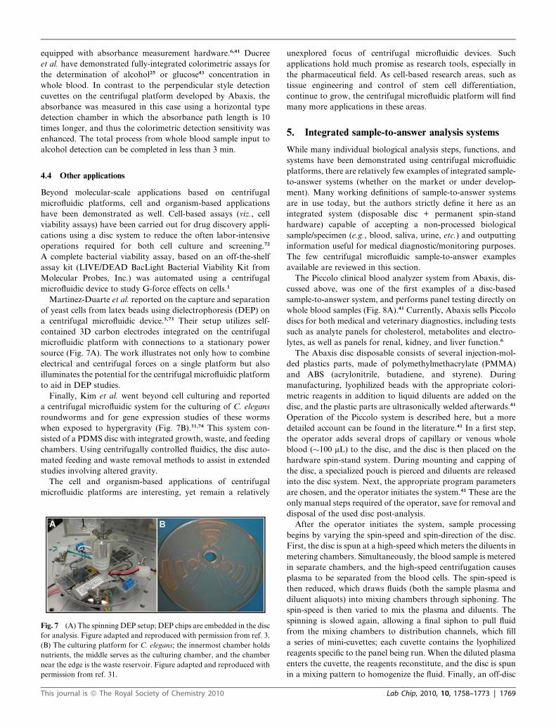
Martinez-Duarte et al. reported on the capture and separation
of yeast cells from latex beads using dielectrophoresis (DEP) on
a centrifugal microfluidic device.3,73 Their setup utilizes self-
contained 3D carbon electrodes integrated on the centrifugal
microfluidic platform with connections to a stationary power
source (Fig. 7A). The work illustrates not only how to combine
electrical and centrifugal forces on a single platform but also
illuminates the potential for the centrifugal microfluidic platform
to aid in DEP studies.
Finally, Kim et al. went beyond cell culturing and reported
a centrifugal microfluidic system for the culturing of C. elegans
roundworms and for gene expression studies of these worms
when exposed to hypergravity (Fig. 7B).31,74 This system con-
sisted of a PDMS disc with integrated growth, waste, and feeding
chambers. Using centrifugally controlled fluidics, the disc auto-
mated feeding and waste removal methods to assist in extended
studies involving altered gravity.
The cell and organism-based applications of centrifugal
microfluidic platforms are interesting, yet remain a relatively
Fig. 7 (A) The spinning DEP setup; DEP chips are embedded in the disc
for analysis. Figure adapted and reproduced with permission from ref. 3.
(B) The culturing platform for C. elegans; the innermost chamber holds
nutrients, the middle serves as the culturing chamber, and the chamber
near the edge is the waste reservoir. Figure adapted and reproduced with
permission from ref. 31.
This journal is ª The Royal Society of Chemistry 2010
unexplored focus of centrifugal microfluidic devices. Such
applications hold much promise as research tools, especially in
the pharmaceutical field. As cell-based research areas, such as
tissue engineering and control of stem cell differentiation,
continue to grow, the centrifugal microfluidic platform will find
many more applications in these areas.
5. Integrated sample-to-answer analysis systems
While many individual biological analysis steps, functions, and
systems have been demonstrated using centrifugal microfluidic
platforms, there are relatively few examples of integrated sample-
to-answer systems (whether on the market or under develop-
ment). Many working definitions of sample-to-answer systems
are in use today, but the authors strictly define it here as an
integrated system (disposable disc + permanent spin-stand
hardware) capable of accepting a non-processed biological
sample/specimen (e.g., blood, saliva, urine, etc.) and outputting
information useful for medical diagnostic/monitoring purposes.
The few centrifugal microfluidic sample-to-answer examples
available are reviewed in this section.
The Piccolo clinical blood analyzer system from Abaxis, dis-
cussed above, was one of the first examples of a disc-based
sample-to-answer system, and performs panel testing directly on
whole blood samples (Fig. 8A).41 Currently, Abaxis sells Piccolo
discs for both medical and veterinary diagnostics, including tests
such as analyte panels for cholesterol, metabolites and electro-
lytes, as well as panels for renal, kidney, and liver function.6
The Abaxis disc disposable consists of several injection-mol-
ded plastics parts, made of polymethylmethacrylate (PMMA)
and ABS (acrylonitrile, butadiene, and styrene). During
manufacturing, lyophilized beads with the appropriate colori-
metric reagents in addition to liquid diluents are added on the
disc, and the plastic parts are ultrasonically welded afterwards.41
Operation of the Piccolo system is described here, but a more
detailed account can be found in the literature.41 In a first step,
the operator adds several drops of capillary or venous whole
blood (�100 mL) to the disc, and the disc is then placed on the
hardware spin-stand system. During mounting and capping of
the disc, a specialized pouch is pierced and diluents are released
into the disc system. Next, the appropriate program parameters
are chosen, and the operator initiates the system.41 These are the
only manual steps required of the operator, save for removal and
disposal of the used disc post-analysis.
After the operator initiates the system, sample processing
begins by varying the spin-speed and spin-direction of the disc.
First, the disc is spun at a high-speed which meters the diluents in
metering chambers. Simultaneously, the blood sample is metered
in separate chambers, and the high-speed centrifugation causes
plasma to be separated from the blood cells. The spin-speed is
then reduced, which draws fluids (both the sample plasma and
diluent aliquots) into mixing chambers through siphoning. The
spin-speed is then varied to mix the plasma and diluents. The
spinning is slowed again, allowing a final siphon to pull fluid
from the mixing chambers to distribution channels, which fill
a series of mini-cuvettes; each cuvette contains the lyophilized
reagents specific to the panel being run. When the diluted plasma
enters the cuvette, the reagents reconstitute, and the disc is spun
in a mixing pattern to homogenize the fluid. Finally, an off-disc
Lab Chip, 2010, 10, 1758–1773 | 1769
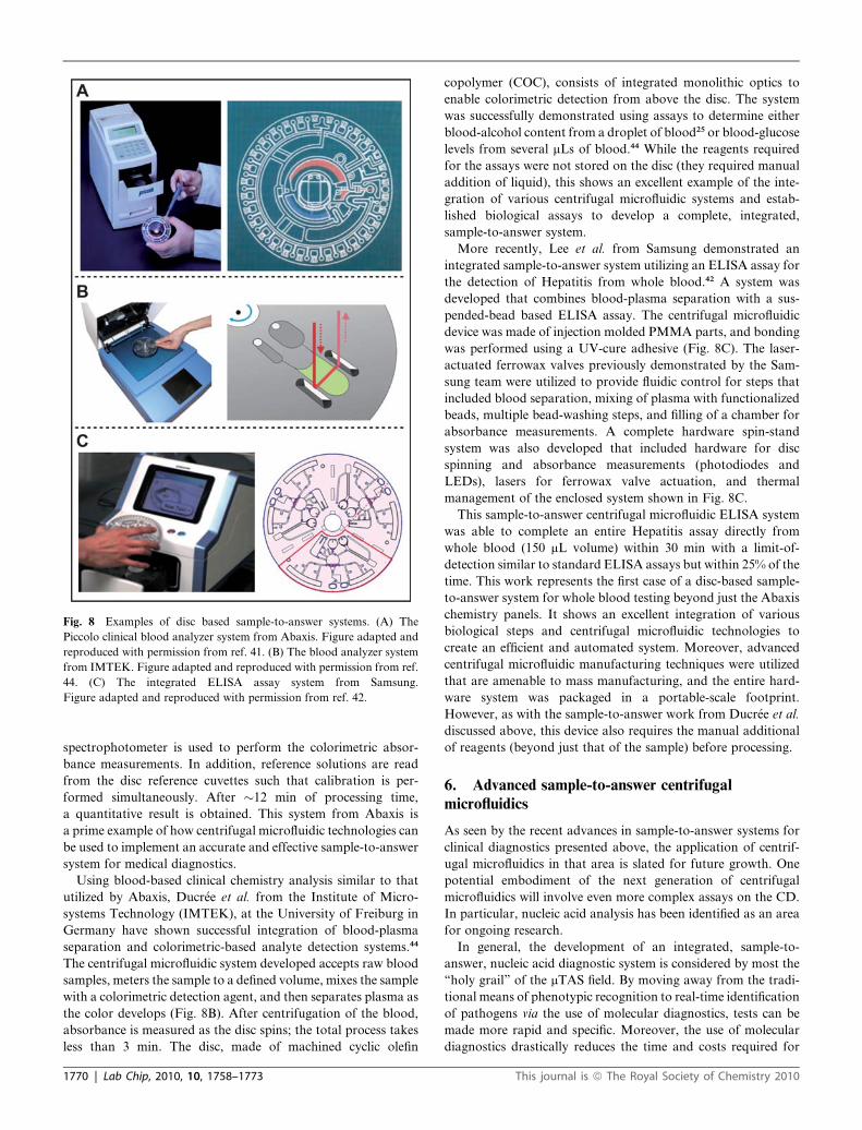
Fig. 8 Examples of disc based sample-to-answer systems. (A) The
Piccolo clinical blood analyzer system from Abaxis. Figure adapted and
reproduced with permission from ref. 41. (B) The blood analyzer system
from IMTEK. Figure adapted and reproduced with permission from ref.
44. (C) The integrated ELISA assay system from Samsung.
Figure adapted and reproduced with permission from ref. 42.
spectrophotometer is used to perform the colorimetric absor-
bance measurements. In addition, reference solutions are read
from the disc reference cuvettes such that calibration is per-
formed simultaneously. After �12 min of processing time,
a quantitative result is obtained. This system from Abaxis is
a prime example of how centrifugal microfluidic technologies can
be used to implement an accurate and effective sample-to-answer
system for medical diagnostics.
Using blood-based clinical chemistry analysis similar to that
utilized by Abaxis, Ducr�ee et al. from the Institute of Micro-
systems Technology (IMTEK), at the University of Freiburg in
Germany have shown successful integration of blood-plasma
separation and colorimetric-based analyte detection systems.44
The centrifugal microfluidic system developed accepts raw blood
samples, meters the sample to a defined volume, mixes the sample
with a colorimetric detection agent, and then separates plasma as
the color develops (Fig. 8B). After centrifugation of the blood,
absorbance is measured as the disc spins; the total process takes
less than 3 min. The disc, made of machined cyclic olefin
1770 | Lab Chip, 2010, 10, 1758–1773
copolymer (COC), consists of integrated monolithic optics to
enable colorimetric detection from above the disc. The system
was successfully demonstrated using assays to determine either
blood-alcohol content from a droplet of blood25 or blood-glucose
levels from several mLs of blood.44 While the reagents required
for the assays were not stored on the disc (they required manual
addition of liquid), this shows an excellent example of the inte-
gration of various centrifugal microfluidic systems and estab-
lished biological assays to develop a complete, integrated,
sample-to-answer system.
More recently, Lee et al. from Samsung demonstrated an
integrated sample-to-answer system utilizing an ELISA assay for
the detection of Hepatitis from whole blood.42 A system was
developed that combines blood-plasma separation with a sus-
pended-bead based ELISA assay. The centrifugal microfluidic
device was made of injection molded PMMA parts, and bonding
was performed using a UV-cure adhesive (Fig. 8C). The laser-
actuated ferrowax valves previously demonstrated by the Sam-
sung team were utilized to provide fluidic control for steps that
included blood separation, mixing of plasma with functionalized
beads, multiple bead-washing steps, and filling of a chamber for
absorbance measurements. A complete hardware spin-stand
system was also developed that included hardware for disc
spinning and absorbance measurements (photodiodes and
LEDs), lasers for ferrowax valve actuation, and thermal
management of the enclosed system shown in Fig. 8C.
This sample-to-answer centrifugal microfluidic ELISA system
was able to complete an entire Hepatitis assay directly from
whole blood (150 mL volume) within 30 min with a limit-of-
detection similar to standard ELISA assays but within 25% of the
time. This work represents the first case of a disc-based sample-
to-answer system for whole blood testing beyond just the Abaxis
chemistry panels. It shows an excellent integration of various
biological steps and centrifugal microfluidic technologies to
create an efficient and automated system. Moreover, advanced
centrifugal microfluidic manufacturing techniques were utilized
that are amenable to mass manufacturing, and the entire hard-
ware system was packaged in a portable-scale footprint.
However, as with the sample-to-answer work from Ducr�ee et al.
discussed above, this device also requires the manual additional
of reagents (beyond just that of the sample) before processing.
6. Advanced sample-to-answer centrifugalmicrofluidics
As seen by the recent advances in sample-to-answer systems for
clinical diagnostics presented above, the application of centrif-
ugal microfluidics in that area is slated for future growth. One
potential embodiment of the next generation of centrifugal
microfluidics will involve even more complex assays on the CD.
In particular, nucleic acid analysis has been identified as an area
for ongoing research.
In general, the development of an integrated, sample-to-
answer, nucleic acid diagnostic system is considered by most the
‘‘holy grail’’ of the mTAS field. By moving away from the tradi-
tional means of phenotypic recognition to real-time identification
of pathogens via the use of molecular diagnostics, tests can be
made more rapid and specific. Moreover, the use of molecular
diagnostics drastically reduces the time and costs required for
This journal is ª The Royal Society of Chemistry 2010
a diagnosis (from days to hours) while greatly increasing the
specificity and accuracy.
While individual process steps towards nucleic acid analysis
and diagnostics have been demonstrated on centrifugal micro-
fluidic platforms, there remains hurdles to overcome before an
integrated sample-to-answer nucleic acid system can be achieved.
Many of these hurdles are not reserved solely for centrifugal
microfluidic platforms, but are endemic to all microfluidic
systems. However, as discussed previously, the advantages of the
centrifugal microfluidic system make it a promising solution to
bridge the gap in this field.
Typical NA analysis consists of a sample preparation step
(including cellular/viral lysis), NA amplification (e.g., PCR), and
detection (e.g., via DNA/RNA microarray). As discussed above,
each of these systems has been independently demonstrated using
centrifugal microfluidic technology, showing many of the
advantages in bringing the current bench-top methods into the
centrifugal microfluidic domain. The integration of these steps,
however, remains complex.
As with any biological diagnostics process, the main source of
complexity and variability comes from the sample itself (blood,
saliva, mucus, urine, etc.), which can vary drastically in terms of
both fluidic characteristics (e.g., viscosity, density, surface
tension) and biological characteristics (e.g., analyte concentra-
tion, inhibitor concentration). In particular, the initial sample
volume required for NA analysis presents unique challenges. In
blood for example, genomic DNA exists at concentrations
around 106–107 DNA mL�1 and pathogenic DNA at concen-
trations as low as 10–102 DNA mL�1. This means that systems
must work with samples as large as several mLs to obtain the
amount of DNA required for detection. In contrast, for clinical
chemistry and immunoassays applications, sample volumes of
less than mLs are usually sufficient.
The integration of macro and micro systems for fluid manip-
ulation remain another challenge for integrated systems; targets
must be isolated from mL volumes then purified and concen-
trated to mL volumes. Furthermore, different sample types
dictate the necessity for distinctive sample preparation steps.
However, centrifugal microfluidic technology has shown itself to
be very adaptable in the area of sample preparation, thus making
it a leading technology choice for the development of an NA
sample-to-answer system.
Besides the inherent difficulties with sample preparation, there
are problems with reagent handling required for assays in micro-
fluidic systems. Automated handling through isolated reagent
packs and robotics to the disc has been demonstrated, and elimi-
nates the need for reagent storage on the disc; however, this option
drastically increases the complexity and cost of the system and
limits its use in point-of-care settings. Another option is to store
reagents on the disc, either in liquid or solid form (e.g., through
drying, lyophilization). However, long-term storage of reagents
on the disc requires robust liquid and vapor valving. Additionally,
methods of preparing reagents are inherently difficult for
advanced assays. For example, lyophilization can be difficult for
the delicate enzymes and reagents required for NA analysis.
Another hurdle to overcome towards integrated NA diag-
nostic systems is the complex and expensive hardware platform
required for complete analysis. Although it is often presented
that inexpensive disc rotors and optical systems like those found
This journal is ª The Royal Society of Chemistry 2010
in widely available computer drives can be implemented for
centrifugal microfluidic analysis systems, there remains addi-
tional instrumentation requirements for propulsion and detec-
tion. While stronger motors are widely available to spin the discs
at higher speeds required for processes like blood separation or
for bead-beating cell lysis, all current detection systems use some
form of adapted optics. These optical systems, like those required
for fluorescence detection, include light sources, detectors, filters,
and electronics. Moreover, heaters/coolers will be required to
perform thermal control for sensitive steps such as lysis, NA
amplification, and NA hybridization. Finally, the entire system
needs to be packaged to include thermal management systems
and user interface systems. All motor control, analysis instru-
mentation, optical systems and other equipment must be inte-
grated and built in a cost effective way.
In regards to the assay platform itself, many of the centrifugal
microfluidic systems discussed are excellent examples of sepa-
rating the disposable from the permanent hardware system,
which helps to keep the cost-per-test low. Additionally, the
motor allows the disc to shift easily for alignment with off-disc
hardware. This minimizes the need for multiplexed hardware
systems, thus reducing costs and complexities. However, other
issues, such as injection molding and FDA approval for the
devices, remain challenges to development of advanced centrif-
ugal microfluidic systems.
While the key components are in place for a disc-based NA
sample-to-answer system, the complex task of robust and effi-
cient integration of both the disposable disc itself and the hard-
ware platform must be carefully considered before success can be
achieved.
It should be noted that the barriers presented in this section are
not limited to NA analysis. Changing the assay type will change
the sample preparation and analysis steps; however, the consid-
erations outlined above are generic. For example, if disc based
immunoassays will ever be accepted for diagnostics, biological
issues of sample complexity and reagent storage as well as
engineering issues of mass production and instrumentation must
still be dealt with. Additionally, while new detection technolo-
gies, like those based on interferometry or SPR, potentially lessen
the burden on analysis by reducing extensive analysis steps and
decreasing the complexity of analysis equipment, they cannot
eliminate the fundamental sample preparation steps that are
universal to all assays from clinical samples (e.g., isolation of
biomolecule targets from the bulk samples through lysis and
purification and preparation for detection). In summary, the
continued work on individual sample preparation systems for
varied clinical specimens can potentially serve to answer tech-
nological issues for centrifugal based assays in general.
7.0 Conclusions
A review of centrifugal microfluidic technologies has been pre-
sented, with an emphasis on biomedical applications both in
industry and academia. Various centrifugal microfluidic func-
tions such as valving, metering, mixing, and flow-switching were
discussed, in addition to a broad range of applications in which
those components and their combinations were incorporated for
biological applications. As discussed, the centrifugal microfluidic
platform provides many advantages over other microfluidic
Lab Chip, 2010, 10, 1758–1773 | 1771
platforms, making it an excellent candidate to tackle complex
biological analysis problems, particularly in the area of in vitro
diagnostics (IVD).
While many advances in centrifugal microfluidic disc tech-
nology have been made, the full potential of the platform has yet
to be realized. With the exception of the few established
companies like Abaxis and Gyros, the market is relatively bare of
centrifugal microfluidic technologies and is limited to the appli-
cations of blood panel analysis and characterization of anti-
bodies and proteins for drug development. While no direct
leaders have emerged in the disc-based NA diagnostics field,
several prominent groups in industry and academia are working
to develop not only advanced disc-based NA analysis devices,
but true sample-to-answer devices for NA IVDs as well (e.g.,
Madou group, Samsung, IMTEK). Moreover, new areas of
interest like cell-based applications for the centrifugal micro-
fluidic platform will likely emerge as valuable research tools in
the near future. There are many areas where centrifugal micro-
fluidic technology can find new applications, and the areas
already established for centrifugal microfluidic devices will
continue to strengthen. Expanded R&D efforts in both industry
and academia are expected to ‘‘burst’’ this barrier in the near
future and bring the centrifugal microfluidic platform to its full
potential.
Acknowledgements
This work was supported by WCU (World Class University)
program (R32-2008-000-20054-0) and Basic Science Research
Program (52-2009-0014) through the National Research Foun-
dation of Korea funded by the Ministry of Education, Science
and Technology. The authors also wish to thank Samsung
Electronics Co. Ltd., Genome Canada and DARPA MF3 for
support. Finally, thanks to Dr Horacio Kido for insightful
guidance and information.
Notes and references
1 M. Madou, J. Zoval, G. Jia, H. Kido, J. Kim and N. Kim, Annu. Rev.Biomed. Eng., 2006, 8, 601–628.
2 J. M. Park, Y. K. Cho, B. S. Lee, J. G. Lee and C. Ko, Lab Chip, 2007,7, 557–564.
3 R. Martinez-Duarte, R. A. Gorkin III, K. Abi-Samra andM. J. Madou, Lab Chip, 2010, 10, 1030–1043.
4 R. M. Rocco, Landmark Papers in Clinical Chemistry, Elsevier,Amsterdam, 2006.
5 C. A. Burtis, J. C. Mailen, W. F. Johnson, C. D. Scott, T. O. Tiffanyand N. G. Anderson, Clin. Chem., 1972, 18, 753–761.
6 Abaxis Inc. USA, (www.abaxis.com).7 M. Madou and G. Kellogg, in Proc. SPIE Systems and Technologies
for Clinical Diagnostics and Drug Discovery, ed. G. E. Cohn andA. Katzir, San Jose, CA. USA, 1998, pp. 80–93.
8 D. C. Duffy, H. L. Gillis, J. Lin, N. F. Sheppard and G. J. Kellogg,Anal. Chem., 1999, 71, 4669–4678.
9 M. Shea, S. Ommert, L. Gleich, T. Towle, H. Haspel, N. Schmid,H. Jindal, G. Kellogg and B. Carvalho, J. Assoc. Lab. Autom.,2003, 8, 74–77.
10 H. Kido, A. Maquieira and B. D. Hammock, Anal. Chim. Acta, 2000,411, 1–11.
11 Gyros AB, Sweden, www.gyros.com.12 M. Zhao, D. Nolte, W. R. Cho, F. Regnier, M. Varma, G. Lawrence
and J. Pasqua, Clin. Chem., 2006, 52, 2135–2140.13 Quadraspec, USA., www.quadraspec.com.14 R. Barathur, J. Bookout, S. Sreevatsan, J. Gordon, M. Werner,
G. Thor and M. Worthington, Psychiatr. Genet., 2002, 12, 193–206.
1772 | Lab Chip, 2010, 10, 1758–1773
15 I. Alexandre, Y. Houbion, J. Collet, S. Hamels, J. Demarteau,J. L. Gala and J. Remacle, Biotechniques, 2002, 33, 435–436, 438–439.
16 J. V. Zoval and M. J. Madou, Proc. IEEE, 2004, 92, 140–153.17 J. Ducr�ee, S. Haeberle, S. Lutz, S. Pausch, F. v. Stetten and
R. Zengerle, J. Micromech. Microeng., 2007, 17, S103–S115.18 S. Haeberle and R. Zengerle, Lab Chip, 2007, 7, 1094–1110.19 S. Lai, S. Wang, J. Luo, L. J. Lee, S.-T. Yang and M. J. Madou, Anal.
Chem., 2004, 76, 1832–1837.20 R. D. Johnson, I. H. A. Badr, G. Barrett, S. Y. Lai, Y. M. Lu,
M. J. Madou and L. G. Bachas, Anal. Chem., 2001, 73, 3940–3946.21 I. H. A. Badr, R. D. Johnson, M. J. Madou and L. G. Bachas, Anal.
Chem., 2002, 74, 5569–5575.22 G. Ekstrand, C. Holmquist, A. E. Orleforn, B. Hellmann, A. Larsson
and P. Andersson, in Proc. in MicroTAS, ed. A. van den Berg andP. Bergveld, Kluwer Academic Publisher, 2000, pp.311–314.
23 P. Andersson, G. Jesson, G. Kylberg, G. Ekstrand and G. Thorsen,Anal. Chem., 2007, 79, 4022–4030.
24 Y. K. Cho, J. G. Lee, J. M. Park, B. S. Lee, Y. Lee and C. Ko, LabChip, 2007, 7, 565–573.
25 J. Steigert, M. Grumann, T. Brenner, L. Riegger, J. Harter,R. Zengerle and J. Ducr�ee, Lab Chip, 2006, 6, 1040–1044.
26 M. C. R. Shastry, S. D. Luck and H. Roder, Biophys. J., 1998, 74,2714–2721.
27 R. H. Liu, J. Microelectromech. Syst., 2000, 9, 190–197.28 A. Groisman and V. Steinberg, Nature, 2001, 410, 905–908.29 E. Biddiss, D. Erickson and D. Q. Li, 2004, 76, pp. 3208–3213.30 H. Kido, M. Micic, D. Smith, J. Zoval, J. Norton and M. Madou,
Colloids Surf., B, 2007, 58, 44–51.31 N. Kim, C. M. Dempsey, J. V. Zoval, J. Y. Sze and M. J. Madou,
Sens. Actuators, B, 2007, 122, 511–518.32 M. Grumann, A. Geipel, L. Riegger, R. Zengerle and J. Ducree, Lab
Chip, 2005, 5, 560–565.33 Z. Noroozi, H. Kido, M. Micic, H. Pan, C. Bartolome, M. Princevac,
J. Zoval and M. Madou, Rev. Sci. Instrum., 2009, 80, 075102.34 S. Haeberle, T. Brenner, H.-P. Schlosser, R. Zengerle and J. Ducree,
Chem. Eng. Technol., 2005, 28, 613–616.35 J. Steigert, M. Grumann, T. Brenner, K. Mittenb€uhler, T. Nann,
J. R€uhe, I. Moser, S. Haeberle, L. Riegger, J. Riegler, W. Bessler,R. Zengerle and J. Ducr�ee, J. Assoc. Lab. Autom., 2005, 10, 331–341.
36 J. Ducr�ee, S. Haeberle, T. Brenner, T. Glatzel and R. Zengerle,Microfluid. Nanofluid., 2006, 2, 97–105.
37 J. Ducr�ee, T. Brenner, S. Haeberle, T. Glatzel and R. Zengerle,Microfluid. Nanofluid., 2006, 2, 78–84.
38 M. A. Bynum and G. B. Gordon, Anal. Chem., 2004, 76, 7039–7044.
39 T. Brenner, T. Glatzel, R. Zengerle and J. Ducree, Lab Chip, 2005, 5,146–150.
40 S. Haeberle, T. Brenner, R. Zengerle and J. Ducr�ee, Lab Chip, 2006, 6,776–781.
41 G. T. Schembri, T. L. Burd, A. R. Kopf-Sill, L. R. Shea andR. Braynin, J. Autom. Methods Manage. Chem., 1995, 17, 99–104.
42 B. S. Lee, J.-N. Lee, J.-M. Park, J.-G. Lee, S. Kim, Y.-K. Cho andC. Ko, Lab Chip, 2009, 9, 1548–1555.
43 C. T. Schembri, V. Ostoich, P. J. Lingane, T. L. Burd and S. N. Buhl,Clin. Chem., 1992, 38, 1665–1670.
44 M. Grumann, J. Steigert, L. Riegger, I. Moser, B. Enderle,K. Riebeseel, G. Urban, R. Zengerle and J. Ducr�ee, Biomed.Microdevices, 2006, 8, 209–214.
45 J. Steigert, M. Grumann, M. Dube, W. Streule, L. Riegger,T. Brenner, P. Koltay, K. Mittmann, R. Zengerle and J. Ducr�ee,Sens. Actuators, A, 2006, 130–131, 228–233.
46 J. L. Zhang, Q. Q. Guo, M. Liu and J. Yang, J. Micromech.Microeng., 2008, 18, 125025.
47 L. Riegger, M. Grumann, J. Steigert, S. Lutz, C. P. Steinert,C. Mueller, J. Viertel, O. Prucker, J. Ruhe, R. Zengerle andJ. Ducree, Biomed. Microdevices, 2007, 9, 795–799.
48 G. J. Kellogg, T. E. Arnold, B. L. Carvalho, D. C. Duffy andN. F. Sheppard, in Proc. in MicroTAS, ed. A. van den Berg,W. Olthuis and P. Bergveld, 2000, pp. 239–242.
49 K. Klep�arnik and M. Horky, Electrophoresis, 2003, 24, 3778–3783.50 J. Kim, S. H. Jang, G. Jia, J. V. Zoval, N. A. Da Silva and
M. J. Madou, Lab Chip, 2004, 4, 516–522.51 J. Siegrist, R. Gorkin, M. Bastien, G. Stewart, R. Peytavi, H. Kido,
M. Bergeron and M. Madou, Lab Chip, 2010, 10, 363–371.
This journal is ª The Royal Society of Chemistry 2010
52 N. Bao and C. Lu, in Principles of Bacterial Detection: Biosensors,Recognition Receptors and Microsystems, ed. M. Zourob, S. Elwaryand A. Turner, Springer New York, 2008, pp. 817–831.
53 R. M. Taskova, H. Zorn, U. Krings, H. Bouws and R. G. Berger,Zeitschrift f€ur Naturforschung C, 2006, 61, 347–350.
54 K. F. Jarrell, D. Faguy, A. M. Hebert and M. L. Kalmokoff, Can. J.Microbiol., 1992, 38, 65–68.
55 S. Morais, J. Carrascosa, D. Mira, R. Puchades and A. Maquieira,Anal. Chem., 2007, 79, 7628–7635.
56 N. Honda, U. Lindberg, P. Andersson, S. Hoffmann and H. Takei,Clin. Chem., 2005, 51, 1955–1961.
57 L. Riegger, M. Grumann, T. Nann, J. Riegler, O. Ehlert, W. Bessler,K. Mittenb€uhler, G. Urban, L. Pastewka, T. Brenner, R. Zengerleand J. Ducr�ee, Sens. Actuators, A, 2006, 126, 455–462.
58 H. Nagai, Y. Narita, M. Ohtaki, K. Saito and S. I. Wakida, Anal. Sci.,2007, 23, 975–979.
59 R. F. Taylor, IVD Technology Magazine, 2008, 39–47.60 J. Tamarit-Lopez, S. Morais, R. Puchades and A. Maquieira, Anal.
Chim. Acta, 2008, 609, 120–130.61 G. M�artensson, M. Skote, M. Malmqvist, M. Falk, A. Asp,
N. Svanvik and A. Johansson, Eur. Biophys. J., 2006, 35, 453–458.62 Qiagen Rotor-Gene Q., www1.qiagen.com.63 S. O. Sundberg, C. T. Wittwer, C. Gao and B. K. Gale, Anal. Chem.,
2010, 82, 1546–1550.
This journal is ª The Royal Society of Chemistry 2010
64 D. Nolte, Rev. Sci. Instrum., 2009, 80, 101101.65 Biosurfit, Portugal., www.biosurfit.com.66 R. Peytavi, F. R. Raymond, D. Gagne, F. J. Picard, G. Jia,
J. Zoval, M. Madou, K. Boissinot, M. Boissinot, L. Bissonnette,M. Ouellette and M. G. Bergeron, Clin. Chem., 2005, 51, 1836–1844.
67 H. Chen, L. Wang and P. C. H. Li, Lab Chip, 2008, 8, 826–829.68 L. Wang, P. C. H. Li, H. Z. Yu and A. M. Parameswaran, Anal. Chim.
Acta, 2008, 610, 97–104.69 X. Y. Peng, P. C. H. Li, H. Yu, M. Parameswaran and W. L. Chou,
Sens. Actuators, B, 2007, 128, 64–69.70 S. Morais, J. Tamarit-Lopez, J. Carrascosa, R. Puchades and
A. Maquieira, Anal. Bioanal. Chem., 2008, 391, 2837–2844.71 M. Ba~nuls, F. Garc�ıa-Pi~n�on, R. Puchades and �A. Maquieira,
Bioconjugate Chem., 2008, 19, 665–672.72 N. Thomas, A. Ocklind, I. Blikstad, M. Griffiths, K. Kenrick,
H. Derand, G. Ekstrand, C. Ellstr€om, A. Larsson andP. Andersson, in Proc. in MicroTAS, Enschede, The Netherlands,2000.
73 R. Martinez-Duarte, R. Gorkin, K. Abi-Samra and M. Madou, inProc. in Transducers, Denver, CO. USA., 2009.
74 N. Kim, C. M. Dempsey, C. Kuan, J. V. Zoval, E. O’Rourke,G. Ruvkun, M. J. Madou and J. Y. Sze, Genetics, 2007, 177,835–845.
Lab Chip, 2010, 10, 1758–1773 | 1773