Embed Size (px)
Citation preview
Functional Morphology of Prey Ingestion by Placetron wosnessenskii Schalfeew Zoeae(Crustacea: Anomura: Lithodidae)Author(s): Jennifer A. CrainSource: Biological Bulletin, Vol. 197, No. 2, Centennial Issue: October, 1899-1999 (Oct., 1999),pp. 207-218Published by: Marine Biological LaboratoryStable URL: http://www.jstor.org/stable/1542616 .
Accessed: 28/06/2014 09:19
Your use of the JSTOR archive indicates your acceptance of the Terms & Conditions of Use, available at .http://www.jstor.org/page/info/about/policies/terms.jsp
.JSTOR is a not-for-profit service that helps scholars, researchers, and students discover, use, and build upon a wide range ofcontent in a trusted digital archive. We use information technology and tools to increase productivity and facilitate new formsof scholarship. For more information about JSTOR, please contact [email protected].
.
Marine Biological Laboratory is collaborating with JSTOR to digitize, preserve and extend access toBiological Bulletin.
http://www.jstor.org
This content downloaded from 185.31.195.48 on Sat, 28 Jun 2014 09:19:49 AMAll use subject to JSTOR Terms and Conditions
Reference: Biol. Bull. 197: 207-218. (October 1999)
Functional Morphology of Prey Ingestion by Placetron wosnessenskii Schalfeew Zoeae
(Crustacea: Anomura: Lithodidae)
JENNIFER A. CRAIN
Shannon Point Marine Center, 1900 Shannon Point Road, Anacortes, Washington 98221
Abstract. The relationship between the morphology and functions of the feeding appendages of first-stage zoeae of the lithodid crab Placetron wosnessenskii Schalfeew during ingestion is explored in this study. The preoral chambers of these zoeae are bordered on all sides, with the labrum and mandibles forming the anterior borders, the paragnaths and sternal projection together creating the posterior boundaries, and the maxillules forming the sides. The maxillules are the sole pair of appendages responsible for prey manipulation immediately preceding ingestion. Maxillules are capable of remarkable plasticity of movement, enabling them to grasp, control, and redirect violently struggling prey (Artemia sp. metanauplii). The asymmetrical mandibles tear and grind the prey, working against each other with rotating motions.
Two separate ratchet-like coordinations of the append- ages were seen, each of which enabled the zoea to maintain a firm grasp on the prey while renewing points of leverage for ingestion. The mandibles hold prey in position while the maxillules regrab it to push it farther into the mouth. Sim-
ilarly, the labrum holds the prey while the mandibles pre- pare for a new grinding rotation.
Capture and ingestion of an algal cell by a rapid outward
flinging and inward clasping of the mouthparts was seen in one videotaped sequence. Gut fluorescence after introduc- tion of various algal species reveals an ability to ingest a
range of particle sizes. This plasticity of feeding behaviors allows the zoeae to ingest a range of food items, and thus meet their nutritional needs.
Received 16 July 1998; accepted 29 July 1999. Current address: College of Oceanic and Atmospheric Sciences, Oregon
State University, Corvallis, OR 97331-5503. E-mail: [email protected]
Introduction
Behavioral observations, especially in conjunction with detailed morphological descriptions, are valuable in relating form and function of the mouthparts of adult crustaceans. Among decapod crustaceans, feeding behavior has been studied by many authors (e.g., Orton, 1927; Roberts, 1968; Greenwood, 1972; Kunze and Anderson, 1979; Manjulatha and Babu, 1991). In adult crabs, the tasks of manipulating, tearing, shredding, and retaining food are divided among their six pairs of feeding appendages. These small, rapidly moving appendages (the mandibles, maxillules, maxillae, and first, second, and third maxillipeds) are obliquely lay- ered, forming the confines and manipulatory structures of the preoral chamber. Gross accounts of feeding behavior in crustaceans commonly include descriptions of the functions of the outer mouthparts, emphasizing the maxillipeds, in manipulation of food items. Detailed accounts of the move- ments of the inner mouthparts during ingestion are less common, because these appendages are usually blocked from view either by the food being ingested or by adjacent appendages. Schembri (1982) accounted for the motions of the inner mouthparts in his description of feeding in the brachyuran crab Ebalia tuberosa (Pennant), and Alexander and Hindley (1985) described the functional morphology of these appendages during food ingestion by the banana
prawn Penaeus merguiensis De Man. To date, studies of the functional morphology of decapod
zoeal mouthparts have focused on descriptions of morpho- logical features, with few or no behavioral observations available. The development of the feeding apparatus in
decapods, with special emphasis on the mandibles and gas- tric mill, has been extensively reviewed by Factor (1989). Lavalli and Factor (1992) used scanning electron micros- copy to detail the functional morphology of mouthparts of
207
This content downloaded from 185.31.195.48 on Sat, 28 Jun 2014 09:19:49 AMAll use subject to JSTOR Terms and Conditions
J. A. CRAIN
juvenile and larval lobsters. Decapod zoeae have fewer pairs of functional mouthparts than the adults. The zoeal maxil- lipeds are used for locomotion, and are not used for feeding until the megalopal stage. In these earlier stages, all the actions involved with manipulation and ingestion of food are therefore divided among the maxillules, mandibles, la- brum, and, possibly, maxillae. Although detailed analyses of the motions and functions of the inner mouthparts of decapod zoeae during ingestion have not been made, it is reasonable to expect a situation similar to that found in copepods (Paffenhofer and Lewis, 1989), with the functions of zoeal mouthparts being quite different from those ob- served in adult crabs.
The species used in this study is Placetron wosnessenskii Schalfeew, a somewhat dorsoventrally flattened, long- legged lithodid crab found at depths from 0 to 110 m in rocky areas from the Aleutian Islands in Alaska to Puget Sound, Washington (Hart, 1982). This species exhibits a reproductive pattern typical of lithodids, with mating in the spring, brooding for nearly a year, and hatching between March and early May (Nyblade, 1987). There is a short
prezoeal stage (several minutes in duration; pers. obs.), followed by four planktonic zoeal stages and one megalopal stage (pers. obs.), during which settlement as a megalopa occurs. A morphological description of the first zoeal stage was published by Haynes (1984), and a description of larval and megalopal development and morphology is currently in
preparation (Crain and McLaughlin, unpubl. data). P. wos- nessenskii was chosen for this study because of the large size of the first-stage zoeae (carapace length is about 3 mm) and their willingness to feed under the conditions necessary for videotape recording.
The present paper relates form to function for the append- ages involved in the ingestion of large prey items (Artemia metanauplii) and unicellular algae by first-stage P. wosnes- senskii zoeae. Feeding appendages and accompanying setal types of newly hatched zoeae are described and related to their functions during prey ingestion. Descriptions of ap- pendage functions are based on direct observations and on analysis of videotaped feeding activity of untethered first- feeding zoeae.
Materials and Methods
Morphological descriptions of the mouthparts and behav- ioral observations were based on zoeae from two separate broods. The first brood was from an ovigerous female Placetron wosnessenskii collected on 5 March 1992 and the second from an ovigerous female collected in late October of 1993. Both ovigerous females were collected from depths of about 13 to 15 m at rocky sites near Anacortes, Wash- ington. Each ovigerous female was transported in a bucket of seawater to Shannon Point Marine Center, where it was transferred to a sea table with continuously flowing natural
seawater and held throughout the hatching period. Upon hatching, normal larvae underwent a brief prezoeal stage, after which they shed the prezoeal cuticles and swam ac- tively in the sea table as first-stage zoeae. Only actively swimming, healthy zoeae were used in the study. A careful comparison showed no morphological differences among the zoeae or between broods.
Specimens were preserved in 70% ethanol. Dissection and mounting of preserved specimens in polyvinyl alcohol lactophenol followed staining with 1% chlorozol black (3% in equal parts lactic acid and phenol). Details of appendages and setae were described with the aid of Nomarski differ- ential interference microscopy. Illustrations of morpholog- ical features were drawn with camera lucida attachments mounted on dissecting and compound microscopes. Setal descriptions follow the system of Lavalli and Factor (1992) in general terminology and categorization. Terminology used for morphological descriptions follows that of McLaughlin et al. (1988).
Appendage movements during ingestion of Artemia meta- nauplii were observed with a dissecting microscope. Be- cause preliminary attempts to tether the zoeae to thin glass rods with cyanoacrylate glue or modeling clay were unsuc- cessful, newly hatched zoeae (less than 24 h old) were videotaped while they swam freely in a small glass dish containing about 7.5 ml of filtered seawater. During obser- vations of feeding behavior, each zoea was isolated indi- vidually in the observation chamber and allowed to adjust to the dish for one to several minutes prior to addition of Artemia metanauplii or, in several cases, algal cells. Feeding sequences were videotaped using a black-and-white CCD (charge-coupled device) video camera mounted on a dis- secting microscope and connected to a VHS video recorder equipped with an onscreen stopwatch. Frame duration of 1/1000 s enhanced the clarity of the images.
Analysis of videotaped feeding sequences was based on review of the tapes both on normal speed (30 frames per second) and in slow motion using a jog shuttle advance mechanism or stepping through individual frames. Dura- tions of sequences were determined either by use of the onscreen stopwatch or by an actual count of the number of frames used for a given sequence.
Selected frames from ingestion sequences were digitized with Bioscan Optimas image analysis software and illus- trated from projected negatives. Illustrations of the ratchet- ing mechanisms observed from the videotapes were pieced together by copying morphological illustrations into the positions in which the appendages were observed for the relevant sequences.
A preliminary experiment to test the ability of first-stage P. wosnessenskii zoeae to capture algal cells was performed using five species of algae. Eight zoeae were isolated in each of six small glass bowls with 75 ml of filtered (2 /um) seawater. Each bowl except the control received one of the
208
This content downloaded from 185.31.195.48 on Sat, 28 Jun 2014 09:19:49 AMAll use subject to JSTOR Terms and Conditions
INGESTION BY LITHODID ZOEAE
i ,i tLK,.
7 , !
serrate I ar
serrate simple
I
/ '
/plumose
plumose
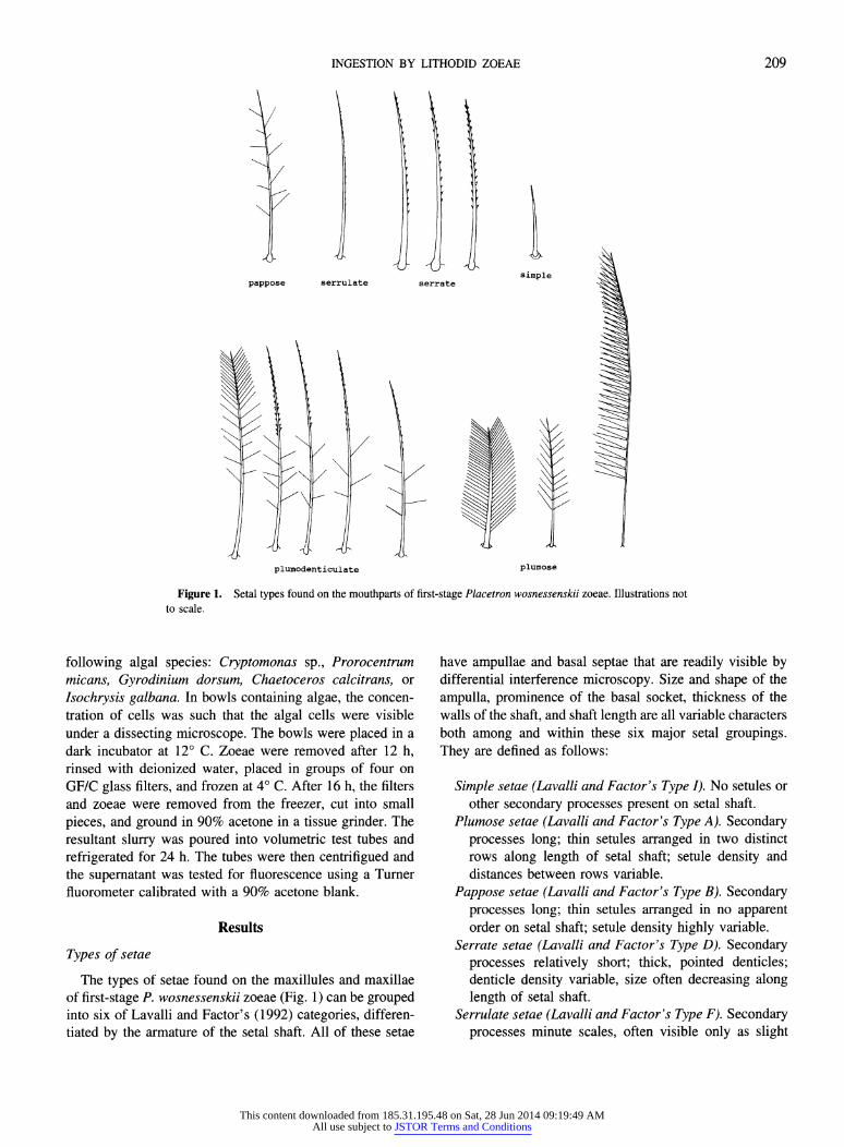
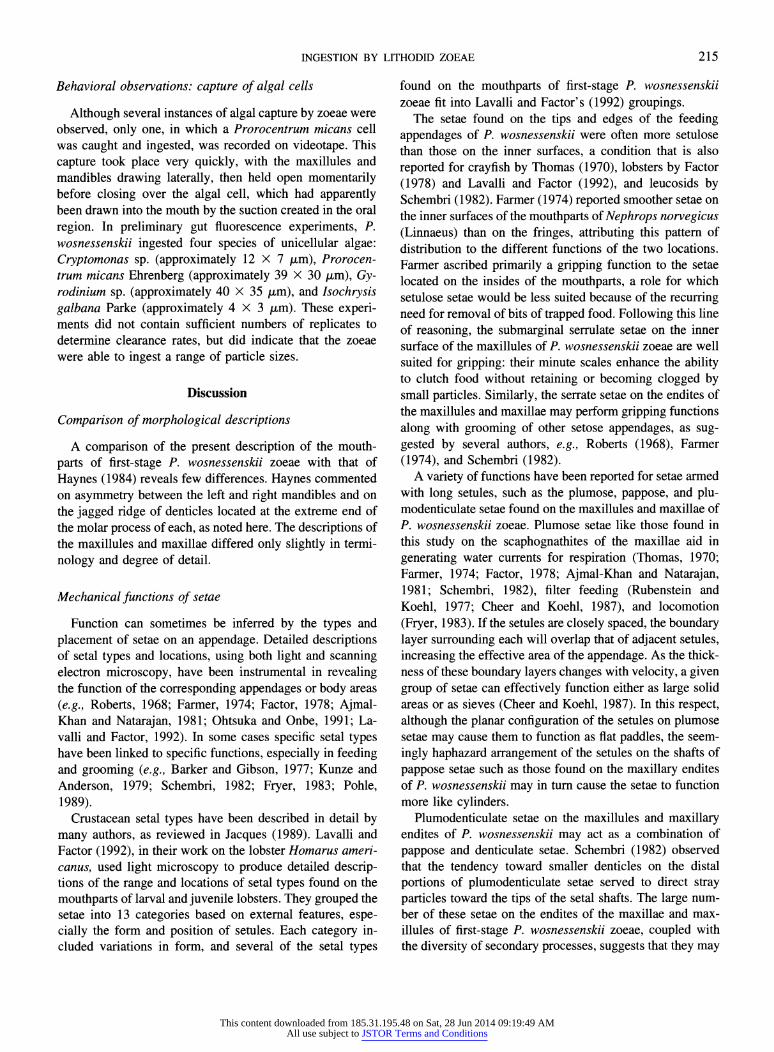
Figure 1. Setal types found on the mouthparts of first-stage Placetron wosnessenskii zoeae. Illustrations not to scale.
following algal species: Cryptomonas sp., Prorocentrum micans, Gyrodinium dorsum, Chaetoceros calcitrans, or Isochrysis galbana. In bowls containing algae, the concen- tration of cells was such that the algal cells were visible under a dissecting microscope. The bowls were placed in a dark incubator at 12? C. Zoeae were removed after 12 h, rinsed with deionized water, placed in groups of four on GF/C glass filters, and frozen at 4? C. After 16 h, the filters and zoeae were removed from the freezer, cut into small
pieces, and ground in 90% acetone in a tissue grinder. The resultant slurry was poured into volumetric test tubes and refrigerated for 24 h. The tubes were then centrifigued and the supernatant was tested for fluorescence using a Turner fluorometer calibrated with a 90% acetone blank.
Results
Types of setae
The types of setae found on the maxillules and maxillae of first-stage P. wosnessenskii zoeae (Fig. 1) can be grouped into six of Lavalli and Factor's (1992) categories, differen- tiated by the armature of the setal shaft. All of these setae
have ampullae and basal septae that are readily visible by differential interference microscopy. Size and shape of the
ampulla, prominence of the basal socket, thickness of the walls of the shaft, and shaft length are all variable characters both among and within these six major setal groupings. They are defined as follows:
Simple setae (Lavalli and Factor's Type I). No setules or other secondary processes present on setal shaft.
Plumose setae (Lavalli and Factor's Type A). Secondary processes long; thin setules arranged in two distinct rows along length of setal shaft; setule density and distances between rows variable.
Pappose setae (Lavalli and Factor's Type B). Secondary processes long; thin setules arranged in no apparent order on setal shaft; setule density highly variable.
Serrate setae (Lavalli and Factor's Type D). Secondary processes relatively short; thick, pointed denticles; denticle density variable, size often decreasing along length of setal shaft.
Serrulate setae (Lavalli and Factor's Type F). Secondary processes minute scales, often visible only as slight
1/
pappose serrulate
plumodenticulate
209
\I
This content downloaded from 185.31.195.48 on Sat, 28 Jun 2014 09:19:49 AMAll use subject to JSTOR Terms and Conditions
J. A. CRAIN
indentations of outer walls, arranged along setal shaft in one or more rows, concentrated on the distal half.
Plumodenticulate setae (Lavalli and Factor's Type C). Secondary processes not homogeneous; proximal half of setal shaft usually bearing long setules arranged in no apparent order; setules increasing in thickness to form one or two rows of denticles on distal half of shaft, often decreasing in size toward the tip. Second- ary process densities variable on all subtypes.
Morphology and movements of oral region and appendages
Comparisons of the mouthparts of the zoeae used in behavioral analysis with those used for morphological de-
scriptions showed no differences between the two broods. The morphology and movements of preoral chamber struc- tures and of the appendages expected to be involved in
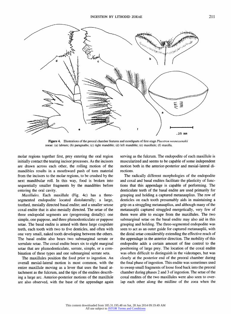
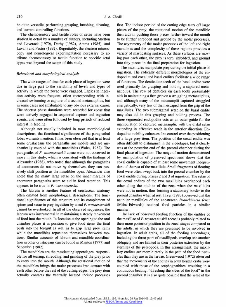
ingestion are as follows: Preoral chamber. The anterior portion of the preoral
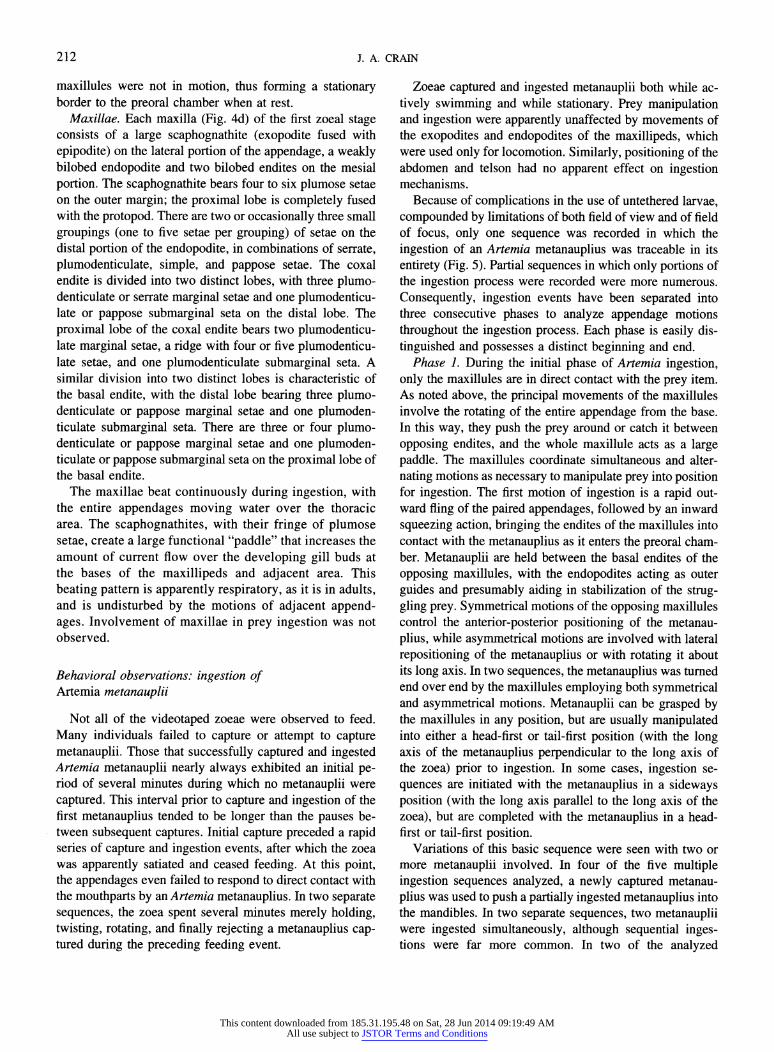
chamber (Figs. 2, 3) is bordered by the labrum (Fig. 4f), a large, subquadrate lobe with two rows of five or six slender
spines medially on the posterior face and a small, dense
patch of posteroventrally directed simple setae located be- tween the rows of spines. The observed range of motion of this lobe was limited to a short ventral-to-dorsal movement in which the spines were tilted toward the mouth.
The two ventrally directed lobes of the paragnaths (Figs. 2, 3, 4e) form the posterior lateral borders of the preoral chamber. Each paragnathal lobe is fringed with many fine, anteriorly directed, simple setae along the inner and distal
margins. The paragnaths were not seen to move during
Figure 2. Ventral view of the oral region of a first-stage Placetron wosnessenskii zoea, showing locations and orientations of the mouthparts. Setae have been omitted from illustration in order to clearly illustrate
appendage positions. Orientation: anterior is top left, posterior is bottom
right.
sternal
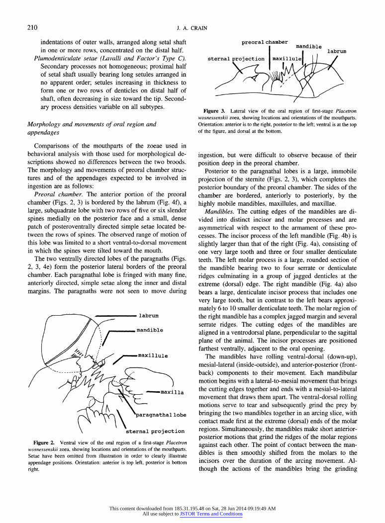
Figure 3. Lateral view of the oral region of first-stage Placetron wosnessenskii zoea, showing locations and orientations of the mouthparts. Orientation: anterior is to the right, posterior to the left; ventral is at the top of the figure, and dorsal at the bottom.
ingestion, but were difficult to observe because of their position deep in the preoral chamber.
Posterior to the paragnathal lobes is a large, immobile projection of the sterite (Figs. 2, 3), which completes the posterior boundary of the preoral chamber. The sides of the chamber are bordered, anteriorly to posteriorly, by the highly mobile mandibles, maxillules, and maxillae.
Mandibles. The cutting edges of the mandibles are di- vided into distinct incisor and molar processes and are asymmetrical with respect to the armament of these pro- cesses. The incisor process of the left mandible (Fig. 4b) is slightly larger than that of the right (Fig. 4a), consisting of one very large tooth and three or four smaller denticulate teeth. The left molar process is a large, rounded section of the mandible bearing two to four serrate or denticulate ridges culminating in a group of jagged denticles at the extreme (dorsal) edge. The right mandible (Fig. 4a) also bears a large, denticulate incisor process that includes one very large tooth, but in contrast to the left bears approxi- mately 6 to 10 smaller denticulate teeth. The molar region of the right mandible has a complex jagged margin and several serrate ridges. The cutting edges of the mandibles are aligned in a ventrodorsal plane, perpendicular to the sagittal plane of the animal. The incisor processes are positioned farthest ventrally, adjacent to the oral opening.
The mandibles have rolling ventral-dorsal (down-up), mesial-lateral (inside-outside), and anterior-posterior (front- back) components to their movement. Each mandibular motion begins with a lateral-to-mesial movement that brings the cutting edges together and ends with a mesial-to-lateral movement that draws them apart. The ventral-dorsal rolling motions serve to tear and subsequently grind the prey by bringing the two mandibles together in an arcing slice, with contact made first at the extreme (dorsal) ends of the molar regions. Simultaneously, the mandibles make short anterior- posterior motions that grind the ridges of the molar regions against each other. The point of contact between the man- dibles is then smoothly shifted from the molars to the incisors over the duration of the arcing movement. Al- though the actions of the mandibles bring the grinding
210
This content downloaded from 185.31.195.48 on Sat, 28 Jun 2014 09:19:49 AMAll use subject to JSTOR Terms and Conditions
INGESTION BY LITHODID ZOEAE
.25 mm
.25 mm
Figure 4. Illustrations of the preoral chamber features and mouthparts of first-stage Placetron wosnessenskii zoeae: (a) labrum; (b) paragnaths; (c) right mandible; (d) left mandible; (e) maxillule; (f) maxilla.
molar regions together first, prey entering the oral region initially contact the tearing incisor processes. As the incisors are drawn across each other, the rolling motion of the mandibles results in a mouthward push of torn material from the incisors to the molar regions, to be crushed by the next mandibular roll. In this way, food is broken into
sequentially smaller fragments by the mandibles before
entering the oral cavity. Maxillules. Each maxillule (Fig. 4c) has a three-
segmented endopodite located distolaterally; a large, toothed, mesially directed basal endite; and a smaller setose coxal endite that is also mesially directed. The setae of the three endopodal segments are (progressing distally): one
simple, one pappose, and three plumodenticulate or pappose setae. The basal endite is armed with three large cuspidate teeth, each tooth with two to five denticles, and often with one very small, naked tooth developing between the others. The basal endite also bears two submarginal serrate or serrulate setae. The coxal endite bears six to eight marginal setae that are plumodenticulate, serrate, simple, or a com- bination of these types and one submarginal serrate seta.
The maxillules position the food prior to ingestion. An overall mesial-lateral motion is most common, with the entire maxillule moving as a lever that uses the basal at- tachment as the fulcrum, and the tips of the endites describ-
ing a large arc. Anterior-posterior motions of the maxillule are also observed, with the base of the appendage again
serving as the fulcrum. The endopodite of each maxillule is muscularized and seems to be capable of some independent motion both in the anterior-posterior and mesial-lateral di- rections.
The radically different morphologies of the endopodite and coxal and basal endites facilitate the plasticity of func- tions that this appendage is capable of performing. The denticulate teeth of the basal endite are used primarily for
grasping and holding a captured metanauplius. The row of denticles on each tooth presumably aids in maintaining a
grip on a struggling metanauplius, and although many of the
metanauplii captured struggled energetically, very few of them were able to escape from the maxillules. The two
submarginal setae on the basal endite may also aid in this
grasping and holding. The three-segmented endopodite was seen to act as an outer guide for captured metanauplii, with the distal setae considerably extending the effective reach of the appendage in the anterior direction. The mobility of this
endopodite adds a certain amount of fine control to the
positioning of large prey. The location of the coxal endite was often difficult to distinguish in the videotapes, but was clearly at the posterior end of the preoral chamber during the final phase of ingestion. This endite was sometimes used to sweep small fragments of loose food back into the preoral chamber during phases 2 and 3 of ingestion. The setae of the coxal endites of the two maxillules were also seen to over- lap each other along the midline of the zoea when the
211
This content downloaded from 185.31.195.48 on Sat, 28 Jun 2014 09:19:49 AMAll use subject to JSTOR Terms and Conditions
J. A. CRAIN
maxillules were not in motion, thus forming a stationary border to the preoral chamber when at rest.
Maxillae. Each maxilla (Fig. 4d) of the first zoeal stage consists of a large scaphognathite (exopodite fused with epipodite) on the lateral portion of the appendage, a weakly bilobed endopodite and two bilobed endites on the mesial portion. The scaphognathite bears four to six plumose setae on the outer margin; the proximal lobe is completely fused with the protopod. There are two or occasionally three small groupings (one to five setae per grouping) of setae on the distal portion of the endopodite, in combinations of serrate, plumodenticulate, simple, and pappose setae. The coxal endite is divided into two distinct lobes, with three plumo- denticulate or serrate marginal setae and one plumodenticu- late or pappose submarginal seta on the distal lobe. The proximal lobe of the coxal endite bears two plumodenticu- late marginal setae, a ridge with four or five plumodenticu- late setae, and one plumodenticulate submarginal seta. A similar division into two distinct lobes is characteristic of the basal endite, with the distal lobe bearing three plumo- denticulate or pappose marginal setae and one plumoden- ticulate submarginal seta. There are three or four plumo- denticulate or pappose marginal setae and one plumoden- ticulate or pappose submarginal seta on the proximal lobe of the basal endite.
The maxillae beat continuously during ingestion, with the entire appendages moving water over the thoracic area. The scaphognathites, with their fringe of plumose setae, create a large functional "paddle" that increases the amount of current flow over the developing gill buds at the bases of the maxillipeds and adjacent area. This beating pattern is apparently respiratory, as it is in adults, and is undisturbed by the motions of adjacent append- ages. Involvement of maxillae in prey ingestion was not observed.
Behavioral observations: ingestion of Artemia metanauplii
Not all of the videotaped zoeae were observed to feed. Many individuals failed to capture or attempt to capture metanauplii. Those that successfully captured and ingested Artemia metanauplii nearly always exhibited an initial pe- riod of several minutes during which no metanauplii were captured. This interval prior to capture and ingestion of the first metanauplius tended to be longer than the pauses be- tween subsequent captures. Initial capture preceded a rapid series of capture and ingestion events, after which the zoea was apparently satiated and ceased feeding. At this point, the appendages even failed to respond to direct contact with the mouthparts by an Artemia metanauplius. In two separate sequences, the zoea spent several minutes merely holding, twisting, rotating, and finally rejecting a metanauplius cap- tured during the preceding feeding event.
Zoeae captured and ingested metanauplii both while ac- tively swimming and while stationary. Prey manipulation and ingestion were apparently unaffected by movements of the exopodites and endopodites of the maxillipeds, which were used only for locomotion. Similarly, positioning of the abdomen and telson had no apparent effect on ingestion mechanisms.
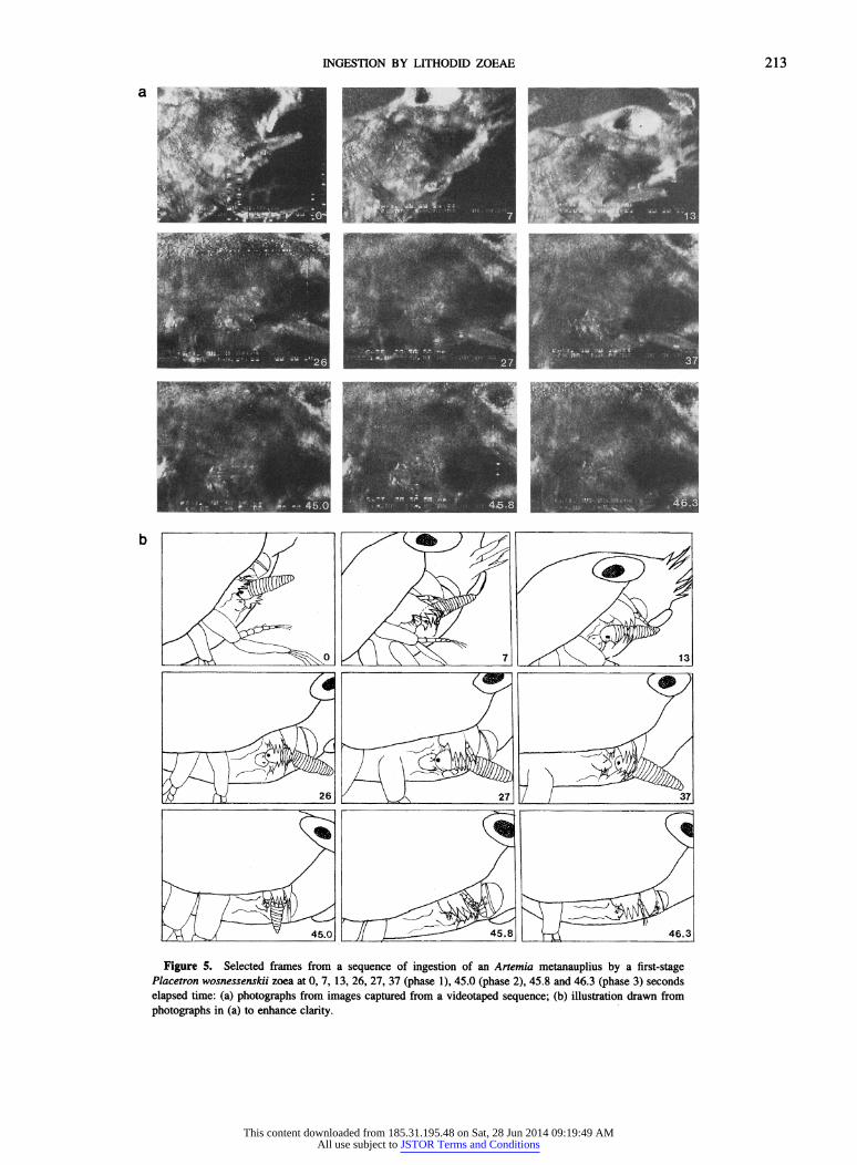
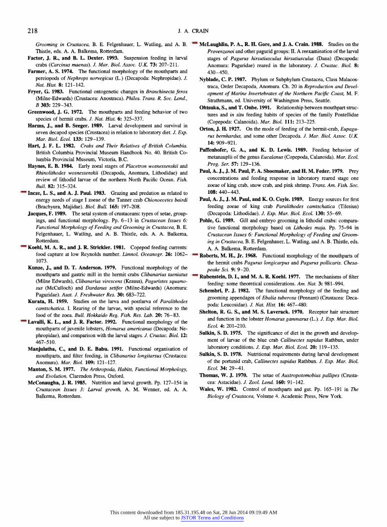
Because of complications in the use of untethered larvae, compounded by limitations of both field of view and of field of focus, only one sequence was recorded in which the ingestion of an Artemia metanauplius was traceable in its entirety (Fig. 5). Partial sequences in which only portions of the ingestion process were recorded were more numerous. Consequently, ingestion events have been separated into three consecutive phases to analyze appendage motions throughout the ingestion process. Each phase is easily dis- tinguished and possesses a distinct beginning and end.
Phase 1. During the initial phase of Artemia ingestion, only the maxillules are in direct contact with the prey item. As noted above, the principal movements of the maxillules involve the rotating of the entire appendage from the base. In this way, they push the prey around or catch it between opposing endites, and the whole maxillule acts as a large paddle. The maxillules coordinate simultaneous and alter- nating motions as necessary to manipulate prey into position for ingestion. The first motion of ingestion is a rapid out- ward fling of the paired appendages, followed by an inward squeezing action, bringing the endites of the maxillules into contact with the metanauplius as it enters the preoral cham- ber. Metanauplii are held between the basal endites of the opposing maxillules, with the endopodites acting as outer guides and presumably aiding in stabilization of the strug- gling prey. Symmetrical motions of the opposing maxillules control the anterior-posterior positioning of the metanau- plius, while asymmetrical motions are involved with lateral repositioning of the metanauplius or with rotating it about its long axis. In two sequences, the metanauplius was turned end over end by the maxillules employing both symmetrical and asymmetrical motions. Metanauplii can be grasped by the maxillules in any position, but are usually manipulated into either a head-first or tail-first position (with the long axis of the metanauplius perpendicular to the long axis of the zoea) prior to ingestion. In some cases, ingestion se- quences are initiated with the metanauplius in a sideways position (with the long axis parallel to the long axis of the zoea), but are completed with the metanauplius in a head- first or tail-first position.
Variations of this basic sequence were seen with two or more metanauplii involved. In four of the five multiple ingestion sequences analyzed, a newly captured metanau- plius was used to push a partially ingested metanauplius into the mandibles. In two separate sequences, two metanauplii were ingested simultaneously, although sequential inges- tions were far more common. In two of the analyzed
212
This content downloaded from 185.31.195.48 on Sat, 28 Jun 2014 09:19:49 AMAll use subject to JSTOR Terms and Conditions
INGESTION BY LITHODID ZOEAE
a ~' , 2 . .'= . Ro - i? ; { _ueg ! .: ' 2 E ...': .. i%' i. =..-.
r . a3 ,: i~Pc._e.... . i...& ..
...
- .',iN A 'I
.
-F~!j L/ / . N
Placetron wosnessenskii zoea at 0, 7, 13, 26, 27, 37 (phase 1), 45.0 (phase 2), 45.8 and 46.3 (phase 3) seconds
i ' 4)
X f C A X '-
213
This content downloaded from 185.31.195.48 on Sat, 28 Jun 2014 09:19:49 AMAll use subject to JSTOR Terms and Conditions

J. A. CRAIN
Figure 6. Coordination of maxillule and mandible movements during phase 2 of ingestion of an Artemia metanauplius by a first-stage Placetron wosnessenskii zoea. Approximate duration: 0.1 to 0.6 s.
sequences, phase 1 of ingestion was shortened to merely the
fling and capture steps, which were followed immediately by the maceration associated with phase 2.
Recorded phase 1 duration ranged from 0.2 to 5.9 s in
sequences involving only a single metanauplius, and from 6.1 to 233.0 s in sequences involving two or more meta- nauplii. Mean durations were 3.9 (n = 14; SD = 5.8) and 56.8 (n = 5; SD = 93.2) s respectively.
Phase 2. This phase of ingestion involves the maxillules, mandibles, and labrum. Phase 2 begins with the first contact of the metanauplius with the mandibles and ends with the loss of direct contact with the prey item by the basal endites of the maxillules. The maxillules, after manipulating the metanauplius into position in phase 1, begin pushing the prey toward the mouth, bringing it into contact with the mandibles. In one sequence, phase 2 was initiated by re-
peated nudging of the metanauplius into and out of range of the mandibles by the maxillules.
The maxillules and labrum press the metanauplius against the mandibles as the latter tear and grind the prey on its way into the mouth. Two separate ratcheting mechanisms were observed, each of which maintains a steady net movement of the metanauplius into the mouth, while enabling the appendages to renew points of contact for leverage. First, the maxillules and mandibles alternate motions, maintaining a firm grip on the metanauplius as it is masticated (Fig. 6). The maxillules press the prey against the mandibles as they shred bits of tissue with their rolling motions. The mandi- bles then hold the metanauplius as the maxillules regrasp it
once every two or three mandibular rotations, or as seen in two sequences, with each mandibular roll. Second, the man- dibles and the labrum work in conjunction to prevent food from escaping from the mouth when the mandibles are on the recovery stroke of their motion (Fig. 7). The labrum moves downward, pinning the metanauplius in the preoral chamber with its spiny processes as the mandibles return to their initial (lateral) position in preparation for the next
grinding roll, with a 1:1 ratio of alternating movements. The recorded total durations of phase 2 range from 6.5 to
150 s, with a mean duration of 49 s (n = 11; SD = 45.7). Phase 3. This phase of ingestion begins when the basal
endites of the maxillules are no longer in contact with the
prey, and continues until the entire metanauplius has passed through the mandibular region into the oral cavity. The mandibles and labrum continue to work together as in phase 2 until the entire metanauplius has been ingested. Although the maxillules are apparently no longer in direct contact with the prey, they often continue to make the sweeping mesial-lateral motions associated with phase 2, and direct small, easily lost fragments of masticated food back into the preoral chamber. A variation of phase 3 was seen in four
sequences, when the maxillules pushed one metanauplius through the final stage of ingestion with a second metanau-
plius. The range of durations recorded for this phase was from
4 to 84 s, with a mean duration of 23.2 s (n = 9; SD =
31.6).
- w . ',
mandible
Figure 7. Coordination of mandible and labrum movements during phases 2 and 3 of ingestion of an Artemia metanauplius by a first-stage Placetron wosnessenskii zoea. Approximate duration: 0.1 to 0.3 s.
214
This content downloaded from 185.31.195.48 on Sat, 28 Jun 2014 09:19:49 AMAll use subject to JSTOR Terms and Conditions
INGESTION BY LITHODID ZOEAE
Behavioral observations: capture of algal cells
Although several instances of algal capture by zoeae were observed, only one, in which a Prorocentrum micans cell was caught and ingested, was recorded on videotape. This
capture took place very quickly, with the maxillules and mandibles drawing laterally, then held open momentarily before closing over the algal cell, which had apparently been drawn into the mouth by the suction created in the oral
region. In preliminary gut fluorescence experiments, P. wosnessenskii ingested four species of unicellular algae: Cryptomonas sp. (approximately 12 X 7 /am), Prorocen- trum micans Ehrenberg (approximately 39 X 30 uam), Gy- rodinium sp. (approximately 40 X 35 /um), and Isochrysis galbana Parke (approximately 4 X 3 uam). These experi- ments did not contain sufficient numbers of replicates to determine clearance rates, but did indicate that the zoeae were able to ingest a range of particle sizes.
Discussion
Comparison of morphological descriptions
A comparison of the present description of the mouth- parts of first-stage P. wosnessenskii zoeae with that of
Haynes (1984) reveals few differences. Haynes commented on asymmetry between the left and right mandibles and on the jagged ridge of denticles located at the extreme end of the molar process of each, as noted here. The descriptions of the maxillules and maxillae differed only slightly in termi-
nology and degree of detail.
Mechanical functions of setae
Function can sometimes be inferred by the types and
placement of setae on an appendage. Detailed descriptions of setal types and locations, using both light and scanning electron microscopy, have been instrumental in revealing the function of the corresponding appendages or body areas
(e.g., Roberts, 1968; Farmer, 1974; Factor, 1978; Ajmal- Khan and Natarajan, 1981; Ohtsuka and Onbe, 1991; La- valli and Factor, 1992). In some cases specific setal types have been linked to specific functions, especially in feeding and grooming (e.g., Barker and Gibson, 1977; Kunze and Anderson, 1979; Schembri, 1982; Fryer, 1983; Pohle, 1989).
Crustacean setal types have been described in detail by many authors, as reviewed in Jacques (1989). Lavalli and Factor (1992), in their work on the lobster Homarus ameri- canus, used light microscopy to produce detailed descrip- tions of the range and locations of setal types found on the mouthparts of larval and juvenile lobsters. They grouped the setae into 13 categories based on external features, espe- cially the form and position of setules. Each category in- cluded variations in form, and several of the setal types
found on the mouthparts of first-stage P. wosnessenskii zoeae fit into Lavalli and Factor's (1992) groupings.
The setae found on the tips and edges of the feeding appendages of P. wosnessenskii were often more setulose than those on the inner surfaces, a condition that is also reported for crayfish by Thomas (1970), lobsters by Factor (1978) and Lavalli and Factor (1992), and leucosids by Schembri (1982). Farmer (1974) reported smoother setae on the inner surfaces of the mouthparts of Nephrops norvegicus (Linnaeus) than on the fringes, attributing this pattern of distribution to the different functions of the two locations. Farmer ascribed primarily a gripping function to the setae located on the insides of the mouthparts, a role for which setulose setae would be less suited because of the recurring need for removal of bits of trapped food. Following this line of reasoning, the submarginal serrulate setae on the inner surface of the maxillules of P. wosnessenskii zoeae are well suited for gripping: their minute scales enhance the ability to clutch food without retaining or becoming clogged by small particles. Similarly, the serrate setae on the endites of the maxillules and maxillae may perform gripping functions
along with grooming of other setose appendages, as sug- gested by several authors, e.g., Roberts (1968), Farmer (1974), and Schembri (1982).
A variety of functions have been reported for setae armed with long setules, such as the plumose, pappose, and plu- modenticulate setae found on the maxillules and maxillae of P. wosnessenskii zoeae. Plumose setae like those found in this study on the scaphognathites of the maxillae aid in
generating water currents for respiration (Thomas, 1970; Farmer, 1974; Factor, 1978; Ajmal-Khan and Natarajan, 1981; Schembri, 1982), filter feeding (Rubenstein and Koehl, 1977; Cheer and Koehl, 1987), and locomotion
(Fryer, 1983). If the setules are closely spaced, the boundary layer surrounding each will overlap that of adjacent setules, increasing the effective area of the appendage. As the thick- ness of these boundary layers changes with velocity, a given group of setae can effectively function either as large solid areas or as sieves (Cheer and Koehl, 1987). In this respect, although the planar configuration of the setules on plumose setae may cause them to function as flat paddles, the seem-
ingly haphazard arrangement of the setules on the shafts of
pappose setae such as those found on the maxillary endites of P. wosnessenskii may in turn cause the setae to function more like cylinders.
Plumodenticulate setae on the maxillules and maxillary endites of P. wosnessenskii may act as a combination of
pappose and denticulate setae. Schembri (1982) observed that the tendency toward smaller denticles on the distal portions of plumodenticulate setae served to direct stray particles toward the tips of the setal shafts. The large num- ber of these setae on the endites of the maxillae and max- illules of first-stage P. wosnessenskii zoeae, coupled with the diversity of secondary processes, suggests that they may
215
This content downloaded from 185.31.195.48 on Sat, 28 Jun 2014 09:19:49 AMAll use subject to JSTOR Terms and Conditions
J. A. CRAIN
be quite versatile, performing grasping, brushing, cleaning, and current-controlling functions.
The chemosensory and tactile roles of setae have been studied in detail by a number of authors, including Shelton and Laverack (1970), Derby (1982), Atema (1985), and Lavalli and Factor (1992). Regrettably, the electron micros- copy and neurological experimentation necessary to at- tribute chemosensory or tactile function to specific setal types was beyond the scope of this study.
Behavioral and morphological analysis
The wide ranges of time for each phase of ingestion were due in large part to the variability of levels and types of activity in which the zoeae were engaged. Lapses in inges- tion activity were frequent, often corresponding with in- creased swimming or capture of a second metanauplius, but in some cases not attributable to any obvious external cause. The shortest phase durations were recorded for zoeae that were actively engaged in sequential capture and ingestion events, and were often followed by long periods of reduced interest in feeding.
Although not usually included in most morphological descriptions, the functional significance of the paragnathal lobes warrants mention. It has been observed that in at least some crustaceans the paragnaths are mobile and are me- chanically coupled with the mandibles (Wales, 1982). The paragnaths of P. wosnessenskii zoeae were not observed to move in this study, which is consistent with the findings of Alexander (1988), who noted that although the paragnaths of anomurans do not move by themselves, they can pas- sively shift position as the mandibles open. Alexander also noted that the many large setae on the inner margins of anomuran paragnaths seem to aid in food retention. This appears to be true in P. wosnessenskii.
The labrum is another feature of crustacean anatomy often omitted from morphological descriptions. The func- tional significance of this structure and its complement of spines and setae in prey ingestion by zoeal P. wosnessenskii cannot be overlooked. In all of the observed sequences, the labrum was instrumental in maintaining a steady movement of food into the mouth. Its location at the opening to the oral chamber places it in position to give food items the final push into the foregut as well as to grip large prey items while the mandibles reposition themselves between mo- tions. Similar accounts of labrum and mandible coordina- tion in other crustaceans can be found in Manton (1977) and Schembri (1982).
The mandibles are the masticating appendages, responsi- ble for all tearing, shredding, and grinding of the prey prior to entry into the mouth. Although the rotational motion of the mandibles brings the molar processes into contact with each other before the rest of the cutting edges, the prey item actually contacts the ventrally located incisor processes
first. The incisor portion of the cutting edge tears off large pieces of the prey; the rotational motion of the mandible then aids in pushing those pieces farther toward the mouth to be further shredded and ground by the molar processes. The asymmetry of the molar processes of the left and right mandibles and the complexity of these regions provides a variety of masticating surfaces. As these surfaces are mov- ing past each other, the prey is torn, shredded, and ground into tiny pieces in the final preparation for ingestion.
The maxillules manipulate prey during the initial phase of ingestion. The radically different morphologies of the en- dopodite and coxal and basal endites facilitate a wide range of functions. The denticulate teeth of the basal endite were used primarily for grasping and holding a captured meta- nauplius. The row of denticles on each tooth presumably aids in maintaining a firm grip on a struggling metanauplius, and although many of the metanauplii captured struggled energetically, very few of them escaped from the grip of the maxillules. The two submarginal setae on the basal endite may also aid in this grasping and holding process. The three-segmented endopodite acts as an outer guide for the manipulation of captured metanauplii, with the distal setae extending its effective reach in the anterior direction. En- dopodite mobility enhances fine control over the positioning of a large prey item. The position of the coxal endite was often difficult to distinguish in the videotapes, but it clearly was at the posterior end of the preoral chamber during the final phase of ingestion. The range of motion demonstrated by manipulation of preserved specimens shows that the coxal endite is capable of at least some movement indepen- dent of the rest of the maxillule. Small fragments of floating food were often swept back into the preoral chamber by the coxal endite during phases 2 and 3 of ingestion. The setae of the coxal endites of the two maxillules overlapped each other along the midline of the zoea when the maxillules were not in motion, thus forming a stationary border to the preoral chamber when at rest. Fryer (1983) observed that the naupliar maxillules of the anostracan Branchinecta ferox (Milne-Edwards) retained food particles in a similar manner.
The lack of observed feeding function of the endites of the maxillae of P. wosnessenskii zoeae is probably related to their more posterior position in the zoeal stages compared to the adults, in which they are presumed to be involved in ingestion. In adult crabs, all of the feeding appendages, including the three pairs of maxillipeds, overlap one another obliquely and are limited in their posterior extension by the sterites of the pereopods. In this arrangement, the maxil- lary endites are more directly in the path of the food parti- cles than they are in the larvae. Greenwood (1972) observed that the movements of the endites in adult hermit crabs were coupled with those of the scaphognathites, resulting in a continuous beating, "threshing the sides of the food" in the preoral chamber. It is also quite possible that the setae of the
216
This content downloaded from 185.31.195.48 on Sat, 28 Jun 2014 09:19:49 AMAll use subject to JSTOR Terms and Conditions

INGESTION BY LITHODID ZOEAE
maxillary endites of both zoeae and adult crabs aid in the ingestion of smaller food items either through physical contact with the food or by the creation of a strong feeding current.
Other possible food sources for zoeae: ecological implications
For some decades, the focus of studies on zoeal diet has been its effect on growth and survival under laboratory and natural conditions (e.g., Kurata, 1959; Sulkin, 1975, 1978; Paul et al., 1979; McConaugha, 1985; Harms and Seeger, 1989; Paul et al., 1989; Epifanio et al., 1991). There is increasing agreement that under natural conditions, zoeae are unlikely to encounter zooplankton prey in the concen- trations routinely used in laboratory rearing experiments, and therefore they can probably utilize a variety of food sources (Incze and Paul, 1983; Harms and Seeger, 1989; Paul et al., 1989; Epifanio et al., 1991). Sulkin found that the zoeae of Callinectes sapidus Rathbun, a brachyuran crab that cannot successfully complete development to metamor- phosis on purely algal diets, nonetheless ingested unicellu- lar organisms (Sulkin, 1975). The lithodid crab Paralith- odes camtschatica (Tilesius) can be reared in the laboratory on polychaete larvae or Artemia nauplii, but not on a diet made up solely of diatoms (Kurata, 1959). However, Paul et al. (1989) found that when first-stage P. camtschatica zoeae ingested phytoplankton soon after hatching, they molted to the second zoeal stage at higher rates than those that did not. The algal diet did not sustain the zoeae through metamor- phosis, and an increasing dependence on carivory through the zoeal stages was hypothesized for this species (Paul et al., 1989). First-feeding P. wosnessenskii zoeae are able to take advantage of a wide range of prey items and probably rely on a variety of planktonic food sources throughout their development.
Factor and Dexter (1993) found that the larvae of the brachyuran crab Carcinus maenas (Linnaeus) could capture suspended algal cells, and hypothesized that the setose mouthparts were involved in suspension feeding. In prelim- inary gut fluorescence experiments, P. wosnessenskii in- gested unicellular algae covering a range of sizes. How these zoeae capture small algal cells is not known, but we assume that the mechanism is similar to that seen for in- gestion of a Prorocentrum micans cell. In the one sequence of particle capture recorded on videotape, a P. micans cell was captured and ingested using a "fling and clap" method similar to that described for copepods by Koehl and Strick- ler (1981). The mouthparts were flung outward, enlarging the space between them, thus drawing the cell into the mouth. The mouthparts were then closed over the cell, squeezing water out through the spaces between the setae and endites, as seen in algal capture by copepods (Koehl and Strickler, 1981).
This study demonstrates that P. wosnessenskii zoeae can utilize prey items ranging from unicellular algae (Crypto- monas sp., Prorocentrum micans, Gyrodinium sp., and Iso- chrysis galbana) to relatively large, active zooplankton (Ar- temia sp. metanauplii). McConaugha (1985) identified three criteria for suitability of prey items as food sources for larval crustaceans: (1) appropriate size for capture and con- sumption, (2) adequate concentration, and (3) essential di- etary nutrients to meet the larvae's needs for survival, growth, and metamorphosis. Natural plankton assemblages are varied in composition both spatially and temporally. The ability to capture and ingest a variety of sizes and shapes, expanding the diversity of prey species that meet Mc- Conaugha's first criterion, increases the probability that the zoea will be able to fulfill its nutritional requirements for successful development.
Acknowledgments
This project was funded by a grant from the PADI Foun- dation. Special thanks go to Drs. P. A. McLaughlin, R. R. Strathmann, S. D. Sulkin, D. Schneider, and C. B. Miller for invaluable advice and assistance. Use of space and equip- ment at Shannon Point Marine Center and Friday Harbor Laboratories is gratefully acknowledged.
Literature Cited
Ajmal-Khan, S., and R. Natarajan. 1981. Functional morphology of mouth parts and feeding behavior in the estuarine hermit crab Cliba- narius longitarsus (De Haan). Indian J. Mar. Sci. 10: 357-362.
Alexander, C. G. 1988. The paragnaths of some intertidal crustaceans. J. Mar. Biol. Assoc. U.K. 68: 581-590.
Alexander, C. G., and J. P. R. Hindley. 1985. The mechanism of food
ingestion by the banana prawn, Penaeus merguiensis. Mar. Behav.
Physiol. 12: 33-46. Atema, J. 1985. Chemoreception in the sea: adaptations of chemorecep-
tors and behavior to aquatic stimulus conditions. Pp. 387-423 in
Physiological Adaptations of Marine Animals, Society of Experimental Biology Symposium 39, M. S. Laverack, ed. Company of Biologists, Cambridge, U.K.
Barker, P. L., and R. Gibson. 1977. Observations on the feeding mechanism, structure of the gut, and digestive physiology of the
European lobster Homarus gammarus (L.) (Decapoda: Nephropidae). J. Exp. Mar. Biol. Ecol. 26: 297-324.
Cheer, A. Y. L., and M. A. R. Koehl. 1987. Paddles and rakes: fluid flow through bristled appendages of small organisms. J. Theor. Biol. 129: 17-39.
Derby, C. D. 1982. Structure and function of cuticular sensilla of the lobster Homarus americanus. J. Crustac. Biol. 2: 1-21.
Epifanio, C. E., J. S. Cope, P. M. Rowe, and F. M. Jenkins. 1991.
Comparison of rates of development of Atlantic mud crab larvae in the laboratory and in field-deployed enclosures. J. Crustac. Biol. 11: 540- 545.
Factor, J. R. 1978. Morphology of the mouthparts of larval lobsters, Homarus americanus (Decapoda: Nephropidae), with special emphasis on their setae. Biol. Bull. 154: 383-408.
Factor, J. R. 1989. Development of the feeding apparatus in decapod crustaceans. Pp. 185-203 in Functional Morphology of Feeding and
217
This content downloaded from 185.31.195.48 on Sat, 28 Jun 2014 09:19:49 AMAll use subject to JSTOR Terms and Conditions
J. A. CRAIN
Grooming in Crustacea, B. E. Felgenhauer, L. Watling, and A. B. Thistle, eds. A. A. Balkema, Rotterdam.
Factor, J. R., and B. L. Dexter. 1993. Suspension feeding in larval crabs (Carcinus maenas). J. Mar. Biol. Assoc. U.K. 73: 207-211.
Farmer, A. S. 1974. The functional morphology of the mouthparts and
pereiopods of Nephrops norvegicus (L.) (Decapoda: Nephropidae). J. Nat. Hist. 8: 121-142.
Fryer, G. 1983. Functional ontogenetic changes in Branchinecta ferox (Milne-Edwards) (Crustacea: Anostraca). Philos. Trans. R. Soc. Lond., B 303: 229-343.
Greenwood, J. G. 1972. The mouthparts and feeding behavior of two
species of hermit crabs. J. Nat. Hist. 6: 325-337. Harms, J., and B. Seeger. 1989. Larval development and survival in
seven decapod species (Crustacea) in relation to laboratory diet. J. Exp. Mar. Biol. Ecol. 133: 129-139.
Hart, J. F. L. 1982. Crabs and Their Relatives of British Columbia. British Columbia Provincial Museum Handbook No. 40. British Co- lumbia Provincial Museum, Victoria, B.C.
Haynes, E. B. 1984. Early zoeal stages of Placetron wosnessenskii and Rhinolithodes wosnessenskii (Decapoda, Anomura, Lithodidae) and review of lithodid larvae of the northern North Pacific Ocean. Fish. Bull. 82: 315-324.
Incze, L. S., and A. J. Paul. 1983. Grazing and predation as related to energy needs of stage I zoeae of the Tanner crab Chionoecetes bairdi
(Brachyura, Majidae). Biol. Bull. 165: 197-208.
Jacques, F. 1989. The setal system of crustaceans: types of setae, group- ings, and functional morphology. Pp. 6-13 in Crustacean Issues 6: Functional Morphology of Feeding and Grooming in Crustacea, B. E.
Felgenhauer, L. Watling, and A. B. Thistle, eds. A. A. Balkema, Rotterdam.
Koehl, M. A. R., and J. R. Strickler. 1981. Copepod feeding currents: food capture at low Reynolds number. Limnol. Oceanogr. 26: 1062- 1073.
Kunze, J., and D. T. Anderson. 1979. Functional morphology of the
mouthparts and gastric mill in the hermit crabs Clibanarius taeniatus (Milne Edwards), Clibanarius virescens (Krauss), Paguristes squamo- sus (McCulloch) and Dardanus setifer (Milne-Edwards) (Anomura: Paguridae). Aust. J. Freshwater Res. 30: 683-722.
Kurata, H. 1959. Studies on the larva and postlarva of Paralithodes camtschatica. I. Rearing of the larvae, with special reference to the food of the zoea. Bull. Hokkaido Reg. Fish. Res. Lab. 20: 76-83.
Lavalli, K. L., and J. R. Factor. 1992. Functional morphology of the
mouthparts of juvenile lobsters, Homarus americanus (Decapoda: Ne-
phropidae), and comparison with the larval stages. J. Crustac. Biol. 12: 467-510.
Manjulatha, C., and D. E. Babu. 1991. Functional organisation of
mouthparts, and filter feeding, in Clibanarius longitarsus (Crustacea: Anomura). Mar. Biol. 109: 121-127.
Manton, S. M. 1977. The Arthropoda, Habits, Functional Morphology, and Evolution. Clarendon Press, Oxford.
McConaugha, J. R. 1985. Nutrition and larval growth. Pp. 127-154 in Crustacean Issues 3: Larval growth, A. M. Wenner, ed. A. A. Balkema, Rotterdam.
McLaughlin, P. A., R. H. Gore, and J. A. Crain. 1988. Studies on the Provenzanoi and other pagurid groups: II. A reexamination of the larval
stages of Pagurus hirsutiusculus hirsutiusculus (Dana) (Decapoda: Anomura: Paguridae) reared in the laboratory. J. Crustac. Biol. 8: 430-450.
Nyblade, C. P. 1987. Phylum or Subphylum Crustacea, Class Malacos- traca, Order Decapoda, Anomura. Ch. 20 in Reproduction and Devel-
opment of Marine Invertebrates of the Northern Pacific Coast, M. F. Strathmann, ed. University of Washington Press, Seattle.
Ohtsuka, S., and T. Onbe. 1991. Relationship between mouthpart struc- tures and in situ feeding habits of species of the family Pontellidae
(Copepoda: Calanoida). Mar. Biol. 111: 213-225.
Orton, J. H. 1927. On the mode of feeding of the hermit-crab, Eupagu- rus bernhardus, and some other Decapoda. J. Mar. Biol. Assoc. U.K. 14: 909-921.
Paffenhofer, G. A., and K. D. Lewis. 1989. Feeding behavior of
metanauplii of the genus Eucalanus (Copepoda, Calanoida). Mar. Ecol.
Prog. Ser. 57: 129-136.
Paul, A. J., J. M. Paul, P. A. Shoemaker, and H. M. Feder. 1979. Prey concentrations and feeding response in laboratory reared stage one zoeae of king crab, snow crab, and pink shrimp. Trans. Am. Fish. Soc. 108: 440-443.
Paul, A. J., J. M. Paul, and K. O. Coyle. 1989. Energy sources for first
feeding zoeae of king crab Paralithodes camtschatica (Tilesius) (Decapoda: Lithodidae). J. Exp. Mar. Biol. Ecol. 130: 55-69.
Pohle, G. 1989. Gill and embryo grooming in lithodid crabs: compara- tive functional morphology based on Lithodes maja. Pp. 75-94 in Crustacean Issues 6: Functional Morphology of Feeding and Groom-
ing in Crustacea, B. E. Felgenhauer, L. Watling, and A. B. Thistle, eds. A. A. Balkema, Rotterdam.
Roberts, M. H., Jr. 1968. Functional morphology of the mouthparts of the hermit crabs Pagurus longicarpus and Pagurus pollicaris. Chesa-
peake Sci. 9: 9-20.
Rubenstein, D. I., and M. A. R. Koehl. 1977. The mechanisms of filter
feeding: some theoretical considerations. Am. Nat. 3: 981-994. Schembri, P. J. 1982. The functional morphology of the feeding and
grooming appendages of Ebalia tuberosa (Pennant) (Crustacea: Deca-
poda: Leucosidae). J. Nat. Hist. 16: 467-480.
Shelton, R. G. S., and M. S. Laverack. 1970. Receptor hair structure and function in the lobster Homarus gammarus (L.). J. Exp. Mar. Biol. Ecol. 4: 201-210.
Sulkin, S. D. 1975. The significance of diet in the growth and develop- ment of larvae of the blue crab Callinectes sapidus Rathbun, under
laboratory conditions. J. Exp. Mar. Biol. Ecol. 20: 119-135.
Sulkin, S. D. 1978. Nutritional requirements during larval development of the portunid crab, Callinectes sapidus Rathbun. J. Exp. Mar. Biol. Ecol. 34: 29-41.
Thomas, W. J. 1970. The setae of Austropotomobius pallipes (Crusta- cea: Astacidae). J. Zool. Lond. 160: 91-142.
Wales, W. 1982. Control of mouthparts and gut. Pp. 165-191 in The
Biology of Crustacea, Volume 4. Academic Press, New York.
218
This content downloaded from 185.31.195.48 on Sat, 28 Jun 2014 09:19:49 AMAll use subject to JSTOR Terms and Conditions