Embed Size (px)
Citation preview

R E V I E W S
NATURE REVIEWS | MICROBIOLOGY VOLUME 2 | JULY 2004 | 541
The degradation of plant cell walls by microorganismshas an important role in the carbon cycle of the earth.Most plant cell walls are composed of approximately15–40% CELLULOSE, 30–40% HEMICELLULOSE and pectin,and 20% lignin. These components are degraded enzy-matically to yield smaller oligomers and eventually glu-cose, pentoses and other small carbon compounds,which are metabolized to CO
2.
Although abundant in nature, cellulose is a partic-ularly difficult polymer to degrade, as it is insolubleand is present as hydrogen-bonded crystalline fibres1.Hemicellulose, pectin and lignin are generally easier todegrade than cellulose. Two types of enzyme systemsfor the degradation of plant cell walls have beenobserved in microorganisms. In the case of aerobicfungi and bacteria, several individual endoglucanases,exoglucanases and ancillary enzymes are secreted thatcan act synergistically to attack plant cell walls. Thebest studied of these enzymes are the glycosyl hydro-lases of Trichoderma reesei. These free enzyme systemshave been well documented and reviewed2,3 and willnot be discussed further in this article. In anaerobicmicroorganisms, a different type of system has evolvedthat involves the formation of a large, extracellularenzyme complex called the CELLULOSOME, which consistsof a scaffolding protein and many bound enzymes4.
Several recent reviews have discussed the structureand function of cellulosomes5–12. In this review, we willfocus on the most recent findings that have added toour knowledge of cellulosome structure, regulationand genetics, and the synergism between CELLULASES
and hemicellulases.
Cellulosome-producing microorganismsExperimental evidence of the presence of cellulosomeshas only been obtained for anaerobic bacteria.Although there is growing evidence that anaerobicfungi also produce cellulosomes13–18, the presence of aSCAFFOLDIN6 gene, which encodes an important compo-nent of the cellulosome, has not yet been documentedfor the anaerobic fungi. A list of cellulosome-producingmicroorganisms is presented in TABLE 1. This list isexpected to grow rapidly in the next few years asmore cellulosome-producing microorganisms arediscovered.
It should be noted that the presence of cellulosome-like structures does not always confer cellulolytic activ-ity to a microorganism. Clostridium acetobutylicumsynthesizes a 650-kDa cellulosome19, which has lowdegradative activity on CMC (carboxymethyl cellulose,a soluble form of cellulose), but cannot grow on cellu-lose and cannot hydrolyse crystalline cellulose20. This
CELLULOSOMES: PLANT-CELL-WALL-DEGRADING ENZYME COMPLEXESRoy H. Doi* and Akihiko Kosugi‡
Cellulose, the main structural component of plant cell walls, is the most abundant carbohydratepolymer in nature. Although abundant, it is extremely difficult to degrade, as it is insoluble and ispresent as hydrogen-bonded crystalline fibres. Anaerobic microorganisms have evolved asystem to break down plant cell walls that involves the formation of a large extracellular enzymecomplex called the cellulosome,which consists of a scaffolding protein and many boundcellulases. Cellulosomes have many potential biotechnological applications as the conversion ofcellulosic biomass into sugars by cellulosomes could result in the production of high-valueproducts such as ethanol or organic acids from inexpensive renewable resources. Rapidadvances in cellulosome research are providing basic information for the development of both in vitro and in vivo systems to achieve such goals.
CELLULOSE
The most abundant plantpolysaccharide consisting of(1→4)β-D-glucan chainshydrogen-bonded to oneanother along their length.
HEMICELLULOSE
Cross-linking glycans thatcomprise up to about 30% ofplant cell walls; the two majorhemicelluloses are xyloglucansand glucuronoarabinoxylans.
*Section of Molecular &Cellular Biology, Universityof California, Davis,California, USA.‡Fine Chemicals Division,Kaneka Corporation,1-8, Miyamae-machi,Takasago-cho Takasago,Hyogo, 676-8688, Japan.Correspondence to R.H.D.e-mail: [email protected]:10.1038/nrmicro925

CELLULOSOME
An extracellular enzymecomplex consisting of ascaffoldin and cellulosomalenzymes that are capable ofdegrading plant cell walls.Cellulosomes are produced byanaerobic microorganisms.
CELLULASE
Glycosyl hydrolases that degradecellulose.
SCAFFOLDIN
A scaffolding protein found incellulosomes containing cohesindomains that bind cellulosomalenzymes.
COHESIN
Domains in the scaffoldin towhich cellulosomal enzymes arebound. There are at least threetypes of cohesins, which vary inamino acid sequence.
DOCKERIN
Duplicated sequences present incellulosomal enzymes that bindto cohesins. There are at leastthree types of dockerins, whichvary in amino acid sequence.
AVICEL
A commercially availablemicrocrystalline cellulose.
542 | JULY 2004 | VOLUME 2 www.nature.com/reviews/micro
R E V I E W S
So, there are interspecies differences among cellulo-somes owing to the variations in the properties of scaf-foldins and intraspecies variations depending on thetype of enzymes that bind to the scaffoldin itself. Thecomposition of cellulosomes is complicated further bythe fact that, although some bacterial species that syn-thesize cellulosomes seem to contain only one type ofscaffoldin, other species have been shown to have mul-tiple scaffoldins22,95, which could also potentially yielddifferent types of cellulosomes.
Experimentally, the heterogeneity in the cellulo-some population has been demonstrated particularlywell with Clostridium papyrosolvens. The cellulosomepopulation from this species was separated into sevendistinct subpopulations by anion-exchange chro-matography, and each subpopulation had morpholog-ical differences as well as functional differences23,24.Electron photomicrographs of C. papyrosolvens cellu-losomes are particularly distinctive as the fractions notonly contain different cellulosomes but, within eachfraction, the morphology of the cellulosomes seems tobe homogeneous. Anion-exchange chromatographyhas also been used to obtain homogeneous subpopula-tions of Clostridium thermocellum cellulosomes thatdiffer with respect to subunit composition and enzymeactivities such as avicelase (the activity that is capableof degrading microcrystalline cellulose or AVICEL) andxylanase activity25. Subpopulations of cellulosomesfrom Clostridium cellulovorans in which the functionand subunit composition of the cellulosomes wereclearly different have also been observed26. One sub-population had much more plant-cell-wall-degradingactivity than the other. This mixture of cellulosomescould allow the bacterial cell to attack various sub-strates more efficiently, as greater functional display ispossible and could facilitate synergism between theenzymes.
Cellulosome assemblyThe observation of heterogeneous populations ofcellulosomes raises an important question: do cellu-losomes assemble by the random assembly of the cel-lulosomal enzyme subunits on the scaffoldins or isthe assembly nonrandom, with the scaffoldins bind-ing the cellulosomal enzymes in a more organizedand specific manner? The fractionation of cellulo-somes into relatively homogeneous fractions indi-cates that some form of organized assembly process isoccurring. In nature and during billion of years ofevolution, most natural events are not random; it ismost likely therefore that the extracellular assemblyof cellulosomes is nonrandom. Exactly how thisoccurs is still far from being understood. As the scaf-foldin and cellulosomal enzymes are secreted fromthe cell, there must be mechanisms to fold them intomature forms and to determine which enzymesassemble on a particular scaffoldin. If this were not thecase, there would be an almost infinite number of com-positionally different cellulosomes and it is unlikelythat relatively homogeneous fractions of cellulosomeswould be observed24.
species, which is used industrially to produce acetone andbutanol, cannot currently be grown on less-expensiveforms of carbon such as cellulosic biomass.
Cellulosome compositionElectron-microscopy studies have shown that extra-cellular protuberances are associated with cellulo-some-producing bacteria7 and it is believed that theseprotuberances contain cellulosomes. Cellulosomesconsist of a fibrillar protein (the scaffolding protein)with masses (enzyme subunits) positioned periodi-cally along the fibrils21. The non-enzymatic scaffold-ing protein or scaffoldin6 contains binding sites(COHESINS)6 for the cellulosomal enzyme subunits,which have various different functions and invariablycontain a cohesin-binding site called a DOCKERIN6
(FIG. 1).The cohesin–dockerin interaction is an important
factor in cellulosome assembly. It is now apparent thatthe cellulosome fraction is not homogeneous in com-position. The main reasons for cellulosome hetero-geneity are species-specific variation in scaffoldinproperties, which allows the assembly and the compo-sition of the cellulosome to differ between bacterialspecies7,10, and the fact that most scaffoldins containbetween six and nine different cohesins, which canbind up to 26 different cellulosomal enzymes (TABLE 2).Depending on which enzymes are bound to the scaf-foldin, there is the potential to make many cellulosomeswith many different compositions within a singlemicroorganism.
Table 1 | Cellulosome-producing anaerobic bacteria and fungi
Microorganism M/T* Source References
Anaerobic bacteria
Acetivibrio cellulolyticus M Sewage 34
Bacteroides cellulosolvens M Sewage 86
Butyrivibrio fibrisolvens M Rumen 87
Clostridium acetobutylicum M Soil 49
Clostridium cellobioparum M Rumen 88
Clostridium cellulolyticum M Compost 39
Clostridium cellulovorans M Wood fermenter 89
Clostridium josui M Compost 33
Clostridium papyrosolvens M Paper mill 23
Clostridium thermocellum T Sewage soil 53
Ruminococcus albus M Rumen 90
Ruminococcus flavefaciens M Rumen 35
Anaerobic fungi‡
Neocallimastix patriciarum M Rumen 91
Orpinomyces joyonii M Rumen 92
Orpinomyces PC-2 M Rumen 93
Piromyces equi M Rumen 94
Piromyces E2 M Faeces 94*M/T indicates the optimum growth temperature; M, mesophilic; T, thermophilic (above 50°C). ‡The anaerobic fungi have been postulated to possess cellulosomes, as their enzymes contain non-catalyticdockerin domains (NCDD)17. The genes encoding scaffoldins from these anaerobic bacteria have beensequenced; however, no scaffoldin-encoding gene has been isolated from the anaerobic fungi so far.Modified with permission from REF. 10 © (2003) American Society for Microbiology.
NATURE REVIEWS | MICROBIOLOGY VOLUME 2 | JULY 2004 | 543
R E V I E W S
Further studies showed that there were other cohesinsthat were not homologous to the type I cohesins, andthese are known as type II29 and type III35 cohesins.Type I cohesins bind to type I dockerins. A dockerinhas been found that does not bind type I cohesins,but binds to type II cohesins that are found on cell-surface-binding proteins; this dockerin is known as atype II dockerin29. So, in C. thermocellum, the type IIdockerin of the scaffoldin is linked to the type II cohesinsof cell-surface-binding proteins that bind to the cellsurface via a particular motif known as surface layerhomology (SLH) domains36 (BOX 1).
A more complex organization has been observed inA. cellulolyticus cellulosomes22. In this system, there aretwo primary scaffoldins, ScaA and ScaC, and an adap-tor scaffoldin, ScaB. ScaA is the primary scaffoldin thatbinds the cellulosomal enzymes and contains a CBM,type I cohesins and a type II carboxy-terminal dock-erin domain. It is linked to ScaC by the adaptor pro-tein, ScaB. ScaB binds to the C-terminal dockerin inScaA through a type II cohesin and binds to a cohesinin the primary anchoring scaffoldin, ScaC, through its C-terminal dockerin. Given that ScaA has sevencohesins and a C-terminal dockerin, ScaB has fourtype II cohesins and a C-terminal dockerin, and ScaChas three cohesins and a C-terminal SLH domain, it ispossible that this complex could bind at least 96 cellu-losomal enzymes to form a large enzyme complex thatis bound to the cell surface by ScaC22 (FIG. 3). This com-plex system found in A. cellulolyticus for binding thecellulosome to the cell surface is somewhat similar tothe SdbA and OlpB anchoring system that is found inC. thermocellum7.
As only a limited number of scaffoldins have beencharacterized, it is likely that many variations are yet tobe discovered. One possibility that has been proposedis the presence of more than one primary scaffoldin,each of which can bind specifically to a different set ofenzymes37. It is interesting that more examples of mul-tiple scaffoldins in a species have not been reported, asthe presence of two or more different scaffoldinswould increase the types and functions of cellulosomesavailable to the cell.
Cohesin–dockerin specificityThe cohesin–dockerin interaction is crucial for cellulo-some assembly. All cellulosomal enzymes contain dock-erin domains that interact with the cohesins that arepresent on the scaffoldins38. There is species speci-ficity in this interaction; for example, the dockerinsthat are found in Clostridium cellulolyticum celluloso-mal enzymes do not interact with the cohesins thatare found in C. thermocellum scaffoldins and vice-versa39. This specific interaction has been analysed bymutational studies of the cohesin40 and dockerin41–43
domains, and X-ray crystallographic studies of thecohesin domain42,44,45 and the cohesin–dockerin com-plex46. The interaction between cohesins and dockerinswas postulated to reside in four specific amino acidresidues in the dockerin domain39,40. This prediction wasvalidated by the fact that mutating these four residues
Polycellulosomes can have a mass of more than100 MDa and consist of large numbers of cellulo-somes. They appear as protuberances on the surface ofcells. How are polycellulosomes assembled? There is onereport of a recombinant cohesin that can bind to a scaf-foldin27. This indicates that it might be possible forinteractions to occur between scaffoldins that could leadto the formation of polycellulosomes that are linkedthrough scaffoldin–scaffoldin interactions. The other,more complex structure that could lead to the assemblyof polycellulosomes is represented by the interaction ofthe scaffoldin of Acetivibrio cellulolyticus with adaptorproteins22. This latter observation will be discussed inmore detail below.
Scaffoldins in different microorganismsThe scaffoldin is a major part of any cellulosome, asits ternary functions include binding cellulosomalenzymes, binding the substrate, cellulose, and bindingcell-surface-associated proteins. FIGURE 2 illustrates thetypes of scaffoldins that have been characterized so far.Each scaffoldin usually contains cohesins and a cellu-lose-binding domain (CBD) or carbohydrate-bindingmodule (CBM)28. However, there are significant varia-tions among the scaffoldins that have been character-ized: scaffoldins can contain different types of cohesin, adockerin29, hydrophilic domains of unknown func-tion30–33, a glycosyl hydrolase domain34, various unchar-acterized domains and one scaffoldin that has beenfound even lacks a CBD35 (FIG. 2). With the characteriza-tion of more scaffoldins, other unique features willundoubtedly be identified.
When scaffoldins were first characterized, thecohesins that were found were called type I cohesinsand were involved in binding cellulosomal enzymescontaining dockerins known as type I dockerins.
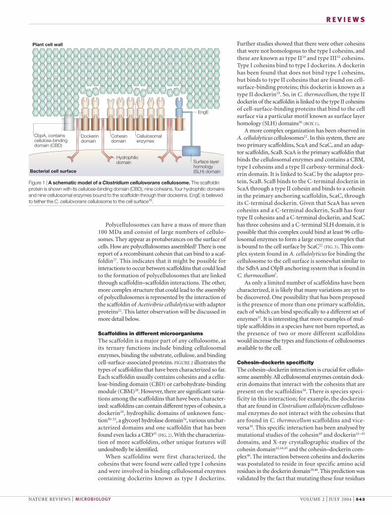
Plant cell wall
Bacterial cell surface
CbpA, contains cellulose-bindingdomain (CBD)
Hydrophilic domain
Cohesin domain
Dockerin domain
Surface layerhomology(SLH) domain
EngE
Cellulosomalenzymes
Figure 1 | A schematic model of a Clostridium cellulovorans cellulosome. The scaffoldinprotein is shown with its cellulose-binding domain (CBD), nine cohesins, four hydrophilic domainsand nine cellulosomal enzymes bound to the scaffoldin through their dockerins. EngE is believedto tether the C. cellulovorans cellulosome to the cell surface10.

544 | JULY 2004 | VOLUME 2 www.nature.com/reviews/micro
R E V I E W S
Table 2 | Cellulosomal subunits of clostridia
Cellulosomal enzymes Function Molecular mass (kDa) Modular structure*
Clostridium acetobutylicumCelA Endoglucanase 54 GH5-DS1CelE Endoglucanase 96 CBD3-Ig-GH9-DS1CelF Exoglucanase 81 GH48-DS1CelG Endoglucanase 77 GH9-CBD3-DS1CelH Exoglucanase 80 GH9-CBD3-DS1CelL Endoglucanase 60 GH9-DS1EngA Endoglucanase 67 GH44-DS1ManA Mannanase 47 GH5-DS1CAC0919 Sialidase 91 GH74-DS1CAC3469 Endoglucanase 110 (SLH)3-GH5-X-DS1Clostridium cellulolyticumCelA Endoglucanase 50 GH5-DS1CelC Endoglucanase 51 GH8-DS1CelD Endoglucanase 63 GH5-DS1CelE Endoglucanase 97 CBD4-Ig-GH9-DS1CelF Exoglucanase 78 GH48-DS1CelG Endoglucanase 80 GH9-CBD3-DS1CelH Endoglucanase 83 GH9-CBD3-DS1CelJ Endoglucanase 85 GH9-CBD3-DS1CelM Endoglucanase 58 GH9-DS1ManK Mannanase 48 DS1-GH5RglY Rhamnogalacturonan lyase 75 GPL11-DS1Clostridium cellulovoransEngB Endoglucanase 49 GH5-DS1EngE Endoglucanase 110 (SLH)3-GH5-X-DS1EngH Endoglucanase 79 GH9-CBD3-DS1EngK Endoglucanase 97 CBD4-Ig-GH9-DS1EngL Endoglucanase 58 GH9-DS1EngM Endoglucanase 96 CBD4-Ig-GH9-DS1EngY Endoglucanase 80 CBD2-GH9-DS1ExgS Exoglucanase 80 GH48-DS1ManA Mannanase 47 DS1-GH5PelA Pectate lyase 94 X-CBD2-GPL9-DS1XynA Xylanase/acetyl xylan esterase 57 GH11-DS1-CE4Clostridium josuiCelB Endoglucanase 51 GH8-DS1CelE Endoglucanase 81 GH9-CBD3-DS1CelD Exoglucanase 80 GH48-DS1AgaA α-Galactosidase 52 GH27-DS1Clostridium thermocellumCbhA Cellobiohydrolase 138 CBD4-Ig-GH9-X-X-CBD3-DS1CelA Endoglucanase 53 GH8-DS1CelB Endoglucanase 64 GH5-DS1CelD Endoglucanase 72 Ig-GH9-DS1CelE Endoglucanase 90 GH5-DS1-CE2CelF Endoglucanase 82 GH9-CBD3-DS1CelG Endoglucanase 63 GH5-DS1CelH Endoglucanase 102 GH26-GH5-CBD11-DS1CelJ Endoglucanase 178 X-Ig-GH9-GH44-DS-XCelK Endoglucanase 101 CBD4-Ig-GH9-DS1CelN Endoglucanase 82 GH9-CBD3-DS1CelO Endoglucanase (cellobiohydrolase) 75 CBD3-PT-GH5-DS1CelP Endoglucanase 58 GH9-DS1CelQ Endoglucanase 80 GH9-CBD3-DS1CelS Exoglucanase 83 GH48-DS1CelT Exoglucanase 65 GH9-DS1CseP Unknown 62 UN-DS1ChiA Chitinase 55 GH18-DS1LicB Lichenase 38 GH16-DS1ManA Mannanase 67 CBD4-GH26-PT-DS1XynA (XynU) Xylanase/acetyl xylan esterase 74 GH11-CBD4-DS1-CE4XynB (XynV) Xylanase 50 GH11-CBD6-DS1XynC Xylanase 70 X-GH10-DS1XynD Xylanase 70 CBD22-GH10-DS1XynY Xylanase/feruloyl esterase 120 CBD22-GH10-CBD22-DS1-CE1XynZ Xylanase/feruloyl esterase 92 CE1-CBD6-DS1-GH10
*The modular structures of cellulosomal subunits are indicated by abbreviations: CBD, cellulose-binding domain; CE, carbohydrate esterasefamily; DS1, dockerin domain type I; GH, glycosyl hydrolase; GPL, polysaccharide lyase family 9 (pectate lyase); Ig, immunoglobulin-like module;PT, proline-rich linker; SLH, surface layer homology domain; UN, unknown domain; X, unknown domain containing a hydrophilic domain. Modifiedwith permission from REF. 10 © (2003) American Society for Microbiology.

NATURE REVIEWS | MICROBIOLOGY VOLUME 2 | JULY 2004 | 545
R E V I E W S
resulted in a change in the cohesin–dockerin recogni-tion specificity43. The mutagenesis studies indicatedthat the specificity of this interaction was stronglyaffected by a single amino acid change (threonine toleucine) at a given position in the dockerin43, althoughit is thought that other subtle interactions are alsoinvolved. The association between dockerin andcohesin domains was shown to be largely dependenton hydrophobic interactions41. The three-dimensionalstructure of the dockerin–cohesin complex indicatesthat the cohesin–dockerin interaction is mediatedmainly by hydrophobic interactions between one ofthe ‘faces’ of the cohesin and α-helices 1 and 3 of thedockerin.
Cellulosomal enzymesThe cellulosomal enzymes include cellulases, hemicellu-lases, pectinase, chitinase and many ancillary enzymesthat can degrade plant cell wall materials. Cellulases arepart of a large group of glycosyl hydrolases that havebeen categorized into several families on the basis oftheir amino acid homology. Hemicellulases are able todegrade hemicelluloses, a class of polysaccharides thatcan form hydrogen bonds with cellulose fibrils andform a network in plant cell walls. Xylans and mannansare examples of hemicelluloses. A list of cellulosomalenzymes representing several glycosyl hydrolase familiesis presented in TABLE 2. Some 26 cellulosomal enzymeshave been identified for C. thermocellum7. Some of theenzymes work in concert to facilitate the degradation ofthe main polymers, for example, xylans and mannans.These include both cellulosomal and non-cellulosomalenzymes that remove various groups from the xylan andmannan backbones.
The endoglucanases, which cleave cellulose inter-nally, primarily belong to glycosyl hydrolase families 5and 9, and the exoglucanases, which can attack cellulosefrom either the reducing or non-reducing ends, belongto family 48. The family 9 endoglucanases are versatileas they not only cleave cellulose molecules internally,but also proceed in a processive manner along the chainfrom the cleavage site, and could be important enzymesin the cellulosome47.
C. cellulovorans48, C. acetobutylicum49 and C. cellu-lolyticum50 contain large gene clusters as well as unlinkedgenes that encode cellulosomal enzymes. The organiza-tion of the gene clusters in these species is related andthe clusters might have evolved from a common set ofgenes. In the case of the C. cellulovorans gene cluster, atransposase gene is located at its 3′ end, indicating thatlateral gene transfer might have occurred48. By contrast,the genes encoding the C. thermocellum cellulosomalenzymes are scattered throughout the chromosome51
and no clusters of cellulosomal enzyme genes have beenobserved, except for several genes involved in bindingthe cellulosome to the cell surface52.
Regulation of cellulosomal genesWhat are the factors that regulate the expression of cel-lulosomal genes? The regulation of cellulosomal geneexpression has been examined at the microscopic,
Clostridium cellulolyticum CipC (155 kDa)
Bacteroides cellulosolvens CipBc (245 kDa)
Clostridium josui CipC (120 kDa)
Clostridium cellulovorans CbpA (189 kDa)
Clostridium acetobutylicum CipA (154 kDa)
ScaA (CipV) (199 kDa)
ScaB (100 kDa)
ScaC (124 kDa)
Acetivibrio cellulolyticus
Type III cohesin
Unknown domain
Clostridium thermocellum
SdbA (69 kDa)
OlpB (178 kDa)
Orf2p (75 kDa)
OlpA (49 kDa)
CipA (196 kDa)
Ruminococcus flavefaciens
ScaB (181 kDa)
ScaA (93 kDa)
Signal peptide
Hydrophilicdomain
Type II cohesin
Type II dockerin
Surface layer homology (SLH) domain
Type I cohesin
Glycosyl hydrolase(family 9)
T, P, S, D, E or K-rich linking segments
Cellulose-bindingdomain (family 3)
Figure 2 | The modular structure of scaffoldins from various microorganisms. The non-enzymatic scaffoldin protein contains binding sites (cohesins) for the cellulosomal enzymesubunits6. Most scaffoldins contain between six and nine different cohesins. The species-specificvariations in scaffoldin properties allow the composition of the cellulosome to differ betweenbacterial species7,10. Some bacterial species that synthesize cellulosomes contain only one typeof scaffoldin, however, other species have been shown to contain multiple scaffoldins22,95, whichcould also potentially yield different types of cellulosomes.

546 | JULY 2004 | VOLUME 2 www.nature.com/reviews/micro
R E V I E W S
The growth medium has been shown to affect bothsubunit structure and function of the cellulosome.When cells are grown on different substrates, such asglucose, cellobiose, xylan, mannan or pectin, and theircellulosomes are fractionated by anion-exchange chro-matography, fractions are obtained that differ in sub-unit composition and enzymatic activity4,56,57. Thisimplies that the cell responds to different substrates byexpressing cellulosomal genes, which results in a popu-lation of cellulosomes with activities that are directedtowards the available substrate. So, the growth sub-strate has a significant effect on cellulosome synthesisand subunit composition.
As the cellulosome comprises a scaffoldin proteinand a large number of enzymatic subunits, it is ofinterest to determine how their genes are regulated,whether there is coordinate expression of the genes toform this multisubunit enzyme complex, and whattype of promoter region controls their expression.Transcription studies of the C. cellulovorans large cellu-losomal gene cluster indicated that there are severaloperons within the gene cluster and that there is coor-dinate expression of several of the operons58. The pro-moters for the genes were similar to those found forSIGMA-A RNA polymerases of Gram-positive bacteria.When cells were grown on different substrates such asglucose, xylan, mannan or pectin and their mRNAanalysed, abundant expression was observed for mostof the genes in the cellulosomal gene cluster as well asfor cellulosomal genes unlinked to the cluster, andmoderate or low levels of expression were observedwhen various monosaccharides were the substrates.The xylanase and pectate lyase genes were specificallyinduced in the presence of xylan and pectin, respec-tively. The results indicated that cellulases and hemi-cellulases were coordinately expressed, that cellulaseexpression was regulated by a CATABOLITE-REPRESSION-likemechanism, and that the presence of hemicellulosesinfluenced cellulose utilization by the cell58. Analysis ofthe transcription of cellulosomal genes that wereunlinked to the large cellulosomal gene cluster indi-cated that most were monocistronic and could beexpressed coordinately with the genes in the large genecluster56. Previous studies with C. thermocellum alsoconcluded that a catabolite-repression-like mechanismwas controlling the expression of cellulosomal genes59.
One of the main enzymes of the C. thermocellumcellulosome is the CelS exoglucanase. The level ofexpression of CelS is much higher when cells aregrown on cellulose than on cellobiose. When thetranscriptional level of celS mRNA was determined, itwas found that transcriptional activity was inverselyproportional to the growth rate. Two transcriptionalstart points were observed upstream of the transla-tional start point that exhibited homology to the σA and σB promoter sites in Bacillus subtilis. The relativeactivity of the promoters remained constant underthe conditions studied. celS is therefore regulated atthe transcriptional level and its expression is modu-lated by the growth rate under conditions of nitrogenand cellobiose limitation60.
physiological and transcription levels. The earliestmicroscopic studies demonstrated the presence of protuberances, which contain polycellulosomes53.In a study using scanning electron microscopy, the pro-tuberances were observed from cellulose-grown cells,but not glucose-, fructose-, CELLOBIOSE- or CMC-growncells54. The formation of protuberances took about 4 hours when C. cellulovorans cells were grown on cel-lulose. Within 5 minutes of the addition of the solublesugars glucose, cellobiose or methylglucose, the protu-berances could no longer be detected. This indicatedthat the dissociation of the protuberances was rapidand that the presence of soluble sugars was responsible.An early transcription study on a cellulosomal gene,engB, indicated that it was transcribed as a single tran-scription unit and that the relative amount of engBmRNA was much higher in cellulose-grown cells thanin cellobiose-grown cells55.
CELLOBIOSE
An individual unit of cellulose.
SIGMA-A
Sigma factors are variableprotein components of thebacterial RNA polymerase thatinfluence transcription bydetermining where thepolymerase binds to DNA. InBacillus, σA is a housekeepingsigma factor, σB an alternativesigma factor that responds tostress and σL the Bacillus subtilishomologue of σ54, the majorvariant sigma factor in E. coli.
Box 1 | Cellulosomes and SLH domains
Many Gram-positive bacteria have a surface-layer protein (SLP) that surrounds theexterior cell wall. This layer of proteins is attached to secondary cell-wall polymers inthe rigid cell-wall layer. The surface layer homology (SLH) domains of severalextracellular enzymes are homologous to regions of the SLP and it is believed that,like the SLP, SLH domains also attach to secondary cell-wall polymers and bind theseSLH-containing enzymes to the cell surface. As the cellulosomes of Clostridiumthermocellum are attached to surface layer SLH- and cohesin-containing proteinsthrough dockerin–cohesin interactions, it is believed that this interaction tethers theC. thermocellum cellulosomes to the cell surface. In the case of Clostridiumcellulovorans, a cellulosomal enzyme, EngE, contains both a dockerin and threetandem SLHs. EngE can bind to the scaffoldin protein through its dockerin as well asto the cell surface through its tandem SLHs. So, the C. cellulovorans cellulosome isbound to the cell surface through EngE, which binds to both the scaffoldin proteinand the cell surface.
ScaB dockerindomain
Unknown domain
SLH domain
Cellulosomal subunits
Bacterial cell surface
Plant cell wall
Dockerin domain(Type II)
ScaB
ScaC
ScaA
CBDGlycosyl hydrolasedomain (Family 9)
Cohesin domain (Type I)
Cohesin domain(Type II)
Cohesin domain(new group)
Figure 3 | A model of the Acetivibrio cellulolyticus cellulosome. This cellulosome has twoprimary scaffoldins, ScaA and ScaC, and an adaptor scaffoldin, ScaB22. The proteins areconnected via dockerin–cohesin interactions.
NATURE REVIEWS | MICROBIOLOGY VOLUME 2 | JULY 2004 | 547
R E V I E W S
30%. The antisense strain also overproduced two non-cellulosomal proteins, P105 and P98. These results showthat Cel48F has an important role in the degradation ofcrystalline cellulose and that the translational inhibitionof the synthesis of Cel48F could reduce the productionof another transcriptionally linked protein, CipC.
Further analysis of cellulolysis has been obtained bydisrupting the cipC gene of C. cellulolyticum66. Thismutant was severely impaired in its ability to degradecrystalline cellulose and it produced a small amount of atruncated CipC (P120), which could complex with cel-lulosomal enzymes. However, none of the enzymesassociated with P120 was encoded by the genes distal tocipC in the large cipC gene cluster. The complex formedwith P120 contained three important proteins desig-nated P98, P105 and P125, and 12 other dockerin-containing enzymes encoded by genes outside the cipCgene cluster. These results indicate that a mutation inthe cipC gene has a strong polar effect and that theenzymes encoded downstream in the cipC gene clusterare essential for the efficient degradation of crystallinecellulose by the cellulosome.
Without question, our relatively recent ability to usedirect genetic analysis techniques to analyse celluloso-mal genes will lead to a much better understanding ofcellulosomal structure and function.
Biotechnological uses of cellulosomesThere is much interest in exploiting the properties ofcellulosomes for practical purposes6. The specificcohesin–dockerin interaction, the strong cellulose-binding property of the CBD domain, the potential fortransforming non-cellulose degraders to cellulosedegraders and the construction and use of ‘designer’cellulosomes for specific degradative activities areimportant properties of the cellulosome that can beused in biotechnology.
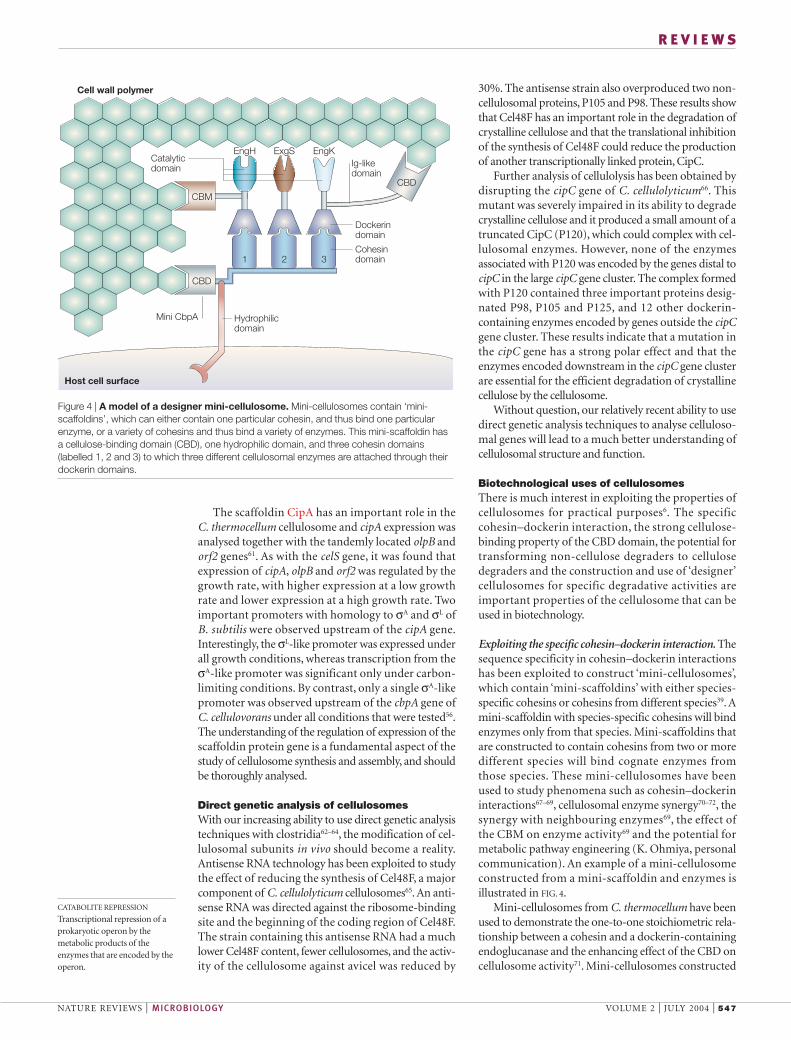
Exploiting the specific cohesin–dockerin interaction. Thesequence specificity in cohesin–dockerin interactionshas been exploited to construct ‘mini-cellulosomes’,which contain ‘mini-scaffoldins’ with either species-specific cohesins or cohesins from different species39. Amini-scaffoldin with species-specific cohesins will bindenzymes only from that species. Mini-scaffoldins thatare constructed to contain cohesins from two or moredifferent species will bind cognate enzymes fromthose species. These mini-cellulosomes have beenused to study phenomena such as cohesin–dockerininteractions67–69, cellulosomal enzyme synergy70–72, thesynergy with neighbouring enzymes69, the effect ofthe CBM on enzyme activity69 and the potential formetabolic pathway engineering (K. Ohmiya, personalcommunication). An example of a mini-cellulosomeconstructed from a mini-scaffoldin and enzymes isillustrated in FIG. 4.
Mini-cellulosomes from C. thermocellum have beenused to demonstrate the one-to-one stoichiometric rela-tionship between a cohesin and a dockerin-containingendoglucanase and the enhancing effect of the CBD oncellulosome activity71. Mini-cellulosomes constructed
The scaffoldin CipA has an important role in the C. thermocellum cellulosome and cipA expression wasanalysed together with the tandemly located olpB andorf2 genes61. As with the celS gene, it was found thatexpression of cipA, olpB and orf2 was regulated by thegrowth rate, with higher expression at a low growthrate and lower expression at a high growth rate. Twoimportant promoters with homology to σA and σL ofB. subtilis were observed upstream of the cipA gene.Interestingly, the σL-like promoter was expressed underall growth conditions, whereas transcription from theσA-like promoter was significant only under carbon-limiting conditions. By contrast, only a single σA-likepromoter was observed upstream of the cbpA gene ofC. cellulovorans under all conditions that were tested56.The understanding of the regulation of expression of thescaffoldin protein gene is a fundamental aspect of thestudy of cellulosome synthesis and assembly, and shouldbe thoroughly analysed.
Direct genetic analysis of cellulosomesWith our increasing ability to use direct genetic analysistechniques with clostridia62–64, the modification of cel-lulosomal subunits in vivo should become a reality.Antisense RNA technology has been exploited to studythe effect of reducing the synthesis of Cel48F, a majorcomponent of C. cellulolyticum cellulosomes65. An anti-sense RNA was directed against the ribosome-bindingsite and the beginning of the coding region of Cel48F.The strain containing this antisense RNA had a muchlower Cel48F content, fewer cellulosomes, and the activ-ity of the cellulosome against avicel was reduced by
CATABOLITE REPRESSION
Transcriptional repression of aprokaryotic operon by themetabolic products of theenzymes that are encoded by theoperon.
Host cell surface
Cell wall polymer
Ig-likedomain
EngH ExgSCatalytic domain
EngK
Dockerindomain
Cohesindomain
Mini CbpA
CBD
1 2 3
Hydrophilic domain
CBM
CBD
Figure 4 | A model of a designer mini-cellulosome. Mini-cellulosomes contain ‘mini-scaffoldins’, which can either contain one particular cohesin, and thus bind one particularenzyme, or a variety of cohesins and thus bind a variety of enzymes. This mini-scaffoldin hasa cellulose-binding domain (CBD), one hydrophilic domain, and three cohesin domains(labelled 1, 2 and 3) to which three different cellulosomal enzymes are attached through theirdockerin domains.
548 | JULY 2004 | VOLUME 2 www.nature.com/reviews/micro
R E V I E W S
containing a cellulose-binding motif (CBM3a), one X2module (a hydrophilic domain of unknown function)and a cohesin (cohesin 1), and a chimeric scaffoldinScaf3 gene containing a cohesin from C. thermocellum(cohesin 3) fused to the C-terminus of CipC1
c(CipC1
c–
Coh3t) were transformed into C. acetobutylicum. Both
mini-scaffoldins were produced and secreted by C. acetobutylicum and both were able to bind their cog-nate enzymes, indicating that the synthesis of activemini cellulosomes by C. acetobutylicum is possible.
Another approach has been to overexpress ahomologous mini-cellulosome containing a CBD, twocohesin domains and a cognate enzyme in C. aceto-butylicum82. This approach produced a mini-cellulo-some of 250 kDa, with two major subunits of 122 kDaand 84 kDa, as opposed to the normal 665 kDa. Themini-cellulosome was unable to degrade avicel or bac-terial cellulose, but did show low activity on CMC andphosphoric-acid-swollen cellulose, as shown previ-ously. These experiments were the first to demonstratethe in vivo expression of mini-cellulosomes and indi-cate that C. acetobutylicum and other species can betransformed with cellulosomal genes to form activecellulosomes in vivo. This finding will allow the devel-opment of many commercially important microor-ganisms that use cellulosic biomass as an inexpensivegrowth substrate.
Heterologous expression of cellulosomal genes. If micro-organisms could use cellulose as a growth substrate,inexpensive biomass such as corn stover, rice straw, saw-dust and wheat straw could be used to produce ‘value-added’ products such as ethanol, amino acids andorganic acids. One approach would be to transformmicroorganisms with cellulosomal genes so that cellu-losic biomass could be directly converted to valuableproducts. The expression of C. cellulovorans EngB in C. acetobutylicum has been reported83. In anotherstudy, the genes for mini-scaffoldins derived from C. cellulolyticum and C. thermocellum were introducedinto C. acetobutylicum and expressed as active mini-scaffoldins81. With further refinements, it is likely thatcellulosomal genes will be expressed in several differ-ent species to allow these organisms to utilize biomassand agricultural wastes to produce products such asethanol, amino acid and organic acids. This willrequire the transfer of a scaffoldin gene and a numberof exoglucanase, endoglucanase and hemicellulasegenes into the microorganism of interest.
Improving cellulosomal enzyme properties: DNAshuffling. A full understanding of the function of thecellulosome should lead to the synthesis of a maximallyefficient cellulosome with specified properties.As well asmaximizing the synergy both between the cellulosomalenzymes and between cellulosomes and non-celluloso-mal enzymes, another approach would be to modify andimprove the properties of the enzymes. The creation of arecombinant cellulase with greater heat stability has beenreported84. In this case, DNA shuffling was carried outbetween two highly homologous endoglucanases from
from only C. cellulovorans components were used toinvestigate the synergy between cellulases70, between cel-lulases and hemicellulases72, and between a cellulosomalenzyme and non-cellulosomal enzymes73,74. In all cases,synergy was observed, indicating that the synergybetween the enzymes in cellulosomes makes the cellulo-some structure more effective in attacking the substrate.The synergy observed between cellulosomes and non-cellulosomal enzymes also suggests that the maximumeffectiveness in degrading natural substrates requires theinteraction of cellulosomes and non-cellulosomaldegradative enzymes73,74.
A scaffoldin with three different cohesins that canbind three enzymes by their cognate dockerins has beenconstructed (K. Ohmiya, personal communication).These researchers hope to show that this designer mini-cellulosome can organize three tandem enzymes into ametabolic pathway that is capable of converting a sub-strate into a desired product by ‘enzyme channelling’ ina similar manner to that found in vivo. This could leadto the future development of artificial metabolic path-ways with controlled activities for the synthesis of anydesired product.
Practical applications of the CBD. The presence ofCBDs in many scaffoldin proteins has led to the devel-opment of some practical applications for the CBDdomain75,76. In a series of recent papers, Shoseyov andcolleagues have shown that fusion proteins containing aCBD and various other proteins are capable of bindingto a cellulose matrix. These fusion proteins can be usedas a bioreactor77, a plant-growth modulator78,79 and aprotein-purification system80. The advantages of using a CBD system are that a relatively inexpensive, inert cel-lulose matrix with excellent physical properties can beused, a large-scale, safe, affinity-purification procedureis possible and it can be used to modify physical andchemical properties in agro-biotechnology, both in vitroand in vivo.
The Clostridium acetobutylicum cellulosome. Whenthe C. acetobutylicum genome was sequenced49, it wasobserved that several cellulosomal genes were present ina gene cluster similar to that found in C. cellulolyticumand C. cellulovorans. However, as mentioned above,C. acetobutylicum, which is an important species for theproduction of acetone and butanol, produces a cellu-losome with a molecular mass of about 665 kDa19
but cannot grow on cellulose20. This is due to eitherextremely low production of cellulosomes or a defi-ciency in the cellulosome that prevents it from degrad-ing crystalline cellulose. At present, much research isfocused on characterizing the C. acetobutylicum cellulo-some, with the aim of converting this microorganisminto an active user of cellulosic biomass.
One approach to this problem is to insert the cellulo-somal genes from an active cellulosome producer, suchas C. cellulolyticum, into C. acetobutylicum to producean active heterologous cellulosome that is capable ofdegrading crystalline cellulose81. For this purpose, amini scaffoldin gene (CipC1
c) from C. cellulolyticum
NATURE REVIEWS | MICROBIOLOGY VOLUME 2 | JULY 2004 | 549
R E V I E W S
Future areas for investigationThe current status of cellulosomal research indicatesthat advances in the following areas are possible.
Cellulosome genetics. Although biochemical studies ofcellulosomes have progressed well, the most limitingaspect of cellulosome research is the lack of geneticanalysis of the cellulosome. The increasing use of, andimprovements in, genetic manipulations in clostridiawill facilitate further studies on the function and regula-tion of cellulosomal genes, the structure of the cellulo-some, and the secretion and extracellular assembly ofthe cellulosome.
Assembly of cellulosomes and polycellulosomes. Theassembly process is one of the most intriguing aspects ofcellulosome and polycellulosome synthesis. How arecellulosomes assembled extracellularly? Are there extra-cellular chaperones or are chaperone-like functionsincorporated into the cellulosomal proteins? Whatdetermines which enzymes are going to bind to a partic-ular scaffoldin molecule? Are there built-in factors thatdetermine cohesin–dockerin interactions? How arepolycellulosomes assembled? One approach to answer-ing these questions is to determine whether specific cel-lulosomal enzymes are bound to specific cohesins presentin the scaffoldin in preference to other cellulosomalenzymes.
DNA shuffling to improve cellulosomal enzyme proper-ties. DNA-shuffling technology can be used to modifythe properties of cellulosomal enzymes and to improvethe function and specificity of cellulosomes. By usingtargeted selection methods, almost any desired enzymecharacteristic can be obtained. It seems likely that con-tinued use of this technology will result in more activeheat-stable enzymes that can function at low and highpH values and under high and low salt concentrations.
Designer cellulosomes — construction of function-specific cellulosomes. Cellulosomes with specific dock-erin-binding capacities can be created by using cohesinsfrom various scaffoldins. By creating chimeric scaffoldinproteins with tandem dockerin-specific cohesins, itshould be possible to construct designer cellulosomesand mini-cellulosomes with enzymatic pathways inwhich efficient substrate channelling occurs. This couldlead to the development of complex and sophisticatedbioreactors and biosensors.
Transformation of cellulosomal genes — conversion ofnon-cellulolytic to cellulolytic organisms. By creatingrecombinant plasmids containing cellulosomal genesand by using transformation techniques, it shouldbecome possible to convert several microorganisms intocellulose degraders. These microorganisms will be ableto use cellulosic biomass and agricultural wastes andconvert them directly to useful products, such asethanol, butanol, amino acids and organic acids, that arecurrently synthesized from expensive substrates by thesemicroorganisms.
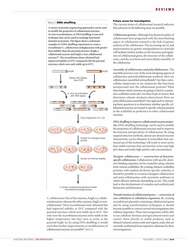
C. cellulovorans. One of the enzymes, EngB, is a cellulo-somal enzyme whereas the other enzyme, EngD, is non-cellulosomal. Three recombinants were obtained thathad improved stability at 55ºC compared with theparental enzymes, which were stable up to 45ºC. Notonly were the recombinant enzymes more stable at thehigher temperature, but they were as active as theparental EngB. So, by using DNA shuffling, it wouldseem that further improvements or modifications ofcellulosomal enzymes are possible85 (BOX 2).
Box 2 | DNA shuffling
A variety of protein-engineering approaches can be usedto modify the properties of cellulosomal enzymes.In vitro recombination, or DNA shuffling, is one suchtechnique that can be used to exchange functionaldomains in proteins. The figure shows a schematicexample of a DNA shuffling experiment to create arecombinant C. cellulovorans endoglucanase with greaterheat stability than the parental enzymes (EngB, acellulosomal enzyme and EngD, a non-cellulosomalenzyme)84. The recombinant clones obtained hadimproved stability at 55°C compared with the parentalenzymes, which were only stable up to 45°C.
Cleave with DNase, then PCR
3 clones containing temperature-stablerecombinant endoglucanase
Clone fragments in E. coli
In vitro recombination with DNA ligase
engB engD
Screen for endoglucanaseactivity
Screen for thermal stability

550 | JULY 2004 | VOLUME 2 www.nature.com/reviews/micro
R E V I E W S
enzymes could reveal the best combinations for effi-cient degradation of plant-cell-wall materials — onecannot assume that only cellulosomal enzymes arenecessary.
In conclusion, our understanding of the basic struc-ture and function of the cellulosome system is now at apoint where cellulosome researchers should be able tomake rapid strides towards the development of severaluseful applications in biotechnology.
Synergy between cellulosomal enzymes and cellulosomaland non-cellulosomal enzymes. To efficiently degradecellulosic biomass, synergy studies should be carriedout between cellulosomal enzymes from purified cel-lulosomes with defined enzymatic activities. Thecombination of specific enzymes should result inactivity that is much greater than the activity of theindividual enzymes. In addition, synergy studiesbetween mixtures of cellulosomes and non-cellulosomal
1. Mansfield, S. D., Mooney, C. & Saddler, J. N. Substrate andenzyme characteristics that limit cellulose hydrolysis.Biotechnol. Prog. 15, 804–816 (1999).
2. Lynd, L. R., Weimer, P. J., van Zyl, W. H. & Pretorius, I. S.Microbial cellulose utilization: fundamentals andbiotechnology. Microbiol. Mol. Biol. Rev. 66, 506–577(2002).
3. Warren, R. A. J. Microbial hydrolysis of polysaccharides.Annu. Rev. Microbiol. 50, 183–212 (1996).
4. Bayer, E. A., Setter, E. & Lamed, R. Organization anddistribution of the cellulosome in Clostridium thermocellum.J. Bacteriol. 163, 552–559 (1985).
5. Bayer, E. A., Belaich, J.-P., Shoham, Y. & Lamed, R. Thecellulosomes: multi-enzyme machines for degradation ofplant cell wall polysaccharides. Annu. Rev. Microbiol. (in thepress).
6. Bayer, E. A., Morag, E. & Lamed, R. The cellulosome — a treasure trove for biotechnology. Trends Biotechnol. 12,378–386 (1994).
7. Bayer, E. A., Shimon, L. J. W., Shoham, Y. & Lamed, R.Cellulosomes — structure and ultrastructure. J. Struct. Biol.124, 221–234 (1998).
8. Bayer, E. A., Shoham, Y. & Lamed, R. Cellulose-decomposing prokaryotes and their enzyme systems. inThe Prokaryotes: An Evolving Electronic Resource for theMicrobiological Community (eds Dworkin, M., Falkow, S.,Rosenberg, E., Schleifer, K.-H. & Stackebrandt, E.) 3rdedition <http://141.150.157.117:8080/prokPUB/chaprender/jsp/showchap.jsp?chapnum=297&initsec=01_00> (Springer–Verlag, New York, 2000).
9. Beguin, P. & Alzari, P. M. The cellulosome of Clostridiumthermocellum. Biochem. Soc. Trans. 26, 178–185(1998).
10. Doi, R. H., Kosugi, A., Murashima, K., Tamaru, Y. & Han, S. O. Cellulosomes from mesophilic bacteria. J. Bacteriol. 185, 5907–5914 (2003).
11. Ohmiya, K., Sakka, K., Kimura, T. & Morimoto, K.Application of microbial genes to recalcitrant biomassutilization and environmental conservation. J. Biosci. Bioeng.95, 549–561 (2003).
12. Schwarz, W. H. The cellulosome and cellulose degradationby anaerobic bacteria. Appl. Microbiol. Biotechnol. 56,634–649 (2001).
13. Wilson, C. A. & Wood, T. M. The anaerobic fungusNeocallimastix frontalis: isolation and properties of acellulosome-type enzyme fraction with the capacity tosolubilize hydrogen-bond-ordered cellulose. Appl. Microbiol.Biotechnol. 37, 125–129 (1992).
14. Ali, B. R. S. et al. Cellulases and hemicellulases of theanaerobic fungus Piromyces constitute a multiproteincellulose-binding complex and encoded by multigenefamilies. FEMS Microbiol. Lett. 125, 15–22 (1995).
15. Fanutti, C., Ponyi, T., Black, G. W., Hazelwood, G. P. &Gilbert, H. J. The conserved noncatalytic 40-residuesequence in cellulases and hemicellulases from anaerobicfungi functions as a protein docking domain. J. Biol. Chem.270, 29314–29322 (1995).
16. Fillingham, I. J., Kroon, P. A., Williamson, G., Gilbert, H. J., &Hazlewood, G. P. A modular cinnamoyl ester hydrolase fromthe anaerobic fungus Piromyces equi acts synergisticallywith xylanase and is part of a multiprotein cellulose-bindingcellulase–hemicellulase complex. Biochem. J. 343,215–224 (1999).
17. Steenbakkers, P. J. M. et al. Non-catalytic docking domainsof cellulosomes of anaerobic fungi. J. Bacteriol. 183,5325–5333 (2001).
18. Steenbakkers, P. J. M. et al. The β-glucosidase in thecellulosome of the anaerobic fungus Piromyces sp. strain E2is a family 3 glycoside hydrolase. Biochem. J. 370, 963–970(2003).
19. Sabathe, F., Belaich, A. & Soucaille, P. Characterization ofthe cellulolytic complex (cellulosome) of Clostridiumacetobutylicum. FEMS Microbiol. Lett. 217, 15–22 (2002).
20. Lee, S. F., Forsberg, C. W. & Gibbins, L. N. Cellulolyticactivity of Clostridium acetobutylicum. Appl. Environ.Microbiol. 50, 220–228 (1985).
21. Madkour, M. & Mayer, F. Structural organization of the intactbacterial cellulosome as revealed by electron microscopy.Cell Biol. Int. 27, 831–836 (2003).
22. Xu, Q. et al. The cellulosome system of Acetivibriocellulolyticus includes a novel type of adaptor protein and acell surface anchoring protein. J. Bacteriol. 185, 4548–4557(2003).
23. Pohlschroder, M. D., Leschine, S. B., & Canale-Parola, E.Multicomplex cellulase–xylanase system of Clostridiumpapyrosolvens C7. J. Bacteriol. 176, 70–76 (1994).
24. Pohlschroder, M., Canale-Parola, E. & Leschine, S. B.Ultrastructural diversity of the cellulase complexes ofClostridium papyrosolvens C7. J. Bacteriol. 177,6625–6629 (1995).One of the best structural studies showing thesubpopulations of cellulosomes.
25. Ali, B. R. S., Romaniec, M. P. M., Hazlewood, G. P. &Freedman, R. B. Characterization of the subunits in anapparently homogeneous subpopulation of Clostridiumthermocellum cellulosomes. Enzyme Microb. Technol. 17,705–711 (1995).
26. Murashima, K., Kosugi, A. & Doi, R. H. Determination ofsubunit composition of Clostridium cellulovoranscellulosomes that degrade plant cell walls. Appl. Environ.Microbiol. 68, 1610–1615 (2002).
27. Park, J.-S., Matano, Y. & Doi, R. H. Cohesin–dockerininteractions of cellusomal subunits of Clostridiumcellulovorans. J. Bacteriol. 183, 5431–5435 (2001).
28. Boraston, A. B. et al. in Carbohydrate Bioengineering (edsGilbert, H. J., Davies, G. J., Henrissat, B. & Svensson, B.)202–211 (The Royal Society of Chemistry, Cambridge,1999).
29. Leibovitz, E. & Beguin, P. A new type of cohesin domain thatspecifically binds the dockerin domain of the Clostridiumthermocellum cellulosome integrating protein CipA. J. Bacteriol. 178, 3077–3084 (1996).
30. Shoseyov, O., Takagi, M., Goldstein, M. & Doi, R. H. Primarysequence analysis of Clostridium cellulovorans cellulosebinding protein A (CbpA). Proc. Natl Acad. Sci. USA 89,3483–3487 (1992).
31. Pages, S. et al. Sequence analysis of scaffolding proteinCipC and ORFXp, a new cohesin-containing protein inClostridium cellulolyticum: comparison of various cohesindomains and subcellular localization of ORFXp. J. Bacteriol.181, 1801–1810 (1999).
32. Gerngross, U., Romaniec, M. P. M., Kobayashi, T.,Huskisson, N. S. & Demain, A. L. Sequencing of aClostridium thermocellum gene (cipA) encoding thecellulosomal SL-protein reveals an unusual degree ofinternal homology. Mol. Microbiol. 8, 325–334 (1993).
33. Kakiuchi, M. et al. Cloning and DNA sequencing of thegenes encoding Clostridium josui scaffolding protein CipAand cellulase CelD and identification of their gene productsas major components of the cellulosome. J. Bacteriol. 180,4303–4308 (1998).
34. Ding, S.-Y., Bayer, E. A., Steiner, D., Shoham, Y. & Lamed, R. A novel cellulosomal scaffoldin from Acetivibriocellulolyticus that contains a family-9 glycosyl hydrolase. J. Bacteriol. 181, 6720–6729 (1999).
35. Ding, S.-Y. et al. Cellulosomal scaffoldin-like proteins fromRuminococcus flavefaciens. J. Bacteriol. 183, 1945–1953(2001).
36. Lemaire, M., Ohayon, H., Gounon,P., Fujino, T., & Beguin, P.OlpB, a new outer layer protein of Clostridium thermocellum,and binding of its S-layer-like domains to components of thecell envelope. J. Bacteriol. 177, 2451–2459 (1995).
37. Rincon, M. T. et al. Novel organization and divergentdockerin specificities in the cellulosome system ofRuminococcus flavefaciens. J. Bacteriol. 185, 703–713(2003).
38. Tokatlidis, K., Salamitou, S., Beguin, P., Dhurjati, P. & Aubert, J.-P. Interaction of the duplicated segment carriedby Clostridium thermocellum cellulases with cellulosomecomponents. FEBS Lett. 291, 185–188 (1991).An early example of a dockerin–cohesin interaction.
39. Pages, S. et al. Species-specificity of the cohesin–dockerininteraction between Clostridium thermocellum andClostridium cellulolyticum: prediction of specificitydeterminants of the dockerin domain. Proteins: Struct.Funct. Genet. 29, 517–527 (1997).An important illustration of the species-specificity ofthe cohesin–dockerin interaction.
40. Miras, I., Schaeffer, F., Beguin, P., & Alzari, P. M. Mapping bysite-directed mutagenesis of the region responsible forcohesin–dockerin interaction on the surface of the seventhcohesin domain of Clostridium thermocellum CipA.Biochemistry 41, 2115–2119 (2002).
41. Schaeffer, F. et al. Duplicated dockerin subdomains ofClostridium thermocellum endoglucanase CelD bind to acohesin domain of the scaffolding protein CipA with distinctthermodynamic parameters and a negative cooperativity.Biochemistry 41, 2106–2114 (2002).
42. Shimon, L. J. et al. A cohesin domain from Clostridiumthermocellum: the crystal structure provides new insightsinto cellulosome assembly. Structure 5, 381–390 (1997).
43. Mechaly, A. S. et al. Cohesin–dockerin recognition incellulosome assembly: experiment versus hypothesis.Proteins 39, 170–177 (2000).
44. Tavares, G. A., Beguin, P. & Alzari, P. M. The crystal structureof a Type I cohesin domain at 1. 7 Å resolution. J. Mol. Biol.273, 701–713 (1997).
45. Spinelli, S. et al. Crystal structure of a cohesin module fromClostridium cellulolyticum: Implications for dockerinrecognition. J. Mol. Biol. 304, 189–200 (2000).
46. Carvalho, A. L. et al. Cellulosome assembly revealed by thecrystal structure of the cohesin–dockerin complex. Proc.Natl Acad. Sci. USA 100, 13809–13814 (2003).A structural analysis of the interaction betweencohesins and dockerins.
47. Gaudin, C., Belaich, A., Champ, S. & Belaich, J.-P. CelE, amultidomain cellulase from Clostridium cellulolyticum: a keyenzyme in the cellulosome? J. Bacteriol. 182, 1910–1915(2000).
48. Tamaru, Y., Karita, S., Ibrahim, A., Chan, H. & Doi, R. H. A large gene cluster for the Clostridium cellulovoranscellulosome. J. Bacteriol. 182, 5906–5910 (2000).
49. Nolling, J. et al. Genome sequence and comparativeanalysis of the solvent-producing bacterium Clostridiumacetobutylicum. J. Bacteriol. 183, 4823–4838 (2001).
50. Belaich, A. et al. Cel9M, a new family 9 cellulase of theClostridium cellulolyticum cellulosome. J. Bacteriol. 184,1378–1384 (2002).
51. Guglielmi, G. & Beguin, P. Cellulase and hemicellulase genesof Clostridium thermocellum from five independentcollections contain few overlaps and are widely scatteredacross the chromosome. FEMS Microbiol. Lett. 161,209–215 (1998).
52. Fujino, T., Beguin, P. & Aubert, J.-P. Organization of aClostridium thermocellum gene cluster encoding thecellulosomal scaffolding protein CipA and a protein possiblyinvolved in attachment of the cellulosome to the cell surface.J. Bacteriol. 175, 1891–1899 (1993).
53. Lamed, R., Setter, E. & Bayer, E. A. Characterization of acellulose-binding cellulase-containing complex inClostridium thermocellum. J. Bacteriol. 156, 828–836(1983).
54. Blair, B. G. & Anderson, K. L. Regulation of cellulose-inducible structures of Clostridium cellulovorans. Can. J.Microbiol. 45, 242–249 (1999).
55. Attwood, G. T., Blaschek, H. P. & White, B. A.Transcriptional analysis of the Clostridium cellulovoransendoglucanase gene, engB. FEMS Microbiol. Lett. 124,277–284 (1994).

NATURE REVIEWS | MICROBIOLOGY VOLUME 2 | JULY 2004 | 551
R E V I E W S
56. Han, S. O., Yukawa, H., Inui, M. & Doi, R. H. Regulation ofexpression of cellulosomal cellulase and hemicellulasegenes in Clostridium cellulovorans. J. Bacteriol. 185,6067–6075 (2003).Illustration of the coordinate expression of cellulasegenes and its regulation by a catabolite-repression-like mechanism.
57. Ali, B. R. S., Romaniec, M. P. M., Hazlewood, G. P. &Freedman, R. B. Characterization of the subunits in anapparently homogeneous subpopulation of Clostridiumthermocellum cellulosomes. Enzyme Microb. Technol. 17,705–711 (1995).
58. Han, S. O., Yukawa, H., Inui, M. & Doi, R. H. Transcription ofClostridium cellulovorans cellulosomal cellulase andhemicellulase genes. J. Bacteriol. 185, 2520–2527 (2003).
59. Mishra, S., Beguin, P. & Aubert, J. P. Transcription ofClostridium thermocellum endoglucanase genes celF andcelD. J. Bacteriol. 173, 80–85 (1991).
60. Dror, T. W. et al. Regulation of the cellulosomal celS (cel48A)gene of Clostridium thermocellum is growth rate dependent.J. Bacteriol. 185, 3042–3048 (2003).
61. Dror, T. W., Rolider, A., Bayer, E. A., Lamed, R. & Shoham, Y.Regulation of expression of scaffoldin-related genes inClostridium thermocellum. J. Bacteriol. 185, 5109–5116(2003).References 60 and 61 show that the expression ofcellulosomal genes is growth-rate-dependent.
62. Jennert, K. C. B., Tardif, C., Young, D. I. & Young, M. Genetransfer to Clostridium cellulolyticum ATCC 35319.Microbiology 12, 3071–3080 (2000).
63. Tardif, C., Maamar, H., Balfin, M. & Belaich, J. P.Electrotransformation studies in Clostridium cellulolyticum.J. Ind. Microbiol. Biotechnol. 27, 271–274 (2001).
64. Tyurin, M. V., Desai, S. G. & Lynd, L. R.Electrotransformation of Clostridium thermocellum. Appl.Environ. Microbiol. 70, 883–890 (2004).
65. Perret, S., Maamar, H., Belaich, J. P. & Tardif, C. Use ofantisense RNA to modify the composition of cellulosomesproduced by Clostridium cellulolyticum. Mol. Microbiol. 51,599–607 (2004).Anti-sense RNA technology was used to illustrate theimportance of exoglucanase Cel48F in the fullexpression of cellulosome activity.
66. Maamar, H. et al. Cellulolysis is severely affected inClostridium cellulolyticum strain cipCMut1. Mol. Microbiol.51, 589–598 (2004).Shows the importance of the expression of thescaffoldin gene in the expression of downstreamgenes of cipC and the enzymes in the cipC genecluster for full expression of the cellulosome activity.
67. Mechaly, A. et al. Cohesin–dockerin interaction incellulosome assembly. J. Biol. Chem. 276, 9883–9888(2001).
68. Fierobe, H.-P. et al. Design and production of activecellulosome chimeras: selective incorporation of dockerin-containing enzymes into defined functional complexes. J. Biol. Chem. 276, 21257–21261 (2001).
69. Fierobe, H.-P. et al. Degradation of cellulose substrates bycellulosome chimeras. J. Biol. Chem. 277, 49621–49630(2002).A good example of the use of chimeric mini-scaffoldins to analyse the functions of enzymes,CBDs and activity.
70. Murashima, K., Kosugi, A. & Doi, R. H. Synergistic effects oncrystalline cellulose degradation between cellulosomalcellulases from Clostridium cellulovorans. J. Bacteriol. 184,5088–5095 (2002).
71. Kataeva, I., Guglielmi, G. & Beguin, P. Interaction betweenClostridium thermocellum endoglucanase CelD andpolypeptides derived from the cellulosome-integratingprotein CipA: stoichiometry and cellulolytic activity of thecomplexes. Biochem. J. 326, 617–624 (1997).
72. Murashima, K., Kosugi, A. & Doi, R. H. Synergistic effects ofcellulosomal xylanase and cellulases from Clostridiumcellulovorans on plant cell wall degradation. J. Bacteriol. 85,1518–1524 (2003).
73. Kosugi, A., Murashima, K. & Doi, R. H. Characterization of non-cellulosomal subunits, ArfA and BgaA fromClostridium cellulovorans, that cooperate with thecellulosome in plant cell wall degradation. J. Bacteriol. 184,6859–6865 (2002).Evidence for interactions between the cellulosomeand non-cellulosomal enzymes.
74. Morag, E., Bayer, E. A. & Lamed, R. Relationship ofcellulosomal and noncellulosomal xylanases of Clostridiumthermocellum to cellulose-degrading enzymes. J. Bacteriol.172, 6098–6105 (1990).
75. Levy, I. & Shoseyov, O. Cellulose-binding domains:biotechnological applications. Biotechnol. Adv. 20, 191–213(2002).Describes the practical applications of CBDtechnology.
76. Tomme, P. et al. Characterization and affinity applications ofcellulose-binding domains. J. Chromatogr. B Biomed. Sci.Appl. 715, 283–296 (1998).
77. Shpigel, E. et al. Immobilization of recombinant heparinase Ifused to cellulose binding domain. Biotechnol. Bioeng. 65,17–23 (1999).
78. Shpigel, E., Roiz, L., Goren, R. & Shoseyov, O. Bacterialcellulose binding domain modulates in vitro elongation ofdifferent plant cells. Plant Physiol. 117, 1185–1194(1998).
79. Shoseyov, O., Levy, I., Shani, Z. & Mansfield, S. D.Modulation of wood fibers and paper by cellulose bindingdomains. in Applications of Enzymes to Lignocellulosics (edsMansfield, S. D. & Saddler, J. N.) 116–131 (AmericanChemical Society, Washington DC, 2003).
80. Shpigel, E., Elias, D., Cohen, I. R. & Shoseyov, O.Production and purification of a recombinant human hsp60eptiope using the cellulose-binding domain in Escherichiacoli. Protein Expr. Purif. 14, 185–191(1998).
81. Perret, S. et al. Production of heterologous and chimericscaffoldins by Clostridium acetobutylicum ATCC 824. J. Bacteriol. 186, 253–257 (2004).
82. Sabathe, F. & Soucaille, P. Characterization of the CipAscaffolding protein and in vivo production of aminicellulosome in Clostridium acetobutylicum. J. Bacteriol.185, 1092–1096 (2003).References 81 and 82 show the potential forconverting C. acetobutylicum to a cellulose degrader.
83. Kim, A. Y., Attwood. G. T., Holt, S. M., White, B. A. &Blaschek, H. P. Heterologous expression of endo-β-1,4-D-glucanase from Clostridium cellulovorans in Clostridiumacetobutylicum ATCC 824 following transformation of theengB gene. Appl. Environ. Microbiol. 60, 337–340(1994).
84. Murashima, K., Kosugi, A. & Doi, R. H. Thermostabilizationof cellulosomal endoglucanase EngB from Clostridiumcellulovorans by in vitro DNA recombination with non-cellulosomal endoglucanase EngD. Mol. Microbiol. 45,617–626 (2002).The DNA-shuffling technique was used to form amore heat-stable endoglucanase from two mesophilicenzymes.
85. Murashima, K. & Doi, R. H. Selection of heat stableClostridium cellulovorans cellulases after in vitrorecombination. Methods in Molecular Biology Series 230,231–237 (eds Arnold, F. H. & Georgiou, G.) (Humana Press,Totowa, New Jersey, USA, 2003).
86. Ding, S.-Y., Bayer, E. A., Steiner, D., Shoham, Y. & Lamed, R.A scaffoldin of the Bacteroides cellulosolvens cellulosomethat contains 11 type II cohesins. J. Bacteriol. 182,4915–4925 (2000).
87. Berger, E., Jones, W. A., Jones, D. T. & Woods, D. R.Sequencing and expression of a cellodextrinase (ced1) genefrom Butyrivibrio fibrisolvens H17c cloned in Escherichia coli.Mol. Gen. Genet. 223, 310–318 (1990).
88. Lamed, R., Naimark. J., Morgenstern, E. & Bayer, E. A.Specialized surface structure in cellulolytic bacteria. J. Bacteriol. 169, 3792–3800 (1987).
89. Sleat, R., Mah, R. A., & Robinson, R. Isolation andcharacterization of an anaerobic, cellulolytic bacterium,Clostridium cellulovorans sp. nov. Appl. Environ. Microbiol.8, 88–91 (1984).
90. Ohara. H., Karita, S., Kimura, T., Sakka, K. & Ohmiya, K.Characterization of the cellulolytic complex (cellulosome)from Ruminococcus albus. Biosci. Biotechnol. Biochem. 64,254–260 (2000).
91. Dalrymple, B. P. et al. Three Neocallimastix patriciarumesterases associated with the degradation complexpolysaccharides are members of a new family of hydrolases.Microbiology 143, 2605–2614 (1997).
92. Qiu, X., Selinger, B., Yanke, L.-J. & Cheng, K.-J. Isolationand analysis of two cellulase cDNAs from Orpinomycesjoyonii. Gene 245, 119–126 (2000).
93. Borneman, W. S., Akin, D. E. & Ljungdahl, L. G.Fermentation products and plant cell wall degradingenzymes produced by monocentric and polycentricanaerobic ruminal fungi. Appl. Environ. Microbiol. 55,1066–1073 (1989).
94. Teunissen, M. J., Op den Camp, H. J. M., Orpin, C. G., Huis in’t Veld, J. H. J. & Vogels, G. D. Comparison of growthcharacteristics of anaerobic fungi isolated from ruminant andnon-ruminant herbivores during cultivation in a definedmedium. J. Gen. Microbiol. 137, 1401–1408 (1991).
95. Xu, Q. et al. Architecture of the Bacteroides cellulosolvenscellulosome: description of a cell surface-anchoringscaffoldin and a family 48 cellulase. J. Bacteriol. 186,968–977 (2004).
AcknowledgementsThe research reported from our laboratory was supported in part bya grant from the US Department of Energy.
Competing interests statementThe authors declare that they have no competing financial interests.
Online links
DATABASESThe following terms in this article are linked online to:Entrez: http://www.ncbi.nlm.nih.gov/Entrez/Clostridium acetobutylicum | Clostridium thermocellum SwissProt: http://www.ca.expasy.org/sprot/CipA
FURTHER INFORMATIONRoy Doi’s laboratory: http://biosci.ucdavis.edu/BioSci/FacultyAndResearch/DisplayFacultyProfile.efm?ResearcherID=1334Access to this links box is available online.