Embed Size (px)
Citation preview
Cellular/Molecular
The Neural Cell Adhesion Molecule Promotes Maturation ofthe Presynaptic Endocytotic Machinery by SwitchingSynaptic Vesicle Recycling from Adaptor Protein 3 (AP-3)- toAP-2-Dependent Mechanisms
Aparna Shetty,1* Vladimir Sytnyk,1,2* Iryna Leshchyns’ka,1,2* Dmytro Puchkov,1 Volker Haucke,3
and Melitta Schachner1,4,5
1Zentrum fur Molekulare Neurobiologie, Universitat Hamburg, 20246 Hamburg, Germany, 2School of Biotechnology and Biomolecular Sciences, TheUniversity of New South Wales, Sydney, New South Wales 2052, Australia, 3Leibniz Institut fur Molekulare Pharmakologie and Freie Universitat Berlin,13125 Berlin, Germany, 4Keck Center for Collaborative Neuroscience, Rutgers University, Piscataway, New Jersey 08854-8082, and 5Center forNeuroscience, Shantou University Medical College, Shantou 515041, People’s Republic of China
Newly formed synapses undergo maturation during ontogenetic development via mechanisms that remain poorly understood. We showthat maturation of the presynaptic endocytotic machinery in CNS neurons requires substitution of the adaptor protein 3 (AP-3) with AP-2at the presynaptic plasma membrane. In mature synapses, AP-2 associates with the intracellular domain of the neural cell adhesionmolecule (NCAM). NCAM promotes binding of AP-2 over binding of AP-3 to presynaptic membranes, thus favoring the substitution ofAP-3 for AP-2 during formation of mature synapses. The presynaptic endocytotic machinery remains immature in adult NCAM-deficient(NCAM�/�) mice accumulating AP-3 instead of AP-2 and its partner protein AP180 in synaptic membranes and vesicles. NCAMdeficiency or disruption of the NCAM/AP-2 complex in wild-type (NCAM�/�) neurons by overexpression of AP-2 binding-defectivemutant NCAM interferes with efficient retrieval of the synaptic vesicle v-SNARE synaptobrevin 2. Abnormalities in synaptic vesicleendocytosis and recycling may thus contribute to neurological disorders associated with mutations in NCAM.
IntroductionCell adhesion molecules are not only important in developmentof the nervous system, but also in the adult regarding their abilityto regulate synaptic plasticity. Prominent examples for their rolesin synaptic functions are the neurexins and neuroligins (Sudhof,2008; Shen and Scheiffele, 2010) and Ig superfamily molecules(Gerrow and El-Husseini, 2006) as well as the synapse-apposedextracellular matrix (Dityatev et al., 2010a,b). A contribution of ad-hesion molecules to influencing synaptic activity has been also rec-
ognized in the novel context of synaptic vesicle (SV) exocytosis andendocytosis (Leshchyns’ka et al., 2006; Andreyeva et al., 2010).
To reach full competency in the maintenance of uninterruptedsynaptic transmission, the machinery for synaptic vesicle recyclingundergoes a complex process of maturation (Hannah et al., 1999;Bonanomi et al., 2006). One of the prominent features of this processis a switch in the adaptor proteins involved in synaptic vesicle endo-cytosis. In mature synapses, endocytosis of synaptic vesicle mem-branes is mediated at least in part by the adaptor protein 2 (AP-2;Kim and Ryan, 2009), which recruits clathrin to the presynapticmembrane to induce membrane internalization into clathrin-coated vesicles. Several accessory proteins, such as AP180 (Koo et al.,2011) and endophilin (Hill et al., 2001), and surface membranephospholipids, such as phosphatidylinositol-4,5-bisphosphate, in-fluence recruitment of AP-2 to the membrane and subsequentclathrin-coated vesicle formation (Slepnev and De Camilli, 2000;Mousavi et al., 2004). Phospholipid-mediated binding of AP-2 to theplasma membrane is further potentiated by interactions of AP-2with integral membrane proteins (Haucke and De Camilli, 1999).However, while AP-2 has been shown to bind to integral membraneproteins of synaptic vesicles, such as synaptotagmin (Zhang et al.,1994; Haucke and De Camilli, 1999), the integral membrane pro-teins that may recruit AP-2 to the presynaptic surface membraneremain unidentified.
AP-3 is another adaptor protein that localizes to endosomesbut has also been found on purified synaptic vesicles (Takamori
Received May 23, 2013; revised Aug. 27, 2013; accepted Sept. 13, 2013.Author contributions: A.S., V.S., and I.L. designed research; A.S., V.S., I.L., and D.P. performed research; A.S., V.S.,
I.L., and D.P. analyzed data; A.S., V.S., I.L., V.H., and M.S. wrote the paper.This work was supported by the Deutsche Forschungsgemeinschaft (M.S., I.L., V.S.; SFB 958/ A01 to V.H.), the
National Health and Medical Research Council (V.S.), the Rebecca L. Cooper Medical Research Foundation (V.S., I.L.),and the Li Ka Shing Foundation at Shantou University Medical College (M.S.). Melitta Schachner is a New Jersey Professor ofSpinal Cord Research. We are grateful to Daria Guseva for help with neuromuscular junction preparation, Patricia Maness(University of North Carolina, Chapel Hill, NC) for full-length NCAM constructs, Gero Miesenbock (University of Oxford,Oxford, UK) for the VAMP2-pHluorin construct, Roger Tsien (University of California, San Diego, CA) for the Cherry construct,Reinhard Jahn (Max-Planck-Institute for Biophysics, Gottingen, Germany) for antibodies against synaptophysin, and UteEicke-Kohlmorgen, Achim Dahlmann, and Eva Kronberg for excellent technical assistance.
*A.S., V.S., and I.L. contributed equally to this work.Correspondence should be addressed to either of the following: Melitta Schachner, Center for Molecular Neurobiology,
University Hospital Hamburg-Eppendorf, Martinistrasse 52, 20246 Hamburg, Germany, E-mail: [email protected]; or Vladimir Sytnyk, School of Biotechnology and Biomolecular Sciences, University of New South Wales,Sydney, NSW, 2052, Australia, E-mail: [email protected].
DOI:10.1523/JNEUROSCI.2192-13.2013Copyright © 2013 the authors 0270-6474/13/3316828-18$15.00/0
16828 • The Journal of Neuroscience, October 16, 2013 • 33(42):16828 –16845
et al., 2006). In contrast to AP-2, AP-3 has been shown to mediateclathrin-independent vesicle formation (Shi et al., 1998) andfunction in immature synaptic vesicle recycling (Zakharenko etal., 1999). Although both AP-2 and AP-3 have been implicated insorting of SV proteins (Kantheti et al., 1998; Nakatsu et al., 2004;Salazar et al., 2004; Seong et al., 2005), the molecular mechanismsthat regulate switching between AP-3- and AP-2-dependentpathways are unknown (Hata et al., 2007).
Studies on neuromuscular junctions showed that deficiencyin the neural cell adhesion molecule (NCAM), a member of the Igsuperfamily, results in a predominantly immature form of neu-rotransmission that is sensitive to brefeldin A, an inhibitor ofAP-3 (and AP-1) function, suggesting that NCAM plays a role insynapse maturation (Polo-Parada et al., 2001, 2004; Hata et al.,2007). The mechanisms of NCAM-dependent synapse matura-tion remain, however, poorly understood. Here we show thatNCAM interacts with AP-2 and promotes its binding to presyn-aptic membranes, thereby inducing a switch from immatureAP-3- to mature AP-2-dependent synaptic vesicle recycling path-ways in favor of AP-2-dependent mechanisms.
Materials and MethodsAntibodies and toxins. Rabbit polyclonal antibodies against the extracel-lular domain of mouse NCAM recognizing all NCAM isoforms(Leshchyns’ka et al., 2003; Westphal et al., 2010), mouse monoclonalantibody (D3) against an NCAM180-specific epitope in the intracellulardomain of NCAM 180 (Pollerberg et al., 1985; Pollerberg et al., 1986;Persohn and Schachner, 1987), and rabbit polyclonal antibodies againstthe extracellular domain of L1 (Rolf et al., 2003) have been describedpreviously. Mouse monoclonal antibodies against the intracellular do-mains of NCAM140 and NCAM180 (clone 5b8), SV2 (clone SV2), deltaadaptin subunit of AP-3 (clone delta SA4), and the �1 subunit of Na,K-ATPase (clone �6F) were from the Developmental Studies HybridomaBank. Mouse monoclonal antibodies against the clathrin heavy chain (clone23, catalog #610499), �-adaptin subunit of AP1 (clone 88, catalog #610385),�-adaptin subunit of AP-2 (clone 8, catalog #610501), neuronal adaptin-likeprotein beta (�NAP) subunit of AP-3 (clone 18, catalog #610892), andPSD-95 (clone 16, catalog #610495) were from BD Biosciences. Mousemonoclonal antibodies against AP180 (clone AP180-I, catalog #A4825) andnonspecific rabbit Ig were from Sigma. Mouse monoclonal antibodiesagainst SNAP25 (clone 71.2), rabbit polyclonal antibodies against syntaxin1B (catalog #110402), synaptotagmin 1 lumenal domain (catalog #105102),and proton ATPase (catalog #109002) were from Synaptic Systems (Gottin-gen, Germany). Mouse monoclonal antibodies against glyceraldehyde-3-phosphate dehydrogenase (GAPDH; clone 6C5, catalog #MAB374) werefrom Millipore Bioscience Research Reagents. Goat polyclonal antibodiesagainst synaptophysin (catalog #sc-7568), C terminus of NCAM (catalog#sc-1507), and cysteine string protein (CSP; catalog #sc-15951) were fromSanta Cruz Biotechnology. Mouse monoclonal antibodies against vesicularacetylcholine transporter (VAChT; clone N6/38) were from UC Davis/NIHNeuroMab Facility. Rabbit polyclonal antibodies against synaptophysinwere a generous gift from Reinhard Jahn (Max-Planck-Institute for Experi-mental Medicine, Gottingen, Germany). Secondary antibodies against rab-bit, rat, goat, and mouse Ig coupled to horseradish peroxidase (HRP), Cy2,Cy3, or Cy5 were from Jackson ImmunoResearch Laboratories. Brefeldin A(BFA) was from Sigma. DL-AP5 and 6-cyano-7-nitroquinoxaline-2,3-dione (CNQX) were from Tocris Bioscience.
DNA constructs and siRNAs. Constructs encoding NCAM140 and itsintracellular domain were as described previously (Leshchyns’ka et al., 2003;Li et al., 2013). The intracellular domain of NCAM140 was mutated usingthe QuickChange II XL site-directed mutagenesis kit from Stratagene.Vesicle-associated membrane protein 2 (VAMP2)-pHluorin construct was akind gift from G. Miesenbock (University of Oxford, Oxford, UK). AP-3�and control siRNAs were from Santa Cruz Biotechnology.
Animals. NCAM�/� mice were provided by Harold Cremer (Cremeret al., 1994) and were inbred for at least nine generations onto theC57BL/6J background. These mice are deficient in expressing all iso-
forms of NCAM. Animals for biochemical experiments were 2- to3-month-old NCAM�/� and NCAM�/� littermates of either sex fromheterozygous breeding. For some studies, younger mice were also used(as indicated in the text). To prepare cultures of hippocampal neurons, 1-to 3-d-old C57BL/6J and NCAM�/� mice of either sex from homozy-gous breeding pairs were used.
Ethics statement. All experiments were conducted in accordance with theGerman and European Community laws on protection of experimental an-imals. All procedures used were approved by the responsible committees ofThe State of Hamburg and the University of New South Wales.
Preparation of brain tissue homogenates. Homogenates of brains wereprepared as described previously (Leshchyns’ka et al., 2006) using a Pot-ter homogenizer in HOMO buffer (1 mM MgCl2, 1 mM CaCl2, 1 mM
NaHCO3, 5 mM Tris, pH 7.4) containing 0.32 M sucrose (HOMO A).Isolation of synaptosomes. Synaptosomes were isolated from homoge-
nates as described previously (Kleene et al., 2001). All steps were per-formed at 4°C. Briefly, homogenates were centrifuged at 1400 � g for 10min. The supernatant and pellet were resuspended separately in HOMOA buffer and centrifuged for 10 min at 700 � g. The resulting superna-tants were combined and centrifuged at 17,500 � g for 15 min. The pelletwas resuspended in HOMO A buffer and applied on top of a step gradientwith interfaces of 0.65, 0.85, 1, and 1.2 M sucrose in HOMO buffer. The700 � g pellets were combined, adjusted to 1 M sucrose in HOMO bufferand layered on 1.2 M sucrose in HOMO buffer. HOMO A buffer wasapplied on the top of the step gradient. Crude synaptosomal fractionswere collected at the 1/1.2 M interface after centrifugation for 2 h at100,000 � g and combined. This crude synaptosomal fraction was againadjusted to 1 M sucrose and layered on top of 1.2 M sucrose in HOMObuffer. HOMO A buffer was applied on top of the step gradient. Aftercentrifugation for 2 h at 100,000 � g, synaptosomes were collected at the1/1.2 M interface, resuspended in HOMO A buffer, and pulled down bycentrifugation for 30 min at 100,000 � g.
Isolation of the soluble synaptic fraction. Soluble synaptic fraction wasobtained as described previously (Whittaker and Barker, 1972). All stepswere performed at 4°C. Brain homogenates were centrifuged at 1000 � gfor 11 min in HOMO A buffer. The supernatant and pellet were resus-pended separately in HOMO A buffer and centrifuged for 11 min at1000 � g. The resulting supernatants were combined and centrifuged at17,500 � g for 1 h. The pellet was lysed in ice cold water (2 ml/g of originaltissue) by repeated pipetting followed by centrifugation at 12,000 � g for30 min. The resulting supernatant was layered on a sucrose step gradientof 0.4, 0.6, 0.8, 1, and 1.2 M sucrose in HOMO buffer and centrifuged at63,000 � g for 2 h. The clear band obtained on top of the gradient wascollected as a soluble synaptic fraction.
Isolation of synaptic plasma membranes and synaptic vesicular organ-elles. Synaptic plasma membranes were isolated from synaptosomes asdescribed previously (Leshchyns’ka et al., 2006). Unless stated otherwise,all steps were performed at 4°C. Briefly, synaptosomes were lysed bydiluting them in nine volumes of ice-cold H2O and then immediatelyadjusted by 1 M HEPES, pH 7.4 to a final concentration of 7.5 mM HEPES.After incubation on ice for 30 min, the mixture was centrifuged at100,000 � g for 20 min. The resulting pellet contained synaptic plasmamembranes. This fraction was highly enriched in the plasma membranemarker Na, K-ATPase and negative for Golgi and trans-Golgi marker�-adaptin (data not shown). The supernatant was centrifuged at230,000 � g for 1 h. The resulting pellet contained synaptic vesicularorganelles.
Isolation of neuromuscular junctions. Neuromuscular junctions wereisolated as described previously (Whelchel et al., 2004). Surgically re-moved diaphragms, soleus muscle, and biceps femoris muscles were ho-mogenized in 255 mM sucrose, 1 mM EDTA, and 20 mM HEPES buffer onice. The homogenate was centrifuged at 1000 � g to obtain a supernatant,which was centrifuged at 10,000 � g. All centrifugation steps wereperformed at 4°C. The resulting supernatant was centrifuged at250,000 � g for 1 h. The pellet enriched in neuromuscular junctions(NMJs) was used for Western blot analysis.
Recombinant protein production. Nonmutated and mutated His6tagged intracellular domains of NCAM140 were expressed in Escherichia
Shetty et al. • NCAM Recruits AP-2 to the Presynaptic Membrane J. Neurosci., October 16, 2013 • 33(42):16828 –16845 • 16829

coli and purified using Ni-NTA beads as described previously (Bodrikovet al., 2005; Chernyshova et al., 2011).
Analysis of AP-2, clathrin, and AP-3 binding to synaptic plasma mem-branes. Recruitment of AP-2, AP-3 and clathrin to synaptic membranesisolated as described above, in Isolation of synaptic plasma membranesand synaptic vesicular organelles, was analyzed using a protocol de-scribed by Krauss et al. (2003) with the following modifications. Synapticplasma membranes were incubated in 0.1 M Na2CO3 for 15 min at 37°Cto strip peripheral proteins. The soluble fraction of brain tissue contain-ing cytosol, which was isolated from the NCAM�/� brain homogenatesby centrifugation at 100,000 � g for 1 h and collecting the supernatant,was used as a source of endogenous AP-2, clathrin, and AP-3. Recruit-ment experiments were performed in a total volume of 500 �l containing100 �g/ml synaptic membrane and 0.5–1 mg/ml brain cytosol diluted incytosolic buffer [containing the following (in mM): 2.5 HEPES-KOH, pH7.4, 25 KCl, 2.5 Mg acetate, 5 EGTA, 150 K-glutamate]. The reactionmixture was incubated for 15 min at 37°C and centrifuged at 100,000 �g for 30 min. The pellet was washed in 500 �l of cytosolic buffer bythorough resuspension and centrifugation at 100,000 � g for 30 min andanalyzed by immunoblotting. When indicated, the synaptic plasmamembrane and cytosol were preincubated with BFA (200 �M) or intra-cellular domains of NCAM140 (55 �M) for 15 min at 37°C before com-bining them.
Isolation and purification of synaptic vesicles. Synaptic vesicles wereisolated as described previously (Huttner et al., 1983; Thoidis et al.,1998). All steps were performed at 4°C with cold buffers. Briefly, 5 to 10adult mouse brains were homogenized in 320 mM sucrose/4 mM HEPES,pH 7.4, and centrifuged at 1100 � g for 10 min. The resulting supernatantand pellet, resuspended in 0.32 M sucrose/4 mM HEPES, pH 7.4, werecentrifuged for 15 min at 9200 � g and 10,500 � g, respectively. Thepellets (crude synaptosomal fraction) were lysed by diluting them in ninevolumes of ice-cold distilled water and immediately adjusted by 1 M
HEPES, pH 7.4, to a final concentration of 7.5 mM HEPES. After incuba-tion on ice for 30 min, the lysate was centrifuged at 25,500 � g for 20 min.The supernatant was then centrifuged for 2 h at 130,000 � g. The result-ing pellet representing a crude synaptic vesicle fraction was resuspendedin 30 mM sucrose/4 mM HEPES, pH 7.4, homogenized by passingthrough a 25 gauge needle, loaded on a continuous gradient of 50 – 800mM sucrose/4 mM HEPES, pH 7.4, and centrifuged at 100,000 � g for 5 h.A broad band in the 200 – 400 mM sucrose region containing synapticvesicles was collected and pooled by centrifugation at 250,000 � g for 2 h.The pellet, containing synaptic vesicles, was resuspended in 30 mM su-crose/4 mM HEPES, pH 7.4. Western blot analysis confirmed that it washighly enriched in the synaptic vesicle marker synaptophysin and nega-tive for plasma membrane marker Na,K-ATPase. To obtain highly puri-fied synaptic vesicles, synaptic vesicles isolated in a sucrose gradient wereincubated in 30 mM sucrose/4 mM HEPES, pH 7.4, with antibodiesagainst synaptophysin or CSP and precipitated with protein A agarosebeads. The beads were then boiled in Laemmli buffer and the elutedmaterial was used for Western blot analysis.
Coimmunoprecipitation. Coimmunoprecipitation experiments wereperformed essentially as described previously (Leshchyns’ka et al., 2006;Westphal et al., 2010). Samples containing 1 mg of total protein werelysed for 40 min at �4°C with lysis buffer, pH 7.5, containing 50 mM Tris� HCl, 150 mM NaCl, 1% Nonidet P-40, 1 mM Na2P2O7, 1 mM NaF, 2 mM
Na3VO4, 0.1 mM PMSF, 2 mM EDTA and EDTA-free protease inhibitorcocktail (Roche Diagnostics). Lysates were centrifuged for 15 min at20,000 � g at 4°C. Supernatants were cleared with protein A/G-agarosebeads (Santa Cruz Biotechnology) applied for 3 h at 4°C and incu-bated with NCAM polyclonal antibodies or nonspecific rabbit IgGapplied overnight at 4°C. NCAM-antibody complexes were then pre-cipitated with protein A/G-agarose beads applied for 1 h at 4°C. Beadswere washed three times with lysis buffer and two times with TBS andused for Western blot analysis. The approximate percentage of AP-2molecules bound to NCAM in synaptosomes (P) was quantified asdescribed previously (Santuccione et al., 2005): P � (AP-2IP/AP-2input) *(NCAMinput/NCAMIP) * 100%, where NCAMinput and NCAMIP arelabeling intensities of NCAM in the input material and NCAM im-munoprecipitates, respectively, and AP-2input and AP-2IP are labeling
intensities of AP-2 in the input material and NCAM immunoprecipi-tates, respectively.
When interactions were analyzed in transfected CHO KO1 or HEK293cells, cells were lysed 48 h after transfection with the lysis buffer andcoimmunoprecipitation was analyzed as described above. In some exper-iments, live HEK293 cells were preincubated with NCAM polyclonalantibodies for 3 h in a CO2 incubator to label surface NCAM only. Cellswere then washed two times with ice-cold PBS and lysed for 30 min on icewith the lysis buffer. NCAM-containing complexes were then precipi-tated with protein A/G-agarose beads as described above. Similar resultswere obtained with both approaches.
Western blot analysis. Proteins were separated by 4 –12% or 8% SDS-PAGE and electroblotted to nitrocellulose transfer membrane(PROTRAN; Schleicher and Schuell; Bodrikov et al., 2005). Immuno-blots were incubated with appropriate primary antibodies followed byincubation with HRP-labeled secondary antibodies and visualized usingECL Western blotting reagents (GE Healthcare) or SuperSignal WestDura Extended Duration reagents (Pierce) on BIOMAX films (Sigma).Molecular weight markers were prestained protein standards from Bio-Rad. All preparations were performed at least three times. The chemilu-minescence quantification was performed using TINA 2.09 software(University of Manchester, Manchester, UK) or Image J (National Insti-tute of Health, Bethesda, MD).
Cultures of hippocampal neurons and CHO and HEK cells. Cultures ofhippocampal neurons were prepared from 1- to 3-d-old mice as de-scribed previously (Puchkov et al., 2011). Neurons were grown for 14 –21d in Neurobasal A medium (Invitrogen) supplemented with 2% B-27(Invitrogen), glutamine (Invitrogen) and FGF-2 (2 ng/ml; R&D Sys-tems) on glass coverslips coated with poly-L-lysine (100 �g/ml). Whenindicated, neurons were transfected 12 d after plating with DNA con-structs or DNA constructs together with siRNA using Lipofectamine2000 (Invitrogen) combined with CombiMag (OzBiosciences) accordingto the manufacturers’ instructions.
CHO KO1 and HEK293 cells were maintained in the presence of 10%fetal calf serum in Glasgow medium or DMEM, respectively. Both cellslines were transfected using Lipofectamine with Plus reagent (Invitro-gen) following the manufacturer’s instructions.
Loading and unloading of FM dyes in synaptic boutons. Dynasore (80�M) and brefeldin A (10 �g/ml) were applied to cultured neurons for 2 hin the culture medium in a CO2 incubator. All procedures then wereperformed at room temperature in the presence of 10 �M CNQX and 50�M AP-5 to prevent recurrent activation elicited by AMPA and NMDAreceptors, respectively. Neurons were washed briefly in modified Tyrodesolution containing the following (in mM): 150 NaCl, 4 KCl, 1 MgCl2, 10glucose, 10 HEPES, and 1 CaCl2, pH 7.4. Synaptic boutons of neuronswere loaded with FM1– 43FX (15 �M; Invitrogen) applied for 90 s in 47mM K � solution (modified Tyrode solution containing equimolar sub-stitution of KCl for NaCl; Leshchyns’ka et al., 2006; Andreyeva et al.,2010). Neurons were then used for the analysis of FM1– 43 release orwashed with modified Tyrode solution and fixed for 15 min in 4% form-aldehyde in PBS, pH 7.4, to be used for the analysis of FM1– 43 uptake.Unloading of synaptic boutons, that is, release of the dye after loading,was achieved by application of 47 mM K � to the cultures (Virmani et al.,2003). Unloading rate was monitored with time by acquiring imagesusing LSM510 with 1 s interval.
Immunofluorescence labeling of recycling synaptic vesicles. Recyclingsynaptic vesicles were labeled by incubating live neurons with antibodiesagainst the lumenal domain of synaptotagmin 1 (Synaptic Systems) ap-plied in culture medium containing 47 mM K � for 5 min in a CO2
incubator. Neurons were then washed with PBS, pH 7.4, fixed for 15 minin 4% formaldehyde in PBS, permeabilized with 0.25% Triton X-100 inPBS for 5 min, and blocked with 1% BSA in PBS for 20 min. Synaptotag-min 1 antibodies were then visualized by corresponding secondary anti-bodies applied for 45 min in 1% BSA in PBS.
Immunofluorescence labeling of fixed neurons. Immunolabeling wasperformed as described previously (Sytnyk et al., 2002). All steps wereperformed at room temperature, and all antibodies were applied in 1%BSA in PBS, pH 7.4. Neurons were fixed for 15 min in 4% formaldehydein PBS. This fixation procedure does not permeabilize membranes (Syt-
16830 • J. Neurosci., October 16, 2013 • 33(42):16828 –16845 Shetty et al. • NCAM Recruits AP-2 to the Presynaptic Membrane

nyk et al., 2002). Neurons were then blocked with 1% BSA in PBS for 20min. NCAM at the cell surface was detected with polyclonal antibodiesagainst NCAM extracellular domain applied for 30 min followed by cor-responding secondary antibodies applied for 30 min. Then neurons werepostfixed for 5 min in 2% formaldehyde in PBS, washed with PBS, per-meabilized with 0.25% Triton X-100 in PBS for 5 min, blocked with 1%BSA in PBS for 20 min, and treated with primary antibodies againstindicated in the text intracellular proteins for 2 h followed by corre-sponding secondary antibodies applied for 45 min. Coverslips were em-bedded in Aqua-Poly/Mount (Polysciences). Images were acquired atroom temperature using a confocal laser scanning microscope LSM510(Zeiss).
FM uptake quantification. In fixed and permeabilized neurons, label-ing with SV2 antibodies was used to identify synaptic boutons that weredefined as SV2 accumulations with a mean intensity of at least 30%higher than background. The background was defined as the mean in-tensity of pixels in the square 30 � 30 pixel area located in the vicinity ofthe synaptic bouton in the part of the image devoid of synaptic boutons(Leshchyns’ka et al., 2006). Synaptic boutons were outlined using athreshold function of the ImageJ software. Within the outlines, meanintensities of the FM dye were measured and expressed in arbitrary unitsdefined as 8 bit pixel values of the gray scale image. In each experiment,images were acquired using identical settings and the same threshold wasused for all groups.
Electron microscopic analysis of HRP uptake. Cultured hippocampalneurons were preincubated with HRP (10 mg/ml) in modified Tyrodesolution for 5 min and stimulated with 47 mM K � solution containingHRP (10 mg/ml) for 90 s. Following stimulation, cultures were washedbriefly with modified Tyrode solution containing HRP and fixed in 3%glutaraldehyde in PBS for 20 min. Where indicated, neurons were pre-treated with BFA (10 �g/ml) applied for 2 h in the culture medium in aCO2 incubator. Cultures were washed in PBS, and HRP was visualized byincubating cultures for 25 min in 3,3�-diaminobenzidine tetrahydro-chloride (0.75 mg/ml) in 0.05 M Tris-HCl, pH 7.6, containing 0.01%hydrogen peroxide (Neale et al., 1999). Neurons were washed in PBS,postfixed with 1% OsO4 for 1 h at room temperature, washed with dis-tilled water, stained en bloc with 1% aqueous solution of uranyl acetate,dehydrated, and embedded into Epoxy resin. Sections (60 nm thick) wereviewed with a Zeiss EM10C transmission electron microscope. Synapticterminals were photographed with a digital camera at 30,000� magnifi-cation. The analysis was performed in a blinded manner, i.e., the observerdid not know the genotype and treatments of the individual samples.Vesicles were considered as HRP positive if the intensity of HRP labelingin the vesicle lumen was at least three times higher when compared to thebackground labeling of nonvesicular compartments in the presynapticterminal. Synapses with asymmetric PSDs presumably representing ex-citatory synapses were taken for analysis. At least 100 synapses from fourto six individual cultures were analyzed per experimental group. Regularshaped vesicles smaller than 50 nm in diameter and 2000 nm 2 in cross-section area were counted as synaptic vesicles. All other larger vesicles ob-served in synaptic terminals were classified as endosomes of different sizes.The cross-section area of vesicles was measured using the UTHSCSA ImageTool program (University of Texas, San Antonio, TX).
pHluorin fluorescence quantification. During recordings, live neuronstransfected with VAMP2-pHluorin were maintained at room temperaturein modified Tyrode solution [containing the following (in mM): 150NaCl, 4 KCl, 1 MgCl2, 10 glucose, 10 HEPES, and 1 CaCl2, pH 7.4].Neurons were cotransfected with cherry fluorescent protein to improveidentification of transfected neurons. When indicated, neurons werestimulated with 47 mM K � buffer (modified Tyrode solution containingequimolar substitution of KCl for NaCl; Virmani et al., 2003). Alterna-tively, neurons were stimulated with 1 ms bipolar current pulses at 10 Hzto yield fields of 10 V/cm. All procedures were performed in the presenceof 10 �M CNQX and 50 �M AP-5 to prevent recurrent activation elicitedby AMPA and NMDA receptors, respectively. Changes in fluorescencewere monitored with time by acquiring images using a laser scanningmicroscope LSM510 (Zeiss) with a 1 s interval. With these recorded timeseries, synaptic boutons were identified as accumulations of pHluorinfluorescence that appeared following the stimulation and were at least
30% brighter in intensity than the background pHluorin fluorescencebefore stimulation. Clusters of pHluorin fluorescence were outlined inImageJ software at the time point when pHluorin fluorescence reachedthe maximum following the stimulation. These outlines were then usedto measure pHluorin fluorescence intensity on all images that were re-corded before and after stimulation.
ResultsSubsynaptic targeting of clathrin, AP-2 and AP-3 is abnormalin NCAM�/� CNS synapsesStudies on the neuromuscular junction have shown immaturityof the synaptic vesicle recycling machinery in NCAM�/� mice(Polo-Parada et al., 2001; 2004), suggesting abnormalities in syn-aptic vesicle genesis. Hence, we started our work by analyzing themolecular machinery responsible for synaptic vesicle formationin NCAM�/� versus NCAM�/� CNS synapses. The AP-2 andAP-3 adaptor proteins are responsible for the clathrin-dependentretrieval of synaptic vesicle membrane from the surface plasmamembrane following synaptic vesicle exocytosis and for clathrin-independent synaptic vesicle formation from intracellular organ-elles of endosomal origin, respectively (Fig. 1A; Faundez et al.,1998; Shi et al., 1998; Hao et al., 1999; Zakharenko et al., 1999;Ford et al., 2001). In cultured NCAM�/� hippocampal neurons,clusters of the integral synaptic vesicle membrane protein synap-tophysin overlapped with accumulations of AP-2 and AP-3 andNCAM (Fig. 1B,C). Synaptophysin positive accumulations ofAP-2 and AP-3 were often apposed to clusters of the postsynapticprotein PSD95 (data not shown), confirming that AP-2 and AP-3accumulate in functional excitatory synapses. Thus, AP-2- andAP-3-dependent synaptic vesicle generating pathways can coexistin NCAM�/� synaptic boutons.
Next, we compared the levels of clathrin and adaptor proteinsin brains and synaptosomes from brains of NCAM�/� mice andmice deficient in all NCAM isoforms (NCAM�/�; Cremer et al.,1994). Synaptosomes were fractionated to obtain the cytosolicfraction, plasma membranes, and the complete pool of vesicularorganelles including synaptic vesicles. Levels of AP-2, clathrin,and the exclusively neuronal presynaptic adaptor protein AP180,which cooperates with AP-2 in clathrin-dependent synaptic ves-icle endocytosis (Zhang et al., 1998; Hao et al., 1999), wereslightly increased in NCAM�/� versus NCAM�/� synapto-somes (Fig. 1E) corresponding to the elevated expression of theseproteins in NCAM�/� brains (Fig. 1D). Despite their elevatedexpression, the levels of clathrin, AP-2, and AP180 were de-creased by �50% in synaptic plasma membranes and synapticvesicular organelles from NCAM�/� versus NCAM�/� brains(Fig. 1E). These observations suggest that recruitment of clathrin,AP-2 and AP180 from the cytoplasm to the synaptic plasmamembrane and formation of AP-2/AP180/clathrin-coated vesi-cles are reduced presynaptically in NCAM�/� synapses. In ac-cordance with this idea, the levels of AP-2, AP180, and clathrinwere increased in the cytosol of NCAM�/� versus NCAM�/�synaptosomes (Fig. 1E), indicating reduced recruitment of theseproteins from the cytoplasm to the plasma membrane and tovesicular compartments.
In contrast, the levels of AP-3 and those of another adaptorprotein AP-1 involved in vesicle budding from the TGN and/ orendosomes were increased in NCAM�/� synaptosomes and insynaptic organelles isolated from NCAM�/� synaptosomes(Fig. 1E). Furthermore, despite the overall increased levels ofAP-3 in NCAM�/� synaptosomes, its levels were reduced incytosol prepared from these synaptosomes (Fig. 1E), again sug-gesting increased recruitment of AP-3 to vesicular organelles. In
Shetty et al. • NCAM Recruits AP-2 to the Presynaptic Membrane J. Neurosci., October 16, 2013 • 33(42):16828 –16845 • 16831

Figure 1. Targeting of AP-2, AP180, and clathrin to the synaptic plasma membrane is reduced in NCAM�/� synapses. A, A scheme showing the subsynaptic localization of the AP-2 and AP-3 adaptorproteins and their role in synaptic vesicle formation. Synaptic vesicles are formed via AP-2/clathrin mediated endocytosis from the presynaptic surface membrane or via AP-3 mediated budding from endosomes.B, Cell surface NCAM was labeled by indirect immunofluorescence of formaldehyde-fixed hippocampal neurons maintained in culture for 12 d. Neurons were then permeabilized and labeled with antibodiesagainstsynaptophysinandAP-2orAP-3.NotethatNCAM-andsynaptophysin-positiveclusterscolocalizewithAP-2andAP-3accumulations(arrows).C,Hippocampalneuronsmaintainedinculturefor12dwerecolabeled with antibodies against synaptophysin, AP-2, and AP-3. Note overlapping accumulations of AP-2 and AP-3, which colocalize with synaptophysin-positive clusters. D, E, NCAM�/�and NCAM�/�brain homogenates (D) and synaptosomes, synaptic plasma membranes, soluble fraction from synaptosomes, and synaptic vesicular organelles (E) were probed by Western blot with the indicated antibodies.Note reduced levels of AP-2, AP180 and clathrin, and increased levels of AP-3 in NCAM�/� synaptic plasma membranes and vesicular organelles. Synaptophysin served as a loading control for synaptosomesand vesicular organelles, while labeling with antibodies against the plasma membrane associated cell adhesion molecule L1 and cytosolic marker protein GAPDH verified equal loading in synaptic plasmamembranes and soluble fraction/brain homogenates, respectively. Graphs show quantitation of blots. Mean levels�SEM in NCAM�/�mice normalized to the levels in NCAM�/�mice set to 100% (dashedlines) are shown. *p � 0.05, paired t test (compared to NCAM�/�; n � 3). Scale bars: 10 �m.
16832 • J. Neurosci., October 16, 2013 • 33(42):16828 –16845 Shetty et al. • NCAM Recruits AP-2 to the Presynaptic Membrane
contrast to AP1, which was not detectablein synaptic plasma membrane fractions ofeither genotype (data not shown), AP-3was present in the synaptic plasma mem-brane fraction. Strikingly, the levels ofAP-3 were sixfold higher in the plasmamembrane fraction of NCAM�/� com-pared to NCAM�/� synaptosomes (Fig.1E). This raises the possibility that AP-3may function at the plasma membrane orplasmalemmal invaginations in agree-ment with a previous report (Voglmaier etal., 2006) and that this function of AP-3 isincreased under conditions of reduced re-cruitment of AP-2 and AP180 to theplasma membrane in NCAM�/� synap-tic boutons.
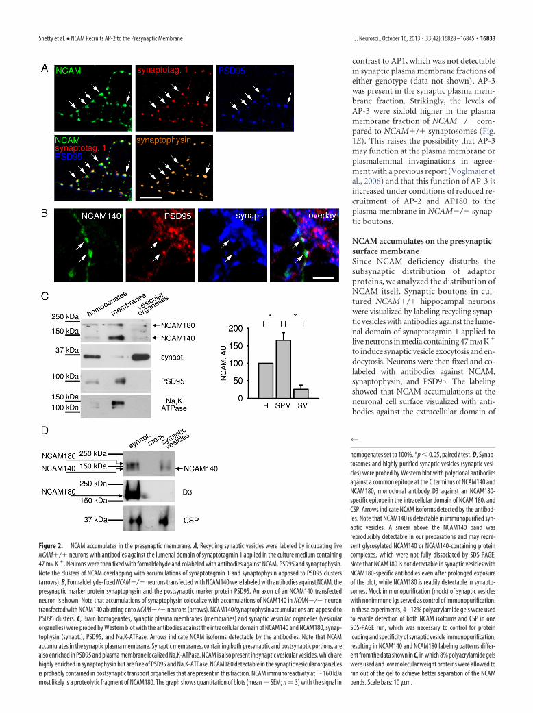
NCAM accumulates on the presynapticsurface membraneSince NCAM deficiency disturbs thesubsynaptic distribution of adaptorproteins, we analyzed the distribution ofNCAM itself. Synaptic boutons in cul-tured NCAM�/� hippocampal neuronswere visualized by labeling recycling synap-tic vesicles with antibodies against the lume-nal domain of synaptotagmin 1 applied tolive neurons in media containing 47 mM K�
to induce synaptic vesicle exocytosis and en-docytosis. Neurons were then fixed and co-labeled with antibodies against NCAM,synaptophysin, and PSD95. The labelingshowed that NCAM accumulations at theneuronal cell surface visualized with anti-bodies against the extracellular domain of
Figure 2. NCAM accumulates in the presynaptic membrane. A, Recycling synaptic vesicles were labeled by incubating liveNCAM�/� neurons with antibodies against the lumenal domain of synaptotagmin 1 applied in the culture medium containing47 mM K �. Neurons were then fixed with formaldehyde and colabeled with antibodies against NCAM, PSD95 and synaptophysin.Note the clusters of NCAM overlapping with accumulations of synaptotagmin 1 and synaptophysin apposed to PSD95 clusters(arrows). B, Formaldehyde-fixed NCAM�/�neurons transfected with NCAM140 were labeled with antibodies against NCAM, thepresynaptic marker protein synaptophysin and the postsynaptic marker protein PSD95. An axon of an NCAM140 transfectedneuron is shown. Note that accumulations of synaptophysin colocalize with accumulations of NCAM140 in NCAM�/� neurontransfected with NCAM140 abutting onto NCAM�/� neurons (arrows). NCAM140/synaptophysin accumulations are apposed toPSD95 clusters. C, Brain homogenates, synaptic plasma membranes (membranes) and synaptic vesicular organelles (vesicularorganelles) were probed by Western blot with the antibodies against the intracellular domain of NCAM140 and NCAM180, synap-tophysin (synapt.), PSD95, and Na,K-ATPase. Arrows indicate NCAM isoforms detectable by the antibodies. Note that NCAMaccumulates in the synaptic plasma membrane. Synaptic membranes, containing both presynaptic and postsynaptic portions, arealso enriched in PSD95 and plasma membrane localized Na,K-ATPase. NCAM is also present in synaptic vesicular vesicles, which arehighly enriched in synaptophysin but are free of PSD95 and Na,K-ATPase. NCAM180 detectable in the synaptic vesicular organellesis probably contained in postsynaptic transport organelles that are present in this fraction. NCAM immunoreactivity at �160 kDamost likely is a proteolytic fragment of NCAM180. The graph shows quantitation of blots (mean � SEM; n � 3) with the signal in
4
homogenates set to 100%. *p � 0.05, paired t test. D, Synap-tosomes and highly purified synaptic vesicles (synaptic vesi-cles) were probed by Western blot with polyclonal antibodiesagainst a common epitope at the C terminus of NCAM140 andNCAM180, monoclonal antibody D3 against an NCAM180-specific epitope in the intracellular domain of NCAM 180, andCSP. Arrows indicate NCAM isoforms detected by the antibod-ies. Note that NCAM140 is detectable in immunopurified syn-aptic vesicles. A smear above the NCAM140 band wasreproducibly detectable in our preparations and may repre-sent glycosylated NCAM140 or NCAM140-containing proteincomplexes, which were not fully dissociated by SDS-PAGE.Note that NCAM180 is not detectable in synaptic vesicles withNCAM180-specific antibodies even after prolonged exposureof the blot, while NCAM180 is readily detectable in synapto-somes. Mock immunopurification (mock) of synaptic vesicleswith nonimmune Igs served as control of immunopurification.In these experiments, 4 –12% polyacrylamide gels were usedto enable detection of both NCAM isoforms and CSP in oneSDS-PAGE run, which was necessary to control for proteinloading and specificity of synaptic vesicle immunopurification,resulting in NCAM140 and NCAM180 labeling patterns differ-ent from the data shown in C, in which 8% polyacrylamide gelswere used and low molecular weight proteins were allowed torun out of the gel to achieve better separation of the NCAMbands. Scale bars: 10 �m.
Shetty et al. • NCAM Recruits AP-2 to the Presynaptic Membrane J. Neurosci., October 16, 2013 • 33(42):16828 –16845 • 16833

NCAM overlapped with synaptic bou-tons identified as overlapping clusters ofsynaptotagmin 1/synaptophysin immuno-reactivity apposed to accumulations ofPSD95 (Fig. 2A). In the CNS, NCAM180expression is predominantly postsynaptic,both in brain tissue (Persohn et al., 1989;Schuster et al., 2001; Fux et al., 2003) and incultured hippocampal neurons (Sytnyk etal., 2006). Presynaptic labeling thus reflectspredominantly the distribution of the sec-ond major neuronal isoform of NCAM,NCAM140. The third major isoform ofNCAM, glycosylphosphatidylinositol (GPI)-linked NCAM120 is expressed in the CNSmainly by glia. To visualize the presynap-tic localization of NCAM140 with suffi-cient spatial resolution (i.e., underconditions where postsynaptic NCAM isabsent), we transfected cultured hip-pocampal neurons from NCAM-deficient(NCAM�/�) mice with an NCAM140expression plasmid. In synapses made byaxons of transfected neurons on dendritesof nontransfected NCAM negative neu-rons, NCAM140 accumulations at theneuronal cell surface overlapped with syn-aptophysin clusters, which were apposedto PSD95 accumulations, consistent witha functional role for NCAM140 in synap-tic vesicle recycling at excitatory synapses(Fig. 2B).
Western blot analysis of synaptosomesand synaptic plasma membranes isolatedfrom adult wild-type (NCAM�/�) miceshowed that NCAM140 and NCAM180are enriched in the synaptic plasma mem-brane fraction when compared to totalbrain homogenates (Fig. 2C). Low levelsof NCAM immunoreactivity were detect-able in the fraction containing synaptic vesicular organelles (Fig.2C), i.e., the total pool of organelles released from synaptosomesby osmotic shock with both NCAM140 and NCAM180 beingdetectable in this fraction. This fraction was enriched in synapto-physin and negative for both Na,K-ATPase, an integral mem-brane protein localized exclusively to the surface membrane,and PSD95, a postsynaptic scaffold protein, highly enriched inthe synaptic plasma membrane fraction (Fig. 2C). To analyzewhether NCAM is present in synaptic vesicles, synaptic vesicleswere first purified on a sucrose gradient and then further immu-nopurified using antibodies against CSP, a protein associatedwith synaptic vesicles via lipid anchors (Fig. 2D), to obtain highlypure synaptic vesicles. NCAM140 but not NCAM180 was detect-able in highly pure synaptic vesicles (Fig. 2D) in accordance withprevious reports (Takamori et al., 2006). The combined observa-tions thus show that at the subsynaptic level, NCAM is predom-inantly present in the synaptic plasma membrane, thus being wellpoised to regulate recruitment of AP-2, AP180, and clathrin tothis place.
NCAM associates with AP-2 via a dileucine motifOne possible explanation for the loss of clathrin/AP-2 fromNCAM�/� synaptic plasma membranes might be a direct inter-
action between NCAM and AP-2. To analyze whether NCAMindeed associates with AP-2 in synapses, NCAM was immuno-precipitated from synaptosomes. Western blot analysis of theseimmunoprecipitates showed that AP-2 but not AP-3 and AP-1coimmunoprecipitated with NCAM (Fig. 3A). Approximately3–5% of all AP-2 molecules present in synaptosomes were foundin NCAM immunoprecipitates.
Analysis of the amino acid sequence of NCAM shows a poten-tial AP-2 binding motif comprising a dileucine (LL) motif atposition 741 of mouse NCAM140 and NCAM180 (at position751 of rat NCAM140 and NCAM180; Fig. 3B). To confirm thatthe LL motif is important for NCAM/AP-2 complex formation inlive cells, nonmutated rat NCAM140 or mutated rat NCAM140with leucine 752 exchanged to alanine were transfected into CHOcells and assayed for their ability to associate with AP-2 by coim-munoprecipitation. Western blot analysis of NCAM immuno-precipitates showed that wild-type NCAM140 forms a complexwith AP-2 (Fig. 3C). Disruption of the LL motif by the LA muta-tion strongly reduced the levels of AP-2 coimmunoprecipitatingwith NCAM140 from transfected CHO cells to 21.1 � 7.1% (n �4; p � 0.01, paired t test) of the levels of AP2 coimmunoprecipi-tating with nonmutated NCAM140 set at 100% (Fig. 3C), indi-
Figure 3. AP-2 binds to the dileucine motif in the intracellular domain of NCAM140. A, NCAM immunoprecipitates (IP) fromsynaptosomes and 13% of the material used for NCAM immunoprecipitation (input, 13%) were probed by Western blot analysiswith the antibodies against the extracellular domain of NCAM, AP-2, AP-3, and AP-1. Proteins were separated using 8% polyacryl-amide gels to achieve better separation of high molecular weight proteins. Mock immunoprecipitation with nonimmune rabbit Igswas performed for control. Note that AP-2 but not AP-3 or AP-1 coimmunoprecipitate with NCAM. B, Amino acid sequence of afragment of NCAM140 comprising transmembrane and membrane adjacent portions of the extracellular and intracellular domainsof NCAM is shown. The LL motif in the intracellular domain of NCAM is shown in bold and underlined. C, NCAM immunoprecipitatesfrom CHO cells transfected with NCAM140, nonmutated or mutated in the membrane-proximal LL dileucine motif, were analyzedby Western blot with antibodies against NCAM and AP-2. Mock immunoprecipitation with NCAM antibodies from CHO cellstransfected with GFP only was performed for control. Note that mutation in the LL dileucine motif inhibits coimmunoprecipitationof AP-2 with NCAM140.
16834 • J. Neurosci., October 16, 2013 • 33(42):16828 –16845 Shetty et al. • NCAM Recruits AP-2 to the Presynaptic Membrane

cating that the LL motif indeed is requiredfor efficient formation of the NCAM/AP-2 complex in live cells.
NCAM promotes recruitment of AP-2to the synaptic plasma membraneTo verify that NCAM induces attachmentof AP-2 to the synaptic plasma mem-brane, we investigated the recruitment ofAP-2 and clathrin from the synaptosomalcytosol of NCAM�/� mice, which servedas a source of these proteins, to synapticmembranes isolated from NCAM�/�and NCAM�/� mice. Before incubationwith cytosol, synaptic membranes wereexposed to high pH levels to strip periph-eral proteins from the membranes (Fig.4A; Haucke and De Camilli, 1999; Cher-nyshova et al., 2011). Western blot analy-sis of the synaptic membranes incubatedwith cytosol showed that cytosolic AP-2and clathrin bound to NCAM�/� andNCAM�/� synaptic membranes (Fig.4A). However, the efficiency of attach-ment of these proteins to NCAM�/�membranes was approximately two tothree times lower than to NCAM�/�membranes (Fig. 4A), suggesting thatNCAM regulates recruitment of AP-2 andclathrin to synaptic membranes. Interest-ingly, recruitment of AP-3 from cytosol toNCAM�/� synaptic membranes was ap-proximately three times higher than toNCAM�/� synaptic membranes (Fig.4A), again suggesting that AP-3 substi-tutes for AP-2 in the absence of NCAM.
To analyze the role of the dileucine(LL) motif in recruitment of AP-2 to syn-aptic membranes, we investigated attach-ment of AP-2 to NCAM�/� synapticmembranes from cytosol that was prein-cubated with nonmutated intracellulardomain of NCAM140 (LL140ID) or withthe intracellular domain of NCAM140 withthe mutated membrane-proximal dileucinemotif (LA140ID). Preincubation of cytosolwith LL140ID inhibited binding of AP-2 tosynaptic membranes (Fig. 4B,C), suggest-ing that recombinant 140ID competes withendogenous NCAM for AP-2. In contrast,LA140ID did not affect membrane associa-tion of AP-2 (Fig. 4B), indicating that thedileucine motif is an important determinantfor NCAM-dependent recruitment of AP-2to synaptic membranes.
Interestingly, when attachment of AP-2to NCAM�/� synaptic membranes was in-hibited by pre-incubation with 140ID, re-cruitment of AP-3 was enhanced (Fig. 4C),thus resembling in this aspect theNCAM�/� phenotype (Fig. 4A). These
Figure 4. NCAM promotes binding of AP-2 versus AP-3 to synaptic plasma membranes. A, NCAM�/� and NCAM�/�synaptic plasma membranes were treated with alkali to strip peripheral proteins. The membranes were then incubated withcytosol and recruitment of AP-2, clathrin, and AP-3 from the cytosol to the membranes was assessed by Western blot. Note thatalkali removed AP-2, clathrin, and AP-3 from the membranes, but not L1, which served as a loading control. Note more efficientrecruitment of AP-2 and clathrin to NCAM�/� synaptic membranes and enhanced recruitment of AP-3 to NCAM�/� synapticmembranes. The graph shows the quantitation of blots. Mean levels � SEM of AP-2, clathrin, and AP-3, recruited to NCAM�/�synaptic membranes, are shown. The signals were normalized to NCAM�/� levels (dashed line) set to 100%. *p � 0.05, pairedt test (compared to NCAM�/�; n � 3). B, C, Recruitment of AP-2 and AP-3 to NCAM�/� synaptic membranes as analyzed byWestern blot. When indicated, the cytosol was preincubated with BFA or competitors, either the nonmutated intracellular domainof NCAM140 (140ID) or 140ID with the mutated membrane-proximal dileucine motif (LA140ID). Note that 140ID but not LA140IDreduces recruitment of AP-2 to the membranes (B). AP-3 recruitment is enhanced from the cytosol preincubated with 140ID, whileAP-2 recruitment is enhanced from the cytosol preincubated with BFA (C). Note that these effects are blocked when the cytosol waspreincubated with 140ID and BFA (C). No recruitment of AP-1 is observed. Brain homogenate (BH) is included to show AP-1 bandsat the expected molecular weight (C). Graphs show quantitation of blots. Mean levels � SEM of AP-2 and AP-3, recruited tosynaptic membranes from cytosol in the presence of BFA or competitors, are shown. The signals were normalized to the signalwithout BFA or competitors (dashed lines) set to 100%. *p � 0.05, paired t test (compared to the signal without BFA or compet-itors; n � 3). L1 (A, C) or syntaxin 1B (syn 1B; B) served as a loading control.
Shetty et al. • NCAM Recruits AP-2 to the Presynaptic Membrane J. Neurosci., October 16, 2013 • 33(42):16828 –16845 • 16835
data suggest that AP-2 and AP-3 can substi-tute for each other, with NCAM favoringbinding of AP-2 to synaptic membranesover that of AP-3. To investigate this ideafurther, we analyzed recruitment of AP-2and AP-3 to NCAM�/� synaptic mem-branes from cytosol preincubated with BFA.AP-3 is recruited to membranes by the smallG protein Arf1 (Ooi et al., 1998). Since BFAblocks nucleotide exchange on Arf1, it in-hibits the ability of Arf1 to interact withmembranes inducing the dispersion of Arf-associated AP complexes such as AP-3 to thecytosol (Robineau et al., 2000). BFA-induced inhibition of AP-3 recruitment toNCAM�/� synaptic membranes was ac-companied by increased attachment ofAP-2 (Fig. 4C), further suggesting that AP-2and AP-3 can substitute for each other. Theeffects of 140ID and BFA were negated withrespect to AP-2 recruitment when they werecoapplied together (Fig. 4C). AP-1 was notrecruited to synaptic membranes either inthe presence or absence of 140ID or BFA(Fig. 4C).
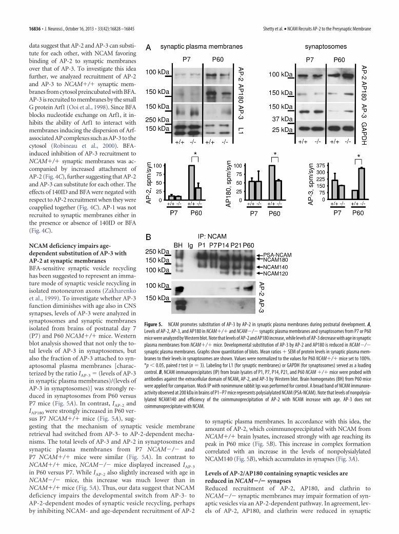
NCAM deficiency impairs age-dependent substitution of AP-3 withAP-2 at synaptic membranesBFA-sensitive synaptic vesicle recyclinghas been suggested to represent an imma-ture mode of synaptic vesicle recycling inisolated motoneuron axons (Zakharenkoet al., 1999). To investigate whether AP-3function diminishes with age also in CNSsynapses, levels of AP-3 were analyzed insynaptosomes and synaptic membranesisolated from brains of postnatal day 7(P7) and P60 NCAM�/� mice. Westernblot analysis showed that not only the to-tal levels of AP-3 in synaptosomes, butalso the fraction of AP-3 attached to syn-aptosomal plasma membranes [charac-terized by the ratio IAP-3 � (levels of AP-3in synaptic plasma membranes)/(levels ofAP-3 in synaptosomes)] was strongly re-duced in synaptosomes from P60 versusP7 mice (Fig. 5A). In contrast, IAP-2 andIAP180 were strongly increased in P60 ver-sus P7 NCAM�/� mice (Fig. 5A), sug-gesting that the mechanism of synaptic vesicle membraneretrieval had switched from AP-3- to AP-2-dependent mecha-nisms. The total levels of AP-3 and AP-2 in synaptosomes andsynaptic plasma membranes from P7 NCAM�/� andP7 NCAM�/� mice were similar (Fig. 5A). In contrast toNCAM�/� mice, NCAM�/� mice displayed increased IAP-3
in P60 versus P7. While IAP-2 also slightly increased with age inNCAM�/� mice, this increase was much lower than inNCAM�/� mice (Fig. 5A). Thus, our data suggest that NCAMdeficiency impairs the developmental switch from AP-3- toAP-2-dependent modes of synaptic vesicle recycling, perhapsby inhibiting NCAM- and age-dependent recruitment of AP-2
to synaptic plasma membranes. In accordance with this idea, theamount of AP-2, which coimmunoprecipitated with NCAM fromNCAM�/� brain lysates, increased strongly with age reaching itspeak in P60 mice (Fig. 5B). This increase in complex formationcorrelated with an increase in the levels of nonpolysialylatedNCAM140 (Fig. 5B), which accumulates in synapses (Fig. 3A).
Levels of AP-2/AP180 containing synaptic vesicles arereduced in NCAM�/� synapsesReduced recruitment of AP-2, AP180, and clathrin toNCAM�/� synaptic membranes may impair formation of syn-aptic vesicles via an AP-2-dependent pathway. In agreement, lev-els of AP-2, AP180, and clathrin were reduced in synaptic
Figure 5. NCAM promotes substitution of AP-3 by AP-2 in synaptic plasma membranes during postnatal development. A,Levels of AP-2, AP-3, and AP180 in NCAM�/� and NCAM�/� synaptic plasma membranes and synaptosomes from P7 or P60mice were analyzed by Western blot. Note that levels of AP-2 and AP180 increase, while levels of AP-3 decrease with age in synapticplasma membranes from NCAM�/� mice. Developmental substitution of AP-3 by AP-2 and AP180 is reduced in NCAM�/�synaptic plasma membranes. Graphs show quantitation of blots. Mean ratios � SEM of protein levels in synaptic plasma mem-branes to their levels in synaptosomes are shown. Values were normalized to the values for P60 NCAM�/� mice set to 100%.*p � 0.05, paired t test (n � 3). Labeling for L1 (for synaptic membranes) or GAPDH (for synaptosomes) served as a loadingcontrol. B, NCAM immunoprecipitates (IP) from brain lysates of P1, P7, P14, P21, and P60 NCAM �/� mice were probed withantibodies against the extracellular domain of NCAM, AP-2, and AP-3 by Western blot. Brain homogenates (BH) from P60 micewere applied for comparison. Mock IP with nonimmune rabbit Igs was performed for control. A broad band of NCAM immunore-activity observed at 200 kDa in brains of P1–P7 mice represents polysialylated NCAM (PSA-NCAM). Note that levels of nonpolysia-lylated NCAM140 and efficiency of the coimmunoprecipitation of AP-2 with NCAM increase with age. AP-3 does notcoimmunoprecipitate with NCAM.
16836 • J. Neurosci., October 16, 2013 • 33(42):16828 –16845 Shetty et al. • NCAM Recruits AP-2 to the Presynaptic Membrane
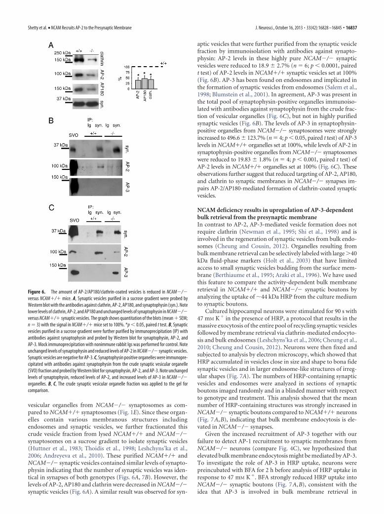
vesicular organelles from NCAM�/� synaptosomes as com-pared to NCAM�/� synaptosomes (Fig. 1E). Since these organ-elles contain various membranous structures includingendosomes and synaptic vesicles, we further fractionated thiscrude vesicle fraction from lysed NCAM�/� and NCAM�/�synaptosomes on a sucrose gradient to isolate synaptic vesicles(Huttner et al., 1983; Thoidis et al., 1998; Leshchyns’ka et al.,2006; Andreyeva et al., 2010). These purified NCAM�/� andNCAM�/� synaptic vesicles contained similar levels of synapto-physin indicating that the number of synaptic vesicles was iden-tical in synapses of both genotypes (Figs. 6A, 7B). However, thelevels of AP-2, AP180 and clathrin were decreased in NCAM�/�synaptic vesicles (Fig. 6A). A similar result was observed for syn-
aptic vesicles that were further purified from the synaptic vesiclefraction by immunoisolation with antibodies against synapto-physin: AP-2 levels in these highly pure NCAM�/� synapticvesicles were reduced to 18.9 � 2.7% (n � 6; p � 0.0001, pairedt test) of AP-2 levels in NCAM�/� synaptic vesicles set at 100%(Fig. 6B). AP-3 has been found on endosomes and implicated inthe formation of synaptic vesicles from endosomes (Salem et al.,1998; Blumstein et al., 2001). In agreement, AP-3 was present inthe total pool of synaptophysin-positive organelles immunoiso-lated with antibodies against synaptophysin from the crude frac-tion of vesicular organelles (Fig. 6C), but not in highly purifiedsynaptic vesicles (Fig. 6B). The levels of AP-3 in synaptophysin-positive organelles from NCAM�/� synaptosomes were stronglyincreased to 496.6 � 123.7% (n � 4; p � 0.05, paired t test) of AP-3levels in NCAM�/� organelles set at 100%, while levels of AP-2 insynaptophysin-positive organelles from NCAM�/� synaptosomeswere reduced to 19.83 � 1.8% (n � 4; p � 0.001, paired t test) ofAP-2 levels in NCAM�/� organelles set at 100% (Fig. 6C). Theseobservations further suggest that reduced targeting of AP-2, AP180,and clathrin to synaptic membranes in NCAM�/� synapses im-pairs AP-2/AP180-mediated formation of clathrin-coated synapticvesicles.
NCAM deficiency results in upregulation of AP-3-dependentbulk retrieval from the presynaptic membraneIn contrast to AP-2, AP-3-mediated vesicle formation does notrequire clathrin (Newman et al., 1995; Shi et al., 1998) and isinvolved in the regeneration of synaptic vesicles from bulk endo-somes (Cheung and Cousin, 2012). Organelles resulting frombulk membrane retrieval can be selectively labeled with large 40kDa fluid-phase markers (Holt et al., 2003) that have limitedaccess to small synaptic vesicles budding from the surface mem-brane (Berthiaume et al., 1995; Araki et al., 1996). We have usedthis feature to compare the activity-dependent bulk membraneretrieval in NCAM�/� and NCAM�/� synaptic boutons byanalyzing the uptake of �44 kDa HRP from the culture mediumto synaptic boutons.
Cultured hippocampal neurons were stimulated for 90 s with47 mM K� in the presence of HRP, a protocol that results in themassive exocytosis of the entire pool of recycling synaptic vesiclesfollowed by membrane retrieval via clathrin-mediated endocyto-sis and bulk endosomes (Leshchyns’ka et al., 2006; Cheung et al.,2010; Cheung and Cousin, 2012). Neurons were then fixed andsubjected to analysis by electron microscopy, which showed thatHRP accumulated in vesicles close in size and shape to bona fidesynaptic vesicles and in larger endosome-like structures of irreg-ular shapes (Fig. 7A). The numbers of HRP-containing synapticvesicles and endosomes were analyzed in sections of synapticboutons imaged randomly and in a blinded manner with respectto genotype and treatment. This analysis showed that the meannumber of HRP-containing structures was strongly increased inNCAM�/� synaptic boutons compared to NCAM�/� neurons(Fig. 7A,B), indicating that bulk membrane endocytosis is ele-vated in NCAM�/� synapses.
Given the increased recruitment of AP-3 together with ourfailure to detect AP-1 recruitment to synaptic membranes fromNCAM�/� neurons (compare Fig. 4C), we hypothesized thatelevated bulk membrane endocytosis might be mediated by AP-3.To investigate the role of AP-3 in HRP uptake, neurons werepreincubated with BFA for 2 h before analysis of HRP uptake inresponse to 47 mM K�. BFA strongly reduced HRP uptake intoNCAM�/� synaptic boutons (Fig. 7A,B), consistent with theidea that AP-3 is involved in bulk membrane retrieval in
Figure 6. The amount of AP-2/AP180/clathrin-coated vesicles is reduced in NCAM�/�versus NCAM�/� mice. A, Synaptic vesicles purified in a sucrose gradient were probed byWestern blot with the antibodies against clathrin, AP-2, AP180, and synaptophysin (syn.). Notelower levels of clathrin, AP-2, and AP180 and unchanged levels of synaptophysin in NCAM�/�versus NCAM�/� synaptic vesicles. The graph shows quantitation of the blots (mean � SEM;n � 3) with the signal in NCAM�/� mice set to 100%. *p � 0.05, paired t test. B, Synapticvesicles purified in a sucrose gradient were further purified by immunoprecipitation (IP) withantibodies against synaptophysin and probed by Western blot for synaptophysin, AP-2, andAP-3. Mock immunoprecipitation with nonimmune rabbit Igs was performed for control. Noteunchanged levels of synaptophysin and reduced levels of AP-2 in NCAM�/� synaptic vesicles.Synaptic vesicles are negative for AP-3. C, Synaptophysin positive organelles were immunopre-cipitated with antibodies against synaptophysin from the crude synaptic vesicular organelle(SVO) fraction and probed by Western blot for synaptophysin, AP-2, and AP-3. Note unchangedlevels of synaptophysin, reduced levels of AP-2, and increased levels of AP-3 in NCAM�/�organelles. B, C, The crude synaptic vesicular organelle fraction was applied to the gel forcomparison.
Shetty et al. • NCAM Recruits AP-2 to the Presynaptic Membrane J. Neurosci., October 16, 2013 • 33(42):16828 –16845 • 16837

NCAM�/� synapses. However, this ef-fect was incomplete, suggesting thatBFA-dependent AP-3-mediated bulkmembrane retrieval coexists with a BFA-independent pathway. Somewhat surpris-ingly, BFA did not inhibit, but ratherincreased HRP uptake into NCAM�/�synaptic boutons (Fig. 7A,B). Accumula-tion of HRP in NCAM�/� terminals fol-lowing BFA application might be relatedto the inhibition of organelle processingwithin the synaptic bouton, a process thatlikely depends on AP-3 (Fig. 7D) and pos-sibly AP-1 [consistent with previous databy Cheung and Cousin (2012)]. In agree-ment, HRP containing organelles werelarger in BFA treated NCAM�/� synapticboutons compared to mock-treated con-trols. This phenotype presumably reflectsimpaired budding of vesicles from synapticvesicle protein containing endosomes in thepresence of BFA (Fig. 7C). More impor-tantly, this observation suggests that AP-2-and AP-3-mediated pathways intersect inNCAM�/� synaptic boutons at the level ofendosomal intermediates (Fig. 7D).
The number of small synaptic vesicles(diameter of �40 nm) per synapse sectionarea was on average slightly lower in BFAtreated compared to untreated controlNCAM�/� neurons (Fig. 7 A, B). Thisreduction could be due to reduced AP-3-dependent reformation of synaptic vesiclesfrom endosomal organelles as suggestedpreviously (Voglmaier et al., 2006), and/oroverall reduced biogenesis of synaptic vesi-cles from the trans-Golgi network (Hannahet al., 1999) over the time of drug appli-cation. Interestingly, the effect of BFAon synaptic vesicle number was reducedin NCAM�/� synaptic boutons (Fig.7A,B). This may reflect BFA-induced sub-stitution of the AP-3-dependent pathway ofsynaptic vesicle recycling by an AP-2/AP180-dependent route as suggested byour biochemical data (Figs. 4C, 8C) or thepresence of AP-2/AP180- and AP-3-independent recycling pathways operat-ing in NCAM�/� synaptic boutons.
NCAM�/� synapses combine AP-2-and AP-3-dependent pathways ofsynaptic vesicle endocytosisNext, we compared the overall efficiencyof synaptic vesicle endocytosis by usingthe lipophilic dye FM1– 43, which labelsthe total recycling pool of vesicles in-cluding those generated by bulk mem-brane retrieval; the latter is thought torepresent a minor fraction of the total recycling vesicle pool(Teng and Wilkinson, 2000). A fixable analog of FM1– 43 wasused in these experiments, and the levels of FM1– 43 dye weremeasured in synaptic boutons visualized with antibodies to
the synaptic vesicle marker SV2 after fixation. FM1– 43 uptakein response to 90 s stimulation with 47 mM K � was reducedby �20% in synaptic boutons from NCAM�/� neurons whencompared to NCAM�/� neurons (Fig. 8A). This reduction
Figure 7. AP-3-mediated bulk membrane retrieval is enhanced in NCAM�/� synaptic boutons. A, NCAM�/� and NCAM�/�cultured hippocampal neurons were allowed to take up HRP in the presence of 47 mM K � applied for 90 s. Where indicated, neurons werepretreated with BFA for 1 h. Representative electron micrographs of synapses are shown. HRP-loaded vesicles are seen as dark corestructures. Scale bar, 300 nm. HRP uptake is enhanced in NCAM�/� synaptic boutons when compared to NCAM�/� synaptic boutons.The number of HRP positive organelles is increased in NCAM�/� synaptic boutons and reduced in NCAM�/� boutons after BFAapplication. Higher magnification of the area outlined in NCAM�/� synapse (right) shows that HRP was taken up by synaptic vesicle-likestructures (arrows) and larger endosome-like structures (arrowheads). B, Graphs show numbers of SV-like structures with the cross-sectionarea �2000 nm 2, endosome-like structures with the cross-section area2000 nm 2, and the total number of SVs including HRP-loadedand nonlabeled SVs counted per synapse section area. C, Graphs show numbers of HRP-loaded vesicles of the indicated size normalized tothe total number of HRP-loaded vesicles per synaptic bouton section (top) and the total cross-section area of all SV-like structures orendosome-like structures normalized to the total cross-section area of all HRP loaded organelles per synaptic bouton section (bottom). In Band C, mean values�SEM are shown. *p�0.05, t test (n100 synapses were analyzed from 4 – 6 coverslips). D, A scheme showing thehypothetical role of AP-2 and AP-3 in bulk membrane retrieval in NCAM�/�and NCAM�/� synaptic terminals. AP-3-dependent stepsblocked by BFA are indicated.
16838 • J. Neurosci., October 16, 2013 • 33(42):16828 –16845 Shetty et al. • NCAM Recruits AP-2 to the Presynaptic Membrane

was not due to reduced exocytosis of synaptic vesicles, sincethe release of FM1– 43 from synaptic boutons in response to 47mM K �, indicative of synaptic vesicle exocytosis, was similarin NCAM�/� synaptic boutons compared to NCAM�/�
neurons (Fig. 8B). Impaired uptake ofFM1– 43 in NCAM�/� synaptic bou-tons was also not caused by reduced totalnumbers of synaptic vesicles, which weresimilar in NCAM�/� and NCAM�/�synapses (Fig. 7). Hence, we conclude thatsynaptic vesicle endocytosis is impaired inNCAM�/� synaptic boutons.
When neurons were preincubated withBFA for 2 h, the uptake of FM1–43 into syn-aptic boutons from NCAM�/� neurons inresponse to 90 s stimulation with 47 mM K�
slightly decreased (Fig. 8A), probably due toa small reduction in the number of syn-aptic vesicles. In agreement, there wasno difference in FM1– 43 uptake betweenuntreated and BFA treated NCAM�/�neurons when FM1– 43 levels were nor-malized to the levels of the synaptic vesiclemarker protein SV2 (Fig. 8A). In contrastto the observations on NCAM�/� NMJs,treatment of NCAM�/� hippocampalneurons with BFA surprisingly increasedthe level of FM1– 43 internalization intosynaptic boutons (Fig. 8A). This increasecould be due to slower processing of syn-aptic vesicles in BFA-treated NCAM�/�synaptic terminals resulting in dye trapping.In agreement with this possibility, FM1-43 release in response to 47 mM K� wasslower and incomplete in synaptic bou-tons from BFA treated versus untreatedNCAM�/� neurons (Fig.8B).BFAhadnoeffect on FM1–43 release in NCAM�/� syn-aptic boutons (Fig. 8B).
Our biochemical data indicate that ap-plication of BFA results in the substitutionof AP-3 with AP-2 at the plasma mem-brane (Fig. 4C), causing a switch fromAP-3- to AP-2-dependent modes of syn-aptic vesicle recycling (Fig. 8C). Hence, itis difficult to judge the role of AP-3 in syn-aptic vesicle endocytosis using data ob-tained with BFA. To compare the role ofAP-3 in synaptic vesicle endocytosis inNCAM�/� and NCAM�/� neurons,cells were treated for 2 h with dynasore, asmall molecule inhibitor of the large GT-Pase dynamin. Such treatment blocksclathrin/AP-2- and dynamin-dependentsynaptic vesicle endocytosis (Newton etal., 2006), while leaving AP-3-dependentroutes unperturbed. Dynasore inhibitedFM1– 43 uptake into NCAM�/� synap-tic boutons by �80% (Fig. 8A), in agree-ment with previous reports (Newton etal., 2006). In contrast, in NCAM�/�neurons dynasore inhibited FM1– 43uptake by only �50% (Fig. 8A), consis-
tent with the upregulation of dynamin-independent AP-3-dependent routes of synaptic vesicle endocytosis in NCAM�/�neurons. In agreement with this interpretation, coapplication ofdynasore with BFA inhibited FM1– 43 uptake in NCAM�/�
Figure 8. AP-3-dependent synaptic vesicle endocytosis and intrasynaptic processing are upregulated in NCAM�/� synapticboutons. A, Presynaptic boutons of NCAM�/� and NCAM�/� cultured hippocampal neurons that were either not treated(control) or treated as indicated with BFA and dynasore were loaded with the fixable analog of FM1– 43 applied in the presence of47 mM K � for 90 s. After washing, neurons were fixed and colabeled with antibodies against the presynaptic marker protein SV2to visualize presynaptic boutons. Graphs show quantification (mean � SEM) of FM1– 43 fluorescence levels in SV2 accumulations(top) or the ratio of FM1– 43 fluorescence levels to SV2 immunofluorescence levels (bottom). Values were normalized to the levelsin control NCAM�/� neurons set to 100%. Compared to NCAM�/� neurons, FM1– 43 uptake in NCAM�/� presynapticboutons is reduced and increased in response to BFA. Note that BFA and dynasore have an additive effect on FM1– 43 uptake inNCAM�/� but not NCAM�/� presynaptic boutons. *p � 0.05 (t test compared to control neurons of the same genotype);§p � 0.05 (t test compared as indicated); n 20 images of neurons with N 200 synapses per image were analyzed. B, Graphsshow quantification (mean � SEM) of FM1– 43 release from presynaptic boutons of NCAM�/� and NCAM�/� culturedhippocampal neurons in response to 47 mM K �. Values [I(t)] were normalized to FM1– 43 levels before stimulation [I(0)]. Whereindicated, neurons were preincubated with BFA. BFA inhibits FM1– 43 release from the presynaptic boutons of NCAM�/�neurons. C, A diagram showing the roles of AP-2 and AP-3 adaptor proteins in synaptic vesicle endocytosis and intrasynapticprocessing in NCAM�/� and NCAM�/� synaptic terminals. AP-3- and AP-2-dependent steps blocked by BFA and dynasore,respectively, are indicated.
Shetty et al. • NCAM Recruits AP-2 to the Presynaptic Membrane J. Neurosci., October 16, 2013 • 33(42):16828 –16845 • 16839

synaptic boutons to a level similar to thatseen in BFA plus dynasore-treatedNCAM�/� neurons.
Together, our data indicate that incontrast to NCAM�/� synapses that pre-dominantly use an AP-2/AP180/dynamin-dependent pathway for synaptic vesicleendocytosis, NCAM�/� synapses combineAP-2- and AP-3-dependent routes for synap-tic vesicle reformation.
NCAM deficiency results in impairedsynaptic vesicle endocytosis inhippocampal neuronsTo analyze synaptic vesicle endocytosiswith higher temporal resolution, we vi-sualized synaptic vesicle recycling in liveNCAM�/� and NCAM�/� culturedhippocampal neurons by transfectingthem with a pH-sensitive form of greenfluorescent protein (GFP) fused to thelumenal domain of synaptobrevin 2,also called VAMP2 (VAMP2-pHluorin).This provides a sensitive optical probeto follow exocytosis and endocytosis ofsynaptic vesicles in real time (Miesen-bock et al., 1998). Stimulation with 47mM K � for 90 s resulted in an increase inVAMP2-pHluorin fluorescence at syn-aptic boutons due to synaptic vesicle ex-ocytosis and exposure of the lumenalpHluorin tag to the neutral extracellularspace (Fig. 9A). Following stimulation,the buffer was exchanged to low (4 mM)K � containing buffer resulting in a re-duction in VAMP2-pHluorin fluores-cence intensity due to endocytosis andvesicle reacidification (Fig. 9B). As seenin Figure 9, B and E, the efficiency ofVAMP2-pHluorin endocytosis characterizedby the difference between pHluorin fluores-cence levels reached during stimulation andafter recovery (Ir) was significantly reduced inNCAM�/� neurons when compared toNCAM�/� littermate neurons. The speed ofVAMP2-pHluorin endocytosis characterizedby t1/2 tended to be slower in NCAM�/�ver-sus NCAM�/� neurons (not statisticallysignificant).
Quenching of pHluorin fluorescence de-pends on the rate of synaptic vesicle endocy-tosis and acidification of the synaptic vesiclelumen. The levels of the largest (116 kDa)subunit of the proton ATPase, which is es-sential for proton pump activity and synap-tic vesicle acidification, were similar insynaptic vesicles isolated from NCAM�/�and NCAM�/� mice (Fig. 9C). This obser-vation renders it unlikely that reducedquenching of VAMP2-pHluorin fluorescence in NCAM�/� synap-tic boutons is due to reduced levels of the vATPase. Hence, the defectin VAMP2-pHluorin quenching most likely results from its im-paired endocytosis in NCAM�/� synaptic boutons.
To analyze whether VAMP2 retrieval is also sensitive to thedisruption of the NCAM/AP-2 complex in wild-type neurons, wealso analyzed VAMP2 retrieval after 90 s stimulation with 47 mM
K� in NCAM�/� neurons transfected with NCAM140LA. Sincethe NCAM140LA mutant contains a wild-type extracellular do-
Figure 9. Endocytosis of VAMP2-pHluorin is reduced in NCAM�/� synaptic boutons. A, NCAM�/� neurons were cotrans-fected with cherry and VAMP2-pHluorin. Neurons were stimulated with 1 ms bipolar current pulses at 10 Hz to yield fields of 10V/cm. Gray scale images show pHluorin fluorescence in neurons before (0 s), during (45 s), and after (420 s) stimulation. Note anincrease in pHluorin fluorescence intensity in synaptic boutons in response stimulation (arrows). B, VAMP2-pHluorin endocytosismonitored in synaptic boutons of NCAM�/� and NCAM�/� cultured hippocampal neurons cotransfected (tr.) with an emptypcDNA3 vector, or NCAM�/� neurons cotransfected with NCAM140LA in the pcDNA3 vector. Neurons were stimulated with 47mM K � and allowed to recover in 4 mM K �. C, Synaptic vesicles purified from NCAM�/� and NCAM�/� brains were probed byWestern blot with antibodies against the largest subunit (116 kDa) of proton ATPase. Note that the levels of proton ATPase are notchanged in NCAM�/� versus NCAM �/� synaptic vesicles. D, VAMP2-pHluorin endocytosis monitored in synaptic boutons ofcultured NCAM�/� and NCAM�/� hippocampal neurons cotransfected with control or AP-3 siRNA or NCAM140. Neurons werestimulated with 1 ms bipolar current pulses at 10 Hz to yield fields of 10 V/cm. In B and D, fluorescence intensity levels beforestimulation were set to 0, and signals during the recovery time were normalized to the peak intensity reached during stimulation.Mean values � SEM are shown (n � 14 –25 synapses from 8 –10 coverslips were recorded in each group). E, Mean � SEM. valuesfor pHluorin fluorescence recovery efficiency (Ir � (1 � Iend) * 100%) and for time constants (t1/2) are shown.*p � 0.05, one-wayANOVA with Dunnett’s multiple comparison post-test.
16840 • J. Neurosci., October 16, 2013 • 33(42):16828 –16845 Shetty et al. • NCAM Recruits AP-2 to the Presynaptic Membrane

main, it is able to compete with endogenous wild-type NCAM forhomophilic and heterophilic interactions in the synaptic mem-brane, which target NCAM to synapses (Leshchyns’ka et al.,2011), and may thus function as a dominant-negative constructby substituting endogenous NCAM at synapses. In agreement,NCAM140LA was detectable in synaptic boutons of transfectedneurons by immunofluorescence labeling (data not shown). En-docytosis of VAMP2-pHluorin was strongly inhibited inNCAM140LA expressing NCAM�/� neurons (Fig. 9B).
To analyze whether NCAM deficiency also affects synapticvesicle endocytosis after a milder stimulation protocol, VAMP2retrieval was analyzed after subjecting neurons to stimulationwith 900 action potentials applied at 10 Hz, a protocol that doesnot activate bulk endocytosis. Also with this stimulation proto-col, VAMP2-pHluorin endocytosis was reduced in NCAM�/�neurons versus NCAM�/� neurons (Fig. 9D,E).
Since removal of AP-3 from the plasma membrane promotesrecruitment of AP-2 (Fig. 4C), we also compared VAMP2 re-trieval after 10 Hz stimulation in NCAM�/� and NCAM�/�neurons transfected with AP-3 siRNA. The difference betweengenotypes was eliminated in neurons transfected with AP-3siRNA (Fig. 9D,E). This effect might be due to reactivation of theAP-2 pathway after inactivation of AP-3 in NCAM�/� neuronsor could reflect other AP-independent mechanisms of bulkmembrane retrieval.
A crucial role for NCAM140 in regulating VAMP2 endocyto-sis was further confirmed by the observation that overexpressionof wild-type NCAM140 in NCAM�/� neurons increasedVAMP2-pHluorin endocytosis (Fig. 9D,E). These data thus in-dicate that NCAM140 regulates endocytic retrieval of VAMP2from the neuronal surface, presumably by facilitating recruit-ment of AP-2/AP180 to the presynaptic plasma membrane (com-pare with Fig. 1).
DiscussionAn inherent feature of neurons is their ability to locally recycle themembranes of neurotransmitter containing vesicles, thus en-abling uninterrupted neurotransmitter release. While exocytosisand endocytosis of synaptic vesicle precursors in immature neu-rons occurs at random sites along axons, following formation ofaxodendritic contacts synaptic vesicle precursors accumulate atthese nascent synapses switching to a spatially restricted andhighly regulated release of neurotransmitters, a process called“maturation” of the presynaptic vesicle release machinery(Zakharenko et al., 1999; Hata et al., 2007). In the present studywe have identified NCAM as an important player in this process:it induces formation of the mature AP-2-dependent synaptic ves-icle endocytotic machinery at nascent synapses.
NCAM accumulates in nascent synapses several minutes fol-lowing an initial axodendritic contact (Sytnyk et al., 2002; Sytnyket al., 2004), thus being well poised for its role in regulating syn-aptic transmission. Our data suggest that NCAM140 at the pre-synaptic membrane then recruits AP-2 and indirectly AP180from the cytosol, thereby stabilizing these adaptors at the endo-cytic periactive zone. The physiological significance of this pro-cess is underscored by our observation of impaired endocyticretrieval of VAMP2, a process known to depend on AP-2/AP180(Koo et al., 2011), in NCAM�/� neurons. We do not exclude,however, that deficiency in NCAM may also be associated withgeneral changes in lipids/lipid dynamics in synapses, which mayadd to the abnormalities in synaptic vesicle recycling. The impor-tance of the interaction between NCAM and AP-2 in endocyticretrieval of VAMP2 is supported by our data showing that
VAMP2 endocytosis is reduced in NCAM�/� neurons overex-pressing NCAM140 with a mutated binding site for AP-2(NCAM140LA). Since NCAM140LA contains the nonmutatedextracellular domain, which mediates adhesion, these data indi-cate that disruption of the interaction between NCAM140 andAP-2 without disruption of NCAM-dependent adhesion is suffi-cient to interfere with VAMP2 endocytosis.
The physiological function of NCAM detectable in synapticvesicles (Fig. 2; Takamori et al., 2006; Morciano et al., 2009)remains to be identified. However, low levels of NCAM in syn-aptic vesicles support the idea that the NCAM/AP-2 complex isdisassembled before synaptic vesicle endocytosis. Dissociation ofthe AP-2/clathrin complex from NCAM may be required to de-tach a nascent endocytic protein complex from the NCAM-associated spectrin cytoskeleton to allow efficient endocytosis,which is otherwise inhibited by the spectrin meshwork (Sato etal., 1995; Puchkov et al., 2011). Such a mechanism would alsoprevent missorting of NCAM, which instead is retained at the cellsurface to promote further rounds of AP-2/AP180 recruitment atthe plasma membrane. Following dissociation from NCAM,AP-2 might bind to synaptic vesicle proteins, such as synaptotag-min (Haucke and De Camilli, 1999), which transiently accumu-lates at the cell surface after synaptic vesicle exocytosis.
We show that AP-3 partially substitutes for AP-2 inNCAM�/� synapses. Not only the attachment of AP-3 to themembranes is increased in NCAM�/� synapses, as shown by thein vitro recruitment assay, but also the overall expression levels ofAP3 are increased in NCAM�/� brains. This may explain thefact that the approximately twofold decline in AP2 levels inNCAM�/� synaptic membranes is accompanied by an approx-imately sixfold increase in levels of AP3. Clathrin-independentbulk endocytosis of synaptic vesicles has been observed followingclathrin inactivation in Drosophila (Heerssen et al., 2008). Ourdata also suggest that AP-3 associates with synaptic membranesand mediates vesicle formation perhaps not only from endo-somes (Faundez et al., 1998), but also from the synaptic surfacemembrane, as proposed previously (Voglmaier et al., 2006). Inaddition to cargo-specific sorting of synaptic vesicle proteins,AP-3 promotes bulk membrane retrieval in NCAM�/� syn-apses, thereby compensating for AP-2 deficiency. Levels of AP-1,an adaptor protein implicated in endosomal synaptic vesiclerecycling (Glyvuk et al., 2010), are also increased in NCAM�/�synaptosomes and synaptic vesicles, suggesting increased synap-tic vesicle recycling via endosomal intermediates.
Our data showing that AP-3 is also present at high levels inNCAM�/� synapses at early stages of neuronal developmentconfirm that AP-3 is a component of the immature synaptic ves-icle endocytotic machinery. AP-3 is recruited to synaptic mem-branes independently of NCAM. This could be an importantfeature in immature axons that assures endocytosis of synapticvesicle precursor membranes being exocytosed at random sitesalong axons in the absence of contacts accumulating NCAM.AP-3 mediated endocytosis is, however, less efficient, as under-scored by our observations in NCAM�/� neurons and suggestedpreviously (Voglmaier et al., 2006).
We show that with progression of brain development the im-mature AP-3-dependent synaptic vesicle recycling pathway issubstituted by an AP-2-dependent mechanism. While the exactmechanisms underlying this switch remain to be determined, ourdata suggest that NCAM is one of the molecules which can induceit. In agreement, the amount of AP-2 in association with NCAMalso increases with age, reflecting the increase in the ratio of ma-ture to immature synapses. We speculate that in the absence of
Shetty et al. • NCAM Recruits AP-2 to the Presynaptic Membrane J. Neurosci., October 16, 2013 • 33(42):16828 –16845 • 16841
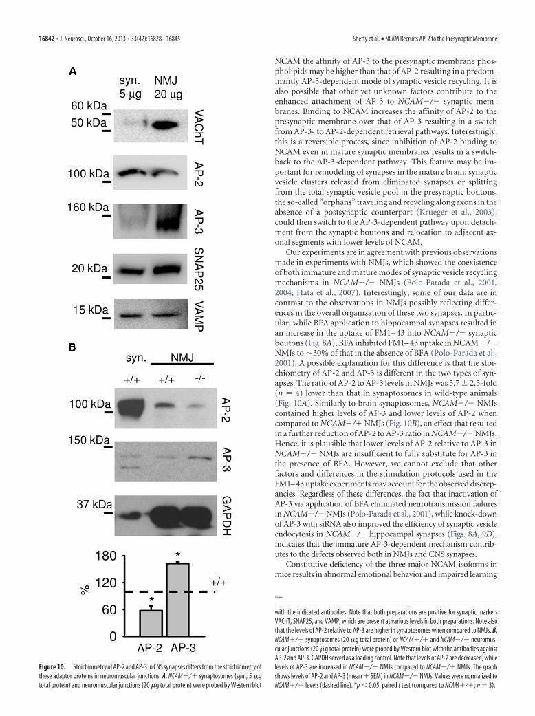
NCAM the affinity of AP-3 to the presynaptic membrane phos-pholipids may be higher than that of AP-2 resulting in a predom-inantly AP-3-dependent mode of synaptic vesicle recycling. It isalso possible that other yet unknown factors contribute to theenhanced attachment of AP-3 to NCAM�/� synaptic mem-branes. Binding to NCAM increases the affinity of AP-2 to thepresynaptic membrane over that of AP-3 resulting in a switchfrom AP-3- to AP-2-dependent retrieval pathways. Interestingly,this is a reversible process, since inhibition of AP-2 binding toNCAM even in mature synaptic membranes results in a switch-back to the AP-3-dependent pathway. This feature may be im-portant for remodeling of synapses in the mature brain: synapticvesicle clusters released from eliminated synapses or splittingfrom the total synaptic vesicle pool in the presynaptic boutons,the so-called “orphans” traveling and recycling along axons in theabsence of a postsynaptic counterpart (Krueger et al., 2003),could then switch to the AP-3-dependent pathway upon detach-ment from the synaptic boutons and relocation to adjacent ax-onal segments with lower levels of NCAM.
Our experiments are in agreement with previous observationsmade in experiments with NMJs, which showed the coexistenceof both immature and mature modes of synaptic vesicle recyclingmechanisms in NCAM�/� NMJs (Polo-Parada et al., 2001,2004; Hata et al., 2007). Interestingly, some of our data are incontrast to the observations in NMJs possibly reflecting differ-ences in the overall organization of these two synapses. In partic-ular, while BFA application to hippocampal synapses resulted inan increase in the uptake of FM1– 43 into NCAM�/� synapticboutons (Fig. 8A), BFA inhibited FM1– 43 uptake in NCAM �/�NMJs to �30% of that in the absence of BFA (Polo-Parada et al.,2001). A possible explanation for this difference is that the stoi-chiometry of AP-2 and AP-3 is different in the two types of syn-apses. The ratio of AP-2 to AP-3 levels in NMJs was 5.7 � 2.5-fold(n � 4) lower than that in synaptosomes in wild-type animals(Fig. 10A). Similarly to brain synaptosomes, NCAM�/� NMJscontained higher levels of AP-3 and lower levels of AP-2 whencompared to NCAM�/� NMJs (Fig. 10B), an effect that resultedin a further reduction of AP-2 to AP-3 ratio in NCAM�/� NMJs.Hence, it is plausible that lower levels of AP-2 relative to AP-3 inNCAM�/� NMJs are insufficient to fully substitute for AP-3 inthe presence of BFA. However, we cannot exclude that otherfactors and differences in the stimulation protocols used in theFM1–43 uptake experiments may account for the observed discrep-ancies. Regardless of these differences, the fact that inactivation ofAP-3 via application of BFA eliminated neurotransmission failuresin NCAM�/� NMJs (Polo-Parada et al., 2001), while knock-downof AP-3 with siRNA also improved the efficiency of synaptic vesicleendocytosis in NCAM�/� hippocampal synapses (Figs. 8A, 9D),indicates that the immature AP-3-dependent mechanism contrib-utes to the defects observed both in NMJs and CNS synapses.
Constitutive deficiency of the three major NCAM isoforms inmice results in abnormal emotional behavior and impaired learning
Figure 10. Stoichiometry of AP-2 and AP-3 in CNS synapses differs from the stoichiometry ofthese adaptor proteins in neuromuscular junctions. A, NCAM�/� synaptosomes (syn.; 5 �gtotal protein) and neuromuscular junctions (20 �g total protein) were probed by Western blot
4
with the indicated antibodies. Note that both preparations are positive for synaptic markersVAChT, SNAP25, and VAMP, which are present at various levels in both preparations. Note alsothat the levels of AP-2 relative to AP-3 are higher in synaptosomes when compared to NMJs. B,NCAM�/� synaptosomes (20 �g total protein) or NCAM�/� and NCAM�/� neuromus-cular junctions (20 �g total protein) were probed by Western blot with the antibodies againstAP-2 and AP-3. GAPDH served as a loading control. Note that levels of AP-2 are decreased, whilelevels of AP-3 are increased in NCAM�/� NMJs compared to NCAM�/� NMJs. The graphshows levels of AP-2 and AP-3 (mean � SEM) in NCAM�/� NMJs. Values were normalized toNCAM�/� levels (dashed line). *p � 0.05, paired t test (compared to NCAM�/�; n � 3).
16842 • J. Neurosci., October 16, 2013 • 33(42):16828 –16845 Shetty et al. • NCAM Recruits AP-2 to the Presynaptic Membrane

and memory (Cremer et al., 1994; Stork et al., 1997; Pillai-Nair et al.,2005). Mutations, abnormal processing or posttranslational modi-fications of NCAM lead to a number of stress-related psychiatricdisorders in humans, such as mood disorders and schizophrenia(Poltorak et al., 1996; Vawter et al., 2000; Arai et al., 2004; Sandi andBisaz, 2007; Sullivan et al., 2007; Tanaka et al., 2007). Abnormalitiesin neuronal network functioning have been considered as importantcontributors to the development of these diseases (Sandi and Bisaz,2007). Our present findings provide insights into the molecularmechanisms that may underlie these abnormalities: inefficient en-docytosis of synaptic vesicles especially under conditions of highactivity accompanied by strong synapse activation and massive exo-cytosis of synaptic vesicles, similar to what is observed in response tohigh K� and electric field stimulation used in our study, may lead toinefficient recovery of synaptic vesicles and disturbances in synapsefunction eventually resulting in psychiatric symptoms. While fur-ther work is required to provide additional support for this idea, ourdata establish NCAM as an important player in the organization ofthe presynaptic machinery in CNS synapses.
ReferencesAndreyeva A, Leshchyns’ka I, Knepper M, Betzel C, Redecke L, Sytnyk V,
Schachner M (2010) CHL1 is a selective organizer of the presynapticmachinery chaperoning the SNARE complex. PLoS One 5:e12018.CrossRef Medline
Arai M, Itokawa M, Yamada K, Toyota T, Arai M, Haga S, Ujike H, Sora I,Ikeda K, Yoshikawa T (2004) Association of neural cell adhesion mole-cule 1 gene polymorphisms with bipolar affective disorder in Japaneseindividuals. Biol Psychiatry 55:804 – 810. CrossRef Medline
Araki S, Yang J, Hashiramoto M, Tamori Y, Kasuga M, Holman GD (1996)Subcellular trafficking kinetics of GLU4 mutated at the N- andC-terminal. Biochem J 315:153–159. Medline
Berthiaume EP, Medina C, Swanson JA (1995) Molecular size-fractionationduring endocytosis in macrophages. J Cell Biol 129:989 –998. CrossRefMedline
Blumstein J, Faundez V, Nakatsu F, Saito T, Ohno H, Kelly RB (2001) Theneuronal form of adaptor protein-3 is required for synaptic vesicle for-mation from endosomes. J Neurosci 21:8034 – 8042. Medline
Bodrikov V, Leshchyns’ka I, Sytnyk V, Overvoorde J, den Hertog J, SchachnerM (2005) RPTPalpha is essential for NCAM-mediated p59fyn activa-tion and neurite elongation. J Cell Biol 168:127–139. Medline
Bonanomi D, Benfenati F, Valtorta F (2006) Protein sorting in the synapticvesicle life cycle. Prog Neurobiol 80:177–217. CrossRef Medline
Chernyshova Y, Leshchyns’ka I, Hsu SC, Schachner M, Sytnyk V (2011) Theneural cell adhesion molecule promotes FGFR-dependent phosphoryla-tion and membrane targeting of the exocyst complex to induce exocytosisin growth cones. J Neurosci 31:3522–3535. CrossRef Medline
Cheung G, Cousin MA (2012) Adaptor protein complexes 1 and 3 are es-sential for generation of synaptic vesicles from activity-dependent bulkendosomes. J Neurosci 32:6014 – 6023. CrossRef Medline
Cheung G, Jupp OJ, Cousin MA (2010) Activity-dependent bulk endocyto-sis and clathrin-dependent endocytosis replenish specific synaptic vesiclepools in central nerve terminals. J Neurosci 30:8151– 8161. CrossRefMedline
Cremer H, Lange R, Christoph A, Plomann M, Vopper G, Roes J, Brown R,Baldwin S, Kraemer P, Scheff S, Barthels D, Rajewsky K, Wille W (1994)Inactivation of the N-CAM gene in mice results in size reduction of theolfactory bulb and deficits in spatial learning. Nature 367:455– 459.CrossRef Medline
Dityatev A, Schachner M, Sonderegger P (2010a) The dual role of the extra-cellular matrix in synaptic plasticity and homeostasis. Nat Rev Neurosci11:735–746. CrossRef Medline
Dityatev A, Seidenbecher CI, Schachner M (2010b) Compartmentalizationfrom the outside: the extracellular matrix and functional microdomainsin the brain. Trends Neurosci 33:503–512. CrossRef Medline
Faundez V, Horng JT, Kelly RB (1998) A function for the AP3 coat complexin synaptic vesicle formation from endosomes. Cell 93:423– 432. CrossRefMedline
Ford MG, Pearse BM, Higgins MK, Vallis Y, Owen DJ, Gibson A, Hopkins
CR, Evans PR, McMahon HT (2001) Simultaneous binding ofPtdIns(4,5)P2 and clathrin by AP180 in the nucleation of clathrin latticeson membranes. Science 291:1051–1055. CrossRef Medline
Fux CM, Krug M, Dityatev A, Schuster T, Schachner M (2003) NCAM180and glutamate receptor subtypes in potentiated spine synapses: an immu-nogold electron microscopic study. Mol Cell Neurosci 24:939 –950.CrossRef Medline
Gerrow K, El-Husseini A (2006) Cell adhesion molecules at the synapse.Front Biosci 11:2400 –2419. CrossRef Medline
Glyvuk N, Tsytsyura Y, Geumann C, D’Hooge R, HuveJ, Kratzke M, Baltes J,Boning D, Klingauf J, Schu P (2010) AP-1/sigma1B-adaptin mediatesendosomal synaptic vesicle recycling, learning and memory. EMBO J 29:1318 –1330. CrossRef Medline
Hannah MJ, Schmidt AA, Huttner WB (1999) Synaptic vesicle biogenesis.Annu Rev Cell Dev Biol 15:733–798. CrossRef Medline
Hao W, Luo Z, Zheng L, Prasad K, Lafer EM (1999) AP180 and AP-2 inter-act directly in a complex that cooperatively assembles clathrin. J BiolChem 274:22785–22794. CrossRef Medline
Hata K, Polo-Parada L, Landmesser LT (2007) Selective targeting of differ-ent neural cell adhesion molecule isoforms during motoneuron myotubesynapse formation in culture and the switch from an immature to matureform of synaptic vesicle cycling. J Neurosci 27:14481–14493. CrossRefMedline
Haucke V, De Camilli P (1999) AP-2 recruitment to synaptotagmin stimu-lated by tyrosine-based endocytic motifs. Science 285:1268 –1271.CrossRef Medline
Heerssen H, Fetter RD, Davis GW (2008) Clathrin dependence of synaptic-vesicle formation at the Drosophila neuromuscular junction. Curr Biol18:401– 409. CrossRef Medline
Hill E, van Der Kaay J, Downes CP, Smythe E (2001) The role of dynaminand its binding partners in coated pit invagination and scission. J Cell Biol152:309 –323. CrossRef Medline
Holt M, Cooke A, Wu MM, Lagnado L (2003) Bulk membrane retrieval inthe synaptic terminal of retinal bipolar cells. J Neurosci 23:1329 –1339.Medline
Huttner WB, Schiebler W, Greengard P, De Camilli P (1983) Synapsin I(protein I), a nerve terminal-specific phosphoprotein. III. Its associationwith synaptic vesicles studied in a highly purified synaptic vesicle prepa-ration. J Cell Biol 96:1374 –1388. CrossRef Medline
Kantheti P, Qiao X, Diaz ME, Peden AA, Meyer GE, Carskadon SL, Kapf-hamer D, Sufalko D, Robinson MS, Noebels JL, Burmeister M (1998)Mutation in AP-3 delta in the mocha mouse links endosomal transport tostorage deficiency in platelets, melanosomes, and synaptic vesicles. Neu-ron 21:111–122. CrossRef Medline
Kim SH, Ryan TA (2009) Synaptic vesicle recycling at CNS snapses withoutAP-2. J Neurosci 29:3865–3874. CrossRef Medline
Kleene R, Yang H, Kutsche M, Schachner M (2001) The neural recognitionmolecule L1 is a sialic acid-binding lectin for CD24, which induces pro-motion and inhibition of neurite outgrowth. J Biol Chem 276:21656 –21663. CrossRef Medline
Koo SJ, Markovic S, Puchkov D, Mahrenholz CC, Beceren-Braun F, MaritzenT, Dernedde J, Volkmer R, Oschkinat H, Haucke V (2011) SNAREmotif-mediated sorting of synaptobrevin by the endocytic adaptors clath-rin assembly lymphoid myeloid leukemia (CALM) and AP180 at syn-apses. Proc Natl Acad Sci U S A 108:13540 –13545. CrossRef Medline
Krauss M, Kinuta M, Wenk MR, De Camilli P, Takei K, Haucke V (2003)ARF6 stimulates clathrin/AP-2 recruitment to synaptic membranes byactivating phosphatidylinositol phosphate kinase type Igamma. J Cell Biol162:113–124. CrossRef Medline
Krueger SR, Kolar A, Fitzsimonds RM (2003) The presynaptic release appa-ratus is functional in the absence of dendritic contact and highly mobilewithin isolated axons. Neuron 40:945–957. CrossRef Medline
Leshchyns’ka I, Sytnyk V, Morrow JS, Schachner M (2003) Neural cell ad-hesion molecule (NCAM) association with PKCbeta2 via betaI spectrin isimplicated in NCAM-mediated neurite outgrowth. J Cell Biol 161:625–639. CrossRef Medline
Leshchyns’ka I, Sytnyk V, Richter M, Andreyeva A, Puchkov D, Schachner M(2006) The adhesion molecule CHL1 regulates uncoating of clathrin-coated synaptic vesicles. Neuron 52:1011–1025. CrossRef Medline
Leshchyns’ka I, Tanaka MM, Schachner M, Sytnyk V (2011) Immobilizedpool of NCAM180 in the postsynaptic membrane is homeostatically re-
Shetty et al. • NCAM Recruits AP-2 to the Presynaptic Membrane J. Neurosci., October 16, 2013 • 33(42):16828 –16845 • 16843
plenished by the flux of NCAM180 from extrasynaptic regions. J BiolChem 286:23397–23406. CrossRef Medline
Li S, Leshchyns’ka I, Chernyshova Y, Schachner M, Sytnyk V (2013) Theneural cell adhesion molecule (NCAM) associates with and signalsthrough p21-activated kinase 1 (Pak1). J Neurosci 33:790 – 803. CrossRefMedline
Miesenbock G, De Angelis DA, Rothman JE (1998) Visualizing secretionand synaptic transmission with pH-sensitive green fluorescent proteins.Nature 394:192–195. CrossRef Medline
Morciano M, Beckhaus T, Karas M, Zimmermann H, Volknandt W (2009)The proteome of the presynaptic active zone: from docked synaptic vesi-cles to adhesion molecules and maxi-channels. J Neurochem 108:662–675. CrossRef Medline
Mousavi SA, Malerød L, Berg T, Kjeken R (2004) Clathrin-dependent en-docytosis. Biochem J 377:1–16. CrossRef Medline
Nakatsu F, Okada M, Mori F, Kumazawa N, Iwasa H, Zhu G, Kasagi Y,Kamiya H, Harada A, Nishimura K, Takeuchi A, Miyazaki T, WatanabeM, Yuasa S, Manabe T, Wakabayashi K, Kaneko S, Saito T, Ohno H(2004) Defective function of GABA-containing synaptic vesicles in micelacking the AP-3B clathrin adaptor. J Cell Biol 167:293–302. CrossRefMedline
Neale EA, Bowers LM, Jia M, Bateman KE, Williamson LC (1999) Botuli-num neurotoxin A blocks synaptic vesicle exocytosis but not endocytosisat the nerve terminal. J Cell Biol 147:1249 –1260. CrossRef Medline
Newman LS, McKeever MO, Okano HJ, Darnell RB (1995) Beta-NAP, acerebellar degeneration antigen, is a neuron-specific vesicle coat protein.Cell 82:773–783. CrossRef Medline
Newton AJ, Kirchhausen T, Murthy VN (2006) Inhibition of dynamin com-pletely blocks compensatory synaptic vesicle endocytosis. Proc Natl AcadSci U S A 103:17955–17960. CrossRef Medline
Ooi CE, Dell’Angelica EC, Bonifacino JS (1998) ADP-Ribosylation factor 1(ARF1) regulates recruitment of the AP-3 adaptor complex to mem-branes. J Cell Biol 142:391– 402. CrossRef Medline
Persohn E, Schachner M (1987) Immunoelectron microscopic localizationof the neural cell adhesion molecules L1 and N-CAM during postnataldevelopment of the mouse cerebellum. J Cell Biol 105:569 –576. CrossRefMedline
Persohn E, Pollerberg GE, Schachner M (1989) Immunoelectron-microscopic localization of the 180 kD component of the neural cell ad-hesion molecule N-CAM in postsynaptic membranes. J Comp Neurol288:92–100. CrossRef Medline
Pillai-Nair N, Panicker AK, Rodriguiz RM, Gilmore KL, Demyanenko GP,Huang JZ, Wetsel WC, Maness PF (2005) Neural cell adhesionmolecule-secreting transgenic mice display abnormalities in GABAergicinterneurons and alterations in behavior. J Neurosci 25:4659 – 4671.CrossRef Medline
Pollerberg EG, Sadoul R, Goridis C, Schachner M (1985) Selective expres-sion of the 180-kD component of the neural cell adhesion moleculeN-CAM during development. J Cell Biol 101:1921–1929. CrossRefMedline
Pollerberg GE, Schachner M, Davoust J (1986) Differentiation state-dependent surface mobilities of two forms of the neural cell adhesionmolecule. Nature 324:462– 465. CrossRef Medline
Polo-Parada L, Bose CM, Landmesser LT (2001) Alterations in transmis-sion, vesicle dynamics, and transmitter release machinery at NCAM-deficient neuromuscular junctions. Neuron 32:815– 828. CrossRefMedline
Polo-Parada L, Bose CM, Plattner F, Landmesser LT (2004) Distinct roles ofdifferent neural cell adhesion molecule (NCAM) isoforms in synapticmaturation revealed by analysis of NCAM 180 kDa isoform-deficientmice. J Neurosci 24:1852–1864. CrossRef Medline
Poltorak M, Frye MA, Wright R, Hemperly JJ, George MS, Pazzaglia PJ, JerrelsSA, Post RM, Freed WJ (1996) Increased neural cell adhesion moleculein the CSF of patients with mood disorder. J Neurochem 66:1532–1538.Medline
Puchkov D, Leshchyns’ka I, Nikonenko AG, Schachner M, Sytnyk V (2011)NCAM/spectrin complex disassembly results in PSD perforation andpostsynaptic endocytic zone formation. Cereb Cortex 21:2217–2232.CrossRef Medline
Robineau S, Chabre M, Antonny B (2000) Binding site of brefeldin A at theinterface between the small G protein ADP-ribosylation factor 1 (ARF1)
and the nucleotide-exchange factor Sec7 domain. Proc Natl Acad SciU S A 97:9913–9918. CrossRef Medline
Rolf B, Lang D, Hillenbrand R, Richter M, Schachner M, Bartsch U (2003)Altered expression of CHL1 by glial cells in response to optic nerve injuryand intravitreal application of fibroblast growth factor-2. J Neurosci Res71:835– 843. CrossRef Medline
Salazar G, Love R, Styers ML, Werner E, Peden A, Rodriguez S, Gearing M,Wainer BH, Faundez V (2004) AP-3-dependent mechanisms controlthe targeting of a chloride channel (ClC-3) in neuronal and non-neuronalcells. J Biol Chem 279:25430 –25439. CrossRef Medline
Salem N, Faundez V, Horng JT, Kelly RB (1998) A v-SNARE participates insynaptic vesicle formation mediated by the AP3 adaptor complex. NatNeurosci 1:551–556. CrossRef Medline
Sandi C, Bisaz R (2007) A model for the involvement of neural cell adhesionmolecules in stress-related mood disorders. Neuroendocrinology 85:158 –176. CrossRef Medline
Santuccione A, Sytnyk V, Leshchyns’ka I, Schachner M (2005) Prion pro-tein recruits its neuronal receptor NCAM to lipid rafts to activate p59fynand to enhance neurite outgrowth. J Cell Biol 169:341–354. CrossRefMedline
Sato K, Saito Y, Kawashima S (1995) Identification and characterization ofmembrane-bound calpains in clathrin-coated vesicles from bovine brain.Eur J Biochem 230:25–31. CrossRef Medline
Schuster T, Krug M, Stalder M, Hackel N, Gerardy-Schahn R, Schachner M(2001) Immunoelectron microscopic localization of the neural recogni-tion molecules L1, NCAM, and its isoform NCAM180, the NCAM-associated polysialic acid, beta1 integrin and the extracellular matrixmolecule tenascin-R in synapses of the adult rat hippocampus. J Neuro-biol 49:142–158. CrossRef Medline
Seong E, Wainer BH, Hughes ED, Saunders TL, Burmeister M, Faundez V(2005) Genetic analysis of the neuronal and ubiquitous AP-3 adaptorcomplexes reveals divergent functions in brain. Mol Biol Cell 16:128 –140.Medline
Shen K, Scheiffele P (2010) Genetics and cell biology of building specificsynaptic connectivity. Annu Rev Neurosci 33:473–507. CrossRef Medline
Shi G, Faundez V, Roos J, Dell’Angelica EC, Kelly RB (1998) Neuroendo-crine synaptic vesicles are formed in vitro by both clathrin-dependent andclathrin-independent pathways. J Cell Biol 143:947–955. CrossRefMedline
Slepnev VI, De Camilli P (2000) Accessory factors in clathrin-dependentsynaptic vesicle endocytosis. Nat Rev Neurosci 1:161–172. CrossRefMedline
Stork O, Welzl H, Cremer H, Schachner M (1997) Increased intermale ag-gression and neuroendocrine response in mice deficient for the neural celladhesion molecule (NCAM). Eur J Neurosci 9:1117–1125. CrossRefMedline
Sudhof TC (2008) Neuroligins and neurexins link synaptic function to cog-nitive disease. Nature 455:903–911. CrossRef Medline
Sullivan PF, Keefe RS, Lange LA, Lange EM, Stroup TS, Lieberman J, ManessPF (2007) NCAM1 and neurocognition in schizophrenia. Biol Psychia-try 61:902–910. CrossRef Medline
Sytnyk V, Leshchyns’ka I, Delling M, Dityateva G, Dityatev A, Schachner M(2002) Neural cell adhesion molecule promotes accumulation of TGNorganelles at sites of neuron-to-neuron contacts. J Cell Biol 159:649 – 661.CrossRef Medline
Sytnyk V, Leshchyns’ka I, Dityatev A, Schachner M (2004) Trans-Golgi net-work delivery of synaptic proteins in synaptogenesis. J Cell Sci 117:381–388. Medline
Sytnyk V, Leshchyns’ka I, Nikonenko AG, Schachner M (2006) NCAM pro-motes assembly and activity-dependent remodeling of the postsynapticsignaling complex. J Cell Biol 174:1071–1085. CrossRef Medline
Takamori S, Holt M, Stenius K, Lemke EA, Grønborg M, Riedel D, Urlaub H,Schenck S, Brugger B, Ringler P, Muller SA, Rammner B, Grater F, HubJS, De Groot BL, Mieskes G, Moriyama Y, Klingauf J, Grubmuller H,Heuser J, et al. (2006) Molecular anatomy of a trafficking organelle. Cell127:831– 846. CrossRef Medline
Tanaka Y, Yoshida S, Shimada Y, Ueda H, Asai K (2007) Alteration in serumneural cell adhesion molecule in patients of schizophrenia. Hum Psycho-pharmacol 22:97–102. CrossRef Medline
Teng H, Wilkinson RS (2000) Clathrin-mediated endocytosis near activezones in snake motor boutons. J Neurosci 20:7986 –7993. Medline
Thoidis G, Chen P, Pushkin AV, Vallega G, Leeman SE, Fine RE, Kandror KV
16844 • J. Neurosci., October 16, 2013 • 33(42):16828 –16845 Shetty et al. • NCAM Recruits AP-2 to the Presynaptic Membrane
(1998) Two distinct populations of synaptic-like vesicles from rat brain.Proc Natl Acad Sci U S A 95:183–188. CrossRef Medline
Vawter MP, Frye MA, Hemperly JJ, VanderPutten DM, Usen N, Doherty P,Saffell JL, Issa F, Post RM, Wyatt RJ, Freed WJ (2000) Elevated concen-tration of N-CAM VASE isoforms in schizophrenia. J Psychiatr Res 34:25–34. CrossRef Medline
Virmani T, Han W, Liu X, Sudhof TC, Kavalali ET (2003) Synaptotagmin 7splice variants differentially regulate synaptic vesicle recycling. EMBO J22:5347–5357. CrossRef Medline
Voglmaier SM, Kam K, Yang H, Fortin DL, Hua Z, Nicoll RA, Edwards RH(2006) Distinct endocytic pathways control the rate and extent of synap-tic vesicle protein recycling. Neuron 51:71– 84. CrossRef Medline
Westphal D, Sytnyk V, Schachner M, Leshchyns’ka I (2010) Clustering ofthe neural cell adhesion molecule (NCAM) at the neuronal cell surfaceinduces caspase-8- and -3-dependent changes of the spectrin meshworkrequired for NCAM-mediated neurite outgrowth. J Biol Chem 285:42046 – 42057. CrossRef Medline
Whelchel DD, Brehmer TM, Brooks PM, Darragh N, Coffield JA (2004)Molecular targets of botulinum toxin at the mammalian neuromuscularjunction. Mov Disord 19:S7–S16. CrossRef Medline
Whittaker VP, Barker LA (1972) The subcellular fractionation of brain tis-sue with special reference to the preparation of synaptosomes and theircomponent organelles. In: Methods of neurochemistry (Fried R, ed), pp1–52. New York: Dekker.
Zakharenko S, Chang S, O’Donoghue M, Popov SV (1999) Neurotransmit-ter secretion along growing nerve processes: comparison with synapticvesicle exocytosis. J Cell Biol 144:507–518. CrossRef Medline
Zhang B, Koh YH, Beckstead RB, Budnik V, Ganetzky B, Bellen HJ (1998)Synaptic vesicle size and number are regulated by a clathrin adaptorprotein required for endocytosis. Neuron 21:1465–1475. CrossRefMedline
Zhang JZ, Davletov BA, Sudhof TC, Anderson RG (1994) Synaptotagmin Iis a high affinity receptor for clathrin AP-2: implications for membranerecycling. Cell 78:751–760. CrossRef Medline
Shetty et al. • NCAM Recruits AP-2 to the Presynaptic Membrane J. Neurosci., October 16, 2013 • 33(42):16828 –16845 • 16845