Embed Size (px)
Citation preview

COMMENTARY ARTICLE SERIES: IMAGING
Cellular structural biology as revealed by cryo-electrontomographyRossitza N. Irobalieva1, Bruno Martins1 and Ohad Medalia1,2,*
ABSTRACTUnderstanding the function of cellular machines requires a thoroughanalysis of the structural elements that underline their function. Electronmicroscopy (EM) has been pivotal in providing information aboutcellular ultrastructure, as well as macromolecular organization.Biological materials can be physically fixed by vitrification andimaged with cryo-electron tomography (cryo-ET) in a close-to-nativecondition. Using this technique, one can acquire three-dimensional(3D) information about the macromolecular architecture of cells, depictunique cellular states and reconstruct molecular networks. Technicaladvancesover the last fewyears, suchas improvedsamplepreparationand electron detection methods, have been instrumental in obtainingdata with unprecedented structural details. This presents an excitingopportunity to explore themolecular architecture of both individual cellsand multicellular organisms at nanometer to subnanometer resolution.In this Commentary, we focus on the recent developments and in situapplications of cryo-ET to cell and structural biology.
KEY WORDS: Cryo-electron tomography, In situ structuraldetermination, Macromolecular assemblies
IntroductionIn cells, intricate ensembles and networks of molecular machinesexecute a multitude of specialized cellular tasks. Many of thecomponents of these networks have been identified by proteomics(mainly mass spectrometry; Passarelli and Ewing, 2013). However,an immense amount of information is needed to decipher thestructure of a myriad of molecular assemblies that comprise thecrowded and complex environment of the cell. In situ structuralinformation is especially crucial in resolving the modus operandi ofthese cellular entities.Electron microscopy (EM) has long been the technique of
choice for visualizing biological specimens and cellular structures.Back in 1934, Ladislaus Marton was the first to use EM forimaging a biological specimen – sundew plant tissue (Marton,1934). Throughout the years, the development of fixation,embedding and sectioning protocols has allowed us to visualizeand characterize various cellular components (McIntosh, 2007).Such tools help reveal the basic organization of cells and set thefoundation for in situ structural biology. In the past couple ofdecades, technical developments in EM (Schröder, 2015), as wellas specimen preparation methods, have had a tremendous impacton cell biology.Electron tomography (ET) is a general approach to obtain three-
dimensional (3D) information. It is based on an old concept, inwhich projection images of vitrified specimens are acquired with an
electron microscope by tilting the specimen through a range of tiltangles (typically −60° to +60°) at a pre-defined interval (Hoppeet al., 1974; Baumeister et al., 1999). In cryo-ET, the stack ofacquired projections can be further processed to yield a 3D volume(a tomogram) that represents the block of vitreous ice in which thespecimen is embedded (Baumeister et al., 1999; Murphy andJensen, 2007). This volume can then be analyzed by visualinspection and segmentation of specific elements of interest (forexample, actin filaments, microtubules and macromolecules), orby extracting and averaging multiple instances of individualcomponents to obtain a medium-to-high resolution structure(reviewed in Briggs, 2013).
Cryo-ET has matured into a reliable method for obtaining astructural blueprint of the unperturbed macromolecular organizationinside cells at a resolution of a few nanometers (Woodward et al.,2015; Stauffer et al., 2014; Elad et al., 2013; Fridman et al., 2012;Patla et al., 2010; Carlson et al., 2010). Here, we focus on the studyof macromolecules, organelles, entire cells and multi-cellspecimens, primarily from the Eukaryotic kingdom, and discusssome of the recent advances and applications of this technique thatmake it possible to obtain a realistic view of cellular landscapes intheir physiological environment.
Cryo-ET of macromolecules – pushing the resolution forstructural determinationSingle-particle cryo-EM has a long-standing reputation in achievingnear-atomic resolution structures, provided that the specimen hasoptimal size, conformational homogeneity (reviewed in Chenget al., 2015), symmetry (as is the case with many viruses) and can bedetected under the electron beam. This method takes advantageof the high number of particles captured in varying orientationswithin two-dimensional (2D) projection images to create a final 3Ddensity map.
Charge-coupled detectors (CCDs) have been instrumental forimage acquisition. In CCDs, the electrons first pass through ascintillator, where they are converted into photons, that thentravel through a fiber-optic bundle before being recognized by thesensor; however, this causes information degradation, which ismanifested at the high frequency ranges. The recent introduction ofdirect detector device (DDD) cameras (Li et al., 2013; Faruqi andMcMullan, 2011; Milazzo et al., 2011; Ruskin et al., 2013; Bammeset al., 2012) is revolutionizing the field of cryo-EM and cryo-ET. Incomparison to CCDs, DDDs have a complementary metal-oxidesemiconductor (CMOS) chip that is able to directly recognizeelectrons. Therefore, DDDs are able to better retrieve high-frequency information. DDDs also benefit from having a highframe rate, which allows for rapid acquisition of multipleprojections of the same area within a short period of time. In turn,this allows for image processing, such as fine-tuning of the data byperforming motion (or drift) correction. With the help of DDDs,single-particle cryo-EM can now routinely resolve molecular
1Department of Biochemistry, University of Zurich, Winterthurerstrasse 190,Zurich 8057, Switzerland. 2Department of Life Sciences and the National Institutefor Biotechnology in the Negev, Ben-Gurion University, Beer-Sheva 84105, Israel.
*Author for correspondence ([email protected])
469
© 2016. Published by The Company of Biologists Ltd | Journal of Cell Science (2016) 129, 469-476 doi:10.1242/jcs.171967
Journal
ofCe
llScience

assemblies to 3–4 Å, with the highest reported resolution to date of2.2 Å (Bartesaghi et al., 2015).For cryo-ET, the resolution has been much lower (∼20 Å), even
when subtomogram averaging is applied. Subtomogram averaginginvolves the alignment and averaging of multiple instances of themacromolecule of interest (for example, ribosomes from a cell).First, the macromolecules are identified within the tomogram andthen computationally extracted, such that each subvolume containsonly one instance of the macromolecule. The computationallyextracted subvolumes are also known as subtomograms.Subtomogram averaging, just as is the case in single-particlecryo-EM, relies on the structural homogeneity of specimens.Subtomograms are aligned in an iterative fashion either to anexternal initial model or to an initial model generated from the dataitself. After the alignment is completed, the subtomograms areaveraged together to generate a final 3D density map. An example ofsubtomogram averaging to subnanometer resolution is the recentwork by Schur et al.; here, cryo-ET and optimized subtomogramaveraging methods were applied to determine the structure ofthe capsid lattice within purified, intact immature humanimmunodeficiency virus type 1 (HIV-1) virions (Fig. 1A; Schuret al., 2015). At 8.8 Å, the structure of the capsid proteins issufficiently resolved to allow for the unambiguous positioning of allα-helices (Fig. 1B–D), which provided a glimpse into the tertiaryand quaternary structural interactions that are involved in theassembly of the virus.Subtomogram averaging can also be applied to molecular
machines that exhibit several conformational states, namelydiscrete heterogeneity. Individual subtomograms can be sortedinto different classes based on how similar or different they lookcompared to one or more models (Shahmoradian et al., 2013;Darrow et al., 2015). However, because of the highly dynamicnature of most cellular components, it remains challenging toachieve subnanometer resolution by cryo-ET. Nevertheless, even atlower resolution, valuable structural and functional information canbe deduced, which often cannot be accomplished with biochemistryalone or by other structural methods. Such information can giveimportant insight into crucial cellular processes. For example, foryears, the integrity of the 26S proteasome as a double-headedstructure was questioned. The 26S proteasome is a key player ineukaryotic protein quality control and in the regulation of numerouscellular processes; however, it is also a highly dynamic molecularmachine. Using quantitative in situ structural studies of intacthippocampal neurons, the ratio between single- and double-cappedproteasomes and the conformational states of individual complexescould be determined. Hence, Asano et al. were able to show that, inthe absence of proteotoxic stress, only 20% of the 26S proteasomeswere engaged in substrate processing, whereas the rest were in thesubstrate-accepting ground state (Asano et al., 2015). These cryo-ET findings suggest that, in the absence of stress, the proteasomesystem is not utilized to its full capacity and that the single-capped26S proteasome is indeed physiologically relevant, rather thanconstituting a biochemical purification artifact.
Cryo-ET of macromolecular assemblies within organelles –
the nuclear pore complexA unique and important task of cryo-ET is to structurally analyzemacromolecular assemblies within organelles and cells, especiallythose that are challenging to purify. One such example is thestructural analysis of the nuclear envelope. Here, cryo-ET has beeninstrumental for studying the structure of the nuclear pore complex(NPC) (Grossman et al., 2012) and the underlying nuclear lamina
(Gruenbaum and Medalia, 2015). The nuclear membrane consistsof two lipid bilayers, which are perforated by NPCs that serve asselective gates between the nucleus and cytoplasm of a cell. Thisenables the selective diffusion of the most essential molecules intothe nucleus and, at the same time, transport of RNA and ribosomalproteins to the cytoplasm. The NPC is composed of multiple copiesof about 30 different proteins (nucleoporins). The extremely largesize (~120 MDa in vertebrates), coupled with the difficulty topurify NPCs biochemically, make the NPC a particularlychallenging specimen for structural studies. However, to fullyappreciate the transport mechanism facilitated by this molecularmachine, a detailed structural analysis is necessary. The study of theNPC by cryo-ET has been of great interest over the last decade(Stoffler et al., 2003; Beck et al., 2004, 2007; Maimon et al., 2012;von Appen et al., 2015); however, advancements in imaging and
A
B C
D
A
B C
D
Fig. 1. The structure of the capsid lattice of immature HIV-1 virions. (A) Atomographic slice of immature HIV-1 particles treated with amprenavir. Theimmature capsid layer is denoted by white and the viral membrane by blackarrowheads. The white arrow shows the hexagonal capsid lattice. Scale bar:50 nm. The structure of the immature capsid lattice viewed from outside (B) andfrom inside the virus (C). (D) Orthogonal view of the capsid lattice. High-resolution structures for the N- (cyan) and C-terminal domains (orange) havebeen fitted into the density. Red and blue represent an individual capsidmonomer. Unfilled densities marked with asterisks correspond to the regionsoccupied by one of the spacer peptides, which is positioned between thecapsid and nucleocapsid domain in the Gag polyprotein. Scale bar: 25 Å.Modified with permission from Schur et al., 2015.
470
COMMENTARY Journal of Cell Science (2016) 129, 469-476 doi:10.1242/jcs.171967
Journal
ofCe
llScience
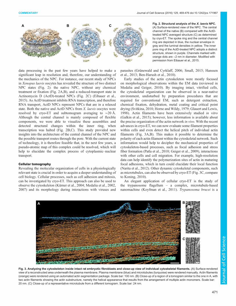
data processing in the past few years have helped to make asignificant leap in resolution and, therefore, our understanding ofthe mechanics of the NPC. For instance, our recent study of NPCsin Xenopus laevis oocytes has revealed the structure of two distinctNPC states (Fig. 2): the native NPC, without any chemicaltreatment or fixation (Fig. 2A,B), and a reduced-transport state inActinomycin D (ActD)-treated NPCs (Fig. 2C) (Eibauer et al.,2015). As ActD treatment inhibits RNA transcription, and thereforeRNA transport, ActD NPCs represent NPCs that are in a relaxedstate. Both the native and ActD NPCs from X. laevis oocytes wereresolved by cryo-ET and subtomogram averaging to ∼20 Å.Although the central channel is mainly composed of flexiblecomponents, we were able to visualize these assemblies anddetected structural changes within the inner ring, whentranscription was halted (Fig. 2B,C). This study provided newinsights into the architecture of the central channel of the NPC andthe possible transport routes through the pore. With the current stateof technology, it is therefore feasible that, in the next few years, apseudo-atomic map of this complex could be resolved, which willhelp to elucidate the complex process of cytoplasmic–nucleartransport.
Cellular tomographyRevealing the molecular organization of cells in a physiologicallyrelevant state is crucial in order to acquire a deeper understanding ofcell biology. Cellular processes, such as cell adhesion and mitosis,can be investigated by cryo-ET. This approach can also be used toobserve the cytoskeleton (Kürner et al., 2004; Medalia et al., 2002,2007) and its morphology during interactions with viruses and
parasites (Grünewald and Cyrklaff, 2006; Small, 2015; Hanssenet al., 2013; Ben-Harush et al., 2010).
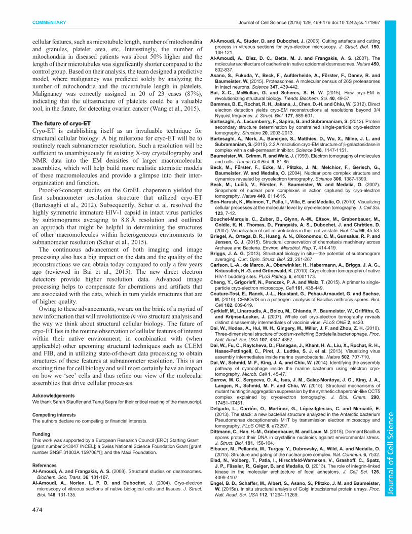
Early studies of the actin cytoskeleton were mostly focusedon morphological observations within the EM data (reviewed inMedalia and Geiger, 2010). By imaging intact, vitrified cells,the cytoskeletal organization can be observed in a near-nativeenvironment, undisturbed by preparation procedures that arerequired for conventional EM, such as detergent extraction,chemical fixation, dehydration, metal coating and critical pointdrying (Svitkina, 2010; Horne andWildy, 1979; Glauert and Lewis,1998). Actin filaments have been extensively studied in vitro(Galkin et al., 2015); however, less information is available aboutthe precise organization of the actin network in vivo. With the recentadvances in cryo-ET, we can now evaluate some filament propertieswithin cells and even detect the helical pitch of individual actinfilaments (Fig. 3A,B). This makes it possible to determine thepolarity of each actin filament within the cytoskeletal network. Suchinformation would help to decipher the mechanical properties ofcytoskeleton-based processes, such as focal adhesion and stressfiber formation (Patla et al., 2010; Geiger et al., 2009), interactionswith other cells and cell migration. For example, high-resolutiondata can help identify the polymerization sites of actin in maturingfocal adhesions, which in turn could elucidate their local function(Narita et al., 2012). Other dynamic cytoskeletal components, suchas microtubules, can also be observed by cryo-ET (Fig. 3C, compareto Koning, 2010).
An elegant application of cellular cryo-ET is the study ofthe trypanosome flagellum – a complex, microtubule-basednanomachine (Koyfman et al., 2011). Trypanosoma brucei is a
A B C Fig. 2. Structural analysis of the X. laevis NPC.(A) Surface-rendered view of the NPC. The centralchannel of the native (B) compared with the ActD-treated NPC averaged structure (C) as determinedby cryo-ET. The spoke ring and the central channelring are depicted in blue, the nuclear envelope ingray and the luminal densities in yellow. The innerpore ring of the ActD-treated NPC adopts a distinctstructure, shown in purple. Channels marked withorange dots are ∼2 nm in diameter. Modified withpermission from Eibauer et al., 2015.
A B
C
B
C
A
Fig. 3. Analyzing the cytoskeleton inside intact rat embryotic fibroblasts and close-up view of individual cytoskeletal filaments. (A) Surface-renderedview of a reconstructed area underneath the plasmamembrane. Plasma membrane (blue) and microtubules (turquoise) were rendered manually. Actin filaments(orange) were rendered using an automated actin segmentation package. Scale bar: 100 nm. (B) Close-up of a region of a tomogram similar to the one in A, withtwo actin filaments showing the actin substructure, namely the helical appearance that results from the arrangement of multiple actin monomers. Scale bar:20 nm. (C) Close-up of a representative microtubule from a different tomogram. Scale bar: 24 nm.
471
COMMENTARY Journal of Cell Science (2016) 129, 469-476 doi:10.1242/jcs.171967
Journal
ofCe
llScience

parasite that causes African sleeping sickness. Its flagellum isessential for the lifecycle and infection ability of the parasite, andit is being explored as a potential drug target. The flagellumcontains a microtubular azoneme, a crystalline paraflagellar rod andconnecting proteins (PFR). Subtomogram averaging revealed thetypical nine microtubule doublets arranged around a centralmicrotubule pair, as well as the structure of the flagellum in threedifferent bending states (Koyfman et al., 2011). Here, analysis of thebent flagella made it possible to suggest a model for the locomotionpattern of the parasite, in which the PFR functions like a jackscrewand modifies the in-plane axoneme motion, resulting in thecharacteristic bihelical motility of the trypanosome.
Improved EM optics impact on what we see inside the cellEM is mainly limited by how far electrons can penetrate throughthe biological sample, restricting the thickness of the specimento be examined to less than 1 μm. This generally limits the type ofspecimens that can be imaged by EM to isolated viruses (Hong et al.,2015; Murata et al., 2010; Dai et al., 2010), protein complexes(Koyfman et al., 2011; Nickell et al., 2007a,b), organelles (Eibaueret al., 2015; Nicastro et al., 2000; Beck et al., 2007, 2004), smallerprokaryotic cells (Briegel et al., 2015; Zhao et al., 2014) and thinperipheral regions of eukaryotic cells (Hu, 2014; Ben-Harush et al.,2010; Patla et al., 2010; Cyrklaff et al., 2007). Cells up to 1 μm inthickness can be imaged by EM, as shown for Ostreococcus tauri,the smallest eukaryotic cell (Henderson et al., 2007); this hasprovided a unique insight into the ultrastructure and characteristics ofthe cells during different stages of their cell cycle. The newlyavailable phase plate technology for EM provides increased imagecontrast and therefore, makes it easier to identify fine cellularcomponents within thicker specimens in situ (Dai et al., 2013, 2014).Phase plates are typically positioned in the back focal plane of theobjective lens of the EM and shift the phase of the scattered electronbeam such that low-frequency information is amplified. This is theinformation that contributes to image contrast, and higher contrastmakes it possible to detect structural features within the data and,therefore, allows for the direct observation and analysis of theinternal 3D structure of cells (Asano et al., 2015; Fukuda et al., 2015;Engel et al., 2015a,b; Dai et al., 2014, 2013) (Fig. 4).
Structural analysis of multicellular systemsIn order to examine bulkier specimens, the sample has to be eithermechanically trimmed or thinned by a focused ion beam. Cryo-sectioning of frozen hydrated specimens (CEMOVIS) is oneapproach to obtain sections that are sufficiently thin for EM. Inthis method, frozen hydrated cells or tissues of interest are sectionedwith a diamond knife resulting in samples with a thickness of40–100 μm (Al-Amoudi et al., 2004). Samples that have beenstudied by CEMOVIS thus far include mitochondria (Hsieh et al.,2006), human skin (Al-Amoudi et al., 2007; Al-Amoudi andFrangakis, 2008), microtubules (Bouchet-Marquis et al., 2007),bacteria (Delgado et al., 2013; Zuber et al., 2008) and bacterialspores (Couture-Tosi et al., 2010; Dittmann et al., 2015). However,this technique has its limitations, mainly due to compression of thethin sections in the direction of cutting, which can complicate theproper interpretation of the data (Al-Amoudi et al., 2005).More recently, cryo-focused ion beam scanning electron
microscopy (cryo-FIB-SEM) has emerged as an alternative toconventional sample-thinning methods. It incorporates specimenvitrification and yields thin lamellae (200–500 nm) from cells grownon EM grids, which can then be imaged by cryo-ET (Marko et al.,2007; Rigort et al., 2012). Cryo-ET of a FIB-milled Dictyostelium
discoideum cell revealed the nuclear envelope with the NPCs, theendoplasmic reticulum, mitochondria, microtubules, vacuolarcompartment and ribosomes all embedded within the lamella region
Fig. 4. Examples of cellular tomography performed with phase plates.(A,B) Cryo-ET projections of a Syn5-infected cyanobacterium cell without(A) and with (B) a phase plate under identical imaging conditions. Scale bar:100 nm. Modified with permission from Dai et al., 2013. (C,D) Projections ofa tomogram of primary cultured neurons without (C) and with phase plate(D). The red arrow in D indicates the lipid bilayer. Scale bar: 100 nm (20 nminset). Modified with permission from Fukuda et al., 2015. (E) A slice of atomogram acquired with a phase plate showing an intracisternal filamentbundle within the cisternae of the trans-Golgi (yellow arrow) and a nearbycoated bud. An acidocalcisome containing a dense aggregate ofpolyphosphate filaments is labeled with ‘ac’. Scale bar is 100 nm.(F) Segmentation of the cisterna in E, with the Golgi membrane in darkorange, the coated bud in yellow and the filament bundle in blue. PanelsE and F were modified with permission from Engel et al., 2015a. (G) A slicefrom a tomogram of a cultured hippocampal neuronal cell. The coloredsquares are zoomed-in views and show magnified slices through individualproteasome volumes of a single-capped (yellow box, middle) and twodouble-capped (red boxes, left and right) 26S proteasomes. Scale bars:100 nm (25 nm inset). Modified with permission from Asano et al., 2015.
472
COMMENTARY Journal of Cell Science (2016) 129, 469-476 doi:10.1242/jcs.171967
Journal
ofCe
llScience

of interest (Rigort et al., 2012). This technique has also recently beenused to acquire 3D structural information for the distribution of ER–mitochondrial contacts in mammalian cells and provide insights intothe localization ofmitochondrial constriction sites and the existence oftwo types ofmembrane associations between the twoorganelles (Ohtaet al., 2014). This procedure can also be applied to tissues such asskeletal muscle (Wagenknecht et al., 2015), thereby enabling thevisualization of natively preserved regions of interest that cannot bepurified. The first 3D-snapshots of the algal chloroplast were alsoobtained by this method, affording a glimpse into photosynthesis,thylakoid biogenesis and carbon fixation (Engel et al., 2015b).FIB milling and imaging by cryo-ET of frozen Caenorhabditis
elegans embryos and adult worms has allowed researchers tovisualizethe intracellular organization during particular stages of developmentin tissues of interest (Harapin et al., 2015). Here, the milled lamellaewere ∼330 nm thick in the case of embryos, and ∼660 nm thick foradult worms, both sufficiently thin to be visualized by cryo-ET(Fig. 5). Furthermore, C. elegans embryos were fluorescently labeledduring the preparation, which allowed the identification of proteins onthe surface of thin cryo-lamellae by correlative light and electronmicroscopy (CLEM). Combining CLEM with cryo-FIB-SEM(Mahamid et al., 2015) and cryo-ET can also be applied to explorethe structure of macromolecular complexes in situ during variousstages of development, which will present further details aboutprocesses occurring inmore complex organisms. Furthermore, the useof FIB-SEM to generate thin enough samples for cryo-ET has thepotential to eventually bridge the gap between developmental andstructural biology, by resolving with high reliability embryonicstructures at different developmental stages.Cryo-FIB can also be combined with phase plate cryo-ET
(Fukuda et al., 2015), as performed in a recent study ofChlamydomonas cells (Engel et al., 2015a). Here, the authors
obtained a remarkable view of the Golgi ultrastructure that revealednew components of theGolgi cisternaewithin unperturbed cells withan unprecedented resolution (Engel et al., 2015a) (Fig. 4E).Although not yet routinely used, we anticipate that in the nearfuture, combining cryo-FIB and phase plates might have atremendous impact on structural studies of multi-cellular specimens.
Combining correlative light and electron microscopyOne limitation of cryo-ET is the inability to unambiguously identifythe position of a protein or a process within the cell because of thelow signal-to-noise ratio of cryo-embedded specimens. Moreover,cryo-tomograms have a limited field of view of 1 to 2 µm2, whichonly covers a very small fraction of a typical eukaryotic cell. UsingCLEM, it has been possible to capitalize on the advantages of bothunderlying techniques (Schorb and Briggs, 2014) and tacklebiological questions that cannot be resolved by either approachalone; specific molecules and thus particular events are localizedwith fluorescence-based microscopy, then cryo-tomograms areacquired to reveal the cellular ultrastructure at these sites.
Cryo-CLEM has the benefit of preserving specimens (e.g. wholecells or bacteria) in a near-native state, while still using fluorescentlabels to identify specific regions of interest. Recently, this methodwas successfully applied to vitrified Streptomyces bacteria to studycell division (Koning et al., 2014).
Cryo-ET as a medical diagnostics toolCryo-ET could be also used as a diagnostics tool and help resolvemolecular differences between healthy and diseased states. The firstreported instance of using human clinical samples for cryo-ETstudies was the structural analysis of human cilia that were isolatedfrom airway epithelial cells from both healthy individuals andpatients with primary ciliary dyskinesia (PCD) (Lin et al., 2014).Diagnosing PCD has traditionally relied on analysis byconventional EM and detection of defects in mobile cilia.However, a third of PCD patients have cilia that appear normalunder the EM, perhaps due to the harsh preparation methodsrequired for conventional imaging. This new study built on theexisting knowledge about human cilia and helped elucidatestructural defects associated with PCD and other ciliopathies.Applying cryo-ET and subtomogram averaging to human ciliaryaxonemes from healthy individuals resulted in an improvedresolution over conventional EM studies that utilized chemicallyfixed samples. The averaged cryo-ET structure showed that theoverall architecture is similar to that of the motile cilia and flagellaof other organisms (Lin et al., 2014). However, subtomogramaveraging of samples derived from patients with mutated ciliapointed to small structural differences compared to healthy cilia,which could explain why the particular variant studied resulted in aclinically attenuated phenotype, where patients experience delayedonset of pulmonary symptoms and maintain better lung functioncompared to patients with mutations in other genes.
Platelet biology is another field that is benefiting from structuralstudies using EM (reviewed in Sorrentino et al., 2015). Recently,Wang et al. have illustrated how cryo-ET can be utilized as a non-invasive method to detect structural biomarkers of diseases that affectplatelets (Wang et al., 2015). For example, it is known that the countand functionality of platelets can be altered in cancer patients. Theteam evaluated frozen-hydrated platelets from patients with newlydiagnosed invasive ovarian cancer and from control subjects (patientswith benign masses and healthy individuals) and analyzed themorphologic features of each sample. The visual differences werequantified by measuring length, area and/or the number of observed
A B
C
M
M
D
Fig. 5. Cryo-FIB milling and cryo-ET of C. elegans. (A) Scanning EM (SEM)image of an adult C. elegans worm that has been vitrified by high-pressurefreezing, using 2-methylpentane as freezing medium. Scale bar: 10 μm. (B) Alamella of the adult worm (arrowheads) shown in A, produced by cryo-FIBmilling. Scale bar: 2 μm. (C) A slice through a tomogram of a FIB-milled C.elegans embryo; here, the plasma membrane (green arrow), mitochondria(yellow arrows; ‘M’), ribosomes (blue arrow) and the nuclear envelope (orangearrow) can be seen. Scale bar: 100 nm. (D) An example of a rendering ofanother tomogram acquired on a C. elegans embryo lamella. Shown here arethe plasma membrane (dark blue), nuclear envelope (pink), ribosomes (gold),NPCs (blue) and filamentous structures near the inner nuclear membrane(green). Modified with permission from Harapin et al., 2015.
473
COMMENTARY Journal of Cell Science (2016) 129, 469-476 doi:10.1242/jcs.171967
Journal
ofCe
llScience
cellular features, such as microtubule length, number of mitochondriaand granules, platelet area, etc. Interestingly, the number ofmitochondria in diseased patients was about 50% higher and thelength of their microtubules was significantly shorter compared to thecontrol group. Based on their analysis, the team designed a predictivemodel, where malignancy was predicted solely by analyzing thenumber of mitochondria and the microtubule length in platelets.Malignancy was correctly assigned in 20 of 23 cases (87%),indicating that the ultrastructure of platelets could be a valuabletool, in the future, for detecting ovarian cancer (Wang et al., 2015).
The future of cryo-ETCryo-ET is establishing itself as an invaluable technique forstructural cellular biology. A big milestone for cryo-ET will be toroutinely reach subnanometer resolution. Such a resolution will besufficient to unambiguously fit existing X-ray crystallography andNMR data into the EM densities of larger macromolecularassemblies, which will help build more realistic atomistic modelsof these macromolecules and provide a glimpse into their inter-organization and function.Proof-of-concept studies on the GroEL chaperonin yielded the
first subnanometer resolution structure that utilized cryo-ET(Bartesaghi et al., 2012). Subsequently, Schur et al. resolved thehighly symmetric immature HIV-1 capsid in intact virus particlesby subtomograms averaging to 8.8 Å resolution and outlinedan approach that might be helpful in determining the structuresof other macromolecules within heterogeneous environments tosubnanometer resolution (Schur et al., 2015).The continuous advancement of both imaging and image
processing also has a big impact on the data and the quality of thereconstructions we can obtain today compared to only a few yearsago (reviewed in Bai et al., 2015). The new direct electrondetectors provide higher resolution data. Advanced imageprocessing helps to compensate for aberrations and artifacts thatare associated with the data, which in turn yields structures that areof higher quality.Owing to these advancements, we are on the brink of a myriad of
new information that will revolutionize in vivo structure analysis andthe way we think about structural cellular biology. The future ofcryo-ET lies in the routine observation of cellular features of interestwithin their native environment, in combination with (whenapplicable) other upcoming structural techniques such as CLEMand FIB, and in utilizing state-of-the-art data processing to obtainstructures of these features at subnanometer resolution. This is anexciting time for cell biology and will most certainly have an impacton how we ‘see’ cells and thus refine our view of the molecularassemblies that drive cellular processes.
AcknowledgementsWe thank Sarah Stauffer and Tanuj Sapra for their critical reading of the manuscript.
Competing interestsThe authors declare no competing or financial interests.
FundingThis work was supported by a European Research Council (ERC) Starting Grant[grant number 243047 INCEL]; a Swiss National Science Foundation Grant [grantnumber SNSF 31003A 159706/1]; and the Maxi Foundation.
ReferencesAl-Amoudi, A. and Frangakis, A. S. (2008). Structural studies on desmosomes.Biochem. Soc. Trans. 36, 181-187.
Al-Amoudi, A., Norlen, L. P. O. and Dubochet, J. (2004). Cryo-electronmicroscopy of vitreous sections of native biological cells and tissues. J. Struct.Biol. 148, 131-135.
Al-Amoudi, A., Studer, D. and Dubochet, J. (2005). Cutting artefacts and cuttingprocess in vitreous sections for cryo-electron microscopy. J. Struct. Biol. 150,109-121.
Al-Amoudi, A., Dıez, D. C., Betts, M. J. and Frangakis, A. S. (2007). Themolecular architecture of cadherins in native epidermal desmosomes.Nature 450,832-837.
Asano, S., Fukuda, Y., Beck, F., Aufderheide, A., Forster, F., Danev, R. andBaumeister, W. (2015). Proteasomes. A molecular census of 26S proteasomesin intact neurons. Science 347, 439-442.
Bai, X.-C., McMullan, G. and Scheres, S. H. W. (2015). How cryo-EM isrevolutionizing structural biology. Trends Biochem. Sci. 40, 49-57.
Bammes, B. E., Rochat, R. H., Jakana, J., Chen, D.-H. andChiu,W. (2012). Directelectron detection yields cryo-EM reconstructions at resolutions beyond 3/4Nyquist frequency. J. Struct. Biol. 177, 589-601.
Bartesaghi, A., Lecumberry, F., Sapiro, G. and Subramaniam, S. (2012). Proteinsecondary structure determination by constrained single-particle cryo-electrontomography. Structure 20, 2003-2013.
Bartesaghi, A., Merk, A., Banerjee, S., Matthies, D., Wu, X., Milne, J. L. andSubramaniam, S. (2015). 2.2 Å resolution cryo-EM structure of β-galactosidase incomplex with a cell-permeant inhibitor. Science 348, 1147-1151.
Baumeister, W., Grimm, R. andWalz, J. (1999). Electron tomography of moleculesand cells. Trends Cell Biol. 9, 81-85.
Beck, M., Forster, F., Ecke, M., Plitzko, J. M., Melchior, F., Gerisch, G.,Baumeister, W. and Medalia, O. (2004). Nuclear pore complex structure anddynamics revealed by cryoelectron tomography. Science 306, 1387-1390.
Beck, M., Lucic, V., Forster, F., Baumeister, W. and Medalia, O. (2007).Snapshots of nuclear pore complexes in action captured by cryo-electrontomography. Nature 449, 611-615.
Ben-Harush, K., Maimon, T., Patla, I., Villa, E. andMedalia, O. (2010). Visualizingcellular processes at the molecular level by cryo-electron tomography. J. Cell Sci.123, 7-12.
Bouchet-Marquis, C., Zuber, B., Glynn, A.-M., Eltsov, M., Grabenbauer, M.,Goldie, K. N., Thomas, D., Frangakis, A. S., Dubochet, J. and Chretien, D.(2007). Visualization of cell microtubules in their native state. Biol. Cell 99, 45-53.
Briegel, A., Ortega, D. R., Huang, A. N., Oikonomou, C. M., Gunsalus, R. P. andJensen, G. J. (2015). Structural conservation of chemotaxis machinery acrossArchaea and Bacteria. Environ. Microbiol. Rep. 7, 414-419.
Briggs, J. A. G. (2013). Structural biology in situ—the potential of subtomogramaveraging. Curr. Opin. Struct. Biol. 23, 261-267.
Carlson, L.-A., de Marco, A., Oberwinkler, H., Habermann, A., Briggs, J. A. G.,Krausslich, H.-G. andGrunewald, K. (2010). Cryo electron tomography of nativeHIV-1 budding sites. PLoS Pathog. 6, e1001173.
Cheng, Y., Grigorieff, N., Penczek, P. A. and Walz, T. (2015). A primer to single-particle cryo-electron microscopy. Cell 161, 438-449.
Couture-Tosi, E., Ranck, J.-L., Haustant, G., Pehau-Arnaudet, G. and Sachse,M. (2010). CEMOVIS on a pathogen: analysis of Bacillus anthracis spores. Biol.Cell 102, 609-619.
Cyrklaff, M., Linaroudis, A., Boicu, M., Chlanda, P., Baumeister, W., Griffiths, G.and Krijnse-Locker, J. (2007). Whole cell cryo-electron tomography revealsdistinct disassembly intermediates of vaccinia virus. PLoS ONE 2, e420.
Dai, W., Hodes, A., Hui, W. H., Gingery, M., Miller, J. F. and Zhou, Z. H. (2010).Three-dimensional structure of tropism-switching Bordetella bacteriophage. Proc.Natl. Acad. Sci. USA 107, 4347-4352.
Dai, W., Fu, C., Raytcheva, D., Flanagan, J., Khant, H. A., Liu, X., Rochat, R. H.,Haase-Pettingell, C., Piret, J., Ludtke, S. J. et al. (2013). Visualizing virusassembly intermediates inside marine cyanobacteria. Nature 502, 707-710.
Dai, W., Schmid, M. F., King, J. A. and Chiu, W. (2014). Identifying the assemblypathway of cyanophage inside the marine bacterium using electron cryo-tomography. Microb. Cell 1, 45-47.
Darrow, M. C., Sergeeva, O. A., Isas, J. M., Galaz-Montoya, J. G., King, J. A.,Langen, R., Schmid, M. F. and Chiu, W. (2015). Structural mechanisms ofmutant huntingtin aggregation suppression by the synthetic chaperonin-like CCT5complex explained by cryoelectron tomography. J. Biol. Chem. 290,17451-17461.
Delgado, L., Carrion, O., Martınez, G., Lopez-Iglesias, C. and Mercade, E.(2013). The stack: a new bacterial structure analyzed in the Antarctic bacteriumPseudomonas deceptionensis M1T by transmission electron microscopy andtomography. PLoS ONE 8, e73297.
Dittmann, C., Han, H.-M., Grabenbauer, M. and Laue, M. (2015). Dormant Bacillusspores protect their DNA in crystalline nucleoids against environmental stress.J. Struct. Biol. 191, 156-164.
Eibauer, M., Pellanda, M., Turgay, Y., Dubrovsky, A., Wild, A. and Medalia, O.(2015). Structure and gating of the nuclear pore complex. Nat. Commun. 6, 7532.
Elad, N., Volberg, T., Patla, I., Hirschfeld-Warneken, V., Grashoff, C., Spatz,J. P., Fassler, R., Geiger, B. and Medalia, O. (2013). The role of integrin-linkedkinase in the molecular architecture of focal adhesions. J. Cell Sci. 126,4099-4107.
Engel, B. D., Schaffer, M., Albert, S., Asano, S., Plitzko, J. M. and Baumeister,W. (2015a). In situ structural analysis of Golgi intracisternal protein arrays. Proc.Natl. Acad. Sci. USA 112, 11264-11269.
474
COMMENTARY Journal of Cell Science (2016) 129, 469-476 doi:10.1242/jcs.171967
Journal
ofCe
llScience
Engel, B. D., Schaffer, M., Kuhn Cuellar, L., Villa, E., Plitzko, J. M. andBaumeister, W. (2015b). Native architecture of the Chlamydomonas chloroplastrevealed by in situ cryo-electron tomography. eLife 4, e04889.
Faruqi, A. R. and McMullan, G. (2011). Electronic detectors for electronmicroscopy. Q. Rev. Biophys. 44, 357-390.
Fridman, K., Mader, A., Zwerger, M., Elia, N. andMedalia, O. (2012). Advances intomography: probing the molecular architecture of cells. Nat. Rev. Mol. Cell Biol.13, 736-742.
Fukuda, Y., Laugks, U., Lucic, V., Baumeister, W. and Danev, R. (2015). Electroncryotomography of vitrified cells with a Volta phase plate. J. Struct. Biol. 190,143-154.
Galkin, V. E., Orlova, A., Vos, M. R., Schroder, G. F. and Egelman, E. H. (2015).Near-atomic resolution for one state of F-actin. Structure 23, 173-182.
Geiger, B., Spatz, J. P. and Bershadsky, A. D. (2009). Environmental sensingthrough focal adhesions. Nat. Rev. Mol. Cell Biol. 10, 21-33.
Glauert, A. M. and Lewis, P. R. (1998). Biological Specimen Preparation forTransmission Electron Microscopy. Practical Methods in Electron Microscopy,Vol. 17. Princeton University Press.
Grossman, E., Medalia, O. and Zwerger, M. (2012). Functional architecture of thenuclear pore complex. Annu. Rev. Biophys. 41, 557-584.
Gruenbaum, Y. and Medalia, O. (2015). Lamins: the structure and proteincomplexes. Curr. Opin. Cell Biol. 32, 7-12.
Grunewald, K. and Cyrklaff, M. (2006). Structure of complex viruses and virus-infected cells by electron cryo tomography. Curr. Opin. Microbiol. 9, 437-442.
Hanssen, E., Dekiwadia, C., Riglar, D. T., Rug, M., Lemgruber, L., Cowman,A. F., Cyrklaff, M., Kudryashev, M., Frischknecht, F., Baum, J. et al. (2013).Electron tomography of Plasmodium falciparum merozoites reveals core cellularevents that underpin erythrocyte invasion. Cell. Microbiol. 15, 1457-1472.
Harapin, J., Bormel, M., Sapra, K. T., Brunner, D., Kaech, A. and Medalia, O.(2015). Structural analysis of multicellular organisms with cryo-electrontomography. Nat. Methods 12, 634-636.
Henderson, G. P., Gan, L. and Jensen, G. J. (2007). 3-D ultrastructure of O. tauri:electron cryotomography of an entire eukaryotic cell. PLoS ONE 2, e749.
Hong, C., Pietila, M. K., Fu, C. J., Schmid, M. F., Bamford, D. H. and Chiu, W.(2015). Lemon-shaped halo archaeal virus His1 with uniform tail but variablecapsid structure. Proc. Natl. Acad. Sci. USA 112, 2449-2454.
Hoppe,W., Gassmann, J., Hunsmann, N., Schramm, H. J. and Sturm, M. (1974).Three-dimensional reconstruction of individual negatively stained yeast fatty-acidsynthetase molecules from tilt series in the electron microscope. Hoppe-SeylersZ. Physiol. Chem. 355, 1483-1487.
Horne, R. W. and Wildy, P. (1979). An historical account of the development andapplications of the negative staining technique to the electron microscopy ofviruses. J. Microsc. 117, 103-122.
Hsieh, C.-E., Leith, A., Mannella, C. A., Frank, J. and Marko, M. (2006). Towardshigh-resolution three-dimensional imaging of native mammalian tissue: electrontomography of frozen-hydrated rat liver sections. J. Struct. Biol. 153, 1-13.
Hu, G.-B. (2014). Whole cell cryo-electron tomography suggests mitochondriadivide by budding. Microsc. Microanal. 20, 1180-1187.
Koning, R. I. (2010). Cryo-electron tomography of cellular microtubules. MethodsCell Biol. 97, 455-473. Academic Press 2010.
Koning, R. I., Celler, K., Willemse, J., Bos, E., van Wezel, G. P. and Koster, A. J.(2014). Correlative cryo-fluorescence light microscopy and cryo-electrontomography of Streptomyces. Methods Cell Biol. 124, 217-239.
Koyfman, A. Y., Schmid, M. F., Gheiratmand, L., Fu, C. J., Khant, H. A., Huang,D., He, C. Y. and Chiu, W. (2011). Structure of Trypanosoma brucei flagellumaccounts for its bihelical motion. Proc. Natl. Acad. Sci. USA 108, 11105-11108.
Kurner, J., Medalia, O., Linaroudis, A. A. and Baumeister, W. (2004). Newinsights into the structural organization of eukaryotic and prokaryoticcytoskeletons using cryo-electron tomography. Exp. Cell Res. 301, 38-42.
Li, X., Mooney, P., Zheng, S., Booth, C. R., Braunfeld, M. B., Gubbens, S.,Agard, D. A. and Cheng, Y. (2013). Electron counting and beam-induced motioncorrection enable near-atomic-resolution single-particle cryo-EM. Nat. Methods10, 584-590.
Lin, J., Yin, W., Smith, M. C., Song, K., Leigh, M. W., Zariwala, M. A., Knowles,M. R., Ostrowski, L. E. and Nicastro, D. (2014). Cryo-electron tomographyreveals ciliary defects underlying human RSPH1 primary ciliary dyskinesia. Nat.Commun. 5, 5727.
Mahamid, J., Schampers, R., Persoon, H., Hyman, A. A., Baumeister, W. andPlitzko, J. M. (2015). A focused ion beam milling and lift-out approach for site-specific preparation of frozen-hydrated lamellas from multicellular organisms.J. Struct. Biol. 192, 262-269.
Maimon, T., Elad, N., Dahan, I. and Medalia, O. (2012). The human nuclear porecomplex as revealed by cryo-electron tomography. Structure 20, 998-1006.
Marko, M., Hsieh, C., Schalek, R., Frank, J. and Mannella, C. (2007). Focused-ion-beam thinning of frozen-hydrated biological specimens for cryo-electronmicroscopy. Nat. Methods 4, 215-217.
Marton, L. (1934). Electron microscopy of biological objects. Nature 133, 911.McIntosh, J. R. (2007). Cellular electron microscopy. Methods Cell Biol. 79, 1-850.Elsevier Science.
Medalia, O. and Geiger, B. (2010). Frontiers of microscopy-based research intocell–matrix adhesions. Curr. Opin. Cell Biol. 22, 659-668.
Medalia, O., Weber, I., Frangakis, A. S., Nicastro, D., Gerisch, G. andBaumeister, W. (2002). Macromolecular architecture in eukaryotic cellsvisualized by cryoelectron tomography. Science 298, 1209-1213.
Medalia, O., Beck, M., Ecke, M., Weber, I., Neujahr, R., Baumeister, W. andGerisch, G., (2007). Organization of actin networks in intact filopodia. Curr. Biol.17, 79-84.
Milazzo, A.-C., Cheng, A., Moeller, A., Lyumkis, D., Jacovetty, E., Polukas, J.,Ellisman, M. H., Xuong, N.-H., Carragher, B. and Potter, C. S. (2011). Initialevaluation of a direct detection device detector for single particle cryo-electronmicroscopy. J. Struct. Biol. 176, 404-408.
Murata, K., Liu, X., Danev, R., Jakana, J., Schmid, M. F., King, J., Nagayama, K.and Chiu, W. (2010). Zernike phase contrast cryo-electron microscopy andtomography for structure determination at nanometer and subnanometerresolutions. Structure 18, 903-912.
Murphy, G. E. and Jensen, G. J. (2007). Electron cryotomography. Biotechniques43, 413-421, 417 passim.
Narita, A., Mueller, J., Urban, E., Vinzenz, M., Small, J. V. and Maeda, Y. (2012).Direct determination of actin polarity in the cell. J. Mol. Biol. 419, 359-368.
Nicastro, D., Frangakis, A. S., Typke, D. and Baumeister, W. (2000). Cryo-electron tomography of neurospora mitochondria. J. Struct. Biol. 129, 48-56.
Nickell, S., Mihalache, O., Beck, F., Hegerl, R., Korinek, A. and Baumeister, W.(2007a). Structural analysis of the 26S proteasome by cryoelectron tomography.Biochem. Biophys. Res. Commun. 353, 115-120.
Nickell, S., Park, P. S.-H., Baumeister, W. and Palczewski, K. (2007b). Three-dimensional architecture of murine rod outer segments determined bycryoelectron tomography. J. Cell Biol. 177, 917-925.
Ohta, K., Okayama, S., Togo, A. and Nakamura, K.-i. (2014). Three-dimensionalorganization of the endoplasmic reticulum membrane around the mitochondrialconstriction site in mammalian cells revealed by using focused-ion beamtomography. Microscopy 63, i34.
Passarelli, M. K. and Ewing, A. G. (2013). Single-cell imaging mass spectrometry.Curr. Opin. Chem. Biol. 17, 854-859.
Patla, I., Volberg, T., Elad, N., Hirschfeld-Warneken, V., Grashoff, C., Fassler, R.,Spatz, J. P., Geiger, B. and Medalia, O. (2010). Dissecting the moleculararchitecture of integrin adhesion sites by cryo-electron tomography.Nat. Cell Biol.12, 909-915.
Rigort, A., Bauerlein, F. J. B., Villa, E., Eibauer, M., Laugks, T., Baumeister, W.and Plitzko, J. M. (2012). Focused ion beam micromachining of eukaryotic cellsfor cryoelectron tomography. Proc. Natl. Acad. Sci. USA 109, 4449-4454.
Ruskin, R. S., Yu, Z. and Grigorieff, N. (2013). Quantitative characterization ofelectron detectors for transmission electron microscopy. J. Struct. Biol. 184,385-393.
Schorb, M. and Briggs, J. A. G. (2014). Correlated cryo-fluorescence and cryo-electron microscopy with high spatial precision and improved sensitivity.Ultramicroscopy 143, 24-32.
Schroder, R. R. (2015). Advances in electron microscopy: a qualitative view ofinstrumentation development for macromolecular imaging and tomography. Arch.Biochem. Biophys. 581, 25-38.
Schur, F. K. M., Hagen, W. J. H., Rumlova, M., Ruml, T., Muller, B., Krausslich,H.-G. and Briggs, J. A. G. (2015). Structure of the immature HIV-1 capsid in intactvirus particles at 8.8 Å resolution. Nature 517, 505-508.
Shahmoradian, S. H., Galaz-Montoya, J. G., Schmid, M. F., Cong, Y., Ma, B.,Spiess, C., Frydman, J., Ludtke, S. J. and Chiu, W. (2013). TRiC’s tricks inhibithuntingtin aggregation. eLife 2, e00710.
Small, J. V. (2015). Pushing with actin: from cells to pathogens. Biochem. Soc.Trans. 43, 84-91.
Sorrentino, S., Studt, J.-D., Medalia, O. and Tanuj Sapra, K. (2015). Roll, adhere,spread and contract: structural mechanics of platelet function. Eur. J. Cell Biol. 94,129-138.
Stauffer, S., Rahman, S. A., de Marco, A., Carlson, L.-A., Glass, B.,Oberwinkler, H., Herold, N., Briggs, J. A. G., Muller, B., Grunewald, K. et al.(2014). The nucleocapsid domain of Gag is dispensable for actin incorporationinto HIV-1 and for association of viral budding sites with cortical F-actin. J. Virol.88, 7893-7903.
Stoffler, D., Feja, B., Fahrenkrog, B., Walz, J., Typke, D. and Aebi, U. (2003).Cryo-electron tomography provides novel insights into nuclear pore architecture:implications for nucleocytoplasmic transport. J. Mol. Biol. 328, 119-130.
Svitkina, T. (2010). Imaging cytoskeleton components by electron microscopy.Methods Mol. Biol. 586, 187-206.
von Appen, A., Kosinski, J., Sparks, L., Ori, A., DiGuilio, A. L., Vollmer, B.,Mackmull, M.-T., Banterle, N., Parca, L., Kastritis, P. et al. (2015). In situstructural analysis of the human nuclear pore complex. Nature 526, 140-143.
Wagenknecht, T., Hsieh, C. and Marko, M. (2015). Skeletal muscle triad junctionultrastructure by Focused-Ion-Beam milling of muscle and Cryo-ElectronTomography. Eur. J. Transl. Myol. 25, 49-56.
Wang, R., Stone, R. L., Kaelber, J. T., Rochat, R. H., Nick, A. M., Vijayan, K. V.,Afshar-Kharghan, V., Schmid, M. F., Dong, J. F., Sood, A. K. et al.(2015). Electron cryotomography reveals ultrastructure alterations in
475
COMMENTARY Journal of Cell Science (2016) 129, 469-476 doi:10.1242/jcs.171967
Journal
ofCe
llScience

platelets from patients with ovarian cancer. Proc. Natl. Acad. Sci. USA 112,14266-14271.
Woodward, C. L., Mendonça, L. M. and Jensen, G. J. (2015). Direct visualizationof vaults within intact cells by electron cryo-tomography. Cell. Mol. Life Sci. 72,3401-3409.
Zhao, X., Norris, S. J. and Liu, J. (2014). Molecular architecture of the bacterialflagellar motor in cells. Biochemistry 53, 4323-4333.
Zuber, B., Chami, M., Houssin, C., Dubochet, J., Griffiths, G. and Daffe, M.(2008). Direct visualization of the outer membrane of mycobacteria andcorynebacteria in their native state. J. Bacteriol. 190, 5672-5680.
476
COMMENTARY Journal of Cell Science (2016) 129, 469-476 doi:10.1242/jcs.171967
Journal
ofCe
llScience