Embed Size (px)
Citation preview

ONLINE ONLY
Cellular, molecular, and tissue-level reactionsto orthodontic forceVinod Krishnana and Ze’ev Davidovitchb
Tamilnadu, India, and Cleveland, Ohio
Remodeling changes in paradental tissues are considered essential in effecting orthodontic tooth movement.The force-induced tissue strain produces local alterations in vascularity, as well as cellular and extracellularmatrix reorganization, leading to the synthesis and release of various neurotransmitters, cytokines, growthfactors, colony-stimulating factors, and metabolites of arachidonic acid. Recent research in the biologicalbasis of tooth movement has provided detailed insight into molecular, cellular, and tissue-level reactions toorthodontic forces. Although many studies have been reported in the orthodontic and related scientificliterature, a concise convergence of all data is still lacking. Such an amalgamation of the rapidly accumulatingscientific information should help orthodontic clinicians and educators understand the biological processesthat underlie the phenomenon of tooth movement with mechanics (removable, fixed, or functionalappliances). This review aims to achieve this goal and is organized to include all major findings from thebeginning of research in the biology of tooth movement. It highlights recent developments in cellular,molecular, tissue, and genetic reactions in response to orthodontic force application. It reviews briefly theprocesses of bone, periodontal ligament, and gingival remodeling in response to orthodontic force. Thisreview also provides insight into the biological background of various deleterious effects of orthodontic
forces. (Am J Orthod Dentofacial Orthop 2006;129:469e.1-460e.32)Tooth movement by orthodontic force applica-tion is characterized by remodeling changes indental and paradental tissues, including dental
pulp, periodontal ligament (PDL), alveolar bone, andgingiva. These tissues, when exposed to varying de-grees of magnitude, frequency, and duration of me-chanical loading, express extensive macroscopic andmicroscopic changes. Orthodontic tooth movement dif-fers markedly from physiological dental drift or tootheruption. The former is uniquely characterized by theabrupt creation of compression and tension regions inthe PDL.1 Physiological tooth movement is a slowprocess that occurs mainly in the buccal direction intocancellous bone or because of growth into corticalbone. In contrast, orthodontic tooth movement canoccur rapidly or slowly, depending on the physicalcharacteristics of the applied force, and the size andbiological response of the PDL.2 These force-inducedstrains alter the PDL’s vascularity and blood flow,resulting in local synthesis and release of various key
aAssistant professor, Department of Orthodontics, Rajas Dental College,Tirunelveli District, Tamilnadu, India.bClinical professor, Department of Orthodontics, Case Western Reserve Uni-versity, Cleveland, Ohio.Reprint requests to: Dr Vinod Krishnan, Gourivilasam, Kudappanakunnu PO,Trivandrum, Kerala State 695043, India; e-mail, [email protected], May 2005; revised and accepted, October 2005.0889-5406/$32.00Copyright © 2006 by the American Association of Orthodontists.
doi:10.1016/j.ajodo.2005.10.007molecules, such as neurotransmitters, cytokines, growthfactors, colony-stimulating factors, and arachidonic acidmetabolites. These molecules can evoke many cellularresponses by various cell types in and around teeth,providing a favorable microenvironment for tissue depo-sition or resorption.3,4
Studies in the early 20th century attempted mainlyto analyze the histological changes in paradental tissuesafter tooth movement. Those studies showed extensivecellular activities in the mechanically stressed PDLinvolving fibroblasts, endothelial cells, osteoblasts, os-teocytes, and endosteal cells.5 Apart from this finding,it was discovered that mechanical stresses alter thestructural properties of tissues at the cellular, molecu-lar, and genetic levels. Current literature has much dataon molecular- and genetic-level cellular responses toorthodontic force. The rapid reactions at the initialstage of mechanotherapy and slower adaptive changeslater are well explained in the literature. The followingdiscussion on cellular, molecular, and tissue reactions isintended to provide basic information about histologi-cal and chemical changes of orthodontic tooth move-ment. It tries to update the readers with recent devel-opments in cellular, molecular, tissue, and geneticreactions in response to orthodontic force applicationalong with a brief description on the processes of bone,PDL, and gingival remodeling in response to orthodon-
tic force. This review also provides insight into the469.e1

American Journal of Orthodontics and Dentofacial OrthopedicsApril 2006
469.e2 Krishnan and Davidovitch
biological background of various deleterious effects oforthodontic forces.
ORTHODONTIC VERSUS ORTHOPEDIC FORCE
Orthodontic force has been defined as “force ap-plied to teeth for the purpose of effecting tooth move-ment, generally having a magnitude lower than anorthopedic force,” whereas orthopedic force is definedas “force of higher magnitude in relation to an orth-odontic force, when delivered via teeth for 12 to 16hours a day, is supposed to produce a skeletal effect onthe maxillofacial complex.”6 These definitions showthat there is no clear distinction between orthodonticand orthopedic forces, even in terms of magnitude;furthermore, many widely variable arbitrary sugges-tions about the characteristics of orthodontic forcesabound in the literature.
Orthodontic mechanotherapy is mainly aimed attooth movement by remodeling and adaptive changes inparadental tissues. To effect this outcome, only smallamounts of force—20 to 150 g per tooth—might berequired. But craniofacial orthopedics is aimed at de-livering higher magnitudes of mechanical forces—more than 300 g—in attempts to modify the form ofcraniofacial bones. The appliances, called craniofacialorthopedic devices, deliver macro-scale mechanicalforces, which produce micro-structural sutural bonestrain and induce cellular growth response in sutures.7
OPTIMAL ORTHODONTIC FORCE
Orthodontic tooth movement is mediated by cou-pling bone resorption and deposition in compressed andstretched sides of the PDL, respectively. Orthodonticforces, by virtue of altering the blood flow and local-ized electrochemical environment, upset the homeo-static environment of the PDL space. This abruptalteration initiates biochemical and cellular events thatreshape the bony contour of the alveolus.8 It is assumedthat an optimal orthodontic force moves teeth effi-ciently into their desired position, without causingdiscomfort or tissue damage to the patient. Primarily,an optimal force is based on proper mechanical princi-ples, which enable the orthodontist to move teethwithout traumatizing dental or paradental tissues, andwithout moving dental roots redundantly (round-trip-ping), or into danger zones (compact plates of alveolarbone). Traditionally, orthodontic forces have been cat-egorized as “light” or “heavy,” and it was assumed thatlight forces are gentler and therefore more physiologicthan heavy forces. However, Burstone9 reported thatorthodontic forces are never distributed equallythroughout the PDL, and Storey10 observed that some
trauma is always associated with applied orthodonticforces, even light ones. Moreover, it is impossible, withthe available instrumentation, to measure precisely theamount of force applied to roots or parts thereof underany mode of treatment. Consequently, at present, it canbe stated that, to engender adequate biological responsein the periodontium, light forces are preferable, becauseof their ability to evoke frontal resorption of bone.Unlike light forces, heavy forces often cause necrosis(hyalinization) of the PDL and undermining boneresorption,11 and have been implicated in root resorp-tion.
The concept of optimal orthodontic force is chang-ing along with the specialty. The classic definition ofoptimal force by Schwarz12 in 1932 was “the forceleading to a change in tissue pressure that approximatedthe capillary vessels’ blood pressure, thus preventingtheir occlusion in the compressed periodontal liga-ment.” According to Schwarz, forces below optimumproduce no reaction, whereas forces above that levellead to tissue necrosis, thus preventing frontal resorp-tion of the alveolar bone. Oppenheim13 and Reitan,11
who recommended applying light forces for toothmovement, demonstrated cell-free compressed areas inthe PDL. Storey and Smith14 also reported the samefinding in 1952. They studied distal movement ofcanines in orthodontic patients and suggested that thereis an optimum range of pressure (150-200 g) on thetooth-bone interface that produces a maximum rate oftooth movement. Pressure below this range producedno tooth movement. When the force was increasedabove optimum, the rate of tooth movement wasdecreased and finally approached zero within a week.
The current concept of optimum force views it as anextrinsic mechanical stimulus that evokes a cellularresponse that aims to restore equilibrium by remodelingperiodontal supporting tissues. So the mechanical inputthat leads to the maximum rate of tooth movement withminimal irreversible damage to root, PDL, and alveolarbone is considered to be optimal. This concept meansthat there is a force of certain magnitude and temporalcharacteristics (continuous v intermitted, constant vdeclining) capable of producing a maximal rate of toothmovement, without tissue damage, and with maximumpatient comfort.15,16 According to this concept, theoptimal force might differ for each tooth and for eachpatient. Clinically, the relationship between orthodonticforce magnitude and rate of tooth movement duringactive treatment is now considered to be a practical toolin identifying optimal forces on an individual basis.
THEORIES OF ORTHODONTIC MECHANISMS
Orthodontic tooth movement has been defined as
the result of a biologic response to interference in the
American Journal of Orthodontics and Dentofacial OrthopedicsVolume 129, Number 4
Krishnan and Davidovitch 469.e3
physiologic equilibrium of the dentofacial complex byan externally applied force.15 The biologic foundationof force-induced tooth movement along with someconcepts related to it was extensively investigated inthe 19th century. This quest led to the proposal of 2main mechanisms for tooth movement—the applicationof pressure and tension to the PDL, and bending of thealveolar bone.
The pressure-tension theory
Classic histologic research about tooth movementby Sandstedt (1904),17 Oppenheim (1911),18 andSchwarz (1932)12 led them to hypothesize that a toothmoves in the periodontal space by generating a “pres-sure side” and a “tension side.” This hypothesis ex-plained that, on the pressure side, the PDL displaysdisorganization and diminution of fiber production.Here, cell replication decreases seemingly due to vas-cular constriction. On the tension side, stimulationproduced by stretching of PDL fiber bundles results inan increase in cell replication (Figs 1 and 2). Thisenhanced proliferative activity leads eventually to anincrease in fiber production.19
Schwarz12 detailed the concept further, by correlat-ing the tissue response to the magnitude of the appliedforce with the capillary bed blood pressure. He con-cluded that the forces delivered as part of orthodontictreatment should not exceed the capillary bed bloodpressure (20-25 g/cm2 of root surface). If one exceedsthis pressure, compression could cause tissue necrosisthrough “suffocation of the strangulated periodontium.”Application of even greater force levels will result inphysical contact between teeth and bone, yieldingresorption in areas of pressure and undermining resorp-tion or hyalinization in adjacent marrow spaces.
The concept of pressure-tension in orthodontictooth movement was evaluated mainly by histologicstudies of the periodontium. It postulated that widthchanges in the PDL cause changes in cell populationand increases in cellular activity. There is an apparentdisruption of collagen fibers in the PDL, with evidenceof cell and tissue damage. The first sign of hyalinizationis the presence of pyknotic nuclei in cells, followed byareas of acellularity, or cell-free zones. The resolutionof the problem starts when cellular elements such asmacrophages, foreign body giant cells, and osteoclastsfrom adjacent undamaged areas invade the necrotictissue. These cells also resorb the underside of boneimmediately adjacent to the necrotic PDL area andremove it together with the necrotic tissue. This processis known as undermining resorption.20,21
Reitan,1,11 in his classic articles on histologic
changes after orthodontic force application, reportedthat hyalinization refers to cell-free areas in the PDL, inwhich the normal tissue architecture and staining char-acteristics of collagen in the processed histologic ma-terial have been lost. He could observe that (1) hyalin-ization occurred in the PDL after the application ofeven minimal force, meant to obtain a tipping move-ment; (2) more hyalinization occurred after applicationof force if a tooth had a short root; (3) during translation
Fig 1. Sagittal section, 6 �m thick, of maxillary canineof 1-year-old female cat, after 14 days of distal tippingwith 80 g force. R, canine root; P, canine PDL; B,alveolar bone. Shown is distal side of canine, wherePDL had been compressed. Compressed PDL containsnecrotic (hyalinized) zone, which is being removed bycells from surrounding viable PDL; adjacent alveolarbone is undergoing undermining and indirect resorp-tion. Hematoxylin and eosin staining; X 320.
Fig 2. Sagittal section, 6 �m thick, of maxillary canineof 1-year-old female cat, after 14 days of distal tippingwith 80 g force. R, canine root, P, canine PDL; B,alveolar bone. Shown is mesial side of canine, wherePDL had been stretched. New bony trabeculae are seenextending into widened PDL space in direction of ap-plied force. Hematoxylin and eosin staining; X 320.
of a tooth, very little hyalinization was observed.

American Journal of Orthodontics and Dentofacial OrthopedicsApril 2006
469.e4 Krishnan and Davidovitch
Reitan11 concluded that the tissue changes observedwere those of degeneration related to force per unitarea, and that attempts should be made to minimizethese changes.
The ongoing discussion suggests that inflammationmight be at least partly responsible for cellular recruit-ment and tissue remodeling in areas of force applica-tion. This process might in turn lead to frontal resorp-tion (where osteoclasts line up in the margin of thealveolar bone adjacent to the compressed PDL, produc-ing direct bone resorption) and undermining resorption.The third phase of bone remodeling consists of loss ofbone mass at PDL pressure areas and apposition attension areas.22 This succession of events formed thecentral theme of the pressure-tension hypothesis.
Baumrind,19 reconsidering the propriety of thepressure-tension hypothesis, pointed out a conceptualflaw in it. He considered the PDL to be a continuoushydrostatic system and suggested that any force deliv-ered to it would be transmitted equally to all regions.He drew support for this concept from Pascal’s law, abasic law in physics. He further stated that the presenceof fibers in the PDL does not modify the operation ofthis law, because of the concomitant existence of acontinuous body of liquefied ground substance. He rec-ognized that only part of the periodontium where differ-ential pressures, as mentioned in the pressure-tensionhypothesis, can be developed, is solid—bone, tooth,and discrete solid fractures of the PDL. Consequently,he proposed an alternative hypothesis in 1969, knownas the bone-bending theory. It states that orthodonticforces routinely produce alveolar bone deflection, andthat these strains are accompanied by changes in thePDL.19
The bone-bending theory
Farrar23 was the first to suggest, in 1888, thatalveolar bone bending plays a pivotal role in orthodon-tic tooth movement. This hypothesis was later con-firmed with the experiments of Baumrind19 in rats andGrimm24 in humans. According to these authors, whenan orthodontic appliance is activated, forces deliveredto the tooth are transmitted to all tissues near forceapplication. These forces bend bone, tooth, and the solidstructures of the PDL. Bone was found to be more elasticthan the other tissues and to bend far more readily inresponse to force application. The active biologic pro-cesses that follow bone bending involve bone turnoverand renewal of cellular and inorganic fractions. Theseprocesses are accelerated while the bone is held in thedeformed position. These authors further stated that“reorganization proceeds not only at the lamina dura of
the alveolus, but also on the surface of every trabacu-lum within the corpus of bone.” The force delivered tothe tooth is dissipated throughout the bone by develop-ment of stress lines, and further force applicationbecomes a stimulus for altered biological responses ofcells lying perpendicular to the stress lines. The alteredactivity of cells in turn modifies the shape and internalorganization of bone, to accommodate the exogenousforces acting on it.
With the help of this theory, and gaining supportfrom Wolff’s law, these authors could explain factssuch as (1) the relative slowness of en-masse toothmovement, when much bone flexion is needed for therapidity of alignment of crowded teeth, and whenthinness makes bone flexion easier; (2) the rapidity oftooth movement toward an extraction site; and (3) therelative rapidity of tooth movement in children, whohave less heavily calcified and more flexible bones thanadults.19,23
The deposition and resorption of bone in responseto its bending by orthodontic forces is evidently anattractive hypothesis, but it seems to contradict thecurrent orthopedic dogma, which states that “any me-chanical compression stimulates bone formation andtension stimulates resorption.”25 Epker and Frost26
described the change in shape of the alveolar bonecircumference resulting from stretching the PDL fibers.This fiber stretching decreases the radius of the alveolarwall, ie, bending bone in the tension zone, whereapposition of bone takes place. They attributed thisresponse to a regional acceleratory phenomenon. Ac-cordingly, any regional noxious stimulus of sufficientmagnitude can evoke a regional acceleratory phenom-enon. The extent of the affected region and intensity ofthe response vary directly with the magnitude andnature of the stimulus. Experimenting with dog mandi-bles in vitro and in vivo, Zengo et al,27 Bassett andBecker,28 and Pollack et al29 demonstrated that orth-odontic canine tipping bends the alveolar bone, creatingon it concave and convex surfaces, identical to thosegenerated in bent long bones. In areas of PDL tension,the interfacing bone surface assumes a concave config-uration, in which the molecules are compressed,whereas, in zones of compressed PDL, the adjacentalveolar bone surface becomes convex. Hence, there isno contradiction between the response of alveolar boneand other parts of the skeleton to mechanical loading.The confusion in this regard has resulted from the usageof the same descriptions for different tissues. Althoughorthodontic tension refers to the PDL, an orthopedistmight say that the area is under compression, because
the bone near the stretched PDL has become concave.
American Journal of Orthodontics and Dentofacial OrthopedicsVolume 129, Number 4
Krishnan and Davidovitch 469.e5
Bioelectric signals in orthodontic tooth movement
In 1962, Bassett and Becker28 proposed that, inresponse to applied mechanical forces, there is gener-ation of electric potentials in the stressed tissues. Thesepotentials might charge macromolecules that interactwith specific sites in cell membranes or mobilize ionsacross cell membranes. Zengo et al27 measured theelectric potential in mechanically stressed dog alveolarbone during in-vivo and in-vitro experiments. Theydemonstrated that the concave side of orthodonticallytreated bone is electronegative and favors osteoblasticactivity, whereas the areas of positivity or electricalneutrality—convex surfaces—showed elevated oste-oclastic activity. It has been proposed by Davidovitchet al30,31 that a physical relationship exists betweenmechanical and electrical perturbation of bone. Bend-ing of bone causes 2 classes of stress-generated elec-trical effects. Their experiments with exogenous elec-trical currents in conjunction with orthodontic forcesdemonstrated enhanced cellular activities in the PDLand alveolar bone, as well as rapid tooth movement(Figs 3-5). Taken together, these findings suggest thatbioelectric responses (piezoelectricity and streamingpotentials) propagated by bone bending incident toorthodontic force application might function as pivotal
Fig 3. Trasversal section, 6 �m thick, of 1-year-old fe-male cat’s mandible, after 7-day exposure to sham elec-trodes (control). Shown is buccal periosteum of secondpremolar opposite sham cathode, stained immunohisto-chemically for cAMP. B, alveolar bone. Bone surface liningcells are flat, and most stain lightly for cAMP; X 640.
cellular first messengers.
Piezoelectricity is a phenomenon observed in manycrystalline materials, in which a deformation of acrystal structure produces a flow of electric current aselectrons are displaced from 1 part of the lattice toanother. Apart from inorganic crystals, it was foundthat organic crystals could also exhibit piezoelectricity.The 2 unusual properties of piezoelectricity, whichseem to not correlate well with orthodontic toothmovement are (1) a quick decay rate, where theelectron transfer from 1 area to another after forceapplication reverts back when the force is removed,which does not or should not happen once orthodontictreatment is over; and (2) production of an equivalentsignal in the opposite direction upon force removal.15
Borgens32 investigated this phenomenon in bonefracture sites by inducing electric current for healingpurposes. He found no correlation to what have beenproposed as piezoelectric effects and showed that thedispersion of current as it enters the lesion is unpredict-able. He attributed this finding to the complexity ofdistribution of mineralized and nonmineralized matri-ces. However, he observed generation of endogenousionic currents evoked in intact and damaged mousebones, and classified these currents as stress-generated
Fig 4. Transversal section, 6 �m thick, of 1-year-oldfemale cat’s mandible (same animal as in Fig 3), afterexposure for 7 days to constant application of 20 �Adirect current to gingival mucosa noninvasively. Shownare tissues near stainless steel cathode, stained immu-nohistochemically for cAMP. B, alveolar bone. Comparedwith cells in Fig 3, bone surface lining cells near cathodeare larger and more darkly stained for camp; X 640.
potentials or streaming potentials, rather than piezo-

American Journal of Orthodontics and Dentofacial OrthopedicsApril 2006
469.e6 Krishnan and Davidovitch
electric currents. In contrast to piezoelectric spikes, thestreaming potentials had long decay periods. Thisfinding led him to hypothesize that the mechanicallystressed bone cells themselves, not the matrix, are thesource of the electric current. His hypothesis receivedsupport from Pollack et al,29 who proposed a mecha-nism by which force-evoked electric potentials canreach the surface of bone cells. According to thisexplanation, an electric double layer surrounds bone,where electric charges flow in coordination with stress-related fluid flow. These stress-generated potentialsmight affect the charge of cell membranes and of macro-molecules in the neighborhood. Davidovitch et al30,31
suggested recently that piezoelectric potentials result fromdistortion of fixed structures of the periodontium—colla-gen, hydroxyapatite, or bone cell surface. But in hydratedtissues, streaming potentials (the electrokinetic effects thatarise when the electrical double layer overlying a chargedsurface is displaced) predominate as the interstitial fluidmoves. They further reported that mechanical perturba-tions of about 1 minute per day are apparently sufficient tocause an osteogenic response, perhaps due to matrixproteoglycan related strain memory.
It is evident from the ongoing discussion thatneither hypothesis provides conclusive evidence on thedetailed nature of the biologic mechanism of toothmovement. Histologic, histochemical, and immunohis-tochemical studies in the 20th century and the early
Fig 5. Occlusal view of maxilla of 1-year-old female catwearing device that delivered constant direct current,20 �A noninvasively to gingival and oral mucosa labialto left canine. Right canine (control) received sameelectrodes, but without electrical current. Both canineswere moved distally by 80 g tipping force. Right canine,which had been subjected only to mechanical force,moved distally smaller distance than left canine, thathad received combined mechanical force and electricalcurrent.
21st century demonstrated that many phenomena, both
physical and biologic, are involved in tooth movement.When mechanical forces are applied, cells, as well asthe extracellular matrix of the PDL and alveolar bone,respond concomitantly, resulting in tissue remodeling.3
During early phases of tooth movement, PDL fluids areshifted, producing cell and matrix distortions, as well asinteractions between these tissue elements. In responseto these physicochemical events and interactions, cyto-kines, growth factors, colony-stimulating factors, andvasoactive neurotransmitters are released, initiating andsustaining the remodeling activity, which facilitatestooth movement.
PHASES OF TOOTH MOVEMENT
In 1962, Burstone9 suggested that, if the rates oftooth movement were plotted against time, there wouldbe 3 phases of tooth movement—an initial phase, a lagphase, and a postlag phase. The initial phase is charac-terized by rapid movement immediately after the ap-plication of force to the tooth. This rate can be largelyattributed to the displacement of the tooth in the PDLspace. Immediately after the initial phase, there is a lagperiod, with relatively low rates of tooth displacementor no displacement. It has been suggested that the lag isproduced by hyalinization of the PDL in areas ofcompression. No further tooth movement occurs untilcells complete the removal of all necrotic tissues. Thethird phase of tooth movement follows the lag period,during which the rate of movement gradually or sud-denly increases.
Two recent studies have proposed a new time/displacement model for tooth movement.33,34 Thesestudies, performed on beagles, divided the curve oftooth movement into 4 phases. The first phase lasts24 hours to 2 days and represents the initial movementof the tooth inside its bony socket. It is followed by asecond phase, when tooth movement stops for 20 to30 days. After the removal of necrotic tissue formedduring the second phase, tooth movement is acceler-ated in the third phase and continues into the fourthphase. The third and fourth phases comprise most of thetotal tooth movement during orthodontic treatment.Researchers33-36 found these patterns well in agreementwith those described in humans by Burstone as theinitial phase, the lag phase, and the postlag phase.
Cellular and tissue reactions start in the initial phaseof tooth movement, immediately after force applica-tion. Because of the compression and stretch of fibersand cells in PDL pressure and tension areas, respec-tively, the complex process of recruitment of osteoclastand osteoblast progenitors, as well as extravasation andchemoattraction of inflammatory cells, begins. The
presence of hyalinized zones in the pressure area was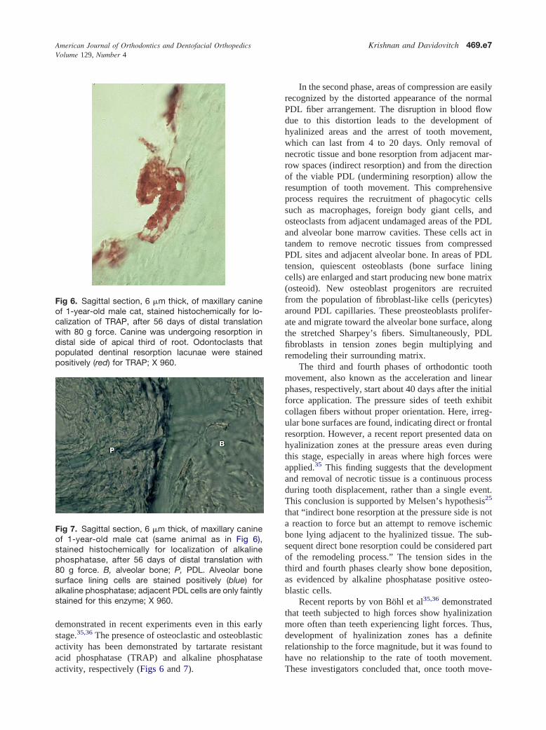
American Journal of Orthodontics and Dentofacial OrthopedicsVolume 129, Number 4
Krishnan and Davidovitch 469.e7
demonstrated in recent experiments even in this earlystage.35,36 The presence of osteoclastic and osteoblasticactivity has been demonstrated by tartarate resistantacid phosphatase (TRAP) and alkaline phosphatase
Fig 6. Sagittal section, 6 �m thick, of maxillary canineof 1-year-old male cat, stained histochemically for lo-calization of TRAP, after 56 days of distal translationwith 80 g force. Canine was undergoing resorption indistal side of apical third of root. Odontoclasts thatpopulated dentinal resorption lacunae were stainedpositively (red) for TRAP; X 960.
Fig 7. Sagittal section, 6 �m thick, of maxillary canineof 1-year-old male cat (same animal as in Fig 6),stained histochemically for localization of alkalinephosphatase, after 56 days of distal translation with80 g force. B, alveolar bone; P, PDL. Alveolar bonesurface lining cells are stained positively (blue) foralkaline phosphatase; adjacent PDL cells are only faintlystained for this enzyme; X 960.
activity, respectively (Figs 6 and 7).
In the second phase, areas of compression are easilyrecognized by the distorted appearance of the normalPDL fiber arrangement. The disruption in blood flowdue to this distortion leads to the development ofhyalinized areas and the arrest of tooth movement,which can last from 4 to 20 days. Only removal ofnecrotic tissue and bone resorption from adjacent mar-row spaces (indirect resorption) and from the directionof the viable PDL (undermining resorption) allow theresumption of tooth movement. This comprehensiveprocess requires the recruitment of phagocytic cellssuch as macrophages, foreign body giant cells, andosteoclasts from adjacent undamaged areas of the PDLand alveolar bone marrow cavities. These cells act intandem to remove necrotic tissues from compressedPDL sites and adjacent alveolar bone. In areas of PDLtension, quiescent osteoblasts (bone surface liningcells) are enlarged and start producing new bone matrix(osteoid). New osteoblast progenitors are recruitedfrom the population of fibroblast-like cells (pericytes)around PDL capillaries. These preosteoblasts prolifer-ate and migrate toward the alveolar bone surface, alongthe stretched Sharpey’s fibers. Simultaneously, PDLfibroblasts in tension zones begin multiplying andremodeling their surrounding matrix.
The third and fourth phases of orthodontic toothmovement, also known as the acceleration and linearphases, respectively, start about 40 days after the initialforce application. The pressure sides of teeth exhibitcollagen fibers without proper orientation. Here, irreg-ular bone surfaces are found, indicating direct or frontalresorption. However, a recent report presented data onhyalinization zones at the pressure areas even duringthis stage, especially in areas where high forces wereapplied.35 This finding suggests that the developmentand removal of necrotic tissue is a continuous processduring tooth displacement, rather than a single event.This conclusion is supported by Melsen’s hypothesis25
that “indirect bone resorption at the pressure side is nota reaction to force but an attempt to remove ischemicbone lying adjacent to the hyalinized tissue. The sub-sequent direct bone resorption could be considered partof the remodeling process.” The tension sides in thethird and fourth phases clearly show bone deposition,as evidenced by alkaline phosphatase positive osteo-blastic cells.
Recent reports by von Böhl et al35,36 demonstratedthat teeth subjected to high forces show hyalinizationmore often than teeth experiencing light forces. Thus,development of hyalinization zones has a definiterelationship to the force magnitude, but it was found tohave no relationship to the rate of tooth movement.
These investigators concluded that, once tooth move-
American Journal of Orthodontics and Dentofacial OrthopedicsApril 2006
469.e8 Krishnan and Davidovitch
ment has started after the second (arrest) phase, boneremodeling takes place at a certain rate, independent offorce magnitude. These findings agree with those ofOwman-Moll et al37 and Van Leeuwen et al,34 who alsoreported the location of hyalinization to be mostlybuccal or lingual to the mesiodistal plane.
SIGNALING MOLECULES AND METABOLITES INORTHODONTIC TOOTH MOVEMENT
The early phase of orthodontic tooth movementalways involves an acute inflammatory response, char-acterized by periodontal vasodilatation and migrationof leucocytes out of the capillaries. These migratorycells produce various cytokines, the local biochemicalsignal molecules, that interact directly or indirectlywith the entire population of native paradental cells.Cytokines, acting as paracrines or autocrines, alwayswith other systemic and local signal molecules, evokethe synthesis and secretion of numerous substances bytheir target cells, including prostaglandins, growth fac-tors, and cytokines. Ultimately, these cells comprise thefunctional units that remodel the paradental tissues andfacilitate tooth movement.
The acute inflammatory process that typifies theinitial phase of orthodontic tooth movement is predom-inantly exudative, in which plasma and leukocytesleave the capillaries in areas of paradental strain. A dayor 2 later, the acute phase of inflammation subsides andis replaced by a chronic process that is mainly prolif-erative, involving fibroblasts, endothelial cells, osteo-blasts, and alveolar bone marrow cells. During thisperiod, leukocytes continue to migrate into the strainedparadental tissues and modulate the remodeling pro-cess.
Chronic inflammation prevails until the next clini-cal appointment, when the orthodontist activates thetooth-moving appliance, thereby starting another periodof acute inflammation, superimposing it on the ongoingchronic inflammation. For the patient, the periods ofacute inflammation are associated with painful sensa-tions and reduced function (chewing). A reflection ofthese phenomena can be found in the gingival crevic-ular fluid (GCF) of moving teeth, where significantelevations in the concentrations of inflammatory me-diators, such as cytokines and prostaglandins, occurtemporally.
Arachidonic acid metabolites
Arachidonic (eicosatetraenoic) acid, the main com-ponent of phospholipids of the cell membrane, isreleased due to the action of phospholipase enzymes.The released acid can be metabolized by 2 pathways—
the cyclooxygenase pathway (with the help of cy-clooxygenase enzymes producing prostaglandins andthromboxanes) and the lipooxygenase pathway leadingto release of leukotriens (originally demonstrated inleucocytes with 3 double bonds (triens) in the backboneof the molecule) and hydroxyeicosatetraenoic acids.38
Evidence for the involvement of these eicosanoids inthe bone remodeling process incident to orthodontictooth movement has been extensively published.
Prostaglandins in tooth movement
Von Euler,39 who first discovered the compound inhuman semen and believed the prostate gland to be themain source of this chemical substance, introduced theterm prostaglandin. However, it was later discoveredthat most cell types in the body produce prostaglandins.Harell et al40 suggested that prostaglandins are impor-tant mediators of mechanical stress. This finding wasfollowed by the work of Yamasaki et al,41 who foundan increase in osteoclast numbers after a local injectionof prostaglandins into the paradental tissues of rodents.This association was demonstrated by the reduced rateof tooth movement after the administration of indo-methocin, an anti-inflammatory agent and a specificinhibitor of prostaglandin synthesis.42
Clinical and animal studies by various authors haveidentified the role of prostaglandins (PGE1 and PGE2)in stimulating bone resorption.43-45 They have reporteda direct action of prostaglandins on osteoclasts in increas-ing their numbers and their capacity to form a ruffledborder and effect bone resorption. Like other bone-resorbing agents, PGE2 also stimulates osteoblastic celldifferentiation and new bone formation, coupling boneresorption in vitro. Studies have also identified otheragents such as growth factors (platelet-derived growthfactors), hormones (parathormone [PTH]), and inter-leukins or other cytokines that induce PGE2 production,to effect bone remodeling and tooth movement.46 Arecent study evaluated the effects of prostacyclin andthromboxane A2 in orthodontic tooth movement andosteoclastic activity on rats. It was found that theseanalogues increase the number of multinuclear oste-oclasts, osteoclastic bone resorption, and the rate oforthodontic tooth movement.47
Within minutes, as paradental tissues become pro-gressively strained by applied forces, the cells aresubjected to other first messengers, the products of cellsof the immune and the nervous systems. The binding ofthese signal molecules to cell membrane receptors leadsto enzymatic conversion of cytoplasmic ATP and GTPinto adenosine 3=, 5=-monophosphate (cyclic AMP[cAMP]), and guanosine 3=, 5=-monophosphte (cyclicGMP [cGMP]), respectively. These latter molecules are
known as intracellular second messengers. Immunohis-
American Journal of Orthodontics and Dentofacial OrthopedicsVolume 129, Number 4
Krishnan and Davidovitch 469.e9
tochemical staining during orthodontic tooth movementin cats showed high concentrations of these moleculesin the strained paradental tissues48 (Figs 8 and 9).
The intracellular second-messenger systems
Sutherland and Rall49 established the second-mes-senger basis for hormone actions in 1958. They discov-ered that free glucose appeared in the bathing media of
Fig 8. Horizontal section, 6 �m thick, of maxillarycanine of 1-year-old female cat. Tooth was not sub-jected to mechanical force and served as control.Section was stained immunohistochemically for cAMP.B, alveolar bone; P, PDL. Alveolar bone surface liningcells are flat; many adjacent PDL cells have roundshape; X 960.
Fig 9. Horizontal section, 6 �m thick, of maxillarycanine of 1-year-old female cat, treated for 24 hourswith distalizing tipping force (same animal as in Fig 8).Zone of force-induced tension in PDL is shown. Sectionwas stained immunohistochemically for cAMP. Com-pared with paradental cells of control canine (Fig 8),both alveolar bone surface cells and neighboring PDLcells were larger, and more intensely stained for camp;X 960.
liver slices exposed to adrenaline. They proposed that
the first messenger (a hormone or another stimulatingagent) binds to a specific receptor on the cell membraneand produces an intracellular chemical second messen-ger. This second messenger then interacts with cellularenzymes, evoking a response, such as protein synthesisor glycogen breakdown. Two main second-messengersystems are now recognized—the cyclic nucleotidepathway and the phosphatidyl inositol (PI) dual signal-ing system.48 These systems mobilize internal calciumstores and activate protein kinase C, respectively. Theactivation of specific protein kinases, together with anincrease in intracellular calcium concentrations, mighttrigger a number of protein phosphorylation events,eventually leading to a cellular response. This responsemight comprise motility, contraction, proliferation,synthesis, and secretion.48
The cAMP pathway
Internal signaling systems are those that translatemany external stimuli to a narrow range of internalsignals or second messengers.38 cAMP and cGMP are2 second messengers associated with bone remodel-ing.50 Bone cells, in response to hormonal and mechan-ical stimuli, produce cAMP in vivo and in vitro.Alterations in cAMP levels have been associated withsynthesis of polyamines, nucleic acids, and proteins,and secretion of cellular products. The action of cAMPis mediated through phosphorylation of specific sub-strate proteins by its dependent protein kinases. Incontrast to this role, cGMP is considered an intracellu-lar regulator of both endocrine and nonendocrine mech-anisms.5 The action of cGMP is mediated throughspecific substrate proteins by cGMP-dependent proteinkinases. This signaling molecule plays a key role insynthesis of nucleic acids and proteins as well assecretion of cellular products.
The PI dual signaling systems
Another second-messenger system reviewed exten-sively in relation to orthodontic tooth movement is thephosphoinositide pathway.51 Interest in this systemstarted with the demonstration of an increase in phos-phate incorporation into cell membrane phospholipidsby Hokin and Hokin in 1953.52 The pathway outlines itas hydrolysis of PI 4,5 biphosphate in response toactivation of cell surface receptors, leading to inositoltriphosphate formation. This reaction in turn leads to arelease of calcium ions from intracellular stores. Fur-ther phosphorylation of inositol triphosphate yieldsIns (1,3,4,5) P4, which controls calcium entry at theplasma membrane through calcium channels. Inositol
triphosphate is a mediator of mitogenesis in mechani-
American Journal of Orthodontics and Dentofacial OrthopedicsApril 2006
469.e10 Krishnan and Davidovitch
cally deformed tissues through an increase in DNAsynthesis.38
The importance of the second-messenger conceptto orthodontics
The second-messenger hypothesis postulates thattarget cells respond to external stimuli, chemicalor physical, by enzymatic transformation of certainmembrane-bound and cytoplasmic molecules to de-rivatives capable of promoting the phosphorylationof cascades of intracellular enzymes. Therefore,temporal increases in the tissue or cellular concen-trations of second messengers are generally viewed asevidence that an applied extracellular first messenger,such as an orthodontic force, has stimulated target cells.The literature includes many reports on significantelevations in the concentrations of intracellular secondmessengers in paradental cells after exposure to appliedmechanical forces.
Vitamin D and diacylglycerol
Another agent that has been identified as an impor-tant factor in orthodontic tooth movement is 1, 25,dehydroxychloecalciferol (1, 25, DHCC).53,54 This agentis a biologically active form of vitamin D and has apotent role in calcium homeostasis. A decrease in theserum calcium level stimulates secretion of parathyroidhormone, which in turn increases excretion of PO4
-3,reabsorption of Ca�� from the kidneys, and hydroxy-lation of 25, hydroxycholecaliferol to 1, 25, DHCC.The latter molecule has been shown to be a potentstimulator of bone resorption by inducing differentia-tion of osteoclasts from their precursors. It is alsoimplicated in increasing the activity of existing oste-oclasts. In addition to bone-resorbing activity, 1, 25DHCC is known to stimulate bone mineralization andosteoblastic cell differentiation in a dose-dependentmanner.46
Kale et al46 compared the effects of local adminis-tration of 1, 25, DHCC and PGE2 on orthodontic toothmovement in rats, and reported that both moleculesenhance tooth movement significantly, when comparedwith the control group. In that study, 1, 25, DHCCwas found to be more effective than PGE2 in mod-ulating bone turnover during tooth movement, be-cause of its well-balanced effects on bone formationand resorption. Another study tried to determine theeffect of 1, 25, DHCC on alveolar bone formationduring tooth movement in rats.55 The researchersobserved significant increases in mineral appositionalrate associated with an elevated osteoblast surface inPDL tension sites of teeth subjected to repeated injec-
tions of 1, 25, DHCC. They concluded that localapplications of 1,25(OH)2D3 could enhance the rees-tablishment of dental supporting tissues, especiallyalveolar bone, after orthodontic treatment.55
Orthodontic forces are applied in patients who candiffer substantially in their biological profiles. VitaminD3 exemplifies this. It is an active participant, withPTH and calcitonin, in the regulation of calcium ho-meostasis. However, its level in the blood of patientsmight be scattered over a wide range, due to variationsin age, sex, and its rates of synthesis and hydroxylation.Moreover, active forms of vitamin D3 are often con-sumed as dietary supplements. Evidently, increasing itsconcentration around paradental cells while they aresubjected to orthodontic forces can evoke synergisticreactions by the cells, leading to rapid tooth movement.Similar responses might occur when other signals areintroduced during orthodontic treatment. These factorsmight originate inside the patient, either locally orsystemically, such as cytokines and hormones or fromexternal sources, such as drugs and electric currents.
Cytoskeleton-extracellular matrix interactions
Cells are motors for tissue modeling and remodel-ing, and most cell types are sensitive to mechanicalloads. This fact provides the scientific foundation oforthodontics.25 The dental and paradental cellular re-sponses to applied mechanical loads involve interplaybetween intra- and extracellular structural elements,and among biochemical messengers. Applied mechan-ical forces are transduced from the strained extracellu-lar matrix (ECM) to the cytoskeleton through cellsurface proteins. The ECM molecules involved in thisprocess include collagen, proteoglycans, laminin, andfibronectin. The transduction occurs by ECM bindingto cell adhesion molecules (integrins) and other cellsurface receptors. Adhesion of the ECM to thesereceptors can induce reorganization of the cytoskeleton,secretion of stored cytokines, ribosomal activation, andgene transcription.56,57
The role of mechanotransducers in transformingmechanical forces into biochemical signals has beenstudied extensively in recent years.38,56-58 Of the 3components of the cytoskeleton—microfilaments, mi-crotubules, and intermediate filaments—microfilamentsare best suited to detect these changes. The majorsubunit protein of microfilaments is actin. Apart fromthis molecule, there are several other associated pro-teins, such as myosin, tropomyosin, vinculin, and talin.Microfilament bundles terminate at special sites of thecell membrane, forming a junctional complex with theECM, known as focal contacts, focal adhesions, or
adhesion plaques.38
American Journal of Orthodontics and Dentofacial OrthopedicsVolume 129, Number 4
Krishnan and Davidovitch 469.e11
The role of cell adhesion molecules in signal trans-duction has received close attention. Explorations of thisissue showed that the cell binding properties of ECMproteins depend on the presence of the peptide se-quence Arg-Gly-Asp (RGD). A family of integralproteins known as integrins, which are present on thecell membrane, connecting the cytoplasm and nucleusto the ECM, recognizes these RGD sites. The integrinsbind to fibronectin extracellularly and talin intracellu-larly, to provide a signal transduction pathway.58 Arecent study identified expressions of integrins (specif-ically �V�3 subunit) in osteoclasts associated withbone resorption and in odontoclasts associated withroot resorption.59 Furthermore, these investigators alsodemonstrated the presence of integrin in epithelial cellrests of Malassez in the PDL. Intracellularly, actin andvinculin microfilaments bind to the talin-integrin com-plex. These details provide an important insight into themechanisms underlying alterations in shape of cells andECM in response to either mechanical forces, cyto-kines, growth factors, or neurotransmitters, that lead tochanges in the attachment apparatus and cellular phe-notype expression.
Role of the extracellular matrix
The ECM is primarily a collection of fibrousproteins embedded in a hydrated polysaccharide gel.57
This important tissue component mainly contains mac-romolecules such as collagen and glycosaminoglycans(GAGs), secreted at a local level by cells such asfibroblasts, osteoblasts, and chondroblasts. In the ECM,GAGs link to a protein with a covalent bond to formproteoglycans. The GAG and proteoglycan moleculesmake a gel-like ground substance, in which other fiberssuch as collagen are embedded. This gel allows diffu-sion of nutrients and hormones, whereas collagenstrengthens the matrix. The roles of the ECM are toprovide a physical framework for the cells that areresponsible for its production and to function as amedium regulating cellular identity, position, prolifer-ation, and fate. It has been reported that all connectivetissues in the body undergo a constant remodeling bysynthesizing and degrading the macromolecular com-ponents of their extracellular matrix.57 The PDL isconsidered to be one of the most highly metabolicallyactive tissues in the body. Sodek60 reported, with thehelp of radio-labeling studies, that the half-life forturnover of mature collagen in the PDL was 2 days,compared with 5 days for the gingiva, 6 days foralveolar bone, and 15 days for skin.
Remodeling of the ECM plays an integral part inorthodontic tooth movement with forces exerted on
the tooth and transmitted to surrounding tissues ofthe periodontium. It is well accepted that cells of theosteoblast lineage, which progress through matura-tional stages, accomplish bone formation. Proteinssynthesized at the mineralization front, such as bonesialoprotein, have proposed roles in osteoblast attach-ment and in coordinating mineral deposition. The laterexpressed proteins, such as osteocalcin, have a possiblerole in preventing hypomineralization via interactionwith osteopontin and osteoclast recruitment.61 Immuno-localization studies recently suggested a profile change inPDL proteoglycans with an increase in immunodetec-tion of chondroitin sulphate-6 epitopes near the com-pressive-side bone surface of tooth movement.62,63
Many enzymes have been implicated in remodeling theECM, including serine proteases, aspartate proteases,and cysteine proteases.58 Recent reports have publisheddata on matrix metalloproteinases (MMPs)—collag-enases, gelatinases, stromolysin, and membrane-typematrix MMPs along with its inhibitor TIMP (tissueinhibitor of MMPs), suggesting that these enzymes playa major role in ECM remodeling.57,64-68 Holliday etal66 reported inhibition of tooth movement with TIMP,thereby providing proof for the mechanistic link be-tween MMP activity and the production of RGDpeptides.
Molecules associated with mechanotransduction
The mechanism behind cellular reactions to me-chanical forces is a matter of profound interest in thearea of orthodontic tooth movement. Signal transductioncan occur through receptors that constitute channels thattraverse the plasma membrane. When interacting with astimulus, these receptors are activated, and signals inthe form of ion movement in or out of the cell producechanges in electric potential. These changes, in turn,enable the signal to be propagated intracellularly. Thecomplexity of cell signaling is further modified by thephysical linkage between cytoskeletal structures andthe ECM, through cell adhesion molecules such asintegrins. The strain produced by mechanical forces inthe ECM causes a change in the cellular shape (Figs 10and 11) and elicits release of signaling moleculesfrom the affected cells. These molecules bind withcell adhesion complexes and start many cellularresponses.56
Histochemical and immunohistochemical studies havedemonstrated that, during the early phases of orthodon-tic tooth movement, PDL fluids are shifted, and cellsand ECM are strained. In areas where tension orcompression evolves under the influence of the orth-odontic appliance, vasoactive neurotransmitters are re-leased from distorted nerve terminals. In the PDL, most
terminals are near blood-vessel walls. Therefore, the
American Journal of Orthodontics and Dentofacial OrthopedicsApril 2006
469.e12 Krishnan and Davidovitch
released neurotransmitters interact first with capillaryendothelial cells. In response, the endothelial cellsexpress receptors that bind circulating leukocytes, pro-moting their migration by diapedesis out of the capil-laries. These migratory cells secrete many signal mol-ecules, including cytokines and growth factors, some ofwhich might be categorized as inflammatory mediators,that stimulate PDL and alveolar bone lining cells to
Fig 10. Sagittal section, 6 �m thick, of untreated max-illary canine of 1-year-old female cat (control). Sectionwas stained with toluidine blue. B, alveolar bone;R, canine root; P, PDL. Cells populating bone and rootsurfaces appear rather flat; PDL cells are predominantlyround; X 320.
Fig 11. Sagittal section, 6 �m thick, of maxillary canineof 1-year-old female cat, that had been tipped distallyfor 12 hours with 80 g force (same animal as in Fig 10).Section was stained with toluidine blue. Tension area inPDL is shown. B, alveolar bone; R, canine root; P, PDL.Major change from Fig 10 (control) is elongated shapeassumed by many PDL cells. Shape alteration is prob-ably due to cells’ attachment to stretched ECM fibers;X 320.
remodel their ECM. This force-induced remodeling
facilitates movement of teeth to areas in which bonehad been resorbed.3
Perhaps the most direct effect of orthodontic forceson paradental cells occurs at sites of focal adhesion. Allparadental cells, with the exception of migratory leu-kocytes, must be attached to the ECM that surroundsthem. This attachment is essential for cell survival.Orthodontic forces, which stretch or compress the PDL,have profound effects on cells in mechanically strainedzones, as can be observed histologically. In suchsituations, cells in PDL tension sites become elongatedas soon as the ECM to which they are attached isstretched. During this phase, their cytoskeleton trans-mits mechanical forces directly to the nucleus. How-ever, the stretched cells continue to react by attemptingto regain their normal, shorter shape. This goal isachieved by detachment and reattachment of the cells totheir ECM. A similar reaction can be seen in PDLcompression sites, where the cells assume a roundshape in the first hours of treatment but regain a normalshape 2 or 3 days later. It can, therefore, be concludedthat the ECM-attachment foci-cytoskeleton-nucleuscombination, which acts in unison in handling mechan-ical loads, is the most powerful biological counterpartof the orthodontist. However, in the orthodontic patient,this force-sensitive mechanism can often be affected byfactors such as neurotransmitters, cytokines, nutrients,and drugs. The extent of these interactions might varyamong patients, and this biological variability is oftenreflected in the quality and pace of the treatment and itsoutcome.
SIGNALING MOLECULES INVOLVEDIN LOAD-INDUCED TISSUE REMODELINGNeurotransmitters
The relationship of nerves to tooth movement hasbeen a matter of considerable research. The PDL isabundantly supplied with 2 kinds of nerve terminals:Ruffini-like endings and nociceptive endings.9 Bothendings can change their structures in response toexternal stimuli, such as orthodontic force.70,71 It hasbeen reported that the mechanoreceptors in the apicalhalf of the dental root have a low threshold and respondto even minor stretching of the PDL.72 In contrast,nociceptors have a high threshold and are activated byheavy forces, tissue injury, and inflammatory media-tors. The force-sensing PDL nerve fibers are eitherunmyelinated C fibers or small myelinated A� fibers.73
The mechanoreceptors are silent in physiological con-ditions but contain various neuropeptides such as sub-stance P, vasoactive intestinal polypeptide, and calcito-nin gene-related peptide (CGRP).74 These neuropeptides
are routinely stored in peripheral and central nerve
American Journal of Orthodontics and Dentofacial OrthopedicsVolume 129, Number 4
Krishnan and Davidovitch 469.e13
terminals, and are released when these terminals arestrained.
Orthodontic tooth movement affects the number,functional properties, and distribution of both mech-anosensitive and nociceptive periodontal nerve fibers.Increased immunoreactivity of substance P has beendemonstrated in the PDL in the early phases of toothmovement. This neuropeptide has been shown to causevasodilatation and increased vascular permeability,contributing to increased local blood flow that accom-panies inflammation.5 It has been demonstrated byDavidovitch et al4 that incubation of substance P withhuman PDL fibroblasts in vitro significantly increasedthe concentration of cAMP in the cells and of PGE2 inthe medium within 1 minute. Another neurotransmitterinvolved in orthodontic tooth movement is CGRP.Kvinnsland and Kvinnsland75 localized CGRP in thePDL and the dental pulp during tooth movement in rats.They could detect an intensification of CGRP immu-noreactivity after 3 days of molar movement in fibro-blasts at PDL tension sites. Norevall et al76 observedintense immunoreactivity for CGRP at PDL tensionsites in cats 1 hour after the onset of treatment. Saito etal77 reported finding intense reactivity to another neu-ropeptide, vasoactive intestinal polypeptide, in thecompressed PDL and in the pulp of moving teeth incats.
It has been proposed that vasoactive neurotransmit-ters released from PDL nerve terminals cause leuco-cytes to migrate out of the capillaries. The experimentsconducted by Rygh and Selvig78 and Storey10 con-firmed these findings in the early days of tooth move-ment. These cells, in addition to their participation inimmune reactions (phagocytosis of necrotic tissues),also produce numerous signal molecules that performvarious functions, from chemoattraction to stimulationof mitogenesis and cytodifferentiation. In addition toleucocytes, other PDL cell types, including osteoblasts,fibroblasts, epithelial cells, endothelial cells, and plate-lets, can also synthesize and secrete these molecules.The products of these cells can be classified intodifferent categories, such as cytokines, growth factors,and colony-stimulating factors. Each of these ligandsmight act in an autocrine or a paracrine fashion, causingactivation of target cells.5
Pain and tooth movement
Tooth movement-associated tissue remodeling, aninflammatory process, might induce painful sensations,particularly after activation of the orthodontic appli-ance. After 24 hours of force application, C-fos (im-munoreactive neurons known to be involved in trans-
mission of nociceptive information) expression is notedipsilaterally in the trigeminal subnucleus caudalis andbilaterally in the lateral parabranchial nucleus. In afollow-up study, fos-like immunoreactive neurons weredistributed in other brain regions such as the neocortex,dorsal raphe, and thalamic nucleus.79 This findingindicates that nociceptive information by tooth move-ment is transmitted and modulated in several regions ofthe brain. These stimuli activate endogenous pain-control systems, including descending monoaminergicpathways.80 Preliminary studies suggested that thenociception is modulated through serotonergic anddopaminergic systems.79 But a subsequent experimentshowed an increase in serotonin turnover in the me-dulla, indicating activation of the bulbospinal seroto-nergic pathway by nociceptive neurological response.80
Thus, there appears to be an indirect nociceptive mecha-nism operating during tooth movement that evokes adelayed and continuous nociceptive response, which isexpected to limit masticatory function during active toothmovement.
A recent report published data on administration ofMK-801 (a noncompetitive antagonist of N-methyl-D-aspartate rececptors), intraperitonially before toothmovement in rats. The results suggest a blockade ofN-methyl-D-aspartate receptors along with neuronalsuppression of trigeminal sensory nuclear complex.These effects were found to increase the neuronal activityin the descending antinociceptive system, including nu-clear raphe magnus, ventrolateral PAG, dorsal raphenucleus, and Edinger-Westphal nucleus. These resultsindicate a pharmacological way to decrease pain percep-tion during orthodontic tooth movement.81
Cytokines in orthodontic tooth movement
Cytokines are extracellular signaling proteins thatact on nearby target cells in low concentrations in anautocrine or paracrine fashion in cell-to-cell communi-cations. Cytokines that were found to affect bonemetabolism, and thereby orthodontic tooth movement,include interleukin 1 (IL-1), interleukin 2 (IL-2) inter-leukin 3 (IL-3), interleukin 6 (IL-6), interleukin 8(IL-8), tumor necrosis factor alpha (TNF�), gammainterferon (IFN�), and osteoclast differentiation factor(ODF). The most potent among these is IL-1, whichdirectly stimulates osteoclast function through IL-1type 1 receptor, expressed by osteoclasts. Secretion ofIL-1 is triggered by various stimuli, including neuro-transmitters, bacterial products, other cytokines, andmechanical forces.5 IL-1 has 2 forms—� and �—thatcode different genes. These interleukins have beenreported to have similar biologic actions, systemi-cally and locally. These actions include attracting leu-
kocytes and stimulating fibroblasts, endothelial cells, os-
American Journal of Orthodontics and Dentofacial OrthopedicsApril 2006
469.e14 Krishnan and Davidovitch
teoclasts, and osteoblasts to promote bone resorption andinhibit bone formation.82 Osteoblasts are target cells forIL-1, which in turn conveys messages to osteoclasts toresorb bone.5 Tuncer et al83 reported increased levels ofIL-8 at PDL tension sites and proposed it to be atriggering factor for bone remodeling.
TNF�, another pro-inflammatory cytokine, wasshown to elicit acute or chronic inflammation andstimulate bone resorption. Recent studies4,84-86 haveshown that TNF� directly stimulates the differentiationof osteoclast progenitors to osteoclasts in the presenceof macrophage colony-stimulating factor (M-CSF).Davidovitch et al4 and Saito et al84 demonstratedmarked increases in the staining intensity for IL-1 andTNF� in cells of the PDL and alveolar bone duringorthodontic tooth movement in cats.
Recent research by Alhashimi et al85,86 focused onthe role of IFN� during bone remodeling as part oforthodontic tooth movement. IFN� is better known as apotent inducer of major histocompatibility complexantigens in macrophages, which is an early marker ofimmune activation during inflammation. It also evokesthe synthesis of other cytokines, such as IL-1 andTNF�. These cytokines were shown to induce produc-tion of nitric oxide, a potentially important osteoblast-osteoclast coupling factor. Alhashimi et al86 reportedthat, during orthodontic treatment, IFN� can causebone resorption by apoptosis of effector T-cells.
The role of cytokines of the RANKL/RANK/OPGsystem in inducing bone remodeling was demonstratedrecently.87 The TNF-related ligand RANKL (receptoractivator of nuclear factor-Kappa ligand) and its 2receptors, RANK and osteoprotegrin (OPG), have beenshown to be involved in this remodeling process.RANKL is a downstream regulator of osteoclast for-mation and activation, through which many hormonesand cytokines produce their osteoresorptive effect. Inthe bone system, RANKL is expressed on osteoblastcell lineage and exerts its effect by binding the RANKreceptor on osteoclast lineage cells. This binding leadsto rapid differentiation of hematopoietic osteoclastprecursors to mature osteoclasts. OPG is a decoyreceptor produced by osteoblastic cells, which competewith RANK for RANKL binding. The biologic effectsof OPG on bone cells include inhibition of terminalstages of osteoclast differentiation, suppression of ac-tivation of matrix osteoclasts, and induction of apopto-sis. Thus, bone remodeling is controlled by a balancebetween RANK-RANKL binding and OPG production.It has been suggested that OPG exists in both mem-brane-bound and soluble forms, and that its expressionis up-regulated by CD40 stimulation. CD40 is a cell
surface receptor that belongs to the tumor necrosisfactor (TNF) receptor family.88 It can be seen in avariety of cells, such as B-lymphocytes, monocytes,dendrite cells, IL-6- and IL-8-secreting cells, such asendothelial cells, basophils, epithelial cells, and fibro-blasts. It was found recently that CD40-CD40L (cellu-lar responses mediated by CD40 are triggered by itscounter receptor CD40L, which also belongs to theTNF gene family) interaction appears to be an activeprocess during orthodontic tooth movement and thatorthodontic force induces T-cell activation.89 Suchactivation might be involved in the induction of inflam-matory mediators and subsequent bone remodeling.Kanzaki et al90 reported recently that OPG gene trans-fer to periodontal tissues inhibited RANKL-mediatedosteoclastogenesis and inhibited experimental toothmovement in rats. The number of reports cited abovemakes it clear that bone remodeling, particularly boneresorption, is regulated by cytokines released in re-sponse to the orthodontic force.
Growth factors
Bone contains abundant amounts of transforminggrowth factor � (TGF�), which includes TGF�1, ac-tivins, inhibins, and bone morphogenetic protein.91
This small polypeptide is produced by several celltypes, such as fibroblasts and osteoblasts, and is depos-ited in the ECM in a latent form. The richest sources ofTGF� are platelets and bone, and it attracts monocytesand fibroblasts, and stimulates angiogenesis in vitro.92
These factors are involved in many biologic activities,including cell growth, differentiation, and apoptosis, aswell as in developmental processes and bone remodel-ing.5 TGF� has been shown to enhance osteoclastdifferentiation in haemopeotic cells stimulated withRANKL and M-CSF.93 More recently, a family ofsignal transducer proteins has been identified, present-ing a mechanism through which TGF� (specificallybone morphogenetic proteins) can signal from the cellmembrane to the nucleus.94 The signal transducerprotein families are phosphorylated by cell surfacereceptors with serine/threonine kinase activity and inthis state translocate to the nucleus. Then, inside thenucleus, the transcription factors produce cellular re-sponses to TGF�.95 Isoforms of TGF� (TGF�1, �2, and�3), which are in latent form, are abundant in bonematrix. ten Dijke et al96 demonstrated increased DNAsynthesis by these 3 isoforms in osteoblast-rich culturesin fetal rats. These isoforms are also shown to enhancesynthesis of collagen and noncollagenous proteins.Davidovitch et al5 demonstrated TGF�1 immunoreac-tivity in cat PDL cells and alveolar bone osteoblastsduring orthodontic tooth movement. They reported that
enhancement can occur as early as 1 hour after force
American Journal of Orthodontics and Dentofacial OrthopedicsVolume 129, Number 4
Krishnan and Davidovitch 469.e15
application. The unstressed PDL and alveolar boneshow negative or no staining for TGF�.
The functions of 2 other growth factors—fibroblastgrowth factor (FGF) and insulin-like growth factor(IGF)—are similar.5 The target cells of FGFs includefibroblasts, endothelial cells, myoblasts, chondrocytes,and osteoblasts. Two forms of FGF were demonstrated:�FGF (acidic PI) and �FGF (basic PI). Since �FGFand �FGF lack a signal sequence, they are sequestratedin the cells responsible for their synthesis and arereleased only when there is a disruption of the plasmamembrane. A recent report discussed plasma mem-brane disruption in PDL tension sites after orthodonticforce application. This effect, demonstrated with thehelp of albumin uptake by PDL cells, suggests thatplasma membrane disruption could promote uptake andrelease of large signaling molecules.97 Globus et al98
reported that bone cells can synthesize �FGF andsecrete it into the surrounding ECM, where it might actas an autocrine or a paracrine signal. Noff et al99
demonstrated increased DNA synthesis, alkaline phos-phatase activity, and formation of bone-like nodules,when rat bone marrow cells were treated with �FGFin vitro.
IGF I and II might also be of relevance during toothmovement. This family of polypeptides promotes cellproliferation and differentiation, and has insulin-likemetabolic effects. The liver is the main organ produc-ing IGF I in humans and rodents, and its production ismodulated by several factors, such as growth hor-mones, estrogen, and insulin, and also by fasting.5 TheIGF type I receptor is structurally similar to the insulinreceptor, a trans-membrane glycoprotein with an extra-cellular ligand binding domain and a cytoplasmicportion with tyrosine kinase activity. However, IGFtype II receptors are identical to the cation independentmannose 6-phosphate receptor, which functions as alysosomal enzyme targeting protein.100 In bone cells,the action of IGF I is regulated by various systemic andlocal factors, including growth hormone, PTH, vitaminD3, corticosteroids, TGF�, IL 1, and platelet-derivedgrowth factor (PDGF). It has been shown that IGF I,when added to PDL cells in culture, causes a dose-related increase in DNA synthesis.101 The evidence isalso increasing regarding the role of IGF II in fibro-blasts, where it influences both calcium influx andDNA synthesis. Coxam et al,102 in experiments innewborn lambs, demonstrated a direct stimulation ofosteoblastic function, by injection of IGF II intrave-nously. The action was mediated through rapid andsustained release of osteocalcin. It has been proventhat PTH and its related peptide enhance IGF I
action.100Each time mechanical damage to the periodontalvasculature is created by orthodontic force, plateletsmigrate from the blood vessels to the extravascularspace. These platelets are a major source of growthfactors for mesenchymal cells, in the form of PDGF.103
Originally, PDGF was isolated from platelets, but itwas later found to be synthesized by various cell types.Two distinct types of PDGF receptors have beenidentified, the � receptor (which binds all 3 isoforms,PDGF AA, PDGF AB, and PDGF BB), and the �receptor (which binds to only PDGF BB). The 2receptors are similar in structure, with an extracellularligand-binding portion, a single transmembrane anchor-ing domain, and a highly conserved intracellular pro-tein, tyrosine kinase. When PDGF binds to the extra-cellular portion, the receptor undergoes dimerizationand autophosphorylation with activation of tyrosinekinases.56 The ligand-receptor complex is then inter-nalized and degraded, leading to (1) activation ofphospholipase A2-release of arachidonic acid, which,via cyclooxygenase and lipooxygenase activity, leadsto formation of prostaglandins and leucotriens; (2)activation of phospholipase-C� through a G-protein,with degradation of PIP2 and formation of IP3 andDAG; and (3) recruitment of substrate proteins to anoligomerized growth factor receptor, with increasedtyrosine kinase activity.104 The proteins include PI3kinase, Ras-GAP, and PLC-�. Davidai et al105 andSandy et al,106 with different experiments, proposedthis pathway to be important in mitogenesis in bonecells. They reached this conclusion by observating thatinhibitors of tyrosine kinases can block PDGF-stimu-lated cell proliferation and receptor phosphorylation.
Connective tissue growth factor (CTGF) is anothersecreted protein that is associated with the ECM duringanabolic bone remodeling.107 This signal moleculeenhances vascular invasion, stimulates proliferation ofosteoblast precursors, and promotes mineralization ofnew bone by osteoblasts. In alveolar bone, CTGF islocalized in osteoblasts and osteocytes near the PDL.After 12 hours of experimental tooth movement, CTGFis expressed in osteoblasts and extends to osteocytesdeep in the bone on both sides of the moving root.108
Colony-stimulating factors
Colony-stimulating factors (CSF) include those re-lated to granulocytes (G-CSF), macrophages (M-CSF),or to both cell types (GM-CSF), and might have particularimplication in bone remodeling through osteoclast forma-tion and thereby during tooth movement.5 These mole-cules are specific glycoproteins, which interact to regulatethe production, maturation, and function of granulocytes
and monocyte-macrophages. Fibroblasts and endothelial
American Journal of Orthodontics and Dentofacial OrthopedicsApril 2006
469.e16 Krishnan and Davidovitch
cells synthesize M-CSF. It was demonstrated by Kahn andSimmons109 that osteoclasts can form as a result ofculturing bone-marrow cells with M-CSF for 10 days. Ithas also been demonstrated that stimulation of fibroblastswith epidermal growth factor, PDGF, FGF, and IL-1induce M-CSF expression by these cells.110 Takahashi etal111 reported that, in terms of potency, M-CSF is the mostpotent in stimulating bone-marrow cells to produce oste-oclasts, followed by GM-CSF, IL-3, and G-CSF.
The above review of signal molecules that modulatevarious steps of tissue remodeling introduces the orth-odontist to the complexity and minute details of eventsthat appear to have major roles in this process. Prom-inent participants are products of the nervous, immune,and endocrine systems, but many locally producedgrowth factors and CSF appear also to participate activelyin remodeling mineralized and nonmineralized connectivetissues. Clinically, orthodontic patients might sense painshortly after appliance activation. However, this feelingis just one of many reactions on the cellular andmolecular levels that typifies orthodontic tissue remod-eling.
Genetic mechanisms
Mechanical activation of bone cells is linked to manygenes, which produce various enzymes, such as gluta-mate/aspartate transporter, inducible nitric oxide synthase,and prostaglandin G/H synthetase.112 In-situ hybridiza-tion under conditions of physiologic tooth movement inrats showed site-specific expression of mRNA forosteonectin, osteocalcin, and osteopontin. Osteoclastand osteoblast progenitor cells had positive signals forosteonectin and osteocalcin. Osteopontin was ex-pressed in osteoblasts and adjacent osteocytes alongbone-resorbing surfaces. In response to orthodonticforce, osteopontin mRNA was elevated in the tissue by12 hours, and this response was found to persist after 48hours.79,113 It was proposed recently that at least 26genes are involved in osteoclast differentiation andregulation, including tyrosine kinase gene, M-CSF,C-fos, Pu.1, and NF-�B (osteoclast formation), andC-tyrosine kinase and micropthalmia transcription fac-tor in osteoclast activity.114 The role of the RANK-RANKL-OPG axis in bone remodeling was discussedin the previous section.87 Kanzaki et al115 observed anincrease in RANKL mRNA expression in the PDL afterexogenous PGE2 treatment and concluded that, inmechanically stressed PDL cells, RANKL up-regula-tion depended on PGE2. In addition, another gene,TREM-2 of TNF, has been implicated in the control ofbone modeling and brain function.116 Wilde et al117
demonstrated periostin (a 90 kDa protein), showing a
divergent expression pattern in PDL fibroblasts andosteoblastic cells in alveolar bone surfaces, even after168 hours of tooth movement.
Pavlin and Gluhak-Heinrich118 used a mouse modelto study the mechanically induced regulation of osteo-blast- and cementoblast-associated genes. They demon-strated a defined temporal pattern of cell-specific generegulation in periodontal osteoblasts, mechanicallystimulated to differentiate and deposit bone matrix.According to these investigators, the primary responsesto osteogenic loading are induction of differentiationand increased cell function, rather than an increase incell numbers. They detected alkaline phosphatase andbone sialoprotein genes after 24 hours of treatment,followed by a concomitant stimulation of osteocalcinand collagen I between 24 and 48 hours, and depositionof osteoid after 72 hours. They reported that differentialgenetic responses to mechanical loading provide func-tional markers for a distinction between the cemento-blast and osteoblast phenotypes.
Recent research has thus begun to unveil the iden-tity of genes that control the synthesis of specificcellular and ECM components during tissue remodel-ing in response to mechanical loads. Continuous re-search in molecular genetics might identify additionalgenes that are activated by applied loads, leadingultimately to the development of new diagnostic, pre-dictive, and therapeutic means in clinical orthodontics.
PATHWAYS OF TOOTH MOVEMENT
On the basis of research in basic biology andclinical observations, Mostafa et al22 proposed anintegrated hypothetical model for tooth movement.This model consists of 2 pathways—I and II—thatwork concurrently to induce tooth movement. Accord-ing to these authors, pathway I represents the morephysiologic response, because it is usually associatedwith normal bone growth and remodeling, whereaspathway II represents the generation of a local inflam-matory response by orthodontic forces.
Pathway I
In pathway I, orthodontic force creates vectors ofpressure and tension, leading to bone bending, genera-tion of tissue bioelectric polarization, and subsequentbone remodeling. With the circumstantial evidence ofprostaglandin synthesis, and with the evoked electricsignals, Mostafa et al22 stated that these phenomena,along with membrane electrical polarization by piezo-electric processes, act on the cell surface cyclic nucle-otide pathway, generating changes in the levels ofintracellular second messengers. This effect, in turn,leads to alterations in cell proliferation, differentiation,
and activation.
American Journal of Orthodontics and Dentofacial OrthopedicsVolume 129, Number 4
Krishnan and Davidovitch 469.e17
This pathway also describes the direction or thecontrol of tooth movement with the help of chargedifferences between the concave and convex sides ofthe strained alveolar bone. This matrix charge polariza-tions—electrically neutral or positive areas—promoteosteoclast activity, whereas electronegativity support-ing osteoblastic activity might change cell membranepolarization, leading to alterations in cAMP levels.
Pathway II
The alternative pathway proposed by Mostafa et al22
attributes orthodontic tooth movement to a classicinflammatory response after force application. Orth-odontic forces, along with increased vascular perme-ability and cellular infiltration, trigger inflammatoryprocesses in the involved dental and paradental tissues.Lymphocytes, monocytes, and macrophages invadethese tissues, enhancing prostaglandin release and hy-drolytic enzyme secretion. The local elevation in pros-taglandins and a subsequent increase in cellular cAMPconcentrations increase osteoclast activity. Secretedhydrolytic enzymes, such as collagenase, dissolve themechanically strained ECM.
Recent model
Recent reports by Jones et al119,120 detailed eventsin bone cells immediately after the application ofmechanical stress. That report is based on the assump-tion that stress in any form—compressive, tensile, orshear—will evoke many reactions in the cell, leading tothe development of strain. In osteoblasts, the firstmeasured responses to physiologic levels of stress areincreases in intracellular free calcium and membranepotential through activation of K� channels. Thisincrease in calcium concentration is related to theactivation of phospholipase C, which releases inositoltriphosphate within 10 seconds. Elevated levels ofphospholipase C maintain the high calcium concentra-tion throughout stress application by keeping the mech-anosensitve ion channels open and by further activationof protein kinase C through diacylglycerol after 3 to 4minutes of force application. Phospholipase A is acti-vated; it acts on stores of arachidonic acid, leading todetection of prostaglandins in the culture medium afterabout 10 minutes. This event is followed by the releaseof products of the lipooxygenase pathway (leucotriensand hydroxyeicosatetraenoic acids), followed by anelevation in the concentration of cAMP. The phosphor-ylation reactions mediated by this cyclic nucleotide inthe nucleus and the cytoplasm lead to cellular syntheticand secretory activities.
A synthesis of the above discussions leads to the
definitive conclusion that inflammation of paradentaltissues occurs every time they are exposed to anorthodontic force, whether the magnitude is light orheavy. The sequence of events after the application ofmechanical forces with the help of orthodontic appli-ances can thus be outlined as:
● Movement of PDL fluids from areas of compressioninto areas of tension.
● A gradual development of strain in cells and ECM ininvolved paradental tissues.
● Direct transduction of mechanical forces to the nu-cleus of strained cells through the cytoskeleton,leading to activation of specific genes.
● Release of neuropeptides (nociceptive and vasoac-tive) from paradental afferent nerve endings.
● Interaction of vasoactive neuropeptides with endo-thelial cells in strained paradental tissues.
● Adhesion of circulating leukocyte to activated endo-thelial cells.
● Plasma extravasation from dilated blood vessels.● Migration by diapedesis of leukocytes into the ex-
travascular space.● Synthesis and release of signal molecules (cytokines,
growth factors, and CSFs) by the leukocytes thathave migrated into the strained paradental tissues.
● Interaction of various types of paradental cells withthe signal molecules released by the migratory leu-kocytes.
● Activation of the cells to participate in the modelingand remodeling of the paradental tissues.
BEHAVIOR OF ORAL SOFT AND HARD TISSUES INRESPONSE TO ORTHODONTIC FORCEBone remodeling and tooth movement
Bone resorption is crucial to orthodontic treatment,by removing alveolar bone from the path of the movingdental root. In this cell-mediated process, the appear-ance of osteoclasts is considered to be the requisite firststep. However, it is unclear whether these cells arisefrom activation of mature osteoclasts already in thePDL or from the proliferation of stem cells in remote orlocal hemopoetic tissues.121 Research on rat PDL hasshown it to be virtually devoid of mature osteoclasts inphysiologic conditions. However, when orthodonticforce is applied, osteoclasts appear within days.122
According to the widely accepted hypothesis by Mundyand Roodman,123 osteoclasts are derived from stemcells in haemopoietic organs, and granulocyte-mac-rophage colony-forming units are the earliest identifi-able precursors of osteoclasts. The proposed pathwaycan be outlined as follows: granulocyte-macrophagecolony-forming units—promonocyte—early preosteo-
clast—late preosteoclast—osteoclast.
American Journal of Orthodontics and Dentofacial OrthopedicsApril 2006
469.e18 Krishnan and Davidovitch
Several lines of evidence exist for the fact that theimmediate precursors of osteoclasts—the late preoste-oclasts—are present in the PDL and are activated ortransformed to mature osteoclasts after orthodonticmechanotherapy. Roberts and Ferguson122 found thatosteoclast numbers per unit bone surface area show apeak level about 50 hours after orthodontic force applica-tion. Additionally, new osteoclasts reach the PDL fromhaemopoietic organs via the blood circulation, and fromalveolar bone marrow cavities, during the orthodontictreatment period, which can last 2 to 3 years.
The bone-resorption cascade involves a series of stepsdirected toward removing both the mineral and the or-ganic constituents of bone matrix by osteoclasts.124 Afterthe differentiation of osteoclasts, the unmineralizedosteoid layer in the bone surface is removed by thelining osteoblasts. These cells produce various enzymessuch as MMPs, collagenases, and gelatinases that helposteoclasts in accessing the underlying, mineralizedbone.125 The next step is osteoclast polarization byattaching itself to specific extracellular bone matrixproteins, such as osteopontin, through RGD sites pro-vided by cell adhesion molecules. The polarizationprocess involves formation of ruffled borders and clearzones, the 2 most characteristic features of osteo-clasts.126 The clear zone is an organelle-free region inthe cytoplasm, which is rich in F-actin filaments. Thesestructures, along with integrin receptors and RGD-containing extracellular proteins from focal adhesionsor podosomes, are responsible for tight cell-to-cellsubstratum interactions. These clear zones provide aseal at the external space beneath the cell, where theruffled border spreads and bone matrix dissolutionoccurs. This extracellular space is called the resorbingcomponent or resorption lacunae.
The next step is osteoclast activation by local andsystemic factors, and production of hydrogen ions (thatdissolve the mineral) and proteolytic enzymes (thatdegrade the organic matrix) in the hemivacuole (local-ized environment) under the ruffled border of thecell.127 In addition to this classic concept, recentfindings have proposed a new concept, in which osteo-blasts can activate osteoclasts through cell-to-cell con-tacts.128 The osteoclasts thus activated produce hydro-gen ions and proteolytic enzymes in the ruffled borderof the cell. It has been proposed that these hydrogenions are generated in the cell by the enzyme carbonicanhydrase, present in the cytoplasm close to the ruffledborder.129
Degradation of the collagenous organic matrix fol-lows the dissolution of the mineralized matrix andmainly involves 2 major classes of enzymes—lyzoso-
mal cysteine proteinases (cathepsin B, L, and K) andMMPs, including collagenase and gelatinase B.124 Os-teoclasts ultimately undergo apoptosis, characterizedby nuclear and cytoplasmic condensation, and fragmen-tation of nuclear DNA into nucleosomal sized units.130
Roodman126 suggested that TGF�, which blocks boneresorption, can induce apoptosis of osteoclasts, whereasosteoclast-stimulating factors, such as PTH and vitaminD3, inhibit osteoclast apoptosis. The progression ofbone remodeling requires continual addition of oste-oclasts, because they have only a limited life span—less than 12.5 days.126 Kobayashi et al131 investigatedthe mechanism controlling the disappearance of oste-oclasts from bone surfaces after bone resorption andobserved a marked increase in TGF beta 1 and OPGmRNA simultaneously with the disappearance of oste-oclasts, suggesting a sequential link in tensional forceapplied to bone lining cells, up-regulation of TGF-beta1/OPG, and the disappearance of osteoclasts.
Once the osteoclasts finish their work of boneremoval, there is a “reversal phase,” when mononuclearcells, which might be of macrophage lineage, are seenon the bone surface. The events during this stage arenot clearly understood but might involve further deg-radation of collagen, deposition of proteoglycans toform the so-called “cement line,” and release of growthfactors to initiate the formation phase.132 The end ofbone resorption and the start of bone formation occursthrough a coupling mechanism, which ensures that anequivalent amount of bone is laid down after theprevious resorption phase. Whether activation of osteo-blasts begins simultaneously with osteoclast recruit-ment or at a later stage during lacunar development isstill controversial. It has been proposed that somegrowth factors and proteinases—TGF�, IGF I and II,and plasminogen activators—play a major role in thiscoupling mechanism.124
Bone formation results from complex events thatinvolve differentiation of osteoblast precursor cellsfrom primitive mesenchymal cells, maturation of osteo-blasts, and matrix formation, followed by its mineral-ization.123 The initial event in this appositional phaseconsists of chemoattraction of osteoblasts or theirprecursors to the site of bone formation. The osteo-blasts that converge at the bottom of a resorptive cavityform osteoid matrix, which mineralizes after 13 days atan initial rate of less than 1 �m per day. The osteoblastscontinue to lay down and mineralize osteoid until thedefect is filled.133 At the bottom of the cavity, osteo-blasts are plump and vigorous, have tall nuclei, andmake a thick layer of osteoid. The cells graduallyflatten and become quiescent lining cells, and somebecome embedded in the newly formed matrix.124
Osteoblasts that become surrounded by calcified matrix

American Journal of Orthodontics and Dentofacial OrthopedicsVolume 129, Number 4
Krishnan and Davidovitch 469.e19
and remain in the lacunae of bone are known asosteocytes. Osteocytes connect with each other bynarrow processes, located in canaliculi throughout thebone matrix. They lose their alkaline phosphataseactivity but maintain high levels of calcium. These cellsare thought to be critical for maintaining fluid flowthrough bone, and any change in fluid flow mightprovide signals that are transmitted to osteoblasts,which then carry out modeling and remodeling activi-ties.134 Bozal et al135 evaluated the response of osteo-cytes when subjected to inflammatory and mechanicalstimuli and observed an enlargement in osteocyticlacunae without changes in cell volume. It was reportedrecently that there is a transient increase in immunore-activity of dentin matrix protein 1, which is mainlyobserved in osteoblasts and osteocytes) after 7 days ofmechanical loading in mouse alveolar bone.136 Theseobservations imply that this protein is involved in theresponses of osteoblasts and osteocytes to mechanicalloading. This finding supports the hypothesis thatosteocytes alter their matrix microenvironment in re-sponse to mechanical loading.136 The factors, systemicand local, affecting the remodeling process are listed inthe Table.
Tissue reaction to orthodontic tooth movementoccurs either through bone or with bone.25 Indirect andundermining bone resorption characterizes tooth move-ment through bone. During these types of alveolar boneresorption, little formative activity takes place at PDLtension sites, because only minor displacement of thetooth occurs initially. Simultaneously, necrosis occursat PDL compression zones, triggering indirect andundermining resorption. When this resorptive activityreaches the PDL and hyalinized tissues are removed,the tooth begins its displacement and becomes mobilebecause of the widened PDL. At this point, appositionrates are markedly increased at PDL tension sites,followed by either renewed hyalinization or continua-tion of tooth movement through direct bone resorption.When teeth are moved with bone, resorption takesplace directly on the wall of the alveolus. In this case,the activity of osteoclasts and osteoblasts is synchro-nized. The width of the PDL is maintained throughoutthe consecutive phases of tooth movement, and thetooth moves along with its alveolus.5 It has beensuggested that, in addition to cell-mediated activitiesthat affect alveolar bone under mechanical stress, thisbone can develop microcracks. A recent study evalu-ated microcracks as a trigger for alveolar bone remod-eling after orthodontic force application in pigs. Anincreased presence of microcracks was found towardthe direction in which the tooth had been moved; this
suggested that it represents the first damage induced byorthodontic force to the bone that must be remod-eled.137
PDL remodeling during tooth movement
PDL and alveolar bone cells are exposed to physicalforces in vivo in response to mastication, para function,and orthodontic tooth movement.138 Orthodontic forcesinduce remodeling of PDL and gingival connectivetissue matrices. Rygh and Brudvik2 described histo-logic and histochemical reactions in the rat PDL afterthe application of orthodontic force. Widening of thePDL was observed in tension sites; the tooth had beendrawn away from the alveolar bone. In the stretchedPDL, several cellular processes are apparently acti-vated, along with an increase in the number of connec-tive tissue cells. This initial phase is followed bydeposition of osteoid tissue at the edge of the socketwall. The blood vessels in the PDL tension site becomedistended, and fibroblasts are rearranged in the direc-tion of strain. The stretched fibroblasts appear spindle-shaped in the middle of the PDL and spherical near the
Table. Factors affecting bone-remodeling process
Hormones PolypeptidesParathyroid hormonesCalcitoninInsulinGrowth hormoneSteroid1,25, dihydroxy vitamin D3
GlucocorticoidsSex steroidsThyroid hormones
Growth factors Insulin-like growth factors I & IITransforming growth factor �Fibroblast growth factorPlatelet derived growth factorConnective tissue growth factors
Cytokines Bone lossInterleukin-1Interleukin-6Interleukin-11Tumor necrosis factorOsteoclast differentiating factorBone formationInterleukin-4Interleukin-13Interleukin-18Interferon-�Osteoprotegrin
Colony-stimulating factors M-CSFG-CSFGM-CSF
Others ProstaglandinsLeukotriensNitric oxide
alveolar bone. Findings by Garant and Cho139 suggest

American Journal of Orthodontics and Dentofacial OrthopedicsApril 2006
469.e20 Krishnan and Davidovitch
that these fibroblasts secrete new Sharpey’s fibers in thePDL simultaneously with the deposition of a newmatrix on the adjacent alveolar bone socket wall. Partof these newly synthesized collagen fibers are incorpo-rated in the newly formed osteoid, whereas the otherpart is embedded in the PDL. Lengthening of fibersseems to occur by incorporation of new fibrils toexisting ones.
Rygh and Brudvik2 observed a great increase invascularity in areas of PDL tension, as indicated by anincrease in the space occupied by blood vessels inhistologic sections of rodent jaws. Transmission elec-tron microscopic (TEM) studies permitted identifica-tion of many cells paravascularly, concomitant with thevascular infiltration. Macrophages and leukocytes,along with proteins and fluids, seemed to have migratedfrom PDL capillaries. These cells are known to be ableto produce various signaling molecules that participatein force-induced tissue remodeling. The pain that isexpressed when the PDL is overstressed is consideredto be the body’s effort to counteract noxious stimuli bylimiting function. This reduced function helps to repairand replace damaged tissue. Beyond a certain level ofstress, the vascular supply to the PDL decreases, withcell death occurring between stretched fibers.
On the pressure side, toward which the tooth isbeing moved, there is narrowing of the PDL space anddeformation of the alveolar crest bone. Depending onthe magnitude of applied force, the reaction at this sitediffers; light pressure produces direct bone resorptionand heavy forces produce hyalinization. In rats, a fewhours after orthodontic force application, osteoclastsappear in the PDL along the alveolar bone surface,evidenced by positive TRAP staining.130,140,141 Withelectron microscope studies, ruffled borders of oste-oclasts were seen to be in close contact with theresorbing bone surface.2,130,142,143 Garant144 observedfibroblasts with increased amounts of intracellular col-lagen profiles near osteoclasts. It is evident that there isextensive remodeling of collagen throughout the PDL,along with new collagen formation and attachment toalveolar bone through localized bone apposition. In-creased pressure in a localized region of the PDL caninhibit differentiation of osteoclasts.145 Instead, a seriesof degenerative tissue reactions takes place, known ashyalinization.2,145
Tissue changes in the compressed PDL are charac-terized by edema, gradual obliteration of blood vessels,and breakdown of the walls of veins, followed byleakage of blood constituents into the extravascularspace. Changes seen in fibroblasts at these sites aremoderate swelling of the endoplasmic reticulum, for-
mation of vacuoles, and rupture and loss of cytoplasm.This disintegration leaves isolated nuclei, which un-dergo lysis over a period of several weeks. As long asthe ground substance remains in the PDL, the tissue hasa glossy appearance. The degenerative process is main-tained as long as the pressure persists. With time,accumulated erythrocyte breakdown products in pres-sure regions might undergo crystallization.78 No toothmovement occurs until the necrotic tissue is removedby the invasion of phagocytosing cells from peripheralundamaged ligament and bone marrow spaces. Thisremoval is completed after 3 to 5 weeks, and theposthyalinized PDL is markedly wider than beforetreatment, perhaps to withstand greater mechanicalinfluences.2,145 A recent article by Attal et al146 re-ported that the periodontal vasculature showed similarchanges in pressure and tension areas, but large-diam-eter vessels are unaffected by mechanical loading. Theyfound a significant decrease in the mean number ofterminal arterioles, whereas there was an increase incapillary and postcapillary venules in the apical partof the tooth. These vascular changes persisted even12 weeks after the removal of orthodontic force, sug-gesting a rebounding effect.
It is clear from this discussion that, as long asankylosis does not occur, the general trend after forceapplication is preservation of the width of the PDL, aremarkable process involving precisely controlled os-teogenic resorption and deposition at specific sites inthe paradental tissues. Conclusions from recent studiesshowed that the PDL might be both the medium offorce transfer and the means by which alveolar boneremodels in response to applied forces.137 Howard etal147 found that mechanical force could induce fi-bronectin and collagen synthesis by PDL cells in astrain magnitude-dependent fashion. Current evidencesuggests the presence of mechanosensory signalingsystems, including adenyl cyclase stretch-activated ionchannels and changes in cytoskeletal organization inresponse to mechanical forces.48,56-59
The PDL is abundantly supplied with 2 kinds ofreceptors: Ruffini-like endings and free nerve end-ings.69 These receptors play key roles in changing thePDL’s structure and cellular functions in response tomechanical loads, such as those applied in orthodon-tics.70 The most rapid response of PDL fibroblasts tomechanical forces involves elevation of the concentra-tion of intracellular calcium ions, and changes in actinfilament polymerization. The influx of calcium ions canthen strongly induce other effectors, including theproteins that regulate the cytoskeleton.148,149 Redlichet al150 reported on a significant time-dependent up-
regulation of tropoelastin gene after external pressure
American Journal of Orthodontics and Dentofacial OrthopedicsVolume 129, Number 4
Krishnan and Davidovitch 469.e21
simulating orthodontic force in cultured human PDLfibroblasts.
Long-term responses to in-vitro mechanical loadingcan include stimulation of cell division, which will leadto increased collagen synthesis, and stimulation of alka-line phosphatase activity as force-induced downstreamchanges.151 Recent studies demonstrated enhancement ofnitric oxide synthase production after mechanical forceapplication in animals152-155 and humans.156 This immu-nohistochemical finding suggests that nitric oxidemight be a key regulator of orthodontic tooth move-ment by regulating the functions of osteoblasts andosteoclasts, and thereby modulating bone metabo-lism.156 Takahashi et al67 demonstrated differentialregulation of the expression of MMP-8 and MMP-13genes, and concluded that this dichotomy could play animportant role in defining the specific characteristics ofPDL remodeling. Current research in this area isdirected toward studying signaling mechanisms evokedthrough mechanotransduction and determining the na-ture of mechanosensors in PDL and bone cells.
Gingival effects of orthodontic force
The cellular and extracellular changes in the PDL,alveolar bone, and cementum after orthodontic forceapplication have been extensively investigated. Al-though gingival changes have also been found to beimportant in the overall response, they have attractedonly minimal attention. A recent review outlined vari-ous changes in the gingiva in response to orthodonticforce application.157 Accordingly, 2 disparate processesoccur in the gingiva after transduction of orthodonticforce. First, there is an injury of the gingival connectivetissue, manifested by torn and ripped collagen fibers;,second, the genes for both collagen and elastin areactivated, whereas those for tissue collagenases areinhibited. The effect of mechanical strain on humangingival fibroblasts was evaluated recently. The studyused as yardsticks the phosphorylation status and local-ization of Forkhead box family members (mediators offibroblast apoptosis) and the level of proliferating cellnuclear antigens (for strain induced proliferation). Theresults indicated that mechanical strain can deliveranti-apoptopic and proliferative stimuli to human gin-gival fibroblasts.158
Gross clinical observation of the gingiva duringorthodontic treatment showed tissue accumulation andenlargement of gingival papillae when extractionspaces are being closed. This tissue accumulation isattributed to both retraction and compression. Adjacentto this tissue accumulation, vertical clefts of bothepithelium and connective tissue, which persisted for
years after treatment, have been reported.159 Histologicstudies showed discontinuation of transseptal fibers atextraction sites and their reestablishment during thehealing phase.160 These newly formed collagen fibersare coiled and compressed, and have a football-shapedappearance after orthodontic approximation of teeth atextraction sites. There are increases in the amount ofoxytalan fibers and GAGs in the transseptal fiber area.It is reported that proline uptake by newly formedcollagen is increased significantly, suggesting an in-crease in the synthesis rate by gingival fibroblasts.161
Ultrastructural analysis with transmission electron mi-croscopy showed increased diameters of newly formedgingival collagen fibers in both compressed and tensionsites. Moreover, there is apparently a slight increase inthe number and size of elastic fibers in gingival pressuresites, when compared to tension sites.157
Studies on gingival fibroblasts in vitro, with reversetranscriptase polymerase chain reaction showed genetranscription levels of collagen type I to be significantlyincreased, wheras that of tissue collagenase (MMP1)is decreased after mechanical loading.162 In a recentreport, Bolcato-Bellemin et al68 subjected human fibro-blasts from the PDL and the gingiva to mechanicalforce to quantify the mRNAs encoding the variousMMPs, TIMPs, and alpha and beta integrin subunits.The results indicated that stretched fibroblasts of bothtypes had the same pattern of mRNAs encoding MMPsand TIMPs, but differed for those encoding variousintegrin subunits, known to act as protein receptors inmechanotransduction. This finding suggests a disturbedequilibrium between collagen synthesis and degrada-tion, required to maintain adequate tissue stability.These data suggest that orthodontic force effects on thegingiva are similar in cases of extraction space closureand rotation corrections. Furthermore, these findingsindicate that the cause of relapse after treatment is mostlikely the increased elasticity of the compressed gin-giva, brought about by biosynthesis of new elasticfibers and GAGs. In this fashion, the gingiva undergoeselastic deformation during orthodontic treatment, capa-ble of causing relapse during the posttreatment reten-tion period.
Biomarkers of bone remodeling in gingivalcrevicular fluid
GCF arises at the gingival margin and can variouslybe described as a transudate or an exudate. Its rate offlow is related to the degree of gingival inflammation,and a rate of 0.05 to 0.20 �l per minute was reported incases of apparent minimal inflammation. The total fluidflow is between 0.5 and 2.4 mL per day.163 Recent studiesin orthodontic tooth movement have used GCF because
of its noninvasive nature and ease of repetitive sam-
American Journal of Orthodontics and Dentofacial OrthopedicsApril 2006
469.e22 Krishnan and Davidovitch
pling from the same site with the help of platinumloops, filter paper strips, gingival washings, and micropipettes. The fluid is used to analyze various biochem-ical markers such as prostaglandin production and theaction of various extracellular and intracellular factors,such as IL-1, IL-6, TNF-�, epidermal growth factors, �2
microglobulin, cathepsin, aspartate aminotransferease, al-kaline phosphatase, and lactate dehydrogenase.
Remodeling changes in the alveolar bone and thePDL induce production of various cell mediators orenzymes that can be used as biomarkers of orthodontictreatment.58,164 The early works by Last et al,165 andEmbery and Waddington58 described many GAGs, andproteoglycan and tissue proteins in GCF, providingevidence for the presence of underlying state of bio-chemical reflections in paradental tissues. Last et al165
were the first to report a finding in this regard bydemonstrating chondroitin-4-sulphate in GCF from thepressure side of tooth movement. Because the orthodonticmodel is a nonplaque, nondisease-related process, it issuggested that the increase in chondroitin-4-sulphate is abiological alteration in deep-seated tissues. Uematsuet al166,167 found several cell mediators, such as IL-1�,IL-6, TNF�, epidermal growth factors, �2 microglobu-lin, and TGF�, to be elevated in the GCF duringorthodontic treatment. Grieve et al168 and Lee et al169
reported similar finding for PGE2 and IL-1�, andLowney et al170 for TNF�.
Griffiths et al171 demonstrated the presence ofosteocalcin in GCF from teeth associated with orth-odontic forces. A study by Insoft et al172 found in-creased alkaline phosphatase levels during the first3 weeks of orthodontic treatment, whereas acid phos-phatase increased in subsequent weeks. Perinetti et al173
also demonstrated alkaline phosphatase along withaspartate aminotransferase activity in GCF. Recently,Serra et al174 observed an increase in lactate dehydro-genase activity in GCF after orthodontic force applica-tion and proposed it to be a sensitive marker forperiodontal metabolism. Sugiyama et al164 reported anincrease in the amount of cathepsin B in GCF andsuggested it to be involved in ECM degradation.Apajalahti et al65 found a significantly higher amountof MMP-8 in GCF after orthodontic force applicationfor 4 to 8 hours. They suggested that the increasedexpression and activation of GCF MMP-8 reflects theenhanced periodontal remodeling changes of orthodon-tic force application. They concluded that the presenceof such markers in GCF might be useful in identifyingthe bone-remodeling activities during orthodontic treat-ment. This fluid can, therefore, be considered a prom-ising and potent area of future research, because these
investigations have already begun to provide insightinto the temporal aspects of resorptive and formativeprocesses in the paradental tissues during orthodontictreatment.
TISSUE REACTIONS WITH VARIED FORCEAPPLICATIONS
Force can be defined as the action of 1 body onanother that tends to change the state of rest or motionof the latter. It is a vectoral quantity, which means that,to adequately describe it, we have to define its magni-tude, direction, and duration, as well as its point ofapplication.6 Orthodontics is based on application offorce on the teeth, under the influence of which toothmovement occurs. The duration and character of forcehave great influence in orthodontic mechanotherapy,alterations in which can produce varied tissue reactions.
Continuous, interrupted, and intermittent forces
Most contemporary fixed orthodontic appliances uselight continuous forces as part of orthodontic mechano-therapy to effect tooth movement. However, a continuousforce can subside rapidly and thus be interrupted after alimited period of time, such as in torquing movements byan edgewise archwire or labial movement of blocked-outmaxillary lateral incisor with the help of ligation. It is notalways possible to distinguish between continuous andinterrupted movements, and the latter act for only com-paratively short durations.175 Nevertheless, it appearsthat this kind of a force, that starts in a continuous modeand then becomes interrupted, is biologically favorable,particularly when its initial magnitude is low. In such acase, hyalinized zones might develop in sites of com-pressed PDL, but, as soon as this necrotic tissue iseliminated and the tooth moves, the force decreasesquickly. Finally, the archwire retains its passivity for awhile, during which time (rest period) there will be anopportunity for calcification of the newly formed os-teoid layer. This rest period between appliance activa-tions is the time used by the tissues for reorganization.This rest can promote favorable cell proliferation forfurther tissue changes when the appliance is activatedagain.176 The characteristic feature of continuous/inter-rupted tooth movement is formation of new bone layersin the richly cellular tissue at the entrance of openmarrow spaces as soon as the tooth movement stops.50
A recent study evaluated the areas of compressed PDLnear rat molars subjected to continuous orthodontic forcewith the help of TEM and TRAP histochemistry.177 Theresults prove that continuous orthodontic forces canresorb the alveolar bone concomitantly with the forma-tion of new bony tissue at PDL tension sites.
Typical intermittent forces act as either an impulse
or a shock of short duration, or for short periods with a
American Journal of Orthodontics and Dentofacial OrthopedicsVolume 129, Number 4
Krishnan and Davidovitch 469.e23
series of interruptions. These forces are mainly pro-duced by removable appliances, which deliver forceperiodically. Examples of such a system are springsresting on tooth surfaces.175 Intermittent force results insmall compression zones in the PDL, short hyaliniza-tion periods, and lengthy rest periods when the appli-ance is removed intermittently. During this time, thetooth moves back to the tension side and remains innormal function. This mode of treatment can improvethe paradental circulation and promote an increase inthe number of PDL cells, because its fibers usuallyretain a functional arrangement.176 Reitan defined thiscondition as “semi-hyalinization,” meaning that in thecompressed PDL not all fibers become compressed, andonly some cells undergo necrosis. Consequently, oste-oclasts might be formed directly along the bone surfacesubjacent to hyalinized tissue, and bone resorption isless disturbed by hyalinization. This situation mightaffect smooth and uniform movement of teeth.175
Light v heavy forces and the rate of toothmovement
The magnitude of force applied for orthodonticmechanotherapy has received significant attention. It isgenerally accepted that light forces produce favorabletooth displacement, resulting in minimal discomfortand pain to the patient, but heavy forces (which exceedcapillary blood pressure of 20-25 gm/cm2 of rootsurface) produce the classic 3-phase reaction—initialstrain, lag phase, and progressive tooth movement.9 Arecent article by Kohno et al178 reported that lightforces can tip teeth without friction, with a constant rateof tooth movement, and without the 3 phases. However,in most cases, this kind of tipping is uncontrolled andcan cause root resorption, despite the small magnitudeof the applied force.
In orthodontic tooth movement, as in all otherbiological responses to mechanical loading, one wouldexpect a dose-response relationship between the mag-nitude of the applied force and the extent of the tissuereaction. Quinn and Yoshikawa179 described 4 alterna-tive models for this relationship. The first proposes anon-off switch that is turned on at a certain force level.All forces above this threshold will lead to the samerate of tooth movement. Several studies have supportedthis hypothesis.37,180 A linear dose response relation-ship with a threshold level force is indicated in thesecond model. This concept was also supported byseveral authors,37,181 who believe that higher forces areefficient in tooth movement. In the third model, it wassuggested that a dose-response relationship exists in thelower force range, up to a certain level. Then a plateau
is reached, and further increase in force level activitydecreases the rate of tooth movement. This hypothesisagrees with Begg’s differential force concept.182 In thefourth model, the decline in the rate of tooth movementin response to an increase in force magnitude isignored, suggesting that merely applying greater orth-odontic forces does not necessarily guarantee a fasterrate of tooth movement.179
These findings suggest that, with increasing magni-tudes of orthodontic forces, a constant rate of toothmovement would be reached, within a broad range offorces. This observation has led to the conclusion thatforce magnitude plays only a subordinate role inorthodontic tooth movement. In support of this hypoth-esis is a study by Pilon et al33 in dogs, in whichapplication of 2 forces (50 and 100 CN) to secondpremolars resulted in the same rate of tooth movement.A clinical experiment by Owman-Moll et al37 in hu-mans produced similar results.
Recent reports by von Böhl et al35,36 also state thatthe models that describe the relationship between themagnitude of the orthodontic force and the rate of toothmovement probably are not valid. Apparently, thedetermining factor is the degree of PDL hyalinization inresponse to the application of heavy forces. The rate ofremoval of this necrotic tissue might depend on thepattern of strain distribution in the paradental tissues,the peculiarities of the PDL, and bone morphology. Itwas concluded that, with present knowledge, an appro-priate model for tooth movement is difficult to propose;additional information is required about the biochemi-cal background of the process, as well as insights intoindividual variations in bone density and the metabo-lism of PDL and bone.
Duration of force application
Clinical experience suggests that successful toothmovement requires a threshold of force duration ofabout 6 hours per day. It was determined in anexperiment on cats that it takes about 3 hours forsignificant elevations in cAMP in extracts of alveolarbone and PDL, after sustained applications of tippingforces to the maxillary canines.183 Recent reports pro-posed a role for nitric oxide as a marker of vascularsignal transduction during the initial state of orthodon-tic tooth movement.152-156 Nitric oxide is produced byvarious cells and is present in blood vessels, nerves, andPDL fibroblasts. This molecule has been reported totake part in bone remodeling184 and in the regulation ofblood vessels and nerves.185 During the initial stages oftooth movement under the influence of light continuousforces, the activity of nitric oxide synthase in paraden-
tal tissues was found to increase as early as 1 to 3
p
American Journal of Orthodontics and Dentofacial OrthopedicsApril 2006
469.e24 Krishnan and Davidovitch
hours152 or within 6 hours15 from the onset of treat-ment.
DELETERIOUS EFFECTS OF ORTHODONTICFORCE
Tissue reactions incident to orthodontic tooth move-ment depend mainly on the pattern of stress-strain distri-bution in the paradental tissues. Different types of toothmovement generate different force distribution pat-terns, and thereby different types of tissue reactions.Various studies, evaluating tissue reactions by radio-logic and histologic means, have detected iatrogenicsequelae to orthodontic force. These effects includecaries, gingivitis, marginal bone loss, pulpal reactions,root resorption, and allergic reactions to appliancematerials. The discussion below is limited to deleteri-ous effects affecting 3 main tissues—gingiva, cemen-tum, and dental pulp.
Gingival problems
Fixed-appliance orthodontics has been shown toproduce deleterious effects on the periodontium, rang-ing from gingivitis to bone loss.186-188 The cementationof orthodontic bands or resin-bonded attachments canevoke local soft tissue response.189 This response ismainly due to plaque accumulation and the proximityof these attachments to the gingival sulcus.186 Anotherlong-term complication of orthodontic treatment isgingival recession. Various authors have reported therange of its occurrence from 1.3% to 10%. It is widelyaccepted that at least 2 mm of keratinized gingivashould be present to withstand orthodontic force andrevent recession.187 Dorfman188 suggested that man-
dibular incisors are most likely to express gingivalrecession in response to orthodontic mechanotherapy.This outcome has been attributed to the thin or nonex-istent labial plate of bone and inadequate or absentkeratinized gingiva that covers labially prominent teeth.This potentially risky and harmful condition receivedmuch attention, because it can lead to poor esthetics,root sensitivity, loss of periodontal support, difficulty inmaintaining oral hygiene, difficulty in successful peri-odontal repair, and increased susceptibility to caries.
Orthodontic treatment often leads to alteration inoral hygiene habits, which might result in plaqueaccumulation and gingival inflammation. The develop-ment of gingival inflammation during orthodonticmechanotherapy is associated with specific bacterialtypes, as listed in many reports.189-191 Huser et al189
studied longitudinally the microbial flora in plaque ofpatients undergoing orthodontic treatment. There weredefinite increases in plaque scores and probing depths
in these patients when compared with controls. Thebacterial plaque was composed mainly of spirochetesand motile rods. Other authors reported increases in thelevels of bacteroids and streptococcus species afterorthodontic banding.190,191 It can, therefore, be con-cluded that orthodontic mechanotherapy produces alocal change in the oral ecosystem, altering the com-position of bacterial plaque, tipping the scale towardthe development of an inflammatory process.
Root resorption
Normally, cementum does not undergo appreciableresorption; it seems to be excluded from remodelingactivities associated with maintenance of calcium ho-meostasis. However, orthodontic force application cansometimes evoke excessive resorption of root cemen-tum, proceeding into the dentin, eventually shorteningthe root length—a process called root resorption.163
Ottolengui (1914)192 and Ketcham (1927)193 were thefirst to report severe root resorption associated withorthodontic tooth movement. It is an undesirable andthe least predictable sequelae of orthodontic treatment.It can occur during the middle or the posttreatmentphase, raising questions about the longevity of thetreated dentition and the stability of the treatmentresults. Along with other factors, orthodontic forces caninitiate and sustain the resorptive process.194 Jarabakand Fizzell,195 after analyzing the effect of forcesystems during mechanotherapy, concluded that themagnitude of an orthodontic force and rigid fixation ofthe archwire to the brackets could be considered themost important factors predisposing a tooth to the rootresorption. Jiggling forces and round tripping of teethduring mechanotherapy were also suggested as majorcauses of orthodontic root resorption.196,197
Comparing treatment mechanics with and withouttooth extraction, there were definite correlation be-tween extraction treatment and root resorption.198-200
Of the various possible orthodontic tooth movements,intrusion and torquing make a tooth root more prone toresorption.198,201 A literature search regarding toothvulnerability to resorption found agreement amongmany authors. They state that maxillary incisors, par-ticularly the central incisors, are the most prone to theprocess, followed by the maxillary molars and thecanines. In the mandibular arch, the most prone teethare the lateral and central incisors. It was also pointedout that tapered roots, compared with the blunted type,exhibit more resorption.202-205
Alignment of impacted canines as a risk factor forresorption has been identified recently.205 Drugs, suchas corticosteroids and alcohol, and systemic diseases,such as asthma and allergies, have been proposed to
predispose tooth roots to resorption after orthodontic
American Journal of Orthodontics and Dentofacial OrthopedicsVolume 129, Number 4
Krishnan and Davidovitch 469.e25
force application.194 Moreover, a previous history ofdentofacial trauma and the detection of root resorptionin pretreatment radiographs have also been proposed aspredisposing factors.205 A recent report suggested that ahypofunctional PDL, associated with nonoccludingteeth, increases the risk for orthodontic root resorp-tion.206
Brezniak and Wasserstein207 classified root resorp-tion according to its severity. Accordingly, it is possibleto identify (1) cemental, or surface resorption, whereonly the outer layers are resorbed, to be fully regener-ated or remodeled later; (2) dentinal resorption withrepair, where the cementum and the outer layers ofdentin are resorbed, and are repaired along with mor-phological alterations; and (3) circumferential rootresorption, where full resorption of the hard tissuecomponents of the root apex occurs, resulting in rootshortening.
Current research in orthodontic root resorption isdirected toward identifying genes involved in the pro-cess, their chromosome loci, and their possible clinicalsignificance. Al Qawasmi et al208 reported on findingevidence of a linkage disequilibrium of IL1� polymor-phism in allele 1 and external apical root resorption.Another study by the same authors209 and one by Lowet al,210 linked RANKL and OPG with the regulation ofroot resorption.
Progress periapical and panoramic radiographs areuseful aids in detecting midtreatment resorptive pro-cesses.203-205,211,212 Whenever such a finding is en-countered, a temporary halt in orthodontic treatment for4 to 6 months is advised.198,203,213,214
Whatever the predisposing or influencing factors,most authors believe that there will be no apparentincrease in resorption after termination of active orthodon-tic treatment.203,205,214-217 Some repair occurs, includingsmoothing and remodeling of cemental surfaces and thereturn of the PDL width to normal. Original rootcontours and lengths are never reestablished, but thefunction of the tooth apparatus is usually not severelyaffected by the loss of root length.214
Several ways to slow down the rate of orthodonticroot resorption during mechanotherapy have been pro-posed, including drugs, hormones, and growth fac-tors.218-223 In a recent experiment, Bialy et al224 studiedthe effect of low-intensity pulsed ultrasound on healingof orthodontically induced root resorption. The resultswere promising, demonstrating a reduction of rootresorption and acceleration in healing of alreadyresorbed sites with reparative cementum over 4 weeks
of low-intensity pulsed ultrasound application.Pulpal reactions
Various dental procedures, including orthodontictooth movement, have been reported to adversely affectthe dental pulp. The literature shows conflicting resultsfor correlation of pulpal changes incident to orthodonticforce. Some reports suggested permanent damage topulpal tissue from orthodontic force, but others claimedno significant long-lasting effects on the dental pulp.225
In 1980, Labart et al226 demonstrated increased pulpalrespiration rate in rat incisor pulp (1-2 times more thancontrols), when subjected to orthodontic stress for 72hours. Harmersky et al227 showed a depression inpulpal respiratory rate after orthodontic force applica-tion in humans. Unsterseher et al225 also had the sameresults.
In recent years, the alterations in pulpal vasculatureand blood flow in response to orthodontic force havegained much attention. Various experiments have dem-onstrated an initial decrease in blood flow, lastingapproximately 32 minutes, followed by an increase inblood flow (lasting 48 hours). Mostafa et al228 reportedcongested and dilated blood vessels, and edema ofpulpal tissue in their histologic observations. A histo-morphometric study on rats by Nixon et al229 contra-indicated some studies and reported an increase in thenumber of functional pulpal vessels after orthodonticforce application. Derringer and Linden,230 whoshowed an increase in angiogenic growth factors indental pulp, supported this result. A recent study byDerringer et al231 identified the specific angiogenicgrowth factors released in response to orthodontic forceapplication to be vascular endothelial growth factor,FGF-2, PDGF, and TGF-beta.
The clinical impact of all these studies was todetermine whether any alterations in pulpal tissue couldjeopardize the long-term vitality of the teeth. Theprogression of the inflammatory process in human pulpfibroblasts apparently depends on stimulation by neu-ropeptides and production of inflammatory cytokines,such as IL-1, IL-3, IL-6, and TNF�. A recent reportdescribed apoptosis in dental pulp tissues of rats un-dergoing orthodontic treatment.232 Perinetti et al233
demonstrated that an enzyme, aspartate aminotransfer-ase (which is released extracellularly upon cell death),is significantly elevated after orthodontic force appli-cation.
POSTTREATMENT CHANGES
Orthodontic forces are known to produce pressureand tension regions in the PDL and alveolar bone. Thisstrain alters the affected tissues’ vascularity and blood
flow, providing a favorable microenvironment for ei-
American Journal of Orthodontics and Dentofacial OrthopedicsApril 2006
469.e26 Krishnan and Davidovitch
ther tissue deposition or resorption. Murrell et al234
reported that removal of orthodontic forces producessignificant changes in the number and density of PDLblood vessels. Normalization of the periodontal vascu-lature was observed during an interval, equivalent tothe duration of orthodontic force application, and it hasbeen suggested to be a main reason for relapse of theend result of orthodontic treatment.
Nakanshi et al70 reported that the response proper-ties of periodontal mechanoreceptors are adapted to thenewly acquired tooth position. After 7 days of forceremoval, the collagen fiber arrangement became verysimilar to that of the control group. Yoshida et al235
evaluated the cellular responses in the relapse ofexperimentally moved rat molars and suggested thatthere will be rapid remodeling of the PDL and alveolarbone after treatment, which can be identified as themain cause of relapse. A recent report pointed out that,after tooth movement, occlusal forces were majorfactors in fostering and expediting periodontal recov-ery.236 It is stated that various types of damage to teethand PDL produced by orthodontic mechanotherapyhealed more rapidly and extensively in functional teeth.During the recovery period, the return of periodontaldimensions to normal values is regulated by the rateand direction of alveolar bone turnover.141
CONCLUSIONS
Rapid advances in all biological fields have enabledus to better understand the mechanisms involved inorthodontic tooth movement. It is evident that, atdifferent stages of tooth movement, different combina-tions of cell-cell and cell-matrix interactions occur;these determine the nature of the remodeling changes.The research trend is now directed toward elucidatingmolecular level interactions during these events. Abetter understanding of the relationship between genesand transcription factors in controlling bone and PDLremodeling will expand our knowledge, and mightstrengthen our clinical capabilities. Above all, thisgrowing body of knowledge on the response of ourcells to mechanical loads should illuminate useful pathsin clinical orthodontics and assist us in identifying anddiscarding harmful methods of mechanotherapy. Futureorthodontics will, therefore, increasingly become bio-logically correct and, consequently, patient-friendly.
At the dawn of the 20th century, Edward H. Angle,the father of modern orthodontics, advocated includingcourses in biological sciences in orthodontic trainingprograms. At that time, these courses were mainlyinside the domain of anatomy and physiology. How-ever, at the onset of the 21st century, the scientific
frontiers in biology have witnessed remarkable ad-vances in molecular biology and molecular genetics.Angle’s recommendation for biological sciences in theorthodontic curriculum most likely stemmed from hisrealization that orthodontic treatment is rendered tohuman patients. This fact has remained unalteredthroughout the passing years, as most orthodonticdepartments adopted the conviction that orthodonticexcellence is derived from a comprehensive knowledgeof mechanics and biology. In reality, though, com-manding knowledge of mechanics, material science,and metallurgy have prevailed, while biological sci-ences continue to play a minor role in clinical orth-odontics. However, future orthodontists will witness animprovement in this imbalance, as has been happeningin all medical fields. Because the face and oral cavityare integral parts of the human body, growing under-standing of the body’s functions on the cellular andmolecular levels will lead to the development of orth-odontic appliances capable of correcting malocclusionssuccessfully in patients with similar morphologicalfeatures but varying biochemical profiles. This ongoingdevelopment will move orthodontics closer to the goalof being optimal, where teeth are moved efficiently,without causing discomfort to the patient or damage tothe teeth and their supporting tissues.
REFERENCES
1. Reitan K. Tissue behavior during orthodontic tooth movement.Am J Orthod 1960;46:881-90.
2. Rygh P, Brudvik P. The histological responses of the periodon-tal ligament to horizontal orthodontic loads. In: Berkovitz BB,Moxham BJ, Newman HN, editors. The periodontal ligament inhealth and disease. St Louis: Mosby; 1995.
3. Davidovitch Z. Tooth movement. Crit Rev Oral Biol Med1991;2:411-50.
4. Davidovitch Z, Nicolay OF, Ngan PW, Shanfeld JL. Neuro-transmitters, cytokines and the control of alveolar bone remod-eling in orthodontics. Dent Clin North Am 1988;32:411-35.
5. Davidovitch Z. Cell biology associated with orthodontic toothmovement. In: Berkovitz BB, Moxham BJ, Newman HN,editors. The periodontal ligament in health and disease. StLouis: Mosby; 1995.
6. Daskalogiannakis J. Glossary of orthodontic terms. Berlin:Quintessence; 2000.
7. Mao JJ, Wang X, Kopher RA. Biomechanics of craniofacialsutures— orthopedic implications. Angle Orthod 2003;73:128-35.
8. Toms SR, Lemons JE, Bartolucci AA, Eberhardt AW. Nonlin-ear stress-strain behavior of periodontal ligament under orth-odontic loading. Am J Orthod Dentofacial Orthop 2002;122:174-9.
9. Burstone CJ. The biomechanics of tooth movement. In: KrausBS, Riedel RA, editors. Vistas in orthodontics. Philadelphia:Lea & Febiger; 1962.
10. Storey E. The nature of tooth movement: Am J Orthod1973;63:292-314.
11. Reitan K. Some factors determining the evaluation of force is
orthodontics. Am J Orthod 1957;43:32-45.
American Journal of Orthodontics and Dentofacial OrthopedicsVolume 129, Number 4
Krishnan and Davidovitch 469.e27
12. Schwarz AM. Tissue changes incident to orthodontic toothmovement. Int J Orthod 1932;18:331-52.
13. Oppenheim A. Human tissue response to orthodontic interven-tion of short and long duration. Am J Orthod Oral Surg1942:28:263-301.
14. Storey E, Smith R. Force in orthodontics and its relation totooth movement. Aust Dent J 1952;56:11-8.
15. Proffit WR. Biologic basis of orthodontic therapy. In: ProffitWR, Fields HW, editors. Contemporary orthodontics. 3rd ed. StLouis: Mosby; 2000.
16. Ren Y, Maltha JC, Kuijpers-Jagtman AM. Optimum forcemagnitude to orthodontic tooth movement—a systematic re-view. Angle Orthod 2003;73:86-92.
17. Sandstedt C. Einige beiträge zur theorie der zahnregulierung.Nord Tandlaeg Tidskr 1904;5:236-56.
18. Oppenheim A. Tissue changes, particularly of the bone, inci-dent to tooth movement. Am Orthod 1911;3:57-67.
19. Baumrind S. A reconsideration of the property of the pressuretension hypothesis. Am J Orthod 1969;55:12-22.
20. Kardos BT, Simpson LO. A new periodontal membrane biologybased on thixotropic concepts. Am J Orthod 1980;77:508-15.
21. Yee JA, Kimmel DB, Jee WSS. Periodontal ligament cellkinetics following orthodontic tooth movement. Cell TissueKinet 1976;9:293-302.
22. Mostafa YA, Weaks-Dybrig M, Osdoby P. Orchestration oftooth movement. Am J Orthod 1983;83:245-50.
23. Farrar JN. Irregularities of the teeth and their correction. Vol 1.New York: DeVinne Press; 1888. p. 658.
24. Grimm FM. Bone bending, a feature of orthodontic toothmovement. Am J Orthod 1972;62:384-93.
25. Melsen B. Biological reaction of alveolar bone to orthodontictooth movement. Angle Orthod 1999;69:151-8.
26. Epker BN, Frost HM. Correlation of bone resorption andformation with the physical behavior of loaded bone. J Dent Res1965;44:33-41.
27. Zengo AN, Bassett CA, Pawluk RJ, Prountzos G. In vivobioelectric potentials in the dentoalveolar complex. Am JOrthod 1974;66:130-9.
28. Basset CAL, Becker RO. Generation of electric potentials bybone in response to mechanical stress. Science 1962;137:1063-5.
29. Pollack SR, Salzstein R, Pienkowski D. The electric doublelayer in bone and its influence on stress generated potentials.Calcif Tissue Int 1984;36(Suppl):S77-81.
30. Davidovitch Z, Finkelson MD, Steigman S, Shanfeld JL,Montgomery PC, Korostaff E. Electric currents, bone remodel-ing and orthodontic tooth movement. I—the effect of electriccurrents on periodontal nucleotides. Am J Orthod 1980;77:14-32.
31. Davidovitch Z, Finkelson MD, Steigman S, Shanfeld JL,Montgomery PC, Korostaff E. Electric currents, bone remodel-ing and orthodontic tooth movement. II—increase in rate oftooth movement and periodontal cyclic nucleotide levels bycombined force and electric current. Am J Orthod 1980;77:33-47.
32. Borgens RB. Endogenous ionic currents traverse intact anddamaged bone. Science 1984;225:478-82.
33. Pilon JJAM, Kuijpers-Jagtman AM, Maltha JC. Magnitude oforthodontic forces and rate of bodily tooth movement: anexperimental study in beagle dogs. Am J Orthod Dentofacial
Orthop 1996;110:16-23.34. Vas Leeuwen EJ, Maltha JC, Kuijpers-Jagtsman AM. Toothmovement with light continuous and discontinuous forces inbeagle dogs. Eur J Oral Sci 1999;107:468-74.
35. von Böhl M, Maltha JC, Von Den Hoff JW, Kuijpers-JagtmanAM. Focal hyalinization during experimental tooth movementin beagle dogs. Am J Orthod Dentofacial Orthop 2004;125:615-23.
36. von Böhl M, Maltha JC, Von den Hoff H, Kuijpers-JagtmanAM. Changes in the periodontal ligament after experimentaltooth movement using high and low continous forces in beagledogs. Angle Orthod 2004;74:16-25.
37. Owman-Moll P, Kurol J, Lundgren P. Effects of doubledorthodontic force magnitude on tooth movement and rootresorption. Eur J Orthod 1996;18:141-50.
38. Sandy JR, Meikle MC. Recent advances in understandingmechanically induced bone remodeling and their relevance toorthodontic therapy and practice. Am J Orthod DentofacialOrthop 1993;103:212-22.
39. Von Euler US. Contributions to the understanding of thepharmacologic effect of secretions and extracts from secondarymale sex glands. Naunym-Schmeideberg’s Archiv fur Experi-mental Pathologie und Pharmakologie 1934;175:78-84.
40. Harell A, Dekel S, Bindermen I. Biochemical effect of mechan-ical stress on cultured bone cells. Calcif Tissue Res 1977;22(Suppl):202-7.
41. Yamasaki K, Shibata Y, Imai S, Tani Y, Shibasaki Y, FukuharaT. Clinical application of prostaglandin E1 (PGE1) upon orth-odontic tooth movement. Am J Orthod 1984;85:508-10.
42. Chumbley AB, Tuncay OC. The effect of indomethacin (asaspirin-like drug) on the rate of orthodontic tooth movement.Am J Orthod 1986;89:312-4.
43. Lee W. Experimental study of the effect of prostaglandinadministration on tooth movement with particular emphasis onthe relationship to the method of PGE1 administration. Am JOrthod Dentofacial Orthop 1990;98:238-41.
44. Leiker BJ, Nanda RS, Currier GF, Howes RI, Sinha PK. Theeffects of exogenous prostaglandins on orthodontic tooth move-ment in rats. Am J Orthod Dentofacial Orthop 1995;108:380-8.
45. Klein DC, Raisz LG. Prostaglandins: stimulation of boneresorption in tissue culture. Endocrinology 1970;86:1436-40.
46. Kale S, Kocadereli I, Atilla P, Asan E. Comparison of theeffects of 1,25 dihydroxycholecalciferol and prostaglandin E2
on orthodontic tooth movement. Am J Orthod DentofacialOrthop 2004;125:607-14.
47. Gurton AU, Akin E, Sagdic D, Olmez H. Effects of PGI2 andTxA2 analogs and inhibitors in orthodontic tooth movement.Angle Orthod 2004;74:526-32.
48. Sandy JR, Farndale RW. Second messengers: regulators ofmechanically induced tissue remodeling. Eur J Orthod 1991;13:271-8.
49. Sutherland EW, Rall TW. Fractionation and characterization ofa cyclic adenine ribonucleotide formed by tissue particles.J Biol Chem 1958;232:1077-91.
50. Reitan K, Rygh P. Biomechanical principles and reactions. In:Graber RM, Vanarsdall RL, editors. Orthodontics—currentprinciples and techniques. 2nd ed. St Louis: Mosby; 1994.
51. Berridge MJ. Inositol triphosphate and diacylglycerol: twointeracting second messengers. Ann Rev Biochem 1987;56:159-93.
52. Hokin MR, Hokin LE. Enzyme secretion and the incorporationof 32p into phospholipids of pancreas slices. J Biol Chem
1953;203:967-77.
American Journal of Orthodontics and Dentofacial OrthopedicsApril 2006
469.e28 Krishnan and Davidovitch
53. Collins M, Sinclair PM. The local use of vitamin D to increaserate of orthodontic tooth movement. Am J Orthod DentofacialOrthop 1988;94:278-84.
54. Takano-Yamamoto T, Kawakami M, Kobayarhi Y, YamashiroT, Sakuda M. The effect of local application of 1,25 dihydroxy-cholecalciferol on osteoclast numbers in orthodontically treatedrats. J Dent Res 1992;71:53-9.
55. Kawakami M, Takano-Yamamoto TT. Local injection of 1,25-dihyroxyvitamin D3 enhanced bone formation for tooth stabi-lization after experimental tooth movement in rats. J BoneMiner Metab 2004;22:541-6.
56. Sandy JR. Signal transduction. Br J Orthod 1998;25:269-74.57. Kerrigan JJ, Mansell JP, Sandy JR. Matrix turnover. J Orthod
2000;27:227-33.58. Waddington RJ, Embery G. Proteoglycans and orthodontic
tooth movement. J Orthod 2001;28:281-90.59. Talic N, Evans CA, Daniel JC, George A, Zaki AM. Immuno-
histochemical localization of �v�3 integrin receptor duringexperimental tooth movement. Am J Orthod Dentofacial Or-thop 2004;125:178-84.
60. Sodek J. A comparison of the rates of synthesis and turnover ofcollagen and non-collagen proteins in adult rat periodontaltissues and skin using a microassay. Arch Oral Biol 1977;22:655-65.
61. Bartold PM, Narayanan AS (eds). Biology of periodontalconnective tissue. Chicago: Quintessence; 1998.
62. Bartold PM. A biochemical and immunohistochemical study ofproteoglycans of alveolar bone. J Dent Res 1990;69:7-19.
63. Smith AJ, Singhrao SK, Newman GR, Waddington RJ, EmberyG. A biochemical and immunoelectron microscopical analysisof chondroitin sulphate rich proteoglycans in human alveolarbone. Histochem J 1997;29:1-9.
64. Reynolds JJ, Meikle MC. Mechanisms of connective tissuematrix destruction in periodontics. Periodontology 2000 1997;14:144-57.
65. Apajalahti S, Sorsa T, Railavo S, Ingman T. The in vivo levelsof matrix metalloproteinase-1 and -8 in gingival crevicular fluidduring initial orthodontic tooth movement. J Dent Res 2003;82:1018-22.
66. Holliday LS, Vakani A, Archer L, Dolce C. Effects of matrixmetalloproteinase inhibitors on bone resorption and orthodontictooth movement. J Dent Res 2003;82:687-91.
67. Takahashi I, Nishimura M, Onodera K, Bae JW, Mitani H,Okazaki M, et al. Expression of MMP-8 and MMP-13 genes inthe periodontal ligament during tooth movement in rats. J DentRes 2003;82:646-51.
68. Bolcato-Bellemin AL, Elkaim R, Abehseia A, Fausser JL,Haikel Y, Tenenbaum H. Expression of mRNAs encoding foralpha and beta integrin subunits, MMPs, and TIMPs instretched human periodontal ligament and gingival fibroblasts.J Dent Res 2000;79:1712-6.
69. Maeda T, Iwanaga T, Fujita T, Takahashi Y, Kobuyashi S.Distribution of nerve fibers immunoreactive to neurofilamentprotein in rat molars and periodontium. Cell Tissue Res1987;249:13-23.
70. Nakanishi H, Seki Y, Kohno T, Muramoto T, Toda K, Soma K.Changes in response properties of periodontal mechanorecep-tors after experimental orthodontic tooth movement in rats.Angle Orthod 2004;74:93-9.
71. Kobayashi H, Ochi K, Saito I, Hanada K, Maeda T. Alterationsin ultra structural localization of growth associated protein 43(GAP-43) in periodontal Ruffini endings of rat molars during
experimental tooth movement. J Dent Res 1998;77:503-17.72. Linden RWA. An update on the innervation of the periodontalligament. Eur J Orthod 1990;12:91-100.
73. Vandevska-Radunovic V. Neural modulation of inflammatoryreactions in dental tissues incident to orthodontic tooth move-ment—a review of literature. Eur J Orthod 1999;21:231-47.
74. Jacobsen EB, Heyerans KJ. Pulpal interstitial tissue fluidpressure and blood flow after denervation and electrical toothstimulation in the ferret. Arch Oral Biol 1997;42:407-15.
75. Kvinnsland I, Kvinnsland S. Changes in CGRP-immunoreac-tive nerve fibers during experimental tooth movement in rats.Eur J Orthod 1990;12:320-9.
76. Norevall LI, Forsgren S, Matsson L. Expression of neuropep-tides (CGRP, substance P) during and after orthodontic toothmovement in the rat. Eur J Orthod 1995;17:311-25.
77. Saito S, Ngan PW, Saito M, Shanfield JL, Davidovitch Z.Interactive effects between cytokines on PGE production byhuman periodontal ligament fibroblasts in vitro. J Dent Res1990;69:1456-62.
78. Rygh P, Selvig KA. Erythrocyte crystallization in rat molarperiodontium incident to tooth movement. Scand J Dent Res1973;81:62-73.
79. Roberts WE, Huja S, Roberts JA. Bone modeling—biomechan-ics, molecular mechanisms and clinical perspectives. SeminOrthod 2004;10:123-61.
80. Yamashiro T, Fukunaga T, Kabuto H, Ogawa N, Takano-Yamamoto T. Activities of the bulbospinal serotonergic systemduring experimental tooth movement in the rats. J Dent Res2001;80:1854-7.
81. Hattori Y, Watanabe M, Iwabe T, Tanak E, Nishi M, AoyamaJ, et al. Administration of MK-801 decreases c-FOS expressionin the trigeminal sensory nuclear complex but increases it inmidbrain during experimental tooth movement in rat molars.Brain Res 2004;1021:183-91.
82. Sabatini M, Boyce B, Aufdemorte T, Bonewald L, Mundy GR.Infusion of recombinant human interleukin 1� and 1�—causehypercalcemia in normal mice. Proc Natl Acad Sci USA1988;85:5235-9.
83. Tuncer BB, Ozmeric N, Tuncer C, Teoman I, Cakilci B, YucelA, et al. Levels of interleukin-8 during tooth movement: AngleOrthod 2005;75:539-44.
84. Saito S, Ngan P, Saito M, Kim K, Lanese R, Shanfeld J, et al.Effect of cytokines of prostaglandin E and cAMP in humanperiodontal ligament fibroblasts in vivo. Arch Oral Biol 1990;35:387-95.
85. Alhashimi N, Frithiof L, Brudvik P, Bakheit M. Orthodonticmovement and de novo synthesis of proinflammatory cytokines.Am J Orthod Dentofacial Orthop 2001;119:307-12.
86. Alhashimi N, Frithiof L, Brudvik P, Bakhiet M. Orthodonticmovement induces high numbers of cells expressing interferon� at mRNA and protein levels. J Interferon Cytokine Res2000;20:7-12.
87. Drugarin D, Drugarin M, Negru S, Cioace R. RANK-RANKL/OPG molecular complex—control factors in bone remodeling.TMJ 2003;53:296-302.
88. Van Kooten C, Banchereau J. CD40-CD40 ligand: a multifunc-tional receptor ligand pair. Adv Immunol 1996:61:1-77.
89. Alhashimi N, Frithiof L, Brudvik P, Bakhiet M. CD 40-CD 40Lexpression during orthodontic tooth movement in rats. AngleOrthod 2004:74:100-5.
90. Kanzaki H, Chiba M, Takahashi I, Haruyama N, Nishimura M,Mitani H. Local OPG gene transfer to periodontal tissue inhibits
orthodontic tooth movement. J Dent Res 2004;83:920-5.
American Journal of Orthodontics and Dentofacial OrthopedicsVolume 129, Number 4
Krishnan and Davidovitch 469.e29
91. Katagiri T, Takahashi N. Regulatory mechanisms of osteoblastand osteoclast differentiation. Oral Dis 2002;8:147-59.
92. Merwin JR, Anderson JM, Kocher O, Van Itallic CM, MadriJA. Transforming growth factor beta l modulates extracellularmatrix organization and cell-cell junctional complex during invitro angiogenesis. J Cell Physiol 1990;142:117-28.
93. Quinn JM, Itoh K, Udagawa N, Hausler K, Yasuda H, Shima N,et al. Transforming growth factor � effects on osteoclastdifferentiation via direct and indirect actions. J Bone Min Res2001;16:1787-94.
94. Heldin CH, Miyazono K, ten Dijke. TGF� signaling from cellmembrane to nucleus through SMAD proteins. Nature 1997;319:511-4.
95. Shi Y. Structural insight on SMAD function in TGF� signaling.Bioessay 2001;23:223-32.
96. ten Dijke P, Iwata KK, Goddard C, Pieler C, Canalis E,McCarthy TL, et al. Recombinant transforming growth factortype �3: biological activities and receptor binding properties inisolated bone cells. Mol Cell Biol 1990;10:4473-9.
97. Orellana MF, Smith AK, Waller JL, DeLeon E Jr, Borke JL.Plasma membrane disruption in orthodontic tooth movement inrats. J Dent Res 2002;81:43-7.
98. Globus KB, Plouet J, Gospodarowicz D. Cultured bovine bonecells synthesize basic fibroblast growth factor and store it inextracellular matrix. Endocrinology 1989;12:1539-47.
99. Noff D, Pitaru S, Savion N. Basic fibroblast growth factorenhances the capacity of bone marrow cells to form bone-likenodules in vitro. FEBS Lett 1989;250:619-21.
100. Cohick WS, Clemmons DR. The insulin-like growth factors.Ann Rev Physiol 1993;55:131-53.
101. Blom S, Holmstrup P, Dabelsteen E. The effect of insulin likegrowth factor 1 and human growth hormone on periodontalligament fibroblast morphology, growth pattern, DNA synthesisand receptor binding. J Periodontol 1992;63:960-8.
102. Coxam V, Davicco MJ, Pastoureau P, Delmas PD, Barlet JP.Insulin-like growth factor II increases plasma osteocalcin con-centration in newborn lambs. Bone Miner 1992;17:177-86.
103. Deuel TF, Kawahara RS, Mustoe TA, Pierce AF. Growthfactors and wound healing: platelet derived growth factor as amodel cytokine. Ann Rev Med 1991;42:567-84.
104. Farndale RW, Sandy JR, Atkinson SJ, Pennington SR, MeghjiS, Meikle MC. Parathyroid hormone and prostaglandin E2
stimulate both inositol phosphates and cyclic Amp accumula-tion on mouse osteoblast cultures. Biochem J 1988;252:263-8.
105. Davidai G, Lee A, Schvartz I, Hazum E. PDGF induce tyrosinephosphorylation in osteoblast like cells—relevance to mitogen-esis. Am J Physiol 1992;263:205-9.
106. Sandy JR, Davies M, Prime S, Farndale R. Singal pathways thattransduce growth factor stimulated mitogenesis in bone cells.Bone 1998;23:17-26.
107. Safadi FF, Xu J, Smock SL, Kanaan RA, Selim AH, OdgrenPR, et al. Expression of connective tissue growth factor inbone—its role in osteoblast proliferation and differentiation invitro and bone formation in vivo. J Cell Physiol 2003;196:51-62.
108. Yamashiro T, Funkunaga T, Kobashi N, Kamioka H, NakanishiT, Takigawa M, et al. Mechanical stimulation induces CTGFexpression in rat osteocytes. J Dent Res 2001;80:461-5.
109. Kahn AJ, Simmons DJ. Investigations of cell lineage in boneusing a chimera of chick and quail embryonic tissue. Nature1975;258:325-7.
110. Falkenberg JHF, Harrington MA, Walsh WK, Daub R, Brox-
meyer HE. Gene expression and release of macrophage colonystimulating factor in quiescent and proliferating fibroblasts:effects of serum, fibroblast growth promoting factors and IL-1.J Immunol 1990;144:4657-62.
111. Takashi N, Udagawa N, Akatsu T, Tanaka H, Shionome M,Suda T. Role of colony stimulating factors in osteoclastdevelopment. J Bone Min Res 1991;6:977-85.
112. Nomura S, Takano-Yamamoto T. Molecular events caused bymechanical stress in bone. Matrix Biol 2000;19:91-6.
113. Takano-Yamamoto T, Takemura T, Kitamura Y, Nomura S.Site-specific expression of RNA for osteonectin, osteocalcinand osteopontin revealed by in situ hybridization in rat peri-odontal ligament during physiological tooth movement. J Histo-chem Cytochem 1994;42:885-96.
114. Shakespeare W, Yang M, Bohacek R, Cerasoli F, Stebbins K,Sundaramoorthi R, et al. Structure based design of anosteoclast-selective non-peptide Src homology 2 inhibitor within vivo antiresorptive activity. Proc Nat Acad Sci USA 2000;97:9373-8.
115. Kanzaki H, Chiba M, Shimizu Y, Mitani H. Periodontalligament cells under mechanical stress induce osteoclastogen-esis by receptor activator of nuclear factor kappa B ligandup-regulation via prostaglandin E2 synthesis. J Bone Miner Res2002;17:210-20.
116. Cella M, Buonsanti C, Strader C, Kondo T, Salmaggi A,Colonna M. Impaired differentiation osteoclasts in TREM-2deficient individuals. J Exp Med 2003;198:645-51.
117. Wilde J, Yokozeki M, Terai K, Kudo A, Moriyama K. Thedivergent expression of periostin mRNA in the periodontalligament during experimental tooth movement. Cell Tissue Res2003;312:345-51.
118. Pavlin D, Gluhak-Heinrich J. Effect of mechanical loading onperiodontal cells. Crit Rev Oral Biol Med 2001;12:414-24.
119. Jones DB, Bingmann D. How do osteoblasts respond tomechanical stimulation? Cells Mater 1991;1:329-40.
120. Jones DB, Nolte H, Scholubbers JG, Turner E, Veltel D.Biochemical signal transudation of mechanical strain in osteo-blast-like cells. Biomaterials 1991;12:101-10.
121. Tsay PT, Chen MH, Oyen OJ. Osteoclast activation andrecruitment after application of orthodontic force. Am J OrthodDentofacial Orthop 1999;115:323-33.
122. Roberts WE, Ferguson DJ. Cell kinetics of periodontal liga-ment. In: Norton LA, Burstone CJ, editors. Biology of toothmovement. Boca Raton, Fla: CRC Press; 1995.
123. Mundy GR, Roodman GD. Osteoclast ontogeny and function.In: Peck WA, editor. Bone and mineral research. Amsterdam:Elsevier; 1987.
124. Hill PA. Bone remodeling. Br J Orthod 1998;25:101-7.125. Meikle MC, Bord S, Hembry RM, Compston J, Croucher PI,
Reynolds JJ. Human osteoblasts in culture synthesize collage-nase and other matrix metalloproteins in response to osteotropichormones and cytokines. J Cell Sci 1992;107:1093-9.
126. Roodman GD. Advances in bone biology: the osteoclast.Endocrine Rev 1996;17:308-32.
127. Martin TJ, Ng K. Mechanisms by which cells of the osteoblastlineage control osteoclast formation and function. J Cell Bio-chem 1994;56:357-66.
128. Fuller K, Gallagher AC, Chambers TJ. Osteoclast resorption—stimulating activity is associated with osteoblast surface and/orextracellular matrix. Biochem Biophys Res Comm 1991;181:67-73.
129. Laitala T, Vaananen HK. Inhibition of bone resorption in vitro
by antisense RNA and DNA molecules targeted against car-
American Journal of Orthodontics and Dentofacial OrthopedicsApril 2006
469.e30 Krishnan and Davidovitch
bonic anhydrase II or two submits of vacolar H (�) ATPase.J Clin Inv 1994;93:2311-8.
130. Noxon SJ, King GJ, Gu G, Huang G. Osteoclast clearance fromperiodontal tissues during orthodontic tooth movement. Am JOrthod Dentofacial Orthop 2001;120:466-76.
131. Kobayashi Y, Hashimoto F, Miyamoto H, Kanaoka K,Miyazaki-Hawashita Y, Nakashima T. Force-induced osteoclastapoptosis in vivo is accompanied by elevation in transforminggrowth factor beta and osteoprotegerin expression. J BoneMiner Res 2000;15:1924-34.
132. Raisz LG. Physiology and pathophysiology of bone remodel-ing. Clin Chem 1999;45:1353-8.
133. Ericksen E, Moselkide L, Melsen F. Trabacular bone remoldingand balance in primary hyperparathyroidism. Bone 1986;7:213-21.
134. Whitson SW. Bone. Oral histology, development structure andfunction. St Louis: Mosby; 1998.
135. Bozal CB, Fiol JA, Ubios AM. Early osteocyte response to boneresorption stimuli. Acta Odontol Latinoam 2001;14:24-9.
136. Gluhak-Heinrich J, Ye L, Bonewald LF, Feng JQ, MacDougallM, Harris SE, et al. Mechanical loading stimulates dentinmatrix protein 1 (DMP1) expression in osteocytes in vivo.J Bone Miner Res 2003;18:807-17.
137. Verna C, Dalstra M, Lee TC, Cattaneo PM, Melsen B. Micro-cracks in the alveolar bone following orthodontic tooth move-ment: a morphological and morphometric study. Eur J Orthod2004;26:459-67.
138. Beersten W, McColloch CA, Sodek J. The periodontal liga-ment: a unique multi functional connective tissue. Periodontol-ogy 2000;1997:13:20-40.
139. Garant PR, Cho MI. Auto radiographic evidence of the co-ordination of the genesis of sharpeys fibres with new boneformation in the periodontium of the mouse. J Periodont Res1979;14:107-14.
140. Keeling SD, King GJ, McCoy EA, Valdez M. Serum andalveolar bone phosphatase changes reflect bone turnover duringorthodontic tooth movement. Am J Orthod Dentofacial Orthop1993;103:320-6.
141. King GJ, Latta L, Ruttenberg AO, Keeling SD. Alveolar boneturnover and tooth movement in male rats after removal oforthodontic appliances. Am J Orthod Dentofacial Orthop 1997;111:266-75.
142. Rody WJ, King GJ, Gu G. Osteoclast recruitment to sites ofcompression in orthodontic tooth movement. Am J OrthodDentofacial Orthop 2001;120:477-89.
143. Hughes B, King GJ. Effect of orthodontic appliance reactiva-tion during the period of peak expansion in the osteoclastpopulation. Anat Rec 1998;251:80-6.
144. Garant PR. Collagen resorption by fibroblasts. J Periodontol1976;47:380-90.
145. Rygh P, Bowling K, Hovlandsdal L, Williams S. Activation ofthe vascular system. A main mediator of periodontal fiberremodeling in orthodontic movement. Am J Orthod 1986;89:453-68.
146. Attal U, Blaushild N, Brin I, Steigman S. Histomorphometricstudy of the periodontal vasculature during and after experi-mental tipping of the rat incisor. Arch Oral Biol 2001;46:891-900.
147. Howard PS, Kucich U, Taliwal R, Korostoff JM. Mechanicalforces alter extracellular matrix synthesis by human periodontal
ligament fibroblasts. J Periodont Res 1998;33:500-8.148. McCulloch CAG, Lekic P, Mckee MD. Role of physical forcesin regulating the form and function of periodontal ligament.Periodontology 2000;2000:24:56-72.
149. Bibby KJ, McCulloch CA. Regulation of cell volume andintracellular calcium in attached human fibroblasts respondingto antiosmotic buffers. Am J Physiol 1994;266:C1639-49.
150. Redlich M, Roos AH, Reichenberg E, Zaks B, Mussig D, et al.Expression of tropoelastin in human periodontal ligamentfibroblasts after simulation of orthodontic force. Arch Oral Biol2001;49:119-24.
151. Bumann A, Carvalho RS, Schwarzer CL, Yen EH. Collagensynthesis from human PDL cells following orthodontic toothmovement. Eur J Orthod 1997;19:29-37.
152. Yoo SK, Wartia H, Soma K. Duration of orthodontic forceaffecting critical response of nitric oxide synthase in ratperiodontal ligament. J Med Dent Sci 2004;51:83-8.
153. Akin E, Gurton AU, Olmez H. Effects of nitric oxide inorthodontic tooth movement in rats. Am J Orthod DentofacialOrthop 2004;126:608-14.
154. Hayashi K, Igarashi K, Miyoshi K, Shinoda H, Mitani H.Involvement of nitric oxide in orthodontic tooth movement inrats. Am J Orthod Dentofacial Orthop 2002;122:306-9.
155. Shirazi M, Nilforoushan D, Alghari H, Dehpour AR. The roleof nitric oxide in orthodontic tooth movement in rats. AngleOrthod 2002;72:211-5.
156. D’Attillio M, Maio FD, D’Arcangela C, Filippi MR, Felaco M,Lohinai Z, et al. Gingival endothelial and inducible nitric oxidesynthase levels during orthodontic treatment: a cross sectionalstudy. Angle Orthod 2004;74:851-8.
157. Redlich M, Shoshan S, Palmon A. Gingival response toorthodontic force. Am J Orthod Dentofacial Orthop 1999;116:152-8.
158. Danciu TE, Gagari E, Adam RM, Damoulis PD, Freeman MR.Mechanical strain delivers anti-apoptopic and proliferativesignals to gingival fibroblasts. J Dent Res 2004;83:596-601.
159. Rivera Circuns AL, Tulloch JF. Gingival invagination inextraction sites of orthodontic patients: their incidence, effectsin periodontal health and orthodontic treatment. Am J Orthod1983;83:468-76.
160. Chase SW, Revesz J. Reestablishment of transseptal fibersfollowing extraction. J Dent Res 1944;23:333-6.
161. Boisson M, Gianelly AA. Collagen synthesis in rat gingivaduring tooth movement. Am J Orthod 1981;80:289-99.
162. Redlich M, Palmon A, Zaks B, Geremi E, Rayzman S, ShoshanS. The effect of mechanical force on the transcription levels ofcollagen type I and collagenase in cultured canine gingivalfibroblasts. Arch Oral Biol 1998;43:313-6.
163. Ferguson DB. Oral bioscience. China: Churchill livingstone;1999.
164. Sugiyama Y, Yamaguchi M, Kanekawa M, Yoshii M, Nozoe T,Nogimura A, et al. The level of cathepsin B in gingivalcrevicular fluid during human orthodontic tooth movement. EurJ Orthod 2002;25:71-6.
165. Last KS, Stanbury JB, Embery G. Glycosaminoglycans inhuman gingival crevicular fluid as indicators of active periodon-tal disease. Arch Oral Biol 1985;30:275-81.
166. Uematsu S, Mogi M, Deguchi T. Interleukin (IL)-1�, IL-6,tumor necrosis factor �, epidermal growth factor and �2
microglobulin are elevated in gingival crevicular fluid duringhuman orthodontic tooth movement. J Dent Res 1996;75:562-7.
167. Uematsu S, Mogi M, Deguchi T. Increase of transformingfactor � in gingival crevicular fluid during human orthodontic
1tooth movement. Arch Oral Biol 1996;41:1091-5.

American Journal of Orthodontics and Dentofacial OrthopedicsVolume 129, Number 4
Krishnan and Davidovitch 469.e31
168. Grieve WG III, Johnson GK, Moore RN, Reinhardt RA, DuboisLM. Prostaglandin E (PGE) and interleukin-1 beta (IL-1beta)levels in gingival crevicular fluid during human orthodontictooth movement. Am J Orthod Dentofacial Orthop 1994;105:369-74.
169. Lee KJ, Park YC, Yu HS, Choi SH, Yoo YJ. Effects ofcontinous and interrupted orthodontic force on interleukin-1�and prostaglandin E2 production in gingival crevicular fluid.Am J Orthod. Dentofacial Orthop 2004;125:168-77.
170. Lowney JJ, Norton LA, Shafer DM, Rossomando EF. Orth-odontic forces increase tumor necrosis factor � in humangingival sulcus. Am J Orthod Dentofacial Orthop 1995;108:19-24.
171. Griffiths GS, Moulson AM, Petrie A, James IT. Evaluation ofosteocalcin and pyridinium cross links of bone collagen asmarkers of bone turnover in gingival crevicular fluid duringdifferent stages of orthodontic treatment. J Clin Periodont1998;25:492-8.
172. Insoft M, King GJ, Keeling SD. The measurement of acid andalkaline phosphatase in gingival crevicular fluid during orth-odontic tooth movement. Am J Orthod Dentofacial Orthop1996;109:287-96.
173. Perinetti G, Paolontonio M, D’Attilio M, D’Archivio D, DolciM, Femminela B, et al. Asparate aminotransferase activity ingingival crevicular fluid during human orthodontic tooth move-ment. A controlled short-term longitudinal study. J Periodont2003;74:145-52.
174. Serra E, Perinetti G, D’Attilio M, Cordella C, Paolantonio M,Festa F, et al. Lactate dehydrogenase activity in gingivalcrevicular fluid during orthodontic treatment. Am J OrthodDentofacial Orthop 2003;124:206-11.
175. Thilander B, Reitan K, et al: Tissue reactions in orthodontics.In: Graber TM, Vanarsdall RL, editors. Orthodontics: currentprinciples and techniques. 3rd ed. St Louis: Mosby; 2000.
176. Gianelly AA. Force induced changes in the vascularity of theperiodontal ligament. Am J Orthod 1969;55:5-11.
177. Bonafe-Oliveira L, Faltin RM, Arana-Chavez VE. Ultrastruc-tural and histochemical examination of alveolar bone at thepressure areas of rat molars submitted to continuous orthodonticforce. Eur J Oral Sci 2003;111:410-416.
178. Kohno T, Matsumoto Y, Kanno Z, Warita H, Soma K.Experimental tooth movement under light orthodontic forces—rates of tooth movement and changes of the periodontium.J Orthod 2002;29:129-35.
179. Quinn RS, Yoshikawa K. A reassessment of force magnitude inorthodontics. Am J Orthod 1985;88:252-60.
180. Iwasaki L, Haack JE, Nickel JC, Morton J, et al. Human toothmovement in response is continuous stress of low magnitude.Am J Orthod Dentofacial Orthop 2000;117:175-83.
181. Hixon EH, Aasen TO, Clark RA, Klosterman R, Miller SS,Odom WM. On force and tooth movement. Am J Orthod1969;55:437-57.
182. Begg PR, Kesling PC. Differential force in orthodontic toothmovement. Am J Orthod 1956;42:481-510.
183. Davidovitch Z, Shanfeld JL. Cyclic AMP levels in alveolarbone of orthodontically treated cats. Arch Oral Biol 1975;20:567-74.
184. Collin-Osdoby P, Nickols GA, Osdoby P, et al. Bone cellfunction, regulation and communication: a role for nitric oxide.J Cell Biochem 1995;57:399-408.
185. Toda N, Okamura T. The pharmacology of nitric oxide in theperipheral nervous system of blood vessels. Pharmacol Rev
2003;55:271-324.186. Alexander SA. Effects of orthodontic attachments on thegingival health of permanent second molars. Am J OrthodDentofacial Orthop 1991;100:337-40.
187. Geiger AM. Mucogingival problems and the movement ofmandibular incisors—a clinical review. Am J Orthod 1980;●●:511-27.
188. Dorfman HS. Mucogingival changes resulting from mandibularincisor tooth movement. Am J Orthod 1978;74:286.
189. Huser MC, Baehni PC, Lang R, et al. Effects on orthodonticbands on microbiologic and clinical parameters. Am J OrthodDentofacial Orthop 1990;97:213-8.
190. Balenseifen JW, Madonia JV. Study of dental plaque inorthodontic patients. J Dent Res 1970;49:320-4.
191. Corbett JA, Brown LR, Keene HJ, Horton IM. Comparison ofstreptococcus mutans concentrations in non-banded and bandedorthodontic patients. J Dent Res 1981;60:1936-42.
192. Ottolengui R. The physiological and pathological resorption oftooth roots. Item of Interest 1914;36:332-62.
193. Ketcham AH. A preliminary report of an investigation of apicalroot resorption of vital permanent teeth. Int J Orthod 1927;13:97-127.
194. Krishnan V. Critical issues concerning root resorption: a con-temporary review. World J Orthod 2005;6:30-40.
195. Jarabak JR, Fizzell JA. Technique and treatment with light wireedgewise appliance. St Louis: C. V. Mosby; 1972.
196. Alexander SA. Levels of root resorption associated with con-tinuous arch and sectional arch mechanics. Am J OrthodDentofacial Orthop 1996;110:21-4.
197. Thompsun WJ. Current applications of Begg mechanics. Am JOrthod 1972;62:245-70.
198. Sameshima GT, Sinclair PM. Predicting and preventing rootresorption—part II. Treatment factors. Am J Orthod Dentofa-cial Orthop 2001;119:11.
199. McNab S, Battistutta D, Taverne A, Symons AL. External apicalroot resorption following orthodontic treatment. Angle Orthod2000;77:227-32.
200. Sharpe W, Reed B, Subtelny JD, Polson A. Orthodontic relapse,apical root resorption and crestal alveolar bone levels. Am JOrthod Dentofacial Orthop 1987;91:252-8.
201. L’Abee EM, Saderink GC. Apical root resorption during Beggtreatment. J Clin Orthod 1985;19:60-1.
202. Beck BW, Harris EF. Apical root resorption in orthodonticallytreated subjects—analysis of edgewise and light wire mechan-ics. Am J Orthod Dentofacial Orthop 1994;105:350-61.
203. Remington DN, Joondeph D, Artun J, Reidel RA, Chapko MK.Long-term evaluation of root resorption occurring during orth-odontic treatment. Am J Orthod Dentofacial Orthop 1989;96:43-6.
204. Sameshima GT, Asgarifer KO. Assessment of root resorptionand root shape—periapicals vs panoramic films. Angle Orthod2001;71:185-9.
205. Linge L, Linge BO. Patient characteristics and treatment vari-ables associated with apical root resorption during orthodontictreatment. Am J Orthod Dentofacial Orthop 1991;99:35-43.
206. Sringkarnboriboon S, Matsumoto Y, Soma K. Root resorptionrelated to hypo functional periodontium on experimental toothmovement. J Dent Res 2003;82:486-90.
207. Brezniak N, Wasserstein A. Orthodontically induced inflamma-tory root resorption—part II: clinical aspects. Angle Orthod2002;72:180-4.
208. Al-Qawasmi RA, Hartsfield JK Jr, Everett ET, Flury L, Liu L,Foroud TM, et al. Genetic predisposition to external apical root
resorption. Am J Orthod Dentofacial Orthop 2003;123:242-52.
American Journal of Orthodontics and Dentofacial OrthopedicsApril 2006
469.e32 Krishnan and Davidovitch
209. Al-Qawasmi RA, Hartsfield JK Jr, Everett ET, Flury L, Liu L,Foroud TM, et al. Genetic predisposition to external apical rootresorption: linkage of chromosome 18 marker. J Dent Res2003;82:350-60.
210. Low E, Kharbanda O, Zoellner H, Darendeliler A. Geneticexpression of RANK/RANKL and OPG during root resorptionfollowing orthodontic treatment. Available at: www.chs.usyd.edu.au/conf2002/minipost/av-low.pdf. Accessed Oct 1, 2005.
211. Janson GRP, Canto GDL, et al. A radiographic comparison ofapical root resorption after orthodontic treatment with 3 differ-ent fixed appliance techniques. Am J Orthod DentofacialOrthop 1999;118:262-73.
212. Sameshima GT, Sinclair PM. Predicting and preventing rootresorption—Part I Diagnostic factors. Am J Orthod DentofacialOrthop 2001;119:505-10.
213. Parker RJ, Harris EF. Directions of orthodontic tooth move-ments associated with apical root resorption of the maxillarycentral incisor. Am J Orthod Dentofacial Orthop 1998;114:677-83.
214. Copeland S, Green LJ. Root resorption in maxillary centralincisors following active orthodontic treatment: Am J OrthodDentofacial Orthop 1986;89:51-5.
215. Levander E, Malmgren O. Long-term follow-up of maxillaryincisor with root end resorption. Angle Orthod 2000;22:25-92.
216. Linge BO, Linge L. Apical root resorption in upper anteriorteeth. Eur J Orthod 1983;5:173-83.
217. Barkana I, Narayanan AS, et al. Cementum attachment proteinenriches putative cementoblastic populations on root surfacesin vitro. J Dent Res 2000;79:1482-8.
218. Igarashi K, Adachi H, Mitani H, Shinoda H. Inhibitory effect oftopical administration of a bisphosphonate (risedronate) on rootresorption incident to orthodontic tooth movement in rats.J Dent Res 1996;75:1644-9.
219. Liu L, Igarashi K, Haruyama N, Saeki S, Shinoda H, Mitani H.Effects of local administration of clodronate on orthodontictooth movement and root resorption in rats. Eur J Orthod2004;26:469-73.
220. Villa PA, Oberti G, Moncada CA, Vasseur O, Jaramillo A,Tobon D, et al. Pulp-dentine complex changes and root resorp-tion during intrusive orthodontic tooth movement in patientsprescribed nabumetone. J Endod 2005;31:61-6.
221. Loberg EL, Engstrom C. Thyroid administration to reduce rootresorption. Angle Orthod 1994;64:395-9.
222. Poumpros E, Loberg E, Engstrom C. Thyroid function and root
resorption. Angle Orthod 1994;64:389-93.223. Shirazi M, Dehpour AR, Jafari F. The effect of thyroid hormoneon orthodontic tooth movement in rats. J Clin Pediatr Dent1999;23:259-64.
224. Bialy TE, El-Shamy I, Graber TM. Repair of orthodonticallyinduced root resorption by ultrasound is humans. Am J OrthodDentofacial Orthop 2004;126:186-93.
225. Unsterseher RE, Nieberg LG, Weimer AD, Dyer JK. Theresponse of human pulpal tissue after orthodontic force appli-cation. Am J Orthod Dentofacial Orthop 1987;92:220-4.
226. Labart WA, Taintor JF, Dyer JK, Weimer AD. The effect oforthodontic forces on pulp respiration in the rat incisor. J Endod1980;6:724-7.
227. Hamersky PA, Weimer AD, Taintor JF. The effect of orthodon-tic force application on pulpal tissue respiration rate in thehuman premolar. Am J Orthod 1980;77:368-78.
228. Mostafa YA, El-Mangoury NH. Iatrogenic pulpal reactions toorthodontic extrusion. Am J Orthod Dentofacial Orthop 1991;99:30-4.
229. Nixon CE, Saviano JA, King GJ, Keeling SD. Histomorpho-metric study of dental pulp during orthodontic tooth movement.J Endod 1993;19:13-6.
230. Derringer KA, Jaggers DC, Linden RWA. Angiogenesis inhuman dental pulp following orthodontic tooth movement. DentRes 1996;75:1761-6.
231. Derringer KA, Linden RWA. Vascular endothelial growthfactor, fibroblast growth factor 2, platelet derived growth factorand transforming growth factor beta released in human dentalpulp during orthodontic force. Arch Oral Biol 2004;49:631-41.
232. Yamaguchi M, Kojima T, Kanekawa M, Aihara N, NogimuraA, Kasai K. Neuropeptides stimulate production of interleukin1 �, interleukin 6 and tumor necrosis factor in human dentalpulp cells. Inflamm Res 2004;53:199-204.
233. Perinetti G, Varvara G, Festa F, Esposito P. Asparatate amino-transferase activity in pulp of orthodontically treated teeth.Am J Orthod Dentofacial Orthop 2004;125:88-92.
234. Murrell EF, Yen EH, Johnson RB. Vascular changes in theperiodontal ligament after removal of orthodontic forces. Am JOrthod Dentofacial Orthop 1996;110:280-6.
235. Yoshida Y, Yoshida Y, Sasaki T, Yokoya K, Hiraide T,Shibasaki Y. Cellular roles in relapse processes of experimen-tally moved rat molars. J Electron Microsc 1999;48:147-57.
236. Terespolsky M, Brin I, Steigman S. The effect of functionalocclusal forces on orthodontic tooth movement and tissuerecovery in rats. Am J Orthod Dentofacial Orthop 2002;121:
620-8.