Embed Size (px)
Citation preview

Cells, gels and mechanics
G. H. Pollack Department of Bioengineering, University of Washington, USA
Abstract
The cell is a gel. Yet, gel-like features of the cytoplasm have hardly been exploited for engineering design. If the cell is a gel, then a logical approach to the understanding of cell function may be through an understanding of gel function. Great strides have been made recently in understanding the principles of gel dynamics. It has become clear that a central mechanism in biology is the polymer-gel phase-transition — a major structural change prompted by a subtle change of environment. Phase-transitions are capable of doing work, and such mechanisms could be responsible for much of the work of the cell. Here, we consider this approach. We set up a polymer-gel-based foundation for cell function, and explore the extent to which this foundation explains how the cell goes about its business, with an eye toward exploiting these principles for engineering design. Keywords: cell, gel-like nature, diffusion, cell membrane, polymer-gel phase-transition, ordered water.
1 Introduction
Engineering design based on cell-biological design has been relatively slow to develop. One reason is the lack of understanding of how the cell goes about its business, which, in turn, stems from limited appreciation of the cell’s physical nature.
The cell is a network biopolymers, including proteins, nucleic acids and sugars, whose interaction with solvent (water) confers a gel-like consistency. This revelation is not new. Even before the classic book by Frey-Wyssling [1] a half-century ago, the cytoplasm’s gel-like consistency had been evident to any who ventured to crack open a raw egg. The gel-like consistency is obvious. The “gel-sol” transition as a central biological mechanism is increasingly debated [2, 3], as are other consequences of the cytoplasm’s gel-like consistency [4, 5].
Design and Nature II, M. W. Collins & C. A. Brebbia (Editors)© 2004 WIT Press, www.witpress.com, ISBN 1-85312-721-3
Such phenomena are well studied by engineers, surface scientists, and polymer scientists, but the fruits of their understanding have made little headway into the biological arena.
Perhaps it is for this reason that virtually all cell biological mechanisms build on the notion of an aqueous solution—or, more specifically on free diffusion of solutes in aqueous solution. One merely needs to peruse representative textbooks to note the many diffusional steps required in proceeding from stimulus to action. These steps invariably include: ions diffusing into and out of membrane channels; ions diffusing into and out of membrane pumps; ions diffusing through the cytoplasm; proteins diffusing toward other proteins; substrates diffusing toward enzymes; etc. A cascade of diffusional steps underlies virtually every intracellular process—notwithstanding the cytoplasm’s character as a gel, where diffusion can be slow enough to be biologically irrelevant. This odd dichotomy between theory and evidence has grown unchecked, in large part because modern cell biology has been pioneered by those with limited familiarity with gel function. The gel-like consistency of the cytoplasm has been largely ignored.
2 Cells as gels
Gels are built around a scaffold of long-chain polymers, often cross-linked to one another and invested with solvent. The cytoplasm is much the same. Cellular polymers such as proteins, polysaccharides, and nucleic acids are long chained molecules, frequently cross-linked to one another to form a matrix. The matrix holds the solvent (water)—which is retained even when the cell is de-membranated. “Skinned” muscle cells, for example, retain water in the same way as gels. Very much, then, the cytoplasm resembles an ordinary gel—as textbooks assert.
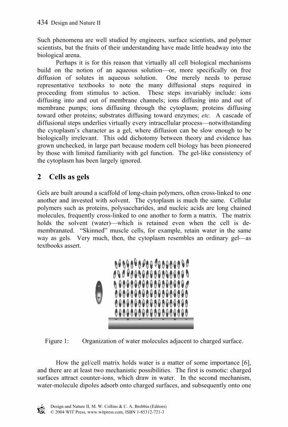
Figure 1: Organization of water molecules adjacent to charged surface.
How the gel/cell matrix holds water is a matter of some importance [6], and there are at least two mechanistic possibilities. The first is osmotic: charged surfaces attract counter-ions, which draw in water. In the second mechanism, water-molecule dipoles adsorb onto charged surfaces, and subsequently onto one
Design and Nature II, M. W. Collins & C. A. Brebbia (Editors)© 2004 WIT Press, www.witpress.com, ISBN 1-85312-721-3
434 Design and Nature II

another to form multilayers. The first mechanism is unlikely to be the prevailing one because: (1) gels placed in a water bath of sufficient size should eventually be depleted of the counter-ions on which water retention depends; yet, the hydrated gel state is retained; and (2), cytoplasm placed under high-speed centrifugation loses ions well before it loses water [7]. The second hypothesis, that charged surfaces attract water dipoles in multilayers, is an old one [8]. The thesis is that water can build layer upon layer (fig. 1). This view had been controversial at one time, but it has been given support by several groundbreaking observations. The first is the now-classical observation by Pashley and Kitchener that polished quartz surfaces placed in a humid atmosphere will adsorb films of water up to 600 molecular layers thick [9]; this implies adsorption of a substantial number of layers, one upon another. The second set of observations are those of Israelachvili and colleagues, who measured the force required to displace solvents sandwiched between closely-spaced parallel mica surfaces [10, 11, 12]. The overall behavior was largely classical, following DLVO theory. However, superimposed on the anticipated monotonic response was a series of regularly spaced peaks and valleys (fig. 2). The spacing between peaks was always equal to the molecular diameter of the sandwiched fluid. Thus, the force oscillations appeared to arise from a layering of molecules between the surfaces.
Figure 2: Effect of separation on force between closely spaced mica plates. Only the oscillatory part of the response is shown. After Horn and Israelachvili [10].
Although the Israelachvili experiments confirm molecular layering near
charged surfaces, they do not prove that the molecules are linked to one another in the manner implied in fig. 1. However, more recent experiments using carbon-nanotube tipped AFM probes approaching flexible monolayer surfaces in water show similar layering [13], implying that the ordering does not arise merely from packing constraints; and, the Pashley/Kitchener experiment implies
Design and Nature II, M. W. Collins & C. A. Brebbia (Editors)© 2004 WIT Press, www.witpress.com, ISBN 1-85312-721-3
Design and Nature II 435

that many layers are possible. Hence, the kind of layering diagrammed in fig. 1 is collectively implied by these experiments.
When two charged polymeric surfaces lie in proximity of one another, the interfacial water layers can bond the surfaces much like glue (fig. 3). This is revealed in common experience. Separating two glass slides stacked face-to-face is no problem; when the slides are wet, however, separation is formidable — sandwiched water molecules cling tenaciously to the glass surfaces and to one another, preventing separation. A similar principle holds in sand: A foot will ordinarily sink deeply into dry sand at the beach, leaving a large imprint; but in wet sand, the imprint is shallow. Water clings to the sand particles, bonding them together with enough strength to support one’s full weight.
Figure 3: Structured water dipoles effectively “glue” charged surfaces to one another.
The picture that emerges, then, is that of a cytoplasmic matrix very much resembling a gel matrix. Water molecules are retained in both cases because of their affinity for the charged (hydrophilic) surfaces and their affinity for one another. The polymer matrix and adsorbed water largely make up the gel. This explains why de-membranated cells retain their integrity. Embodied in this gel-like construct are many features that have relevance for cell function. One important one is ion partitioning. The prevailing explanation for the ion gradients found between extracellular and intracellular compartments lies in a balance between passive flow through channels and active transport by pumps. Thus, the low sodium concentration inside the cell relative to outside is presumed to arise from the activity of sodium pumps, which transport sodium ions against their concentration gradient from the cytoplasm across the cell membrane. The gel construct invites an alternative explanation. It looks toward differences of solubility between extracellular bulk water and intracellular layered, or “structured” water—as well as differences of affinity of various ions for the cell’s charged polymeric surfaces [14]. Na+ has a larger hydrated diameter than K+, and is therefore more profoundly excluded from the cytoplasm than K+; and, because the hydration layers require more energy to remove from Na+ than from K+, the latter has higher affinity for the cell’s negatively charged polymeric surfaces. Hence, the cytoplasm has considerably more potassium than
Design and Nature II, M. W. Collins & C. A. Brebbia (Editors)© 2004 WIT Press, www.witpress.com, ISBN 1-85312-721-3
436 Design and Nature II
sodium. A fuller treatment of this fundamental biological feature is given in the recent book by the author [15].
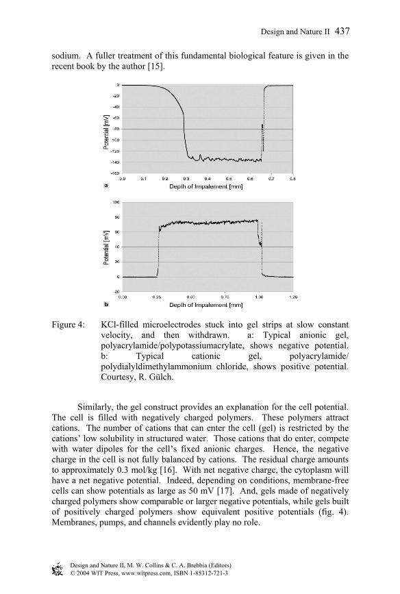
Figure 4: KCl-filled microelectrodes stuck into gel strips at slow constant velocity, and then withdrawn. a: Typical anionic gel, polyacrylamide/polypotassiumacrylate, shows negative potential. b: Typical cationic gel, polyacrylamide/ polydialyldimethylammonium chloride, shows positive potential. Courtesy, R. Gülch.
Similarly, the gel construct provides an explanation for the cell potential. The cell is filled with negatively charged polymers. These polymers attract cations. The number of cations that can enter the cell (gel) is restricted by the cations’ low solubility in structured water. Those cations that do enter, compete with water dipoles for the cell’s fixed anionic charges. Hence, the negative charge in the cell is not fully balanced by cations. The residual charge amounts to approximately 0.3 mol/kg [16]. With net negative charge, the cytoplasm will have a net negative potential. Indeed, depending on conditions, membrane-free cells can show potentials as large as 50 mV [17]. And, gels made of negatively charged polymers show comparable or larger negative potentials, while gels built of positively charged polymers show equivalent positive potentials (fig. 4). Membranes, pumps, and channels evidently play no role.
Design and Nature II, M. W. Collins & C. A. Brebbia (Editors)© 2004 WIT Press, www.witpress.com, ISBN 1-85312-721-3
Design and Nature II 437
Hence, the gel paradigm can go quite far in explaining the cell’s most fundamental attributes—distribution of ions, and the presence of a cell potential. These are equilibrium processes; they require no energy for maintenance.
3 Cell dynamics
The cell is evidently not a static structure but a machine designed to carry out a multitude of tasks. Such tasks are currently described by a broad variety of mechanisms, apparently lacking any single identifiable underlying theme — at least to this author. For virtually every process, there appears to be another mechanism.
Whether a common underlying theme might govern the cell’s many operational tasks is a question worth asking. After all, the cell began as a simple gel, and evolved from there. As it specialized, gel structure and processes gained in intricacy. Given such lineage, the potential for a simple, common, underlying, gel-based theme should not necessarily be remote. Finding a common underlying theme has been a long-term quest in other fields. In physics, for example, protégés of Einstein continue the search for a unifying force. That nature works in a parsimonious manner, employing variations of a few simple principles to carry out multitudinous actions, is an attractive notion, which I do not believe has yet been seriously pursued in the realm of cell function, although simplicity is a guiding principle in engineering.
If the cell is a gel, then a logical approach to the question of a common underlying principle of cell function is to ask whether a common underlying principle governs gel function. Gels do “function.” They undergo transition from one state to another. The process is known as a polymer-gel phase-transition—much like the transition from ice to water—a small change of environment causing a huge change in structure.
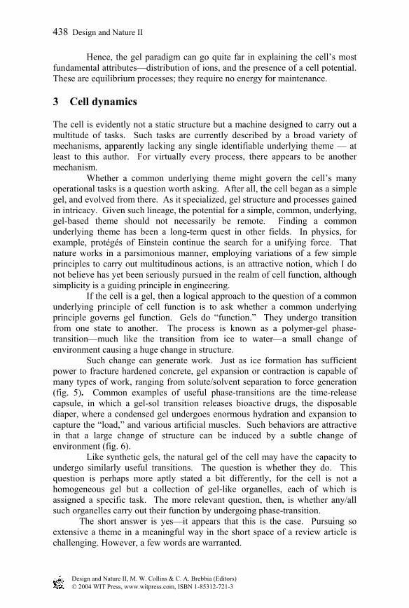
Such change can generate work. Just as ice formation has sufficient power to fracture hardened concrete, gel expansion or contraction is capable of many types of work, ranging from solute/solvent separation to force generation (fig. 5). Common examples of useful phase-transitions are the time-release capsule, in which a gel-sol transition releases bioactive drugs, the disposable diaper, where a condensed gel undergoes enormous hydration and expansion to capture the “load,” and various artificial muscles. Such behaviors are attractive in that a large change of structure can be induced by a subtle change of environment (fig. 6).
Like synthetic gels, the natural gel of the cell may have the capacity to undergo similarly useful transitions. The question is whether they do. This question is perhaps more aptly stated a bit differently, for the cell is not a homogeneous gel but a collection of gel-like organelles, each of which is assigned a specific task. The more relevant question, then, is whether any/all such organelles carry out their function by undergoing phase-transition. The short answer is yes—it appears that this is the case. Pursuing so extensive a theme in a meaningful way in the short space of a review article is challenging. However, a few words are warranted.
Design and Nature II, M. W. Collins & C. A. Brebbia (Editors)© 2004 WIT Press, www.witpress.com, ISBN 1-85312-721-3
438 Design and Nature II
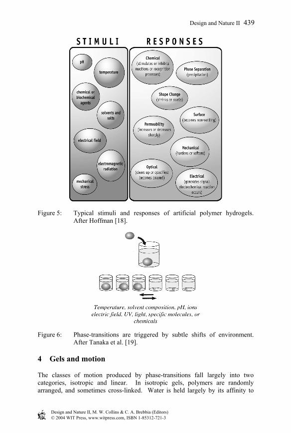
Figure 5: Typical stimuli and responses of artificial polymer hydrogels. After Hoffman [18].
Figure 6: Phase-transitions are triggered by subtle shifts of environment. After Tanaka et al. [19].
4 Gels and motion
The classes of motion produced by phase-transitions fall largely into two categories, isotropic and linear. In isotropic gels, polymers are randomly arranged, and sometimes cross-linked. Water is held largely by its affinity to
Design and Nature II, M. W. Collins & C. A. Brebbia (Editors)© 2004 WIT Press, www.witpress.com, ISBN 1-85312-721-3
Design and Nature II 439

polymers (or proteins, in the case of the cell). The gel is thus well hydrated—and may in the extreme contain as much as 99.97% water [20]. In the transitioned state, the dominant polymer-water affinity gives way to a higher polymer-polymer affinity, condensing the gel into a compact mass and expelling solvent. Thus, water moves, and polymer moves.
Linear polymers also undergo transition—from extended to shortened states. The extended state is stable because it maximizes the number of polymer-water contacts and therefore minimizes the system’s energy. Water builds layer upon layer. In the shortened state the affinity of polymer for itself exceeds the affinity of polymer for water, and the polymer folds. It may fold entirely, or it may fold regionally, along a fraction of its length. As it folds, polymer and water both move. And, if a load is placed at the end of the shortening filament, the load can move as well.
Figure 7: Calcium and other divalent cations can bridge the gap between negatively charged sites, resulting in zipper-like condensation.
Phase-transitions are inevitably cooperative: once triggered, they go to
completion. The reason lies in the transition’s razor-edge behavior: once the polymer-polymer affinity (or the polymer-water affinity) begins to prevail, its prevalence increases; hence the transition goes to completion. An example is illustrated in figure 7. In this example, the divalent ion, calcium, cross-links the polymer strands. Its presence thereby shifts the predominant affinity from polymer-water to polymer-polymer. Once a portion of the strand is bridged, flanking segments of the polymer are brought closer together, increasing the proclivity for additional calcium bridging. Thus, local action enhances the proclivity for action in a neighboring segment, ensuring that the reaction proceeds to completion. In this way, transitions propagate toward completion.
Evidently, the polymer-gel phase-transition can produce different classes of motion. If the cell were to exploit this principle, it could have a simple way of producing a broad array of motions, depending on the nature and arrangement of constituent polymers. In all cases, a small shift of some environmental variable such as pH, chemical content, etc. could give rise to a cooperative, all-or-none response, which could produce massive mechanical action.
That phase-transitions have more than theoretical relevance for cellular organelles is considered in the above-mentioned book [15]. These mechanisms are for the most part simpler than accepted mechanisms, and at least as
Design and Nature II, M. W. Collins & C. A. Brebbia (Editors)© 2004 WIT Press, www.witpress.com, ISBN 1-85312-721-3
440 Design and Nature II

consistent with experimental evidence. It is possible that the phase-transition may be a generic mechanism for generating motion.
Acknowledgement
The consent of Ebner and Sons to reprint figures from Pollack, Cells, Gels and the Engines of Life: A New, Unifying Approach to Cell Function [15], is gratefully acknowledged.
References
[1] Frey-Wyssling, A., Submicroscopic Morphology of Protoplasm. Elsevier: Amsterdam, 1953.
[2] Jones, D. S., Dynamic mechanical analysis of polymeric systems of pharmaceutical and biomedical significance. Int. J. Pharm., 179(2), pp. 167-178, 1999.
[3] Berry, H., Pelta, J., Lairez, D., and Larreta-Garde, V., Gel-sol transition can describe the proteolysis of extracellular matrix gels. Biochim Biophys Acta., 1524(2-3), pp. 110-117, 2000.
[4] Janmey, P.A., Shah, J.V., Tang, J.X., and Stossel, T.P., Actin filament networks. Results Probl. Cell Differ., 32, pp. 181-99, 2001.
[5] Hochachka, P. W., The metabolic implications of intracellular circulation. Proc Natl Acad Sci U S A., 96(22), pp. 12233-12239, 1999.
[6] Rand, R.P., Parsegian V.A., Rau, D. C., Intracellular osmotic action. Cell Mol. Life Sci., 57(7), pp. 1018-1032, 2000.
[7] Ling, G. N., and Walton, C. L., What retains water in living cells? Science, 191, pp. 293-295, 1976.
[8] Ling, G. N., The physical state of water in living cell and model systems. Ann. N. Y. Acad Sci., 125, p. 401, 1965.
[9] Pashley, R. M. and Kitchener, J. A., Surface forces in adsorbed multilayers of water on quartz. J. Colloid Interface Sci., 71, pp. 491-500, 1979.
[10] Horn, R.G., and Israelachvili, J.N., Direct measurement of astructural forces between two surfaces in a nonpolar liquid. J. Chem. Phys., 75(3), pp. 1400-1411, 1981.
[11] Israelachvili, J.N., and McGuiggan, P.M., Forces between surfaces in liquids. Science, 241, pp. 795-800, 1988.
[12] Israelachvili, J.N., and Wennerström, H., Role of hydration and water structure in biological and colloidal interactions. Nature, 379, pp. 219-225, 1996.
[13] Jarvis, S. P., et al., Local solvation shell measurement in water using a carbon nanotube probe. J. Phys. Chem. B, 104, pp. 6091-6097, 2000.
[14] Ling, G. N. A Revolution in the Physiology of the Living Cell, Krieger, Publ. Co.: Malabar, FL, 1992.
[15] Pollack, G. H. Cells, Gels and the Engines of Life: A New, Unifying Approach to Cell Function, Ebner and Sons: Seattle, 2001.
Design and Nature II, M. W. Collins & C. A. Brebbia (Editors)© 2004 WIT Press, www.witpress.com, ISBN 1-85312-721-3
Design and Nature II 441

[16] Wiggins, P. M., Role of water in some biological processes. Microbiol. Rev., 54(4), pp. 432-449, 1990.
[17] Collins, E.W., Jr., and Edwards, C., Role of Donnan equilibrium in the resting potentials in glycerol-extracted muscle. Am. J. Physiol., 22(4), pp. 1130-1133, 1971.
[18] Hoffman, A. S., Conventionally and environmentally sensitive hydrogels for medical and industrial use: a review paper. Polymer Gels, 268(5), pp. 82-87. 1991.
[19] Tanaka, T., Anaka, M., et al., Phase transitions in gels: Mechanics of swelling. NATO ASI Series Vol. H64, Springer Verlag: Berlin, 1992.
[20] Osada, Y. and Gong, J., Stimuli-responsive polymer gels and their application to chemomechanical systems. Prog. Polym. Sci., 18, pp. 187-226, 1993.
Design and Nature II, M. W. Collins & C. A. Brebbia (Editors)© 2004 WIT Press, www.witpress.com, ISBN 1-85312-721-3
442 Design and Nature II