Embed Size (px)
Citation preview
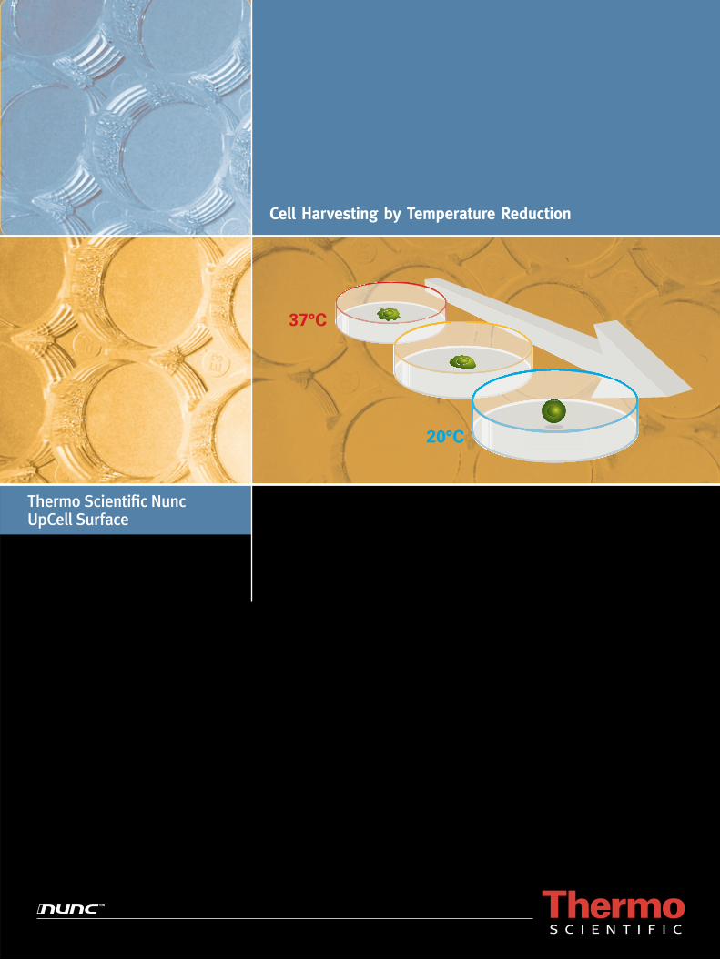
Cell Harvesting by Temperature Reduction
Thermo Scientifi c Nunc UpCell Surface
20°C
37°C

2 UpCell Surface | Cell Harvesting by Temperature Reduction
Features and Benefi ts
Cell harvesting using enzymatic digestion, such as trypsinization, results in degradation of cell surface proteins. These proteins are important for the interactions between the cell and the environment. For example, cell surface proteins are involved in the cell’s response to the extracellular matrix, to other cells and to growth factors and other soluble mediators. Some cell surface proteins are involved in the ion homeostasis of the cell, whereas other cell surface proteins are used as antigens or markers in cell analysis and enrichment procedures.
The Thermo Scientifi c Nunc UpCell Surface enables harvesting of cells with high viability and intact surface proteins for culture passaging, single - cell analyses and cell transplantation research.
No trypsin – preserves cell surface proteins•
No scraping – get high cell viability•
Minimal hands-on time•
Quick, clean and simple - just reduce the temperature•
Preserving Cell Surface Proteins
In tissue engineering, three-dimensional (3D) tissue models or transplants are typically prepared by seeding a cell suspension on a pre-fabricated scaffold. Scaffold materials are not produced by the cells to reside in the engineered tissue, and are most often materials foreign to the body or from another species (xenogeneic), such as, poly lactic acid (PLA), poly glycolic acid (PGA), alginate, gelatin and collagen. Problems often encountered using scaffolds for tissue engineering include uneven cell distribution and diffi culties in controlling the spacial distribution of different cell types. After transplantation, there can be host infl ammatory reactions and fi brous tissue formation due to the exogenous scaffold material.
The UpCell™ Surface enables harvesting of cell sheets and creation of tissue models held together by normal cell junctions and extracellular matrix deposited by the cells.
No scaffold – make 3D tissue models without exogenous material•
No uneven cell distribution – control the spatial distribution of cells in 3D•
Endless possibilities for mixing cell types and creating 3D co - cultures•
Just harvest and stack cell sheets•
Creating 3D Tissue Models
Cells harvested by trypsinization
Cells harvested from UpCell Surfaceby temperature reduction
Tissue engineered using UpCell Surface
Tissue engineered using scaffold

Cell Harvesting by Temperature Reduction | UpCell Surface 3
UpCell Surface is designed to respond to changes in temperature. It releases adherent cells by a simple reduction of the temperature of the cell culture. Products with UpCell Surface include Thermo Scientifi c Nunc MicroWell plates, multidishes and dishes.
The covalently immobilized polymer poly(N- isopropylacrylamide), or PIPAAm, forms an even and thin layer on the cultureware. The PIPAAm layer is slightly hydrophobic at 37°C, allowing cells to attach and grow. When the temperature of the culture is reduced to below 32°C, the PIPAAm layer becomes very hydrophilic, binds water and swells, resulting in the release of adherent cells.
Thermo Scientifi c Nunc UpCell SurfaceTemperature-Responsive Cell Culture Surface
Depending on the degree of confl uence of the culture, and the harvesting technique, single cells or cell sheets can be harvested from the UpCell Surface. Because the extracellular matrix under the cultured cells is harvested with the cells, cell sheets are naturally adhesive to other cell sheets and to cell surfaces in the body.
Extracellular Matrix is Harvested with the Cells
20°C
37°C
Extra-cellularMatrix
PIPAAm
Hydrophilic(Cell detachment)
~60
nm
(below 32°C)
UpCell Surface
At 20°C
~20
nm
UpCell Surface
Hydrophobic(Cell adhesion)
(above 32°C)
At 37°C

4 UpCell Surface | Cell Harvesting by Temperature Reduction
Single-cell suspensions harvested from cultureware with Thermo Scientifi c Nunc UpCell Surface can be
Analyzed • in vitro, for example, by fl ow cytometry Manipulated to purify certain cell types• Re-seeded in cultureware with UpCell Surface or traditional • cell cultureware as part of a passaging procedure Used in cell transplantation research •
Grow your cells in cultureware with UpCell Surface, reduce the temperature, and harvest your cells. It is that simple!
The Nunc UpCell Surface enables harvesting of cells with high viability and intact cell surface receptors and antigens. Even harvest cell types that are diffi cult to detach by other methods, and keep unwanted cell activation to a minimum.
Traditional cell harvesting by enzymatic and mechanical methods often compromises the integrity of surface proteins and the viability of harvested cells. By contrast, UpCell Surface allows cell harvesting by simply reducing the temperature of the cell culture, resulting in cell populations with preserved cell surface proteins and high cell viability – and there is no need for enzyme removal or inhibition.
Examples of applications using single-cell suspensions harvested by temperature reduction
Cell Type Application Reference
Macrophages (mouse) Detachment of cells that are otherwise diffi cult to detach Application Note 1
Bone marrow cells & preadipocytes (human) Cell surface protein preservation (fl ow cytometry) Application Note 2
Microglia (rat) Analysis (detachment and function) Nakajima et al., 2001
Monocytes and macrophages (human) Re-seeding/passaging Collier et al., 2002
Monocytes and macrophages (human) Activation & analysis (structural) Gordon and Freedman, 2006
Basophilic cell line RBL-2H3 (rat) Antigen-mediated degranulation measured by surface plasmon resonance Yanase et al., 2007
Working withSingle Cells
Seed cells in medium in cultureware with
UpCell Surface
Place cells at 37°C
Grow cells (up to 70% confluence, depending on cell type)
Place cells at 20-25°C (10-40 min, depending on cell type)
Harvest cells and medium

Cell Harvesting by Temperature Reduction | UpCell Surface 5
Cell sheets harvested from cultureware with Thermo Scientifi c Nunc UpCell Surface can be
Analyzed • in vitro, for example, by electron microscopy Re-plated to cultureware with UpCell Surface or traditional cell cultureware• Used in different transplantation models• Stacked on top of another cell sheet (see next section, • Tissue Models - Cell Sheet Engineering)
The UpCell Surface enables harvesting of contiguous cell sheets with preserved cell polarization and held together by normal cell junctions and extracellular matrix. Grow your cells to confl uence in cultureware with UpCell Surface, apply the supplied membrane (only Cat. Nos. 174901 6 well multidish and 174904 3.5 cm dish), reduce the temperature, and harvest the cell sheet. It is that easy!
Anchorage-dependent cells in culture initially attach to proteins adsorbed to the cultureware from the medium, and produce and deposit a subcellular matrix during the course of the cultivation (Brevig et al., 2006; Kushida et al., 1999; Pompe et al., 2003). Cells in culture are held together by cell junctions and the matrix deposited by the cells. Traditional enzymatic or mechanical harvesting disrupts these cell-to-cell and cell-to-matrix contacts, as well as the subcellular matrix, and destroys the integrity and polarization of the culture. With the UpCell Surface, cells can be detached as contiguous sheets from the cultureware. The subcellular matrix and the supplied membrane provides the mechanical strength necessary for the handling of the detached cell sheet.
Examples of applications using cell sheets harvested by temperature reduction
Cell Type Application Reference
Aortic endothelial cells (cow) Analysis (structural and matrix deposition) Kushida et al., 1999
Keratinocytes (human) Analysis (electron microscopy) Yamato et al., 2001
Urothelial cells (human) Analysis (electron microscopy) Shiroyanagi et al., 2003
Retinal pigment epithelial cell line ARPE-19 (human) Analysis (light microscopy) Kubota et al., 2006
Kidney epithelial cells (human and dog) Re-plating to traditional cultureware & analysis (electron and fl uorescence microscopy)
Application Note 3Kushida et al., 2005
Lung cells (rat) Re-plating to traditional cultureware & analysis (fl uorescence microscopy) Nandkumar et al., 2002
Smooth muscle cells & fi broblasts (human) Re-plating to PIPAAm surface & analysis (functional) & transplantation Hobo et al., 2008
Mesenchymal stem cells & skin fi broblasts (rat) Analysis (structural and functional) & transplantation Miyahara et al., 2006
Corneal stem cells (human and rabbit) Analysis (structural) & transplantation Nishida et al., 2004
Corneal endothelial cells (human) Analysis (structural and functional) & transplantation Sumide et al., 2006
Oral mucosal epithelial cells (dog) Analysis (structural) & transplantation Ohki et al., 2006
Tracheal epithelial cells (rabbit) Transplantation Kanzaki et al., 2006
Periodontal ligament cells (human) Transplantation Hasegawa et al., 2005
Working withCell Sheets
Grow cells to absolute confluence in cultureware
with UpCell Surface
Transfer membrane with cell layer to new cultureware or graft site
and remove membrane
Aspirate the medium and add fresh medium
Place cells at 20-25˚C for 5-6 min
Place
mem
brane
on top o
f cel
l lay
er
With
draw
mem
brane
with
cell
laye
r
Detach cell layer aroundedge of membrane

6 UpCell Surface | Cell Harvesting by Temperature Reduction
Cell sheet constructs prepared in cultureware with Thermo Scientifi c Nunc UpCell Surface can be
Analyzed • in vitro, for example, by functional tissue-specifi c tests Cultivated • in vitro, for example, as 3D co-cultures Used in different transplantation models, where cell sheets are stacked • before or during the transplantation procedure
The UpCell Surface enables harvested cell sheets to be stacked in order to form 3D tissue models. Grow your cells to confl uence in cultureware with UpCell Surface, harvest the cell sheet, and transfer the cell sheet to another cell sheet. No scaffold is needed!
Stacking of cell sheets, also known as Cell Sheet Engineering, was pioneered by Okano and colleagues (Yamada et al., 1990; Yang et al., 2005 and 2007). The preserved subcellular matrix of a harvested cell sheet provides the adhesive necessary for stacking. It functions as a natural glue to bond the cell sheet to an underlying recipient cell sheet or to a recipient site in a transplantation model, without the use of fi brin glue or sutures.
Examples of Cell Sheet Engineering using cell sheets harvested by temperature reduction
Cell Type Application Reference
Aortic endothelial cells (human) & hepatocytes (rat) Cultivation (3D co-culture) & analysis (structural) Harimoto et al., 2002
Hepatocytes (mouse and human) Analysis (structural and functional) & stacking during transplantation
Ohashi et al., 2007
Skeletal myoblast (dog) Analysis (structural) & stacking during transplantation
Hata et al., 2006
Lung and skin fi broblasts (rat) Analysis (structural) & stacking during transplantation
Kanzaki et al., 2007
Cardiomyocytes (rat) Cultivation & analysis (structural and functional) & stacking before transplantation
Sekine et al., 2006; Sekiya et al., 2006; Shimizu et al., 2002 and 2006
Working withTissue Models - Cell Sheet Engineering
Transfer cell sheet (with membrane) harvested from UpCell Surface to
a confluent culture where the medium has been aspirated
Add fresh medium and incubate at 37˚C or
Aspirate the medium and transfer another cell sheet to construct
Add fresh medium
Place cells at 37˚C for ~30 min
With
draw
mem
brane

Cell Harvesting by Temperature Reduction | UpCell Surface 7
Cultureware with UpCell Surface is intended for research purposes only and single-use only. Any other use is not warranted by Thermo Fisher Scientifi c. Do not use the product for clinical or diagnostic purposes. UpCell Surface is licensed from CellSeed Inc. Made in Japan.
Quality is inherent in our culture. From product development and sourcing raw materials to manufacturing and customer service, quality is refl ected in every Thermo Scientifi c Nunc product.
A certifi cate of quality is packed in each box of cultureware with Thermo Scientifi c Nunc UpCell Surface. This certifi cate is your guarantee that the product has been validated according to the following tests:
Cell growthEach manufacturing lot is sampled and subjected to performance testing for growth with the 3T3-Swiss Albino cell line (derived from a mouse embryo fi broblast) in accordance with standard operating procedures. Acceptance level: minimum of 80% confl uence.
Cell detachmentThe manufacturing lot is sampled and subjected to performance testing for cell detachment by temperature reduction with the 3T3-Swiss Albino cell line in accordance with standard operating procedures. Adherent cells are detached by temperature decreasing treatment (under 32°C) and the degree of detachment is measured. Acceptance level: detachment of 50% or more of the cells.
SterilitySterility is obtained by using ethylene oxide gas according to ISO 11135-1 (Sterilization of health care products, Ethylene oxide, Part 1: Requirements for development, validation and routine control of a sterilization process for medical devices).
Non-pyrogenicRepresentative samples of products with UpCell Surface are tested according to the principles of the LAL-test described in the FDA guidelines and certifi ed nonpyrogenic with a documented endotoxin level of less than 20 endotoxin units/device (0.5 Endotoxin units/mL) as stated in the USP.
ToxicityThe material has successfully passed the USP biological reactivity Class VI test - 50°C (7 days implant). Cytotoxicity test according to ISO 10993-5: Biological evaluation of medical devices - Part 5: Tests for in vitro cytotoxicity.
Cultureware with UpCell SurfaceWith lid. Sterile
Cat. No. 174897 174898 174899 174900 174901 174902 174905 174903 174906 174904
Format 96 MicroWell Plate w/ fl at bottom
48 well Multidish
24 well Multidish
12 well Multidish
6 well Multidish
10 cm Dish 10 cm Dish with grid
6 cm Dish 6 cm Dish with grid
3.5 cm Dish
Number of wells 96 48 24 12 6 1 1 1 1 1
Culture area, cm²/well
0.33 1.1 1.9 3.5 9.6 56.7 56.7 21.5 21.5 8.8
Max. external dimensions, mm
128x86 128x86 128x86 128x86 128x86 92x17 92x17 60x15 60x15 40x12
Suggested working volume, mL/well
0.2 0.5 1 2 3 12.5 12.5 5 5 3
Airvent + + + + + + + + + +
Units per pack/case 1/8 1/6 1/6 1/6 1/6 1/6 1/6 5/30 5/30 5/30
Products
Quality Assurance

8 Nunc UpCell Surface | Cell Harvesting by Temperature Reduction
Cells that are diffi cult to detach from traditional cultureware by enzymatic or mechanical methods may be harvested from cultureware with UpCell Surface simply by reducing the temperature of the cell culture.This application note compares the recovery of mouse peritoneal macrophages harvested from the UpCell Surface using temperature reduction with those harvested from traditional cultureware (tissue culture-treated polystyrene) using trypsinization or scraping.
Methods
Mouse peritoneal macrophages in RPMI 1640 medium supplemented with 10% fetal bovine serum (FBS) were seeded in one dish with UpCell Surface and two traditional cultureware dishes at 2.4 x 10⁵ cells/cm2. The cells were incubated at 37°C in a humidifi ed atmosphere of 5% CO2 in air. After 2 hours of incubation, non-adherent cells were removed by washing with phosphate-buffered saline (PBS). Cells were then cultured for 2 days in RPMI 1640 medium supplemented with 10% FBS and harvested using one of the following procedures:
Harvest of cells from the UpCell Surface using temperature reduction
Non-adherent cells were removed by washing • with Ca2+– and Mg2+–free PBS
4.0 mL RPMI 1640 medium supplemented with • 10% FBS was added and the dish was incubated at 20°C for 30 min.
Detached cells were harvested•
Harvest of cells from traditional cultureware using trypsinization
Non-adherent cells were removed by washing with • Ca2+– and Mg2+–free PBS
1.0 mL of 0.25% trypsin/EDTA was added, and the dish • was incubated at 37°C for 5 min.
3.0 mL RPMI 1640 medium supplemented with 10% • FBS was added
Detached cells were harvested•
Harvest of cells from traditional cultureware using EDTA and scraping
Non-adherent cells were removed by washing • with Ca2+– and Mg2+–free PBS
4.0 mL of 2.5 mM EDTA/PBS was added and the • dish was incubated on ice for 20 minThe cells were detached by scraping and harvested•
The harvested cells were counted and the recovery ratio was calculated.
Results
Figure 1. Photomicrographs of mouse peritoneal macrophages on the UpCell Surface before (a) and after (b) temperature reduction. After temperature reduction, the cells detached from the surface and became spherical. After harvesting of the cells by pipetting, only a few cells remained on the UpCell Surface (c).
0
20
40
60
80
100
Traditional culturewareUpCell Surface
Rec
over
y ra
tio
(%
)
Temperature reduction Scraping Trypsinization
Figure 2. Recovery ratio of mouse peritoneal macrophages harvested from the UpCell Surface was compared with recovery ratios of these cells harvested by either enzymatic (trypsinization) or mechanical (scraping) methods. The recovery of cells from the UpCell Surface was signifi cantly higher than the recovery of cells harvested from traditional cultureware by trypsinization or scraping. Mean and SD is shown.
a b c
Application Note 1UpCell Surface versus Trypsinization and Scraping in Cell Detachment

Cell Harvesting by Temperature Reduction | Nunc UpCell Surface 9
Enzymatic cell harvesting often compromises the integrity of cell surface proteins. By contrast, the UpCell Surface allows cell harvesting simply by reducing the temperature of the cell culture, resulting in cell populations with preserved cell surface proteins. This application note compares the integrity of CD140a (a cell surface tyrosine kinase receptor for members of the platelet-derived growth factor family) on human bone marrow cells and preadipocytes harvested from the UpCell Surface by temperature reduction and from traditional cultureware (tissue culture-treated polystyrene) by trypsinization. The cells were stained using a phycoerythrin (PE)-conjugated antibody against human CD140a and subsequently analyzed by fl ow cytometry.
Methods
Human bone marrow cells or human preadipocytes in Dulbecco’s Modifi ed Eagle’s Medium (DMEM) supplemented with 10% fetal bovine serum (FBS) were seeded at 6.8 x 10³ cells/cm2 in a 6 cm dish with UpCell Surface and also in a traditional 6 cm dish.
Cells were incubated for 24 hours at 37°C in a humidifi ed atmosphere of 5% CO2 in air, then harvested using one of the following procedures:
Harvest of cells from the UpCell Surface using temperature reduction
The dish was incubated at 20°C for 30 min. • (no change of culture medium)
Detached cells were transferred to a 15 mL • conical-bottom tube
Harvest of cells from traditional cultureware using trypsinization
Cells were gently washed once with Ca• 2+– and Mg2+–free phosphate-buffered saline (PBS)
2.0 mL of 0.25% trypsin/EDTA was added, and • the dish was incubated at 37°C for 3 min.
10 mL culture medium was then added to the dish, • and the cells were transferred to a 15 mL conical-bottom tube
Cells harvested by temperature reduction or trypsinization were washed twice by centrifugation (300 x g, 5 min.). Supernatants were discarded, and aliquots of 5.0 x 10⁵ cells were incubated with 200 μL of PE-conjugated mouse monoclonal antibody against human CD140a (5 μg/mL; BD Pharmingen, NJ, USA) or PE-conjugated mouse IgG2a
isotype-control antibody (5 μg/mL; BD Pharmingen). After incubation at 4°C for 60 min. cells were washed with PBS and analyzed by fl ow cytometry (FC500; Beckman Coulter, CA, USA).
Results
Human bone marrow cells and preadipocytes harvested from the UpCell Surface by temperature reduction had preserved cell surface CD140a, whereas CD140a on cells harvested from traditional cultureware by a short (3 min) trypsinization could barely be detected. This demonstrates that using UpCell Surface and temperature reduction preserves the integrity of cell surface proteins to a higher degree than using traditional cultureware and enzymatic cell harvesting.
UpCell Surface Preserves the Integrity of CD140a
Grey: anti-CD140aWhite: isotype control for non-specifi c binding (background)
Cells harvested from UpCell Surface by
temperature reduction
Cells harvested from traditional cultureware
by trypsinization
Human bone marrow cells
Human preadipocytes
Application Note 2UpCell Surface versus Trypsinization in Preservation of Surface Proteins During Cell Harvesting

10 Nunc UpCell Surface | Cell Harvesting by Temperature Reduction
A contiguous cell sheet can be harvested from the UpCell Surface without compromising the subcellular protein matrix and cell-cell junctions. In this application note, the transfer of a cell sheet from the UpCell Surface to a cell culture insert is described and the integrity of cell-cell junctions of the transferred cell sheet is demonstrated. This application note is based on Kushida et al., A non-invasive transfer system for polarized renal tubule epithelial cell sheets using temperature-responsive culture dishes. European Cells and Materials 2005; 10:23-30.
Methods
Madin-Darby Canine Kidney (MDCK) cells in Dulbecco’s Modifi ed Eagle Medium (DMEM) supplemented with 10% fetal bovine serum (FBS), penicillin (100 units/mL) and streptomycin (100 μg/mL) were seeded at 5.7 x 10⁴ cell/cm2 in a 3.5 cm dish with UpCell Surface. The cells were incubated for 21 days at 37°C in a humidifi ed atmosphere of 5% CO2 in air. The cells were then harvested as an intact sheet:
The dish was placed at 20°C for 60 min.•
A supporting membrane was placed on top of the cell • layer and the culture medium was aspirated
The edge of the membrane was gently released from • the dish using forceps, and the membrane with the attached cell layer was then transferred to a cell culture insert and incubated at 20°C for 30 min.
Suffi cient culture medium was added to cover the cell • sheet and membrane and the membrane was removed from the cell layer using forceps
Results
A layer of MDCK cells (Figure 1a) was detached from the UpCell Surface by temperature reduction and harvested as a single contiguous cell sheet, using a membrane as a carrier. After harvesting, no cells were observed on the UpCell Surface (Figure 1b). The harvested cell sheet was transferred to a cell culture insert, and adhered readily (Figure 1c).
Figure 1. Phase contrast microscopy of MDCK cells before and after cell sheet transfer. Scale bar, 100 μm. Printed with permission from European Cells and Materials (ecmjournal.org).
The cell sheet was examined by transmission electron microscopy after transfer. Tight junctions (Figure 2; arrow heads) were intact and the cell layer had maintained its apical-basal polarity.
Figure 2. Transmission electron microscopy of MDCK cell sheet transferred to a cell culture insert. a: Cell layer immediately after transfer. b: Cell layer 5 hours after transfer. Scale bar, 1 μm. Printed with permission from European Cells and Materials (ecmjournal.org).
Application Note 3Transfer of Cell Sheet with Preserved Polarization and Cell-Cell Junctions
Cell Harvesting by Temperature Reduction | Nunc UpCell Surface 11
Brevig T, Røhrmann JH, Riemann H. Oxygen reduces accumulation of type IV collagen in endothelial cell subcellular matrix via oxidative stress. Artif. Organs. 2006; 30:915-921.
Collier TO, Anderson JM, Kikuchi A, Okano T. Adhesion behavior of monocytes, macrophages, and foreign body giant cells on poly (N-isopropylacrylamide) temperature-responsive surfaces. J. Biomed. Mater. Res. 2002; 59:136-143.
Gordon IO, Freedman RS. Defective antitumor function of monocyte-derived macrophages from epithelial ovarian cancer patients. Clin. Cancer Res. 2006; 12:1515-1524.
Harimoto M, Yamato M, Hirose M, Takahashi C, Isoi Y, Kikuchi A, Okano T. Novel approach for achieving double-layered cell sheets co-culture: overlaying endothelial cell sheets onto monolayer hepatocytes utilizing temperature-responsive culture dishes. J. Biomed. Mater. Res. 2002; 62:464-470.
Hasegawa M, Yamato M, Kikuchi A, Okano T, Ishikawa I. Human periodontal ligament cell sheets can regenerate periodontal ligament tissue in an athymic rat model. Tissue Eng. 2005; 11:469-478.
Hata H, Matsumiya G, Miyagawa S, Kondoh H, Kawaguchi N, Matsuura N, Shimizu T, Okano T, Matsuda H, Sawa Y. Grafted skeletal myoblast sheets attenuate myocardial remodeling in pacing-induced canine heart failure model. J. Thorac. Cardiovasc. Surg. 2006; 132:918-924.
Hobo K, Shimizu T, Sekine H, Shin’oka T, Okano T, Kurosawa H. Therapeutic angiogenesis using tissue engineered human smooth muscle cell sheets. Arterioscler. Thromb. Vasc. Biol. 2008; 28:637-643.
Kanzaki M, Yamato M, Hatakeyama H, Kohno C, Yang J, Umemoto T, Kikuchi A, Okano T, Onuki T. Tissue engineered epithelial cell sheets for the creation of a bioartifi cial trachea. Tissue Eng. 2006; 12:1275-1283.
Kanzaki M, Yamato M, Yang J, Sekine H, Kohno C, Takagi R, Hatakeyama H, Isaka T, Okano T, Onuki T. Dynamic sealing of lung air leaks by the transplantation of tissue engineered cell sheets. Biomaterials 2007; 28:4294-4302.
Kubota A, Nishida K, Yamato M, Yang J, Kikuchi A, Okano T, Tano Y. Transplantable retinal pigment epithelial cell sheets for tissue engineering. Biomaterials 2006; 27:3639-3644.
Kushida A, Yamato M, Konno C, Kikuchi A, Sakurai Y, Okano T. Decrease in culture temperature releases monolayer endothelial cell sheets
together with deposited fi bronectin matrix from temperature-responsive culture surfaces. J. Biomed. Mater. Res. 1999; 45:355-362.
Kushida A, Yamato M, Isoi Y, Kikuchi A, Okano T. A noninvasive transfer system for polarized renal tubule epithelial cell sheets using temperature-responsive culture dishes. Eur. Cell Mater. 2005;10:23-30.
Miyahara Y, Nagaya N, Kataoka M, Yanagawa B, Tanaka K, Hao H, Ishino K, Ishida H, Shimizu T, Kangawa K, Sano S, Okano T, Kitamura S, Mori H. Monolayered mesenchymal stem cells repair scarred myocardium after myocardial infarction. Nat. Med. 2006; 12:459-465.
Nakajima K, Honda S, Nakamura Y, López-Redondo F, Kohsaka S, Yamato M, Kikuchi A, Okano T. Intact microglia are cultured and non-invasively harvested without pathological activation using a novel cultured cell recovery method. Biomaterials 2001; 22:1213-1223.
Nandkumar MA, Yamato M, Kushida A, Konno C, Hirose M, Kikuchi A, Okano T. Two-dimensional cell sheet manipulation of heterotypically co-cultured lung cells utilizing temperature-responsive culture dishes results in long-term maintenance of differentiated epithelial cell functions. Biomaterials 2002; 23:1121-1130.
Nishida K, Yamato M, Hayashida Y, Watanabe K, Maeda N, Watanabe H, Yamamoto K, Nagai S, Kikuchi A, Tano Y, Okano T. Functional bioengineered corneal epithelial sheet grafts from corneal stem cells expanded ex vivo on a temperature-responsive cell culture surface. Transplantation 2004; 77:379-385.
Ohashi K, Yokoyama T, Yamato M, Kuge H, Kanehiro H, Tsutsumi M, Amanuma T, Iwata H, Yang J, Okano T, Nakajima Y. Engineering functional two- and three-dimensional liver systems in vivo using hepatic tissue sheets. Nat. Med. 2007; 13:880-885.
Ohki T, Yamato M, Murakami D, Takagi R, Yang J, Namiki H, Okano T, Takasaki K. Treatment of oesophageal ulcerations using endoscopic transplantation of tissue-engineered autologous oral mucosal epithelial cell sheets in a canine model. Gut 2006; 55:1704-1710.
Pompe T, Kobe F, Salchert K, Jørgensen B, Oswald J, Werner C. Fibronectin anchorage to polymer substrates controls the initial phase of endothelial cell adhesion. J. Biomed. Mater. Res. A. 2003; 67:647-657.
Sekine H, Shimizu T, Yang J, Kobayashi E, Okano T. Pulsatile myocardial tubes fabricated with cell sheet engineering. Circulation 2006; 114 (Suppl):I87-I93.
Sekiya S, Shimizu T, Yamato M, Kikuchi A, Okano T. Bioengineered cardiac cell sheet grafts have intrinsic angiogenic potential. Biochem. Biophys. Res. Commun. 2006; 341:573-582.
Shimizu T, Yamato M, Isoi Y, Akutsu T, Setomaru T, Abe K, Kikuchi A, Umezu M, Okano T. Fabrication of pulsatile cardiac tissue grafts using a novel 3Dimensional cell sheet manipulation technique and temperature-responsive cell culture surfaces. Circ. Res. 2002; 90:e40-e48.
Shimizu T, Sekine H, Isoi Y, Yamato M, Kikuchi A, Okano T. Long-term survival and growth of pulsatile myocardial tissue grafts engineered by the layering of cardiomyocyte sheets. Tissue Eng. 2006; 12:499-507.
Shiroyanagi Y, Yamato M, Yamazaki Y, Toma H, Okano T. Transplantable urothelial cell sheets harvested noninvasively from temperature-responsive culture surfaces by reducing temperature. Tissue Eng. 2003; 9:1005-1012.
Sumide T, Nishida K, Yamato M, Ide T, Hayashida Y, Watanabe K, Yang J, Kohno C, Kikuchi A, Maeda N, Watanabe H, Okano T, Tano Y. Functional human corneal endothelial cell sheets harvested from temperature-responsive culture surfaces. FASEB J. 2006; 20:392-394.
Yamada N, Okano T, Sakai H, Karikusa F, Sawasaki Y, Sakurai Y. Thermo-responsive polymeric surfaces; control of attachment and detachment of cultured cells. Makromol. Chem. Rapid Commun. 1990; 11:571-576.
Yamato M, Utsumi M, Kushida A, Konno C, Kikuchi A, Okano T. Thermo-responsive culture dishes allow the intact harvest of multilayered keratinocyte sheets without dispase by reducing temperature. Tissue Eng. 2001; 7:473-480.
Yanase Y, Suzuki H, Tsutsui T, Uechi I, Hiragun T, Mihara S, Hide M. Living cell positioning on the surface of gold fi lm for SPR analysis. Biosens. Bioelectron. 2007;23:562-567.
Yang J, Yamato M, Kohno C, Nishimoto A, Sekine H, Fukai F, Okano T. Cell sheet engineering: recreating tissues without biodegradable scaffolds. Biomaterials 2005; 26:6415-6422.
Yang J, Yamato M, Shimizu T, Sekine H, Ohashi K, Kanzaki M, Ohki T, Nishida K, Okano T. Reconstruction of functional tissues with cell sheet engineering. Biomaterials 2007; 28:5033-5043.
References

Cell Culture Excellence™
Essential products for the cell culture laboratory
Our comprehensive portfolio includes advanced tools designed to helpyou achieve excellence at every stage of your cell culture process –from growth and passage to experimentation through characterization, analysis and storage.
To learn about our full array of cell culture products and services, go to:www.thermo.com/cellculture
North America - Tel: 1-800-625-4327 - technical.nunc@thermofi sher.comAsia Pacifi c - Tel: +65 6770 2807 - intlmktg@thermofi sher.comChina - Tel: 86-21-68654588 - info.nnichina@thermofi sher.comEurope (Nalgene) - Tel: +44 (0) 1432 263933 - [email protected] (Nunc) - Tel: +45 4631 2000 - info.nunc@thermofi sher.comIndia - Tel: +91-22-67162200 - Toll Free: 1800 22 8374 - contact.lpg.in@thermofi sher.comJapan - Tel: +81 3 3816 3355 - [email protected] - www.nalgenunc.co.jpAll other locations (USA, International Department) - Tel: +1 585 899 7198 - intlmktg@thermofi sher.com
www.thermo.com/upcell
© 2008 Thermo Fisher Scientifi c Inc. All Rights Reserved. 74061/N20230 - Ver. 1.0 - 09/2008 - YNI.
UpCell is the trademark of CellSeed Inc. All other trademarks are property of Thermo Fisher Scientifi c Inc. and its subsidiaries.