Embed Size (px)
Citation preview

Cell Growth and Division:
Mitosis vs. Meiosis and What’s going on the rest of the time

Why does a cell need to divide?• As an object grows, the volume increases at a faster rate
than the surface area
Cell Growth and Division:

To Survive
• SA:V ratio must be “right” T– More cytoplasm means more energy needed.
– By limiting the ratio of membrane to cytoplasm you
limit the “doorways” into the cell.
RESULT: Cell can’t get enough materials to support its large size
• Cell dies, unless it divides in half!

In order to divide, a cell must:
• Grow (includes protein synthesis)
• Replicate DNA
• Replicate organelles
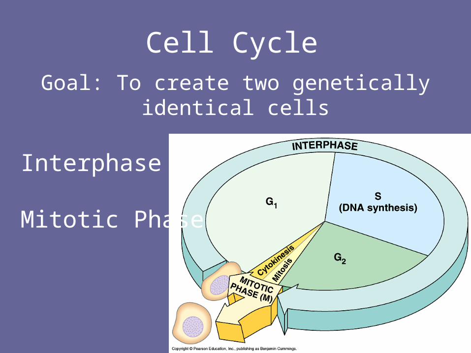
Cell Cycle
Interphase
Mitotic Phase
Goal: To create two genetically identical cells

How Long is a Cell Cycle
• Dependent on the organism
– Bacteria 20 minutes for some
– Yeast 90 – 120 minutes
– Mammalian 24 hours or more

Cell Cycle
INTERPHASE
G1 – growth and protein synthesis
S – DNA replication (copying the DNA)
G2 – Make organelles
M PHASE
Mitosis (Nuclear division)
Cytokinesis – division of cytoplasm and
membrane
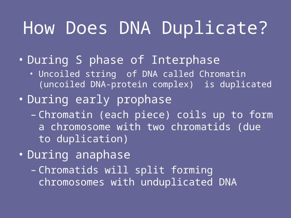
How Does DNA Duplicate?
• During S phase of Interphase• Uncoiled string of DNA called Chromatin (uncoiled
DNA-protein complex) is duplicated
• During early prophase– Chromatin (each piece) coils up to form a
chromosome with two chromatids (due to duplication)
• During anaphase– Chromatids will split forming chromosomes with
unduplicated DNA

How does DNA Duplicate?
• Each daughter cell gets 1 copy of each chromosome
• At the end of mitosis, chromosomes unravel (back to chromatin)

• THE STRUCTURE OF DNA• Most of the time DNA is in
the form of chromatin: strings of DNA wrapped around proteins called histones
• DNA is in chromatin form in G1
• In S, each strand of chromatin is copied
• Then, at the beginning of mitosis, the chromatin is folded into a duplicated chromosome

Remember DNA contains the info needed to build an organism
• Segments of DNA which contain information for a step (a protein or enzyme) are called a genes. There are many genes on each chromosome.
• Each organism has a specific number of chromosomes.

Human Chromosomes
• Humans have 23 types of chromosomes and 2 of each type = total of 46 chromosomes
• One of each type came from your mom the other from your dad.
• Every cell in your body has all 46 chromosomes with the exception of egg/sperm cells (only 1 of each type = 23)

Diploid cell(human)
23 “types” of chromosomes
2 of each type
1 from Mom1 from Dad

Haploid cellsCells that contain
one of each
chromosome are
called Haploid
cells.
Egg and sperm
cells are haploid.
This egg has an
extra chromosome

INTERPHASE: G1 cell:
- all DNA is UNDUPLICATED and in stringy chromatin form
- One piece of chromatin for every chromosome Chromatin/ Chromosomes from mom
Chromatin/ Chromosomes from dad
Green, blue and red represent the TYPES of chromosomes.
- So a dotted red chromosome is homologous to solid red chromosome

INTERPHASE: S phase cell:
- Every piece of chromatin is copied
- At the end each piece of chromatin is attached to its identical copy

G2 phase cell:
- DNA does not change at all
- Cell prepares for Mitosis by duplicating organelles such as centrosomes .

Mitosis
• One Fluid Event; no stopping and starting.
• BUT: for ease of study, we break it into 4 stages
• REMEMBER: all phases are continuous and may, in part, overlap

M Phase – Mitosis
Early Prophase:
- Chromatin coils/folds up into dense chromosomes. Chromosomes have duplicated DNA - “sister chromatids”
- Nuclear membrane and nucleolus begin breaking down
- Centrosomes start moving toward opposite sides of the cell. They are parts of the cells that make microtubule fibers
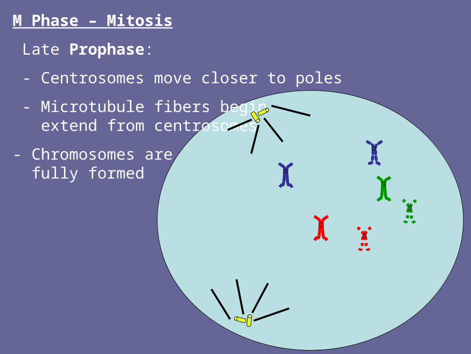
M Phase – Mitosis
Late Prophase:
- Centrosomes move closer to poles
- Microtubule fibers begin extend from centrosomes
- Chromosomes are fully formed

M Phase – Mitosis
Metaphase:
- Duplicated chromosomes line up on metaphase plate
-Microtubule “Cage” (spindle) extends from the centrosome to the chromosomes and attach to the centromeres (middles) of each chromosome –
- Each “bar” of the cage is called a spindle fiber

M Phase – Mitosis
Anaphase:
- Sister chromatids (identical copies) are pulled apart by the spindle fibers
- One copy of each chromosome is pulled towards each end of the cell

M Phase – Mitosis
Telophase:
- A nuclear membrane reforms around each group of chromosomes
- TWO nuclei at this point!!
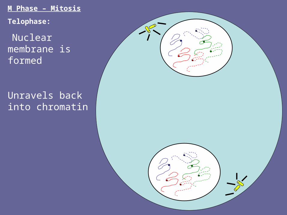
M Phase – Mitosis
Telophase:
Nuclear membrane is formed
Unravels back into chromatin

M Phase – Mitosis
Telophase:
- Chromosomes all uncoil/unfold back into chromatin.
- Notice that each nucleus has UNDUPLICATED chromatin like it did during G1
Cytokinesis
- Sides of membrane start pinching in forming cleavage furrow
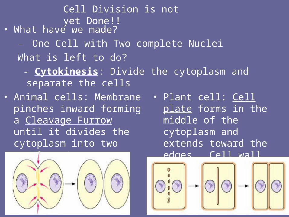
Cell Division is not yet Done!!
• What have we made?– One Cell with Two complete Nuclei
What is left to do?
- Cytokinesis: Divide the cytoplasm and separate the cells
• Animal cells: Membrane pinches inward forming a Cleavage Furrow until it divides the cytoplasm into two equal parts
• Plant cell: Cell plate forms in the middle of the cytoplasm and extends toward the edges. Cell wall forms from this cell plate

M Phase – Mitosis
Cytokinesis:
- Cleavage furrow grows until two cells are entirely separate
M phase is complete and you now have two cells that look JUST like the cell you started with!!!

Cell division is done!
• Now you have Two Identical daughter cells

Controlling Cell Division
• Cells know when they need to divide
When?– During growth– Repair – injury– Replacement (cells are not immortal!)– Reproduce

Controlling Cell Division
• Cells know when the don’t need to divide (or when to stop dividing)
• Cell division “stops” when cells touch other cells.

Identifying the Signal
EXPERIMENT
• A sample of the cytoplasm from a cell in mitosis is taken and injected into an interphase cell.
• Cell division will begin in the interphase cell
• CONCLUSION: Suggests there is a signal that triggers mitosis

Cyclins
• Mystery signal is one of a family of proteins they call “cyclins”
• Some cyclins are “Go” signals– Trigger the cell division process
• Some cyclins are “Stop” signals– shut down the cell division process
Scarring – see handout
• Basically occurs due to the body’s “emergency medicine response”
• Rather than being “neat and organized”, it simply wants to “cover the hole”
• RESULT: unorganized collagen matrix scarring (will “soften” over time..but still be present).

What if control is LOST????
• Cells begin to divide uncontrollably
• Pile up on top of each other
• Form big balls of cells called?????
TUMORS!!!
Tumor response
• Tumor cells do not respond to (or do not have) the body’s control signals
• Missing a “stop” signal so cell division doesn’t stop OR
• Hyperactive “go” signal so cell is constantly dividing

Definition of a Tumor• A tumor or tumour is commonly used as a
synonym for a neoplasm (a solid or fluid-filled (cystic) lesion that may or may not be formed by an abnormal growth of neoplastic cells) that appears enlarged in size.[1] Tumor is not synonymous with cancer. While cancer is by definition malignant, a tumor can be benign, pre-malignant, or malignant, or can represent a lesion without any cancerous potential whatsoever.

Tumors

Tumors vs. Cancer
• Tumor = uncontrolled but isolated growth of cells
• Tumor cells become cancer when they start to invade healthy tissue
• What if 1 cancer cell breaks off and enters the blood stream?
• Where ever it “lands” = new tumor = metastasis (spreading of cancer from one site to the next)

Cancer Terms
• Invasive:• Cancer has spread from the layer (tissue)
in which it developed and is growing into (and replacing) other “healthy” tissues that surround it.
• Metastatis - spread of cancer from one organ or part to a non-adjoining area or organ. We can look at cells to determine whether or not the cancer is metastatic

RECAP
• Body cells are made through mitosis
• Most of the time they “know what to do”,but sometimes we have an “out of control situation Cancer.

Asexual Reproduction
• Remember that mitosis is also used to create new organisms (not just repair)

Mitosis is also utilized in asexual reproduction
• Examples
– BuddingHydra

Cell division – Sexual Reproduction
• When cells participate in sexual reproduction they are “joining”.
• Joining means that all of the DNA will combine
• Too much DNA is usually an issue– Unless, of course you are a seedless
watermelon! (3N)

Gametes
• What would happen if we made Gametes (Egg and Sperm) cells this way?
– Way too much DNA!!!! (since two combine together)

Meiosis
– Happens ONLY in sex cells– Reduces information by ½– Requires two different divisions
• How many cells at the end??
• Meiosis begins the same as Mitosis– Cell in G1 enters S phase. – ALL DNA is copied– Chromatin folds up to form 46 duplicated
chromosomes

Now lets imagine that we need to make Gametes! Begins the same
G1 cell:
- all DNA is UNDUPLICATED and in stringy chromatin form
- One piece of chromatin for every chromosome Chromatin/ Chromosomes from mom
Chromatin/ Chromosomes from dad
Green, blue and red represent the TYPES of chromosomes.
- So a dotted red chromosome is holomogous to solid red chromosome

S phase cell:
- Every piece of chromatin is copied
- At the end each piece of chromatin is attached to its identical copy

G2 phase cell:
- DNA does not change at all
- Cell prepares for Mitosis

M Phase – Meiosis
Prophase I:
- Chromatin Coils/folds up into dense chromosomes. Each copy of a piece of chromatin becomes a “sister chromatid”
- Nuclear membrane and nucleolus begin breaking down
- Centrosomes start moving toward opposite sides of the cell. They are parts of the cells that make microtubule fibers

M Phase – Meiosis
Prophase:
-New event: Homologous chromosomes pair up to form Tetrads

M Phase – Meiosis
Prophase:
-Crossing over happens in each tetrad
- homologous chromosomes swap parts of their chromosomes
- Creates 4 DIFFERENT chromatids, each has a different combination of info
A AA A aaaa
B BB b bBbb

Meiosis I
• Crossing over in prophase
• Line up in homologous pairs (metaphase
• Homologous pairs separate BUT NOT SISTERS!
• RESULT: 2N (with double DNA) 1 N (double DNA)

M Phase – Meiosis
Prophase I:
- Centrosomes move closer to poles
- Microtubule fibers extend from centrosomes
M Phase – Meiosis
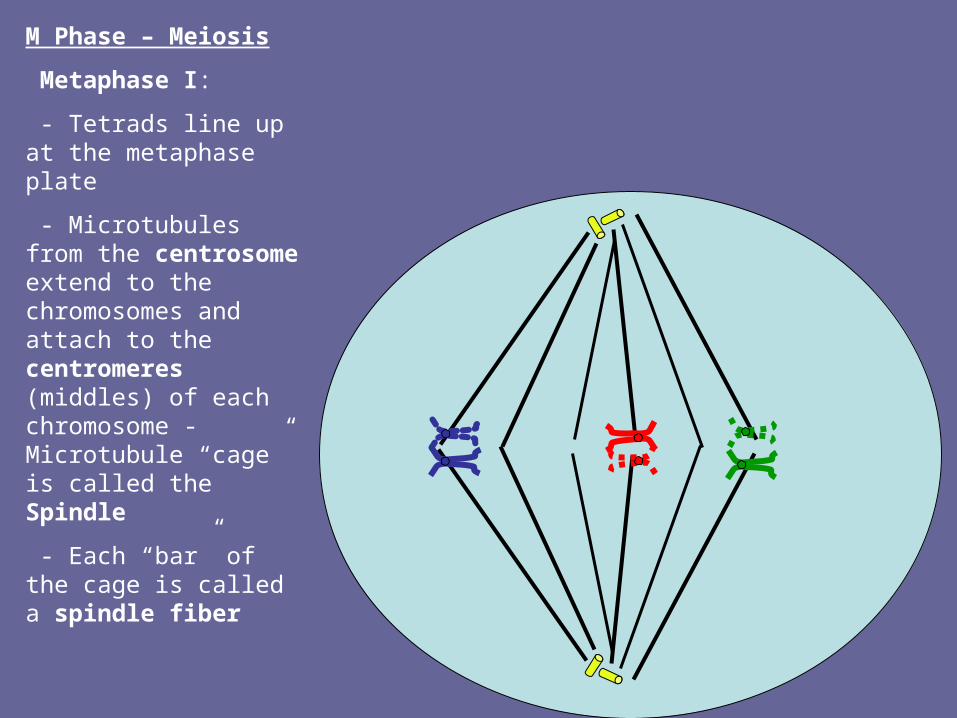
Metaphase I:
- Tetrads line up at the metaphase plate
- Microtubules from the centrosome extend to the chromosomes and attach to the centromeres (middles) of each chromosome - Microtubule “cage” is called the Spindle
- Each “bar” of the cage is called a spindle fiber

M Phase – Meiosis
Anaphase I :
- Tetrads are separated
- any combination of mom/dad chromosomes can be pulled to either side

M Phase – Meiosis
Telophase I:
- A nuclear membrane reforms around each group of chromosomes
- cell will have TWO nuclei at this point!!

Meiosis II
• Can occur without telophase or two separate cells redivide
• Chromosomes line up “single file
• Sisters split (1N 1 N)
• RESULT: 4 cells, all 1N, all unique (due to crossing over)

M Phase – Meiosis II
Prophase II:
- Same events as before
- Only this time it is happening in each of the two cells

M Phase – Meiosis II
Metaphase II:
- Remaining chromosomes line up at metaphase plate of each cell
- spindle fibers attach

M Phase – Meiosis II
Anaphase II:
- Attached chromatids are pulled apart in each cell

M Phase – Meiosis II
Telophase II:
- Nuclear membranes reform and spindle breaks down

M Phase – Meiosis II
Telophase II:
- chromosomes unwind back to chromatin
Cytokinesis
- Four cells are formed
- each one has a completely different combination of DNA thanks to crossing over and the division of homologous chromosomes

Possible sets of chromosomes in each of the 4 cells made during meiosis
a
aA
A
b
c
d
E
F
C
d
e
F
E
f
c
DB
e
f
C
DB
b
Humans and Meiosis
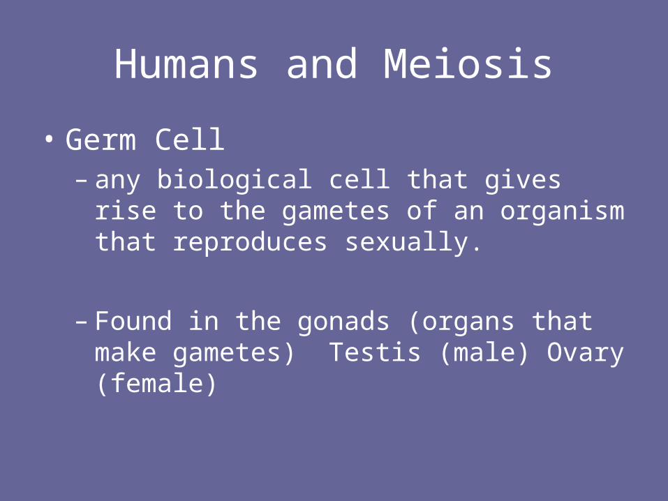
• Germ Cell – any biological cell that gives rise to the
gametes of an organism that reproduces sexually.
– Found in the gonads (organs that make gametes) Testis (male) Ovary (female)

Spermatogenesis
• Germ cell is called Spermatogonium – 2N– Makes sperm (1N) through meiosis– Makes additional spermatogonia (2N) through
mitosis
– Meiosis I• Reduces chromosome number from 2N 1N• BUT.. Still have duplicated chromosomes
– Meiosis II• Splits sister chromatids each chromatid becomes a
chromosome


Oogenesis
• Asymmetrical cell division leads to formation of the ovum and 3 polar bodies
• Polar bodies eventually disintegrate.


Karyotype
• Photomicrograph (microscopic picture) of chromosomes in a normal dividing cell
• Organized based on size of the chromosome and position of the centromere. They are grouped by homologous pairs. Sex chromosomes are the 23rd pair
• Can use chromosomes in telophase (before they uncoil) or prophase (duplicated)


Types of Chromosomes
• Sex chromosomes– Determine the gender of an organism and
may also carry other genes
• X or Y XX XY
• Autosomes– All other chromosomes (pairs 1-22)

Chromosomal Mutations
Mutation
a change in the nucleotide-base sequence of a gene or DNA molecule.

Possible Effects:
Germ cell mutations - occur in gametes
Do not affect the organism, but affect the offspring.
Somatic cell mutations – affect the body cells (but not gametes) and do affect the organism.

Classification of Mutations
Lethal mutations– Cause death, often before birth
Beneficial mutations– Mutations which provide higher survivability or
reproducibility
Silent mutations– neither harmful nor beneficial

Chromosomal vs. Gene Mutations
• Chromosomal Mutations – Involve changes in the structure of a chromosome or the
loss or gain of a chromosome.– Involve more than one gene– Generally seen in a karyotype
• Gene mutations – WE WILL STUDY LATER– The addition or removal of a single nucleotide within a
single gene– AKA point mutation.
Karyotyping and Chromosomal Mutations
• Karyotypes can identify chromosomal mutations
• Can be seen as a change in banding pattern, shape or size of a chromosome
• Can involve losing or gaining a “whole” chromosome.

Chromosome Mutations• Deletion
– Loss of a piece of chromosome due to breakage (genetic information is missing)
• Inversion– Chromsome segment breaks off, flips around (upside
down) and reattaches• Translocation
– Broken off piece of a chromosome attaches to a non-homologous chromosome
• Non-disjunction – non-separation of homologues or sister chromatids

Chromosomal Diseases
• Learn about these at the following site
• http://learn.genetics.utah.edu/content/begin/traits/
• Know the karyotype of:– Down syndrome, trisomy 18,, Turner
syndrome (XO, Klinefelter syndrome (XXY)

Questions to Answer
• What is the difference between monosomy and trisomy? How can both occur “at the same time”?
• What are two possible ways deletions can occur?

Stem Cells
• have the ability to either divide again and again to create more of itself (exact copies), OR to differentiate into another type of cell
• . Because of their ability to develop into any cell type, they could potentially provide an unlimited source of adult cells, such as bone, muscle, liver, or blood cells.
Three types
• Totipotent– Occur during cleavage (first few hours after
fertilization)
• Pleuripotent– One “step” differentiated– Can form many but not all cell types (most
researched currently for medical use)
• Multipotent– Bone marrow cells for all blood cells. Best
understood.

Medical Technology and Controversy
• Can we “grow an organ”?
• Controversy:– Source of stem cells– Limitations of adult stem cells– Cord blood – only blood diseases.

Embryology
• Know definitions
• Know difference between

Fertilization
Egg and sperm join
Sperm cell breaks down but leaves the chromosomes
Zygote:
A Fertilized Egg
Zygote begins to divide (mitosis!)
2-cell stage 4-cell stage (Day 2)
8-cell stage
(Day 3)
Morula: solid ball of 10 to 30 cells (Day 4)
Blastula: Hollow, fluid filled ball of
cells;
(Day 5)
Cells continue to divide and start to
rearrange themselves
Still dividing, outer cells start to fold inward
GASTRULATION
More dividing, three distinct layers of cells are formed
Ectoderm (outer)
Nervous
System and
skin
Mesoderm (middle)
Muscle, skeleton
Dermis, Heart, Kidneys
Endoderm (inner)
Lining of digestive system
Gastrula (W3)
No G1 or G2 phases just M and S during cleavage

1. Are the egg and sperm haploid or diploid? What about the zygote?
HAPLOID. DIPLOID
2. What type of division is occurring to create a morula? A blastula? What term is used to describe the first divisions of the zygote?
MITOSIS, Again MITOSIS, CLEAVAGE
3. In which stage do you see definite differentiation? What causes this?
4. Where would you find totipotent cells? Pleuripotent cells?
Different view of previous slide

Other Important Terms:
• Gamete – a haploid reproductive cell that unites with another haploid
reproductive cell to form a zygote.
• Zygote - The cell that results from the union of gametes; also called a
fertilized egg
Fertilization – the union of a male and female gamete to form a zygote
• Embryo – an organism in the early stages of development . A developing
human is called an embryo from two weeks after conception until the end of
the eighth week.
• Differentiation – the structural and functional specialization of cells during an
organisms’s development
SEE NEXT PAGE FOR TWINS

Other Important Terms:
• Identical twins (monozygotic)- --
• - single egg fertilized by one sperm.
• Fertilized egg splits into two embryos
early in development.
• Most IT share the same placenta. (get
oxygen and nutrients from the mother
and get rid of wastes through the
placenta.)
• Usually in separate amniotic sacs. Fraternal twins (dizygotic) - Fraternal
twins develop when two eggs are fertilized by two
separate sperms. The fetuses have separate
placentas and amniotic sacs.