Embed Size (px)
Citation preview

i
Immunol. Res. 5:117-128 (1986)
I I I I
�9 1986 S. Karger AG, Basel 0 2 5 7 - 2 7 7 X / 8 6 / 0 0 5 2 - 4 ) 1 1 7 5 2 . 7 5 , ' 0
Cell-Cell Interaction Responsible for the Induction of First Order Suppressor T Cells in Hapten-Specific Contact Sensitivity Reactions
Yuko Nakamura, Reiko M. Nakamura, Tohru Tokunaga
Department of Cellular Immunology, National Institute of Health, Kamiosaki, Tokyo, Japan
Introduction
In positive immune responses, it is generally accepted that in order to effectively stimulate an immune reaction, antigens must be pre- sented to helper T cells by antigen-presenting cells (APC) which express the I-A or I-A/I-E subregion gene products [ 1--4]. On the contrary, the mechanisms of the cell-cell interaction nec- essary for the induction of suppressor T cells (Ts) have not yet been elucidated.
It has been reported that the intravenous administration of hapten-coupled splenic cells into syngeneic recipients is capable of inducing the production of Ts which inhibit the generation of the hapten-specific contact sensitivity or delayed-type hypersensitivity reaction [5, 6]. Sherr et al. [7] have shown that Ia-positive APC are necessary for the induction of Ts. Furthermore, several re- searchers have described I-J restrictions con- trolling the induction of Ts and the interac- tion between T cell subsets [8-10]. Takaoki et al. [11] reported that I-J matching is required between the second order suppressor factor (TsF2) and the cells to be coupled with azo- benzenearsonate (ABA) to activate the third
order suppressor T cells (Ts3) in vivo. Lowy et al. [12] showed that I-A-I-J § cells coupled with ABA are necessary to activate Ts3 against the hapten-specific cytotoxic T cell response or delayed-type hypersensitivity (DTH). Nakamura et al. [13, 14] succeeded in the in vitro induction of Ts which were effective in inhibiting the induction of DTH to Mycobacterium boris BCG (BCG). In this experimental system, they demonstrated that I-J-positive splenic adherent cells play an es- sential role in the induction of Ts. These cells were considered as first order Ts (Tsl) be- cause they suppressed the immune response in the inductive phase [13, 14]. We asked therefore, whether Tsl cells in the ABA sys- tem are induced also in the context of the I-J antigen. We used in vivo and in vitro systems to activate Tsl against the contact sensitivity to ABA. In the present paper, we provide evi- dence that I-J positive APC are required for Tsl induction in the ABA system. These findings suggest that the necessity of I-J-posi- tive APC in the generation of Ts in DTH is not limited to the BCG/purified prdtein de- rivative system, but is also applicable to the other antigen, ABA.

t18 Nakam ura/Nakam ura/Tokunaga
M a t e r i a l s a n d M e t h o d s
Mice C3H/He mice of 4-6 week-old males were pur-
chased from Shizuoka Laboratory Animals Inc. Ltd., Hamamatsu, Japan. CBA mice of 6-7 week-old males were purchased from Ohmura Experimental Animals Inc. Ltd., Sagamihara, Japan. They were kept in a clean isorack to maintain SPF conditions.
Antibody and Complement Anti-l-A k (Ia.2) monoclonal antibody and anti-I-
jk alloantiserum, prepared by immunizing B I0.A (3R) mice with BI0.A (5R) lymphocytes, were purchased from Cedarlane Laboratories, Ltd., Ont., Canada. Monoclonal anti-Thy-l .2 antibody was purchased from New England Nuclear, Boston, Mass., USA. Low toxic rabbit complement was also obtained from Ce- darlane Laboratories.
Preparation of Spleen Cells A single cell suspension of splenic cells was pre-
pared in Hanks" balanced salt solution (HBSS) by passing the splenic cells through a stainless steel mesh No. 200. Erythrocytes were lysed in a Tris-ammo- nium chloride solution, pH 7.2. The cells were washed in HBSS twice and then used for separation into adherent and nonadherent fractions or for direct cou- pling with ABA.
Separation of Spleen Cells Spleen cell suspensions were plated onto plastic
tissue culture dishes (NUNC 50350) at a concentra- tion of 1 • 10 ~ cells/plate for 90 min at 37~ Non- adherent cells were collected from the dishes after gentle agitation. The nonadherent cells were reincu- bated in antimouse immunoglobulin antibody-coated plastic dishes (Falcon 3003) for 60 min at 37~ to enrich T cells through the removal of B cells and any contaminating adherent cells. The cells that remained nonadherent were pooled and used as the T cell frac- tion.
The adherent cells from the first incubation were gently scraped off the dishes with a rubber policeman after the dishes had been washed well to remove resid- ual nonadherent cells. To avoid contamination with T cells, the adherent cells were treated with anti-Thy-1.2 antibodies plus complement. These cells were used as the adherent cell fraction.
Antibody Treatment qf Adherent Cells To treat spleen cells with anti-I-A k monoclonal
antibody, 1 • 10 7 adherent cells were suspended either in 1 ml of a 1:40 dilution ofant i - I -A k antibody or in 1 ml of a 1:10 dilution ofant i- l -J k alloantiserum and incubated for 30 rain at 4 ~ The cells were then sedimented and resuspended in 1 ml of 1:15 diluted low toxic rabbit complement (C) and incubated for 3 0 m i n at 37~ in 5% COz.
Preparation qf ABA-Coupled Cells Preparation of Activated ABA. A 40-raM solution
of ABA was prepared from para-arsanilic acid (Wako Pharmaceutical Co., Tokyo, Japan). To prepare 100 ml of this solution, 932 mg ofp-arsanilic acid was dissolved in 3 ml of 6 N t-ICI, and this solution was added to 80 ml ofdistilled water. While stirring with a magnetic stirrer, 10% sodium nitrite was added drop- wise, until free nitrite could be demonstrated by an instantaneous color change on starch iodide paper. The solution was volumed to 100 ml and aliquoted in 2 ml, then stored at - 7 0 *C until used.
ABA solution must be activated just prior to cou- pling with the cells. The activation was accomplished by diluting the aliquot at 1:3 (vol/vol) in borate-buff- ered saline (BBS), pH 8.4. The solution was adjusted to pH 8.4 with I N sodium hydroxide. The color of activated ABA turns rapidly from yellow to orange- red. The final solution had a concentration of 10 m M of ABA.
ABA Coupling to Cells. A counted single-cell sus- pension was centrifuged and the supernatant dis- carded. A solution of ABA was activated and imme- diately added to the packed cells in such a volume thal the concentration of cells to ABA was 4 • 107/ml. The coupling reaction proceeded at room temperature for 10 rain.
The reaction was stopped by the addition of cold HBSS, and the cells were centrifuged at 4 ~ the supernatant was discarded, and the cells were washed twice in cold HBSS. The cells were counted once again before experimental use.
Assay for Contact Sensitivity Response Mice were immunized by skin painting with 0.1 ml
of ABA solution ofdimethytsulfoxide (DMSO) on the shaved abdomen. A 100 mM AI3A solution in DMSO was used for C3H:He mice which had received 100 ml/kg of cyclophosphamide (CY) intraperitone-
I-J-Positive Antigen-Presenting Ceils for First Order Suppressor T Cell Induction I t9
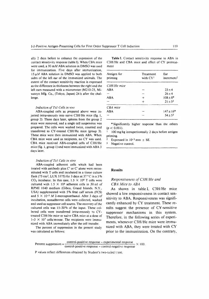
ally 2 days before to enhance the expression of the contact sensitivity response (table I). When CBA mice were used, a 30 m M A B A solution in DMSO was used for immunization. Five days after immunization, 15 g M ABA solution in DMSO was applied to both sides of the left ear of the immunized animals. The extent of the contact sensitivity reaction is expressed as the difference in thickness between the right and the left ears measured with a micrometer (M210-25, Mi- sutoyo Mfg. Co., (Tokyo, Japan) 24 h after the chal- lenge.
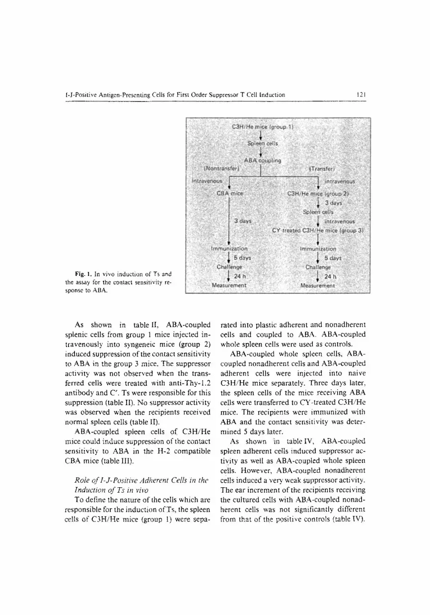
Induction of Tsl Cells in vivo ABA-coupled ceils as prepared above were in-
jected intravenously into naive C3H/He mice (fig. 1, group 2). Three days later, spleens from the group 2 mice were removed, and a single celt suspension was prepared. The cells were washed twice, counted and transferred to CY-treated C3H/He mice (group 3). These mice were then immunized with ABA. When CBA mice were used as recipients, no CY was used. CBA mice received ABA-coupled cells of C3H/He mice (fig. 1, group t) and were immunized with ABA 3 days later.
T a b l e I. Contact sensitivity response to ABA in C3H/He and CBA mice and effect of CY pretreat- ment
Antigen for Treatment Ear priming with CY l increment 2
C3H/He mice ABA - 23 _+ 4 - - 2 6 + 4 ABA + 108 -+-+ 8* -- + 21+53
CBA mice ABA - 147__+ 16" _ _ 54__.53
*Significantly higher response than the others (p < 0.001). 1 100 mg/kg intraperitoneally 2 days before antigen priming. 2 Expressed in 10 - 3 m m _+ SE. 3 Negative control.
Induction of Tsl Cells in vitro ABA-coupled adherent cells which had been
treated with antibody plus C' or C' alone were recon- stituted with T cells and incubated in a tissue culture flask (75 cm 2, LUX 5375) for 3 days at 37 ~ in a 5% CO2 incubator. In this case, 1.5 • 108 T cells were cultured with 1.5 X 10 s adherent cells in 30 ml of RPMI 1640 medium (Gibco, Grand Islands. N.Y., USA) supplemented with 5 % fetal calf serum (FCS) and 5 • 10 -s M 2-mercaptoethanol. After 3 days of incubation, nonadherent cells were collected, washed and used as suppressor cell source. The recovery of the cultured cells was 15-30% of the input. These col- lected cells were transferred intravenously to CY- treated C3H/He mice or naive CBA mice at a dose of 1-3 X 10⁷ cells/mouse. The recipients were immu- nized with ABA immediately after the cell transfer.
The percent of suppression in the present study was calculated as follows:
R e s u l t s
Responsiveness o f C3H/He and
CBA Mice to ABA
A s s h o w n in t a b l e I, C 3 H / H e m i c e
s h o w e d a l ow r e s p o n s i v e n e s s in c o n t a c t sen -
s i t i v i t y to A B A . R e s p o n s i v e n e s s was s ign i f i -
c a n t l y e n h a n c e d by C Y t r e a t m e n t . T h e s e re -
su l t s sugges t t h e p r e s e n c e o f C Y - s e n s i t i v e
s u p p r e s s o r m e c h a n i s m s in t h i s s y s t e m .
T h e r e f o r e , in t h e f o l l o w i n g se r i e s o f e x p e r i -
m e n t s , w h e n e v e r C 3 H / H e m i c e w e r e i m m u -
n i z e d w i t h A B A , t h e y we re t r e a t e d w i t h C Y
p r i o r to the i m m u n i z a t i o n . O n t h e c o n t r a r y ,
control-positive response - experimental response Percent suppression -
control-positive response - control-negative response
P values reflect differences obtained by Student's two-tailed t test.
• 100.

120 Nakam ura/Nakamura/Tokunaga
Table lI, Induction of Ts against contact sensitivity to ABA with ABA-coupted spleen ceils in C3H/He mice I
Cells for Ts ABA Treatment with Ear increment 2 Suppression induction priming anti-Thy-1 + C' %3
Experiment l Normal spleen cells + - 82 _+ 16 ABA spleen cells + - 32 _+ 7*
- - + -- 78• _ _ _ 21+_.55
81
Experiment 2 ABA spleen cells + - 30 z 4* ABA spleen cells + + 69 + 8 -- + -- 65+__74 . . . . . 21_+55
79 9
* Significant suppression in the contact sensitivity (p < 0.05). Recipients were pretreated with CY.
2 Expressed in I0 -3 m m + SE. 3 Percent suppression was calculated according to the formula given in 'Materials and Methods'. 4 Positive control. 5 Negative control.
Table III. ABA-coupled C3H/He spleen cells can also induce suppressor activity in H-2-compatible CBA mice
Cells for Ts ABA Ear Suppression induction priming increment I %e
Normal spleen cells + 114 _ 27 17 ABA spleen ceils + 55 + 2* 97 -- + 105_+53 _ _ 54_+54
* Significantly suppressed response (p < 0.05). 1 Expressed in 10 -3 mm _+ SE. 2 See footnote 3 in table II. 3 Positive control. 4 Negative control.
C B A m i c e w h i c h share the s a m e H - 2 h a p l o -
t ype wi th C 3 H / H e s h o w e d a h igh r e s p o n s i v e -
ness to A B A (table I).
Induction of Ts in vivo It has a l ready been d e m o n s t r a t e d that A B A -
coup l ed syngeneic cells a d m i n i s t e r e d i n t r ave -
nous ly can induce Ts [ 12]. To test w h e t h e r this
m e t h o d can also induce Ts in C 3 H / H e mice ,
the fo l lowing p rocedure was unde r t aken .
G r o u p 2 mice (fig. 1) were in j ec ted in t r ave -
nous ly wi th 5 • 107 A B A - c o u p l e d splenic cells
f rom a syngeneic strain group 1). Af t e r 3 days,
5 • 107 spleen ceils f rom the g roup 2 m i c e were
a d o p t i v e l y t ransfe r red i n t r a v e n o u s l y to C Y -
t rea ted C 3 H / H e mice (group 3). Af t e r t ransfer ,
the g roup 3 m i c e were i m m u n i z e d to A B A by
skin paint ing.
I-J-Positive Antigen-Presenting Cells for First Order Suppressor T Cell Induction t 2 1
Fig. 1. In vivo induction of Ts and the assay for the contact sensilivity re- sponse to ABA.
~:~,~;~,~:~2...--" ~ .-5.?": . . . . . .
:: ! " ~ ' i n t r a v e n ~ u s
�9 : I m m u n i z a t i o n �9 [ m i i : i u n i z a t i o n ~,~';
. j 2 h" ...... ; �9
- " ! ' if- f, M e a s u r e m e n t "- M e a s u r e m e n t , - : ~,~:!~:"~':-':-:
As shown in table II, ABA-coupled splenic cells from group 1 mice injected in- travenously into syngeneic mice (group 2) induced suppression of the contact sensitivity to ABA in the group 3 mice. The suppressor activity was not observed when the trans- ferred cells were treated with anti-Thy-l.2 antibody and C'. Ts were responsible for this suppression (table II). No suppressor activity was observed when the recipients received normal spleen cells (table II).
ABA-coupled spleen cells Of C3H/He mice could induce suppression of the contact sensitivity to ABA in the H-2 compatible CBA mice (table III).
Role of I-J-Positive Adherent Cells in the Induction of Ts in vivo To define the nature of the cells which are
responsible for the induction of Ts, the spleen cells of C3H/He mice (group I) were sepa-
rated into plastic adherent and nonadherent cells and coupled to ABA. ABA-coupled whole spleen cells were used as controls.
ABA-coupled whole spleen cells, ABA- coupled nonadherent cells and ABA-coupled adherent cells were injected into naive C3H/He mice separately. Three days later, the spleen cells of the mice receiving ABA cells were transferred to CY-treated C3H/He mice. The recipients were immunized with ABA and the contact sensitivity was deter- mined 5 days later.
As shown 'in table tV, ABA-coupted spleen adherent cells induced suppressor ac- tiyity as well as ABA-coupled whole spleen cells. However, ABA-coupled nonadherent cells induced a very weak suppressor activity. The ear increment of the recipients receiving the cultured ceils with ABA-coupled nonad- herent cells was not significantly different from that of the positive controls (table IV).
122 Nakamura/Nakamura/Tokunaga
Table IV. Induction of suppressor activity by ABA-coupled splenic ceils in vivo
Recipient Cells for Ts ABA Ear Suppression induction priming increment i 0/o2
Experiment 1 C3H/He ABA spleen cells + 41 +_ 17" 77
ABA adherent cells + 42 +_ 13* 76 ABA nonadherent cells + 82 _+ 12 30 - - + 109+ 183 _ _ 21+-54
Experiment 2 CBA ABA adherent cells + 39 __. 2* 127
ABA nonadherent cells + 91 _+ 9 32 - - + 109+ 163 _ _ 54+_54
* Significantly suppressed response (p<0.05). Expressed in 10 -3 mm +_ SE.
2 See footnote 3 in table If. 3 Positive control. 4 Negative control.
T a b l e V . I-J-positive APC are required for the induction of Ts in vivo
Treatment of Ear Suppression APC increment ~ %2
C' 51 _+_5* 55 Anti-I-J k + C' 74 _-2- 1 20 Anti-l-A k + C" 18 +5* 104 Positive control 88 +- 8 Negative control 21 + 5
Recipients are C3H/He mice. * Significant difference from the positive control (p < 0.05).
Expressed in t0 -3 mm +- SE. ? See footnote 3 in table II.
The suppressor cells induced by ABA-cou-
pled cells o f C 3 H / H e mice could also inhibi t
the contact sensi t ivi ty to A B A in C B A mice
(table IV, exper iment 2),
To character ize the adherent cell popula-
t ion required for the Ts induct ion, the splenic
adherent cells which had been t reated with
a n t i - T h y - l . 2 ant ibody and C' were further
t reated with ei ther an t i - I -A k or an t i - l - J k anti-
body plus C ' or with C ' alone. These cells
were coupled with ABA and injected in t rave-
nously into group 2 mice, and the Ts induc-
t ion was de termined. The results are shown
in table V. T rea tmen t o f the adheren t cells
with ant i - I -A k ant ibody plus C" induced a
stronger suppressive act ivi ty than that with

l-J-Positive Antigen-Presenting Cells for First Order Suppressor T Cell Induction 123
Table VI. Effect of antibody treatment of APC on the induction of Ts in vitro
Recipient Treatment of Treatment of Ear Suppression APC transferred cells incremenO %2
Experiment I C3H/He C' - 77 • 25 13
Anti-l-A k + C' - 44+_6* 65 Anti-I-J k + C' - 85 +- 20 1 Positive control 86 +- 11 Negative control 21 + 5
Experiment 2 CBA C' - 125 + _ 17 1
Anti-l-A k + C" - 71 +- 12* 76 Anti-I-A k + C' Anti-Thy-l.2 + C' 119 + 10 9 Positive control 126 +- 7 Negative control 54 __+ 5
* Significantly suppressed response (p < 0.05). Expressed in I0 -3 mm • SE.
2 See footnote 3 in table II.
C ' a lone. However , the t r ea tmen t o f the ad-
herent cells wi th an t i - I - J k a n t i b o d y plus C"
e l imina ted the ab i l i ty o f the cells to induce
the suppressor act ivi ty . These resul ts indica te
tha t I - J -pos i t ive adhe ren t cells p lay an im-
po r t an t role in the Ts induc t ion in vivo. On
the o the r hand, I - A pos i t ive adheren t cells
are no t necessary for the Ts induc t ion in this
system.
Induction o f Ts in vitro To conf i rm the resul ts o f the in v ivo
expe r imen t , the splenic adheren t cells f rom
C 3 H / H e mice were coup led with A B A after
va r ious a n t i b o d y t r ea tmen t and recons t i tu ted
with n o r m a l T cells and cu l tured in vi t ro .
The abi l i ty o f the adheren t cells to induce Ts
was d e t e r m i n e d by t ransferr ing the cul tured
cells in to group 3 mice which were i m m u -
n ized with A B A immedia t e ly . T h e adheren t
cells, e i ther t rea ted with C ' a lone, an t i - I -A k
plus C' , o r an t i - I - J k plus C' , were m i x e d with
T cells at a 1:100 rat io (1.5 • 108 T
c e l l s / 3 0 m l m e d i u m / b o t t l e ) and incubated
for 3 days.
As shown in table VI, adhe ren t cells
t r ea ted with C ' a lone or an t i - I - J k a n t i b o d y
plus C ' failed to induce suppressor act ivi ty.
On ly the adhe ren t cells t rea ted with an t i - I -A k
a n t i b o d y plus C" induced suppressor ac t iv i ty
agains t the contac t sens i t iv i ty to ABA. Trea t -
men t o f the t ransferred cells wi th an t i -Thy-

124 Nakamura/Nakamura/Tokunaga
1.2 plus C' in this group e l imina ted the sup-
pressor activity, indica t ing that Ts cells were
generated (table VI, exper iment 2). There
was no difference between the abil i ty of the
adherent cells t reated with ant i -I-J k plus C '
and C" alone. In this regard, the results dif-
Table VII. I-J-positive APC are required for the induction of Ts in vitro
Treatment of Ear Suppression APC increment I %2
Anti-l-A k + C' 82_+9* 68 Anti-i-Ak+ anti-I-J k + C' 135 + 11 7 Positive control 141 *__ l 6 Negative control 54 _ 5
Recipients are CBA mice. * Significant difference from the positive control (p < 0.05). 1 Expressed in 10 -3ram +_ SE. 2 See footnote 3 in table II.
fered from the in vivo observa t ion where
ABA-coupled adherent cells treated with
C 'a lone induced Ts cells.
Necessity of 1-J-Positive Adherent Cells for Ts Induction in vitro In the previous exper iment , it was sug-
gested that at least I -A-posi t ive adherent cells
are not required for the Ts induct ion . To con-
firm the role of I-J-posi t ive adheren t cells in
Ts induct ion , the adherent cells treated with
an t i - I -A an t ibody were further treated with
ant i - I -J k an t ibody plus C', and their abili ty to
induce Ts was compared with that of the
adherent cells treated with an t i - I -A k plus C'.
These adherent cells were coupled with ABA
and reconst i tuted with T cells and cultured
for 3 days in vitro. The cultured cells were
de te rmined for their suppressor activity
against the induc t ion of contact sensi t ivi ty to
ABA.
As shown in table VII, the adherent cells
treated with an t i - I -A k and ant i - I -J k plus C"
Table VIII. Antigen specificity of Ts induced by ABA-coupled adherent cells
Antigen for Cells for Ts Antigen Ear Suppression priming induction challenge increment~ %2
ABA ABA-APC 3 ABA 63 _+ 9* 83 ABA - ABA 107 • 64 -- -- ABA 54 _+ 55 PCI ABA-APC 3 PC1 76 +_ 7 0 PCI - PC1 76 • 13 ~
Recipients are CBA mice. * Significantly different from the positive control (p < 0.05). l Expressed in l0 -3 mm +__ SE. 2 See footnote 3 in table It. 3 Adherent cells were treated with anti-l-A k plus C" and then coupled with activated ABA. 4 Positive control. 5 Negative control.

I-J-Positive Antigen-Presenting Cells for First Order Suppressor T Cell Induction 125
before ABA-coupling lost the ability to in- duce Ts, whereas the adherent cells treated with anti-I-A k plus C' retained the Ts induc- ing ability. Thus, it was suggested that the antigen should be associated with I-J-posi- tive APC for the induction of Tsl. This is consistent with the character of the APC responsible for the Ts induction in vivo.
Antigen Specificity of Ts The antigen specificity of Ts induced with
ABA-coupled cells was determined. Ta- ble VIII shows that the suppressor activity induced by ABA-coupled adherent cells di- rected to the contact sensitivity to ABA but not to that to picryl chloride (PC1). These findings suggest that the Ts induced here are antigen-specific.
Discussion
In several mouse systems, it has been sug- gested that the I-J subregion products on T~ or Ts factors regulate the cell-cell or factor. cell interaction in the Ts circuit [8-11, 15- 17]. In some systems, however, it was pro. posed that I-J molecules on the APC play th~ regulatory role in the activation or generatior of Ys [12-14, 19].
This study showed that C3H/He mice are low responders to ABA in the contact sensi. tivity response and that ABA-specific Tsl cells were induced in C3H/He mice whict had received an intravenous injection o:" ABA-coupled spleen cells. The carrier cell, for hapten were shown to be I-J-positive plastic adherent cells which induced Ts 1 (ta. ble V). Plastic nonadherent cells did not acl as APC for the Tsl induction (table IV) I-A-positive adherent cells were not required for the Tsl induction because anti-I-A treat-
ment of the adherent cells before ABA-cou- pling even enhanced the Ts induction. Fur- thermore, the participation of I-A-positive adherent cells for the Ts induction was ne- glected by the in vitro experiment, where Ts were induced only when I-A-positive adher- ent cells were eliminated with anti-I-A plus C" (table VI). Double treatment of the adher- ent cells with anti-I-A and anti-I-J antibodies abrogated the ability of the adherent cell pop- utation to induce Ts in vitro (table VII), con- firming the necessity of I-J-positive APC for Tsl induction.
Splenic adherent cells treated with C" alone did not induce Ts in vitro (table VI). This result differed from that in the BCG/PPD system, where the splenic adher- ent cells treated with C' alone could induce Ts in vitro [13]. The reason for this discrep- ancy is not clear. Difference in the antigen presentation for ABA and PPD may be one of the explanations: ABA is considered to conjugate randomly with surface protein on the cell membrane, while PPD would be in- ternalized into the cell. If the expression of the I-A antigen is dominant over that of the I-J antigen, ABA would be presented in asso- ciation with I-A in more chances than with I-J. Concentration of I-J-positive APC by eliminating I-A-positive APC with anti-I-A treatment seems necessary for the effective association of ABA with the I-J antigen on APC. However, this explanation does not correspond to the difference between in vivo and in vitro experiments in ABA systems (ta- bles IV, V). Further studies are necessary to elucidate this point.
The Ts induced in this study were consid- ered to be Tsl because their activity was demonstrated during the induction phase of the contact sensitivity to ABA. Together with the results of Lowy" et al. [12], it is suggested

126 Nakamura/Nakam ura/Tokunaga
that the I-J molecules on APC act as re- stricting elements either in the first or third order step of the suppressor pathway.
Usui et al. [18] demonstrated that the existence of I-A positive APC responsible for the in vivo induction of Ts cells. They sug- gested that I-J restriction also existed in this process. Noma et al. [19] supported this idea by demonstrating the functional elimination of the APC involved in Ts induction by anti- I-A and anti-I-J antibodies. They concluded that the APC responsible for Ts induction are I-A+I-J + [19]. These data differ from our con- clusion that I-A-J-J + APC are responsible for Ts I induction [13, 14, and this paper]. This disparity may have been caused by the differ- ences in the assay systems and materials used. For example, in some strains of mice, most of the APC might be I-A+I-J + as shown by Habu et al. [20]. In such strains, the coex- istence of I-A and I-J molecules on APC would result in the removal of APC for the induction of Ts by the treatment either with anti-I-A or anti-I-J. However, in C3H/He mice, it appears that there is a significant population of I-A-/I-J + APC which are re- sponsible for the Ts induction both in ABA and BCG/PPD systems, and it seems most likely that I-J, but not I-A, molecules are functionally necessary, for the suppressor pathway.
In many systems, it has been reported that antigens detected by anti-I-J atloantisera are expressed on Ts or TsF, and immune re- sponses are regulated by such q-J' molecules [17, 21]. On the other hand, there has been a great deal of controversy regarding the sero- logical evidence for I-J molecules on APC. Also, mechanisms of immune regulation by I-J on APC have not yet been made clear. Asano et al. [22] reported that l-J determi- nants found on Ts were not detected on APC.
Murphy et al. [23] showed that I-J antigens expressed on APC were detected by some, but not all, lots of anti-I-J alloantisera. We have evidence that polyclonal anti-I-J alloan- tisera reacted with either Ts precursors or APC, but the monoclonal anti-I-J antibody which reacts on Ts precursors did not react with APC [14]. Therefore, we hypothesize that I-J epitopes on APC and T cells are dif- ferent and that the polyclonal anti-I-J antise- rum may contain the antibodies o f both types. In recent studies, it was suggested that I-J on the T cell is an inducible T cell receptor for the 'self" on APC or environmental non-T cell [24, 25]. These investigators consider that the I-J on the T cell is an idiotypic marker or an internal image of the "self" mol- ecule [26, 27]. These concepts seem to be consistent with our hypothesis. We have not yet obtained structural or biochemical evi- dence concerning these I-J moelcules on APC. Further functional and structural char- acterization of I-J moelcules on APC using monoclonal antibodies are under investiga- tion.
Participation of I-E antigens on APC in the Ts induction was not clear in the present study. Monoclonal anti-I-E k (Ia.7) antibody did not affect the ability of APC in the Ts induction either in the ABA or BCG/PPD system (data not shown). In the BCG/PPD system, I-J matching, but not I-E matching, is required between APC and T cells for the Ts induction in vitro [14]. Sumida et al. [24] excluded the involvement of the I-E mole- cule on APC in the Ts generation by the radiation chimera experiment. However, it is yet to be determined ifI-J expression is regu- lated in relation to the I-E gene family [28, 29],
Ts induced in vivo and in vitro using spienic adherent cells of C3H/He mice could

I-J-Positive Antigen-Presenting Cells for First Order Suppressor T Cell Induction 127
c lear ly inh ib i t the induc t ion o f the contact
sens i t iv i ty response to A B A in H-2 c o m p a t -
ib le CBA mice ( table III, VI). This suppresso r
ac t iv i ty was not shown when n o r m a l spleen
cells o f C 3 H / H e mice were in jec ted into CBA
mice ( table III), ind ica t ing that it was not a
case o f nonspeci f ic suppress ion m e d i a t e d by
a l loant igens .
In add i t ion , the A P C used in this s tudy
m a i n t a i n e d the i r ab i l i ty to induce Ts after
t r e a t m e n t wi th an t i -Thy-1 or a n t i - I - A ant i -
b o d y , ind ica t ing tha t they are ne i the r T no r B
cells. A l though we canno t exclude the poss i -
b i l i ty that these cells were dendr i t i c cells, evi-
dence tha t c loned macrophages are capab le
o f induc ing Ts in the B C G / P P D system in
v i t r o suggests tha t mos t l ikely the A P C in the
i n d u c t i o n o f Ts are macrophages [14].
Summary
C3H/He mice were found to be low responders in the contact sensitivity response to ABA. Intravenous injection of ABA-coupled syngeneic spleen cells in- duced hapten-specific Ts in C3H/He mice. These cells were Tsl because they acted on the inductive phase of the contact sensitivity. They could suppress the con- tact sensitivity in H-2-compatible CBA mice which were known to be high responders to ABA. Using in vivo and in vitro systems for the induction of Ts, it was shown that I-A-I-J*Thy-1- adherent cells were necessary as APC for the induction of Tsl.
Acknowledgement
We thank Drs. A. Kojima and T. Taniyama for valuable discussions and Miss M. Takekawa for her excellent technical help. This work was supported in part by a Grant-in-Aid from the Ministry of Health and Welfare, Japan, and by WHO grant for Vaccine Control.
References
1 Schwartz, R.H.: Yano, A.; Paul, W.E.: Interactions between antigen-presenting ceils and primed T lymphocytes: an assessment of Ir gene expression in the antigen-presenting cell. Immunol. Rev. 40: 153 (1978).
2 Unanue, E.R.: The regulation of lymphocyte func- tions by the macrophage. Immunol. Rev. 40:227 (1978).
3 Cowing, C.; Pineus, S.H.; Sachs, D.H.; Dickter, H.B.: A subpopulation of adherent accessory cells bearing both I-A and I-E or C subregion is required for antigen-specific routine T lymphocyte prolifer- ation. J. Immun. 121." 1680 (t978).
4 Nepom, J.T.; Benacerraf. B.: Germain, R.N.: Analysis of Ir gene function using monoclonal an- tibodies: independent regulation of GAT and GLPhe T cell responses by I-A and I-E subregion products on a single accessory cell population. J. Immun. 127:31 (t981).
5 Claman, H.N.; Miller, S.D.; ST, M.-S.; Moorhead, J.W.: Suppressive mechanisms involving sensiti- zation and tolerance in contact allergy. Immunol. Rev. 50:105 (1980).
6 Greene. M.I.: Benacerraf, B.: Studies on hapten- specific T cell immunity and suppression. Immu- nol. Rev. 50:163 (1980).
7 Sherr, D.H.; Heghinian, K.M.: Benacerraf, B.; Doff, M.E.: Immune suppression in vivo with antigen-modified syngeneic cells. IV. Requirement for la § adherent cells for induction. J. Immun. 124: 1389 (1980).
8 Zembata, M,; Asherson, G.L.; Colizzi, V.: Hapten- specific T suppressor factor recognizes both hapten and I-J region products on haptenized spleen cells. Nature 297:411 (1982).
9 Minami, M.; Honji. N.; Doff, M.E.: Mechanism responsible for the induction of I-J restrictions on Ts3 suppressor cells. J. exp. Med. 156:1502 (1982).
10 Germain. R.N.: Benacerraf, B.: A single major pathway of T-lymphocyte interactions in antigen- specific immune suppression. Scand. J. Immunol. 13:1 (1981).
11 Takaoki. M.; Sy, M.-S.; Yominaga, A.; Lowy, A.; Tsurufuji, M.; Finberg, R.; Benacerraf, B.; Greene, M.I.: I-J-restricted interactions in the generation of azobenzenearsonate-specific suppressor T cells. J. exp. Med. 156:1325 (1982).

128 Nakamura/Nakamura/Tokunaga
12 Lowy, A,; Tominaga, A.: Drebin, J.A.; Takaoki, M.; Benacerraf, B.: Greene, M.I.: Identification of an I-J + antigen-presenting cell required for third order suppressor cell activation. J. exp. Med. 157: 353 (1983).
13 Nakamura, R.M.; Tanaka, H.; Tokunaga, T.: In vitro induction of suppressor T cells in delayed- type hypersensitivity to BCG and an essential role of I-J positive accessory cells. Immunol. Lett. 4: 295 (1982).
14 Nakamura, R.M.; Nakamura, Y.; Nagayama, A.; Tokunaga, T.: [-J positive cloned macrophages as accessory cells for the induction of suppressor T cells in vitro. Immunol. Res. 5:106-116 (1986).
15 Tada, T.; Okumara, K.: The role of antigen-spe- cific T cell factors in the immune response. Adv. Immunol. 28:1 (1979).
16 Minami, M.; Furusawa, S.; Dorf, M.E.: l-J restric- tions on the activation and interaction of parental and Fl-derived Ts3 suppressor cells. J. exp. Med. 156:465 (1982).
17 Dorf, M.E.; Benacerraf, B.: l-J as a restriction ele- ment in the suppressor T cell system, lmmunol. Rev, 83:23 (1985).
18 Usui, M.; Aoki, I.; Sunshine, G.H.; Dorf, M.E.: A role for macrophages in suppressor cell induction. J. Immun. 132:1728 (1984).
19 Noma, T.; Usui, M.; Dorf, M.E.: Characterization of the accessory cells involved in suppressor T cell induction. J. Immun. 134:1374 (1985).
20 Habu, S.; Yamauchi, K.; Gershon, R.K.; Murphy, D.B.: A non-T: non-B cell bears I-A, I-E, I-J, and T la (Qa-l?) determinants. Immunogenetics 13: 215 (198t).
21 Murphy, D.B.; Horowitz, M.C.; Homer, R.J.; Flood, P.M.: Genetic, serological and functional analysis of l-J molecules. Immunol. Rev. 83:79 (1985).
22 Asano, Y.: Okumura. K.: Tada, T.: Ia antigens on antigen-presenting cells do not carry the same Ia specificities as detected on suppressor and helper T cells. Scand. J. Immunol. 13:353 (1981).
23 Murphy, D.B.; Yamauchi, K.; Habu, S.; Eardley, D.D.; Gershon, R.K.: T cells in a suppressor cir- cuit and non-T:non-B cells bear different I-J deter- minants. Immunogenetics 13:205 ( 1981).
24 Sumida, T.; Sado, T.; Kojima, M.; Ono, K.; Kami- saku, H.; Taniguchi, M.: l-J as an idiotype of the recognition component of antigen-specific sup- pressor T cell factor. Nature 316:738 (1985).
25 Uracz, W.; Asano, Y.; Abe, R.; Tada, T.: I-J epi- topes are adaptively acquired by T cells differen- tiated in the chimaeric condition. Nature 316:741 (1985).
26 Taniguchi, M.; Sumida, T.: ' l -J ' as an idiotypic marker on the antigen-specific suppressor T cell factor. Immunol. Rev. 83:125 (1985).
27 Tada, T.; Uracz, W.; Abe, R.; Asano. Y.: l-J as an inducible T cell receptor for self. lmmunol. Rev. 83:106 (1985).
28 Ikezawa, Z.; Baxevanis, C.N.; Arden, B.; Tada, T.; Waltenbaugh, C.R.; Nagy. Z.A.; Klein, J.: Evi- dence for two suppressor factors secreted by a sin- gle cell suggests a solution to the J-locus paradox. Proc. natn. Acad. Sci. USA 80." 6637 (1983).
29 Hayes, C.E.; Klyczek, K.K.; Krum, D.P.; Whir- comb, R.M.; Hullet, D.A.: Cantor, H.: Chromo- some 4 Jt gene controls murine T cell surface I-J expression. Science 223. 559 (1984).
Reiko M. Nakamura. PhD, Department of Cellular Immunology. National Institute of Health, Kamiosaki, Shinagawa-ku, Tokyo 141 (Japan)

![Antibodi Semester 1 .ppt [Read-Only] - ocw.usu.ac.idocw.usu.ac.id/course/download/...of-cell-2/bbc215_slide_antibodi.pdf · – Bereaksi den gggpan determinan Anti gen dan hapten](https://img.dokumen.tips/doc/110x75/5b9ebbd609d3f2083f8c2097/antibodi-semester-1-ppt-read-only-ocwusuacidocwusuacidcoursedownloadof-cell-2bbc215slide.jpg)