Embed Size (px)
Citation preview

Supplementary ACHD Echo Acquisition Protocol for
Ebsteins Anomaly
The following protocol for echo in adult patients with Ebsteins anomaly is a guide for performing a comprehensive assessment of this group of patients. It is intended as a supplementary guide to the ISACHD echo protocol and sequential analysis and all regular measurements should be included. It highlights areas of interest in each view specific to Ebstein’s patients.
Definition:
• Apical displacement of the septal leaflet of the tricuspid valve into the right ventricle. The posterior leaflet is sometimes also displaced. The leaflets have failed to fully delaminate from the septum and are often dysplastic with thickened, rolled & shortened chordae and under-developed papillary muscles
• Anterior leaflet is elongated and redundant with abnormal chordal attachments directly to the lateral wall.
• “Atrialization” of the basal portion of the right ventricle (aRV) with abnormal ventricular septal motion.
Diagram 1 Diagram of Ebstein anomaly. Adapted from Ebstein W. Ueber einen sehr seltenen Fall von Insufficienz der Valvula tricuspidalis, bedingt durch eine angeborene hochgradige Missbildung derselben. Arch Anat Physiol. 1866; 238–255.
Fenestrations
Patent foramen ovale
Coronary sinus ostium
Anterior leaflet
Posterior leaflet
Annulus
Septal leaflet

Common associations:
ASD or PFO (bi-directional shunting is common) VSD Pulmonary atresia with intact ventricular septum MV prolapse Coarctation of aorta LV non compaction
Carpentier’s Classification in Ebstein Anomaly
Diagram 2 Carpentier’s Classification. Type A: the volume of the true right ventricle (RV) is adequate; Type B: a large atrialized component of the RV exists, but anterior leaflet of the tricuspid valve moves freely; Type C, the anterior leaflet is severely restricted in its movement and may cause significant obstruction of the right ventricular outflow tract; Type D, almost complete atrialization of the RV except for a small infundibular component. (Carpentier A, et al. A new reconstructive operation for Ebstein’s anomaly of the tricuspid valve. J Thorac Cardiovasc Surg. 1988;96: 92–101. )
Diagram 3. Different types of Ebstein anomaly. Diagram adapted from Popelova et al.
A.
B.
C.
D.
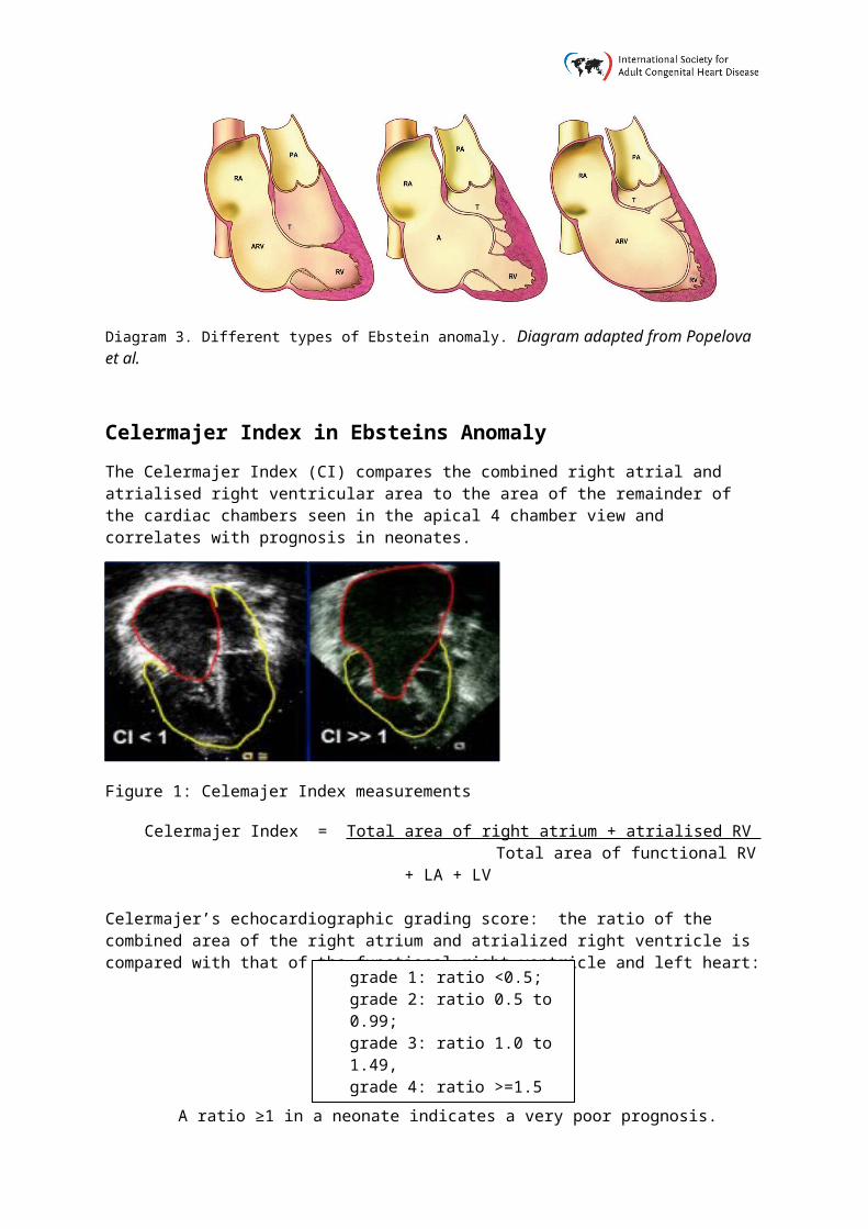
Celermajer Index in Ebsteins Anomaly
The Celermajer Index (CI) compares the combined right atrial and atrialised right ventricular area to the area of the remainder of the cardiac chambers seen in the apical 4 chamber view and correlates with prognosis in neonates.
Figure 1: Celemajer Index measurements
Celermajer Index = Total area of right atrium + atrialised RV Total area of functional RV + LA + LV
Celermajer’s echocardiographic grading score: the ratio of the combined area of the right atrium and atrialized right ventricle is compared with that of the functional right ventricle and left heart:
A ratio ≥1 in a neonate indicates a very poor prognosis.
(Celermajer DS, et al. Ebstein’s anomaly: presentation and outcome from fetus to adult. J Am Coll Cardiol. 1994;23:170 –176.)
grade 1: ratio <0.5;grade 2: ratio 0.5 to 0.99;grade 3: ratio 1.0 to 1.49, grade 4: ratio >=1.5

Imaging protocol for Ebstein anomaly
Subcostal views Establish abdominal and atrial situs, cardiac position & direction of apex Assess IVC size & collapsing Assess atrial septum RV SAX may offer biplane FAC of RV
PLAX/RV inflow
Assess displacement of septal +/- posterior leaflet involvement Assess TR severity Measure LV size TV leaflets seen in RVOT in standard PLAX suggest anterior rotation of the
tricuspid orifice
Parasternal short axis
Assess rotation of tricuspid valve closer in position to the pulmonary valve Assess TR severity Assess RVOT dilatation & function Assess size of pulmonary arteries when repair is being considered Assess interatrial septum
Apical views Zoom on cardiac crux to establish & measure abnormal septal displacement (>2 cm or >8mm/m²)
Assess tricuspid valve anatomy including degree of direct attachment of anterior leaflet into lateral wall of RV
Assess functional right ventricular function Assess degree of atrialisation of right ventricle (Celemajer Index) Assess TR severity at origin of jet (large compliant RA may mask hepatic
flow reversal) Assess left ventricular function
Suprasternal views Assess aortic arch
SVC flow may show flow reversal in cases of severe TR.

Ebstein Anomaly Reports:
Key points to include in transthoracic echo report: Valve anatomy including displacement towards apex and rotation towards the RV
outflow tract Functional RV size & function. In severe cases, this may be only the RVOT Degree of atrialisation of the right ventricle Severity of tricuspid regurgitation Size of pulmonary arteries LV size & systolic function Atrial septal integrity

Key views specific to Ebsteins Anomaly:
A
B
B
Figure 4. Severe TR from 4 chamber view with tricuspid valve Doppler profile. Note the laminar flow of tricuspid regurgitation indicating severe deficiency of valve function. Estimation of pulmonary pressures is not reliable in this scenario.
Fig ure 2. PLAX: note the abnormal enface orientation of the tricuspid valve. This demonstrates the abnormal anterior rotation of the valve. The RV is also dilated.
Figure 3. Apical 4 chamber view demonstrating significant apical displacement of the TV septal leaflet (arrow). A large component of the RV is atrialised. .
AoV
A
P S
Figure 5. Carpentier type IV Ebsteins: in the 4 chamber view (A), there are no discernible leaflets due to marked anterior rotation of the valve, only the moderator band is seen. In the 5 chamber view (B), with the aortic valve as a landmark, all 3 leaflets are seen enface.