Embed Size (px)
Citation preview

CATALYSTS FOR THE HYDROLYSIS OF THIOPHOSPHATE TRIESTERS
A Dissertation
by
ALEXANDRE PICOT
Submitted to the Office of Graduate Studies of Texas A&M University
in partial fulfillment of the requirements for the degree of
DOCTOR OF PHILOSOPHY
December 2004
Major Subject: Chemistry
CATALYSTS FOR THE HYDROLYSIS OF THIOPHOSPHATE TRIESTERS
A Dissertation
by
ALEXANDRE PICOT
Submitted to Texas A&M University in partial fulfillment of the requirements
for the degree of
DOCTOR OF PHILOSOPHY
Approved as to style and content by:
François P. Gabbaï Dragomir B. Bukur (Chair of Committee) (Member)
Kevin Burgess (Member)
Victoria J. DeRose (Member)
Emile A. Schweikert (Head of Department)
December 2004
Major Subject: Chemistry

iii
ABSTRACT
Catalysts for the Hydrolysis of Thiophosphate Triesters.
(December 2004)
Alexandre Picot, B.S.; M.S., Université Paul Sabatier at Toulouse, France
Chair of Advisory Committee: Dr. François P. Gabbaï
The degradation of phosphate triesters is efficiently catalyzed by organophosphate
hydrolases (OPH). While a number of recent studies have focused on optimizing the
rate of hydrolysis observed with the native enzyme, no dinuclear complexes that mimic
the function of OPH have been reported or investigated. Our present research focuses on
the synthesis of dinuclear metal complexes and on the study of their catalytic abilities.
An important aspect of this research concerns the investigation of the coordination
chemistry of dinuclear ligands designed to hold two metal cations in well defined
positions. The ability of the different complexes to catalyze the degradation of
thiophosphate triester is presented. Out of several complexes studied, ortho-metallated
Pd (II) complexes have been found to display the highest catalytic activity for the
hydrolysis of parathion.

iv
DEDICATION
To Aurore
v
ACKNOWLEDGEMENTS
First, I’m really grateful to Dr. François P. Gabbaï for allowing me to work on a
new and challenging project with a great environment. Thank you François for your
guidance and understanding.
I wish to also express my gratitude to my committee members, Professor Dragomir
B. Bukur, Professor Kevin Burgess, Professor Victoria J DeRose, and Professor Donald
J. Darensbourg for their insightful suggestions during my graduate career.
I would like to extend my thanks to the Gabbaï group: Charlotte N. Burress, Ching-
Wen Chui, Samuel Cozzens, Mason R. Haneline, James D. Hoefelmeyer, Julie B. King,
Mieock Kim, Mohand-Ameziane Melaimi, Stephane Sole, Thomas J. Taylor, and
Huadong Wang. I have very much appreciated sharing help, support, suggestions, and
laughs in the lab with all of them.
I would like to express my gratitude to Béatrice Martres and all my friends Basiou,
Chris, Cieut, Coco, Delphine, Evelyne, Franky, George, Guillaume, Jacques, Jérome,
Lolo, Nat, Pull up’s, Queb, Sandrine, Seb, Solange, Sylvere, Tahar, Titac, Titus,
Vanessa, le NO, et j’en passe, for their support through letters and during my stay back
home.
I would like to thank mamoun’s family, Alain, Alfred, Bernadette, Christian, Coye,
Damien, Davidou, Geneu, Jean Claude, Louise, Martine, Maryse, Patrick, Pierrette,
Roger, Yvonne, for taking good care of me and keeping me well hydrated during all
family reunions.

vi
I will surely not forget Primal, and all my friends from }i{nstagiber, even you Dr.
Speth, who did a wonderful job keeping me amused at all times.
I would like to particularly thank my best friend and roommate Aurore Loudet. I’m
really grateful for her standing up to me during all these years.
Finally, I would like to thank my parents, Bernadette and Daniel Picot, my sister
Bérengère Charrier, my grandmothers Jacqueline Durrieu, and Jeanne Picot, for all their
amazing support, and endless love.
vii
TABLE OF CONTENTS
ABSTRACT.............................................................................................................
DEDICATION.........................................................................................................
ACKNOWLEDGEMENTS.....................................................................................
TABLE OF CONTENTS.........................................................................................
LIST OF FIGURES.................................................................................................
LIST OF TABLES...................................................................................................
CHAPTER
Page
iii
iv v
vii
ix
xv
I
II
III
INTRODUCTION AND RESEARCH OBJECTIVES…….........................
1.1 Acetylcholine esterase inhibitors: Pesticides and chemical warfare agents.................................................................................................. 1.2 General decontamination procedures…............................................. 1.3 o-Iodocarboxylates............................................................................. 1.4 Related nucleophiles...........................................................................
1.4.1 α effect nucleophiles............................................................... 1.4.2 Alkoxydes................................................................................
1.5 Enzymes catalysed hydrolysis and mechanisms................................ 1.6 Organometallic catalysis and mechanisms......................................... 1.7 Objectives….......................................................................................
EXPERIMENTAL PROCEDURES.............................................................
2.1 General considerations....................................................................... 2.2 Spectroscopy......................................................................................
LIGAND DESIGN........................................................................................
3.1 Introduction........................................................................................ 3.2 Dinuclear pyridazine based ligands....................................................
3.2.1 Ligands derived from 3,6-dihydroxymethylpyridazine.......... 3.2.2 Dinuclear bis Schiff base ligands............................................
3.3 Dinuclear (iminomethyl)benzene ligands........................................... 3.4 Summary…........................................................................................ 3.5 Experimental.......................................................................................
1 1 3 6 11 11 12 13 18 24
27
27 28
30
30 34 34 43 50 56 57
viii
CHAPTER
IV COORDINATION CHEMISTRY................................................................
4.1 Introduction........................................................................................ 4.2 Investigation of the coordination chemistry of the bis-Schiff base ligands................................................................................................ 4.3 Coordination chemistry of the ligands derived from 3,6-bis- (hydroxymethyl)-pyridazine...............................................................
4.3.1 NMR studies............................................................................ 4.3.2 Synthesis of dinuclear copper complexes...............................
4.4 Summary............................................................................................ 4.5 Experimental.......................................................................................
V ORTHOMETALATED PD (II) COMPLEXES............................................
5.1 Introduction........................................................................................ 5.2 Mononuclear palladium (II) complexes............................................. 5.3 Dinuclear palladium (II) complexes................................................... 5.4 Summary............................................................................................ 5.5 Experimental.......................................................................................
VI METAL CATALYSED ORGANOPHOSPHATE ESTER
HYDROLYSIS..............................................................................................
6.1 Introduction........................................................................................ 6.2 Dinuclear Cu (II) complexes catalysis............................................... 6.3 Metal ion screening............................................................................ 6.4 Dinuclear Pd (II) complexes catalysis................................................ 6.5 Mononuclear Pd (II) complexes catalysis.......................................... 6.6 Summary…........................................................................................ 6.7 Experimental.......................................................................................
VII GENERAL CONCLUSIONS.......................................................................
REFERENCES.......................................................................................................
APPENDIX............................................................................................................
VITA......................................................................................................................
Page
72
72
72
80 80 83 90 91
92
92 94 103 108 108
115
115 116 118 120 124 139 140
142
146
158
160

ix
LIST OF FIGURES
FIGURE
1
2
3
4
5
6
7
8
9
10
11
12
13
14
15
16
17
Structure of GA, VX, GB, and GD.........................................................
Detoxification of G- agents and VX with Bleach...................................
Detoxification of G- agents with DS2....................................................
Structure and reactivity of various Heterocyclic iodanes with PNPDPP.................................................................................................. Structure of INA and various analogues.................................................
Cleavage of 10-5 M parathion by 10-4 M IBA (■) or INA (○) as a function of cetyltrimethylammonium chloride (CTACl) (M) at pH 8.0 and 25°C.................................................................................................. Structure of duplex-IBA and silica-bound IBA......................................
Structure of various oximes....................................................................
Structure of 18-22...................................................................................
Representation of the binuclear Zn2+/Zn2+ metal center at the active site of OPH.............................................................................................. Cartoon of the possible enzymatic hydrolysis mechanisms................... Proposed mechanisms for the hydrolysis of warfare agents at the active site of OPH................................................................................... Structure of diisopropyl phosphorofluoridate and 2,4-dinitrophenyl diethyl phosphate.................................................................................... Structure of related lanthanides and Cu (II) diamine complexes............
Structure of related Zinc (II) and Cu (II) complexes..............................
Structure of related Pd (II) and Pt(II) orthometalated aryl oximes.........
Proposed mechanisms for the hydrolysis of parathion with Pd (II) and Pt (II) complexes.............................................................................
Page
2
4
5
7 8
9
10
12
13
15
16
18
19
20
21
23
24

x
FIGURE
18
19
20
21
22
23
24
25
26
27
28
29
30
31
32
33
34
35
Maruoka, and Hawthorne’s catalysts......................................................
Shibasaki, Trost, and Jacobsen’s catalysts..............................................
Pfaltz’s binuclear ligand design..............................................................
Synthesis of 3,6-di(hydroxymethyl)pyridazine......................................
ORTEP view of 3,6-distyrylpyridazine and 41 (50% ellipsoids)...........
Synthesis of 3,6-di(hydroxymethyl)pyridazine di-p-tosylate.................
3,6-di[-N-(1R,2R,3R,5S)-(-)-isopinocampheylaminomethyl]-pyridazine................................................................................................ Synthesis of 3,6-di[N-(1R-2S)-norephedrinylmethyl]pyridazine and 3,6-di[-N-(1R-2S)-1-amino-2-indanolylmethyl]pyridazine................... ORTEP view of 48 in the crystal (50% ellipsoids).................................
Reaction of 3,6-di(hydroxymethyl)pyridazine di-p-tosylate with various amino-alcohols in MeCN/Na2CO3............................................. ORTEP view of 55 in the crystal (50% ellipsoids).................................
Reaction of 3,6-di(hydroxymethyl)Pyridazine di-p-tosylate with 2-[N-(1R,2R,3R,5S)-(-)-isopinocampheylaminomethyl]pyridine...........
Synthesis of ((1R,2S)-(2-hydroxy-1-methyl-2-phenylethyl)-amino-methylene)pyridazine.............................................................................. ORTEP view of 60 in the crystal (50% ellipsoids).................................
((1R,2S)-(2-hydroxy-1-methyl-2-phenylethyl)-aminomethylene)-pyridazine isomer equilibrium................................................................ Synthesis of 62........................................................................................
ORTEP view of 62 in the crystal (50% ellipsoids).................................
Synthesis of ((1R,2S)-(2-O-benzylhydroxy-1-methyl-2-phenylethyl)-aminomethylene)pyridazine....................................................................
Page
31
33
34
35
36
37
38
39
40
42
42
43
44
45
45
46
47
48
xi
FIGURE
36
37
38
39
40
41
42
43
44
45
46
47
48
49
50
51
52
53
54
ORTEP view of 65 in the crystal (50% ellipsoids).................................
Synthesis of ((1R,2S)-(2-O-tbutyldimethylsilylhydroxy-1-methyl-2-phenylethyl)-aminometylene)pyridazine................................................ Dinuclear organometallic design............................................................
Synthesis of 2,3-dimethoxy-benzene-1,4-dicarbaldehyse......................
Synthesis of 3,6-di[-N-(1R,2R,3R,5S)-(-)isopinocampheylamino-methyl]-1,2-dimethoxy-benzene............................................................. Synthesis of 3,6-di[-N-(2,4,6-trimethylphenyliminomethyl)]-1,2-dimethoxy-benzene................................................................................. ORTEP view of 71 in the crystal (50% ellipsoids).................................
Synthesis of 3,6-di[-N-(2,6-diisopropylphenyliminomethyl)]-1,2-dimethoxy-benzene................................................................................. ORTEP view of 73 in the crystal (50% ellipsoids).................................
Synthesis of 3,6-di[-N-t-butyliminomethyl]-1,2-dimethoxy-benzene....
NMR spectra of 66 in the presence of increasing amount of BiCl3........
Coordination reaction of 66 with BiCl3..................................................
Coordination chemistry of BiCl3............................................................
Coordination reaction of 66 with uranyl nitrate......................................
NMR spectra of 66 in the presence of increasing amount of UO22+.......
Coordination chemistry of UO2 (II)........................................................
Coordination reaction of 66 with Sc3......................................................
NMR spectra of 66 in the presence of increasing amount of Sc3+..........
Coordination reaction of 66 with Y3+.....................................................
Page
48
49
50
51
52
53
53
54
55
56
73
74
75
76
76
77
78
78
79
xii
FIGURE
55
56
57
58
59
60
61
62
63
64
65
66
67
68
69
70
71
72
73
74
NMR spectra of 66 in the presence of increasing amount of Y3+...........
Coordination reaction of 44 with AlMe3................................................
NMR resolution of 82 from C6D6 to pyridine-d5....................................
Coordination reaction of 46 with Y and Sc triflate.................................
NMR spectra of 46 in the presence of increasing amount of Sc3+..........
Spectrometric titration of 46 (10-4 M) with CuCl2 in EtOH...................
Spectrometric titration of 48 (10-4 M) with CuCl2 in EtOH...................
Coordination reaction of 46 and with Cu2+.............................................
Coordination reaction of 48 and with Cu2+.............................................
ESI mass spectrometry of 48 and with Cu2+...........................................
Spectrometric titration of 86 (10-2 M) with CuCl2 in EtOH...................
Coordination reaction of 86 and with Cu2+.............................................
Spectrometric titration of 87 (8x10⁻³ M) with CuCl2 in EtOH...............
Coordination reaction of 87 and with Cu2+.............................................
Ryabov’s cyclopalladated Complex, catalytic process...........................
Coordination reaction of [2-(2-pyridyl)-phenyl-C,N]palladium(II) with methylparathion.............................................................................. 31P {1H} NMR spectra of a solution of methylparathion (5 mg) in the presence of 90 (1 mg) in THF................................................................. Proposed structure of 92.........................................................................
Structure of complex 91 in the crystal (50% ellipsoid)..........................
Solid state 31P{1H} NMR spectra of 91..................................................
Page
79
80
81
82
83
84
85
86
86
87
88
89
89
90
93
94
95
96
97
99

xiii
FIGURE
75
76
77
78
79
80
81
82
83
84
85
86
87
88
89
90
91
92
Regio-isomers 93-94 equilibrium...........................................................
Coordination reaction of di-µ-(dimethylthiophosphate-S,S)-bis[2-(2-pyridyl)phenyl-C,N]dipalladium(II) with triphenylphosphine............... Structure of complex 95 in the crystal (50% ellipsoid)..........................
Reaction of 71 with Pd(OAc)2................................................................
Reaction of 71 with Na2PdCl4................................................................
ORTEP view of 97 in the crystal (50% ellipsoids).................................
Reaction of 69 with Na2PdCl4................................................................
Reaction of 73 with Na2PdCl4................................................................
Hydrolysis of methylparathion, primary tests.........................................
Hydrolysis of methylparathion by 85, and 89 at 60oC and pH 8............
Hydrolysis of methylparathion by metal ions at 60oC and pH 8............
Hydrolysis of methylparathion by 97, 98, and 99 at RT at pH 9............
Pseudo first order kinetic resolution of the hydrolysis of 20 (3.5 10-5 M) by 97 at pH 9......................................................................
Pseudo first order kinetic resolution of the hydrolysis of 20 by 97 (1.75 10-6 M) at pH 9..............................................................................
Hydrolysis reaction of methylparathion by 91 at RT..............................
Pseudo first order kinetic resolution of the hydrolysis of methylparathion (3.5 10-5 M) by 91 at pH 7........................................... Pseudo first order kinetic resolution of the hydrolysis of methylparathion by 91 (1.75 10-6 M) at pH 7......................................... 1H PGSE analysis of 91 (1 mg) in DMSO-d6.........................................
Page
100
101
102
103
105
106
107
107
117
116
119
121
123
124
125
126
127 128
xiv
FIGURE
93
94
95
96
97
98
99
100
101
102
Proposed mechanism for the hydrolysis of 20 by 91 at neutral pH........
Pseudo first order kinetic resolution of the hydrolysis of methylparathion by 91 (1.75 10-6 M) at pH 9......................................... Pseudo half order kinetic resolution of the hydrolysis of 20 (3.5 10-5 M) by 91 at pH 9......................................................................
Equilibrium constant Keq = [monomer]2 /[Dimer])................................
Expression of the Kobs vs pH for the hydrolysis of methylparathion by 91 ([methylparathion] = 3.5 10-5 M; [91] = 1.75 10-6 M)..................
UV recording of 91 (35 µmol) in aqueous buffer with 16% dioxane from pH 7 to pH 9.5................................................................................ Complex 91 equilibrium between neutral and basic pH.........................
1H PGSE analysis of 91 (1 mg) in a 50% DMSO/D2O mixture............
Formation of the dimeric hydroxide bridged complex...........................
Proposed mechanism for the hydrolysis of 20 by 91 at basic pH...........
Page
129
131
132
132
133
134
134
135
137
138
xv
LIST OF TABLES
TABLE Page
1 Crystal data for 3,6-distyrylpyridazine and 41...................................... 67
2 Crystal data for complexes 48 and 55................................................... 68
3 Crystal data for complexes 60 and 62................................................... 69
4 Crystal data for complexes 65 and 71................................................... 70
5 Crystal data for complex 73.................................................................. 71
6 Crystal data for complexes 91 and 95................................................... 113
7 Crystal data for complex 97.................................................................. 114

1
CHAPTER I
INTRODUCTION AND RESEARCH OBJECTIVES
1.1 Acetylcholine esterase inhibitors: Pesticides and chemical warfare agents.
Organophosphorus pesticides are acetylcholinesterase inhibitors. These species,
which typically consist of phosphate or thiophosphate triester derivatives, were
introduced to replace organochlorine pesticides, and have become widely used in the
agricultural industry around the world.1 While most of these pesticides have been
designed to decompose rapidly, their solubility in water facilitates their transport into
rivers2. As a result, these pesticides are often detected in streams and ground waters
across the U.S. with diazinon, malathion, and chloropyrifos as the most abundant.3
According to a recent survey, these pesticides have also been detected in ground water
used for drinking water as well as in air, and rain.4 Other important concerns come from
the fact that several water treatment facilities in the U.S. have failed toxicity tests as a
result of organophosphorus pesticides contamination.5 With an increasing need for
agricultural products, it can be expected that the amount of organophosphorus pesticides
present in the environment will keep on growing.
World War II witnessed the appearance of organophosphorus nerve agents. These
agents are also acetylcholinesterase inhibitors which have been designed to exhibit a This dissertation follows the style and format of the Journal of the American Chemical Society.
2
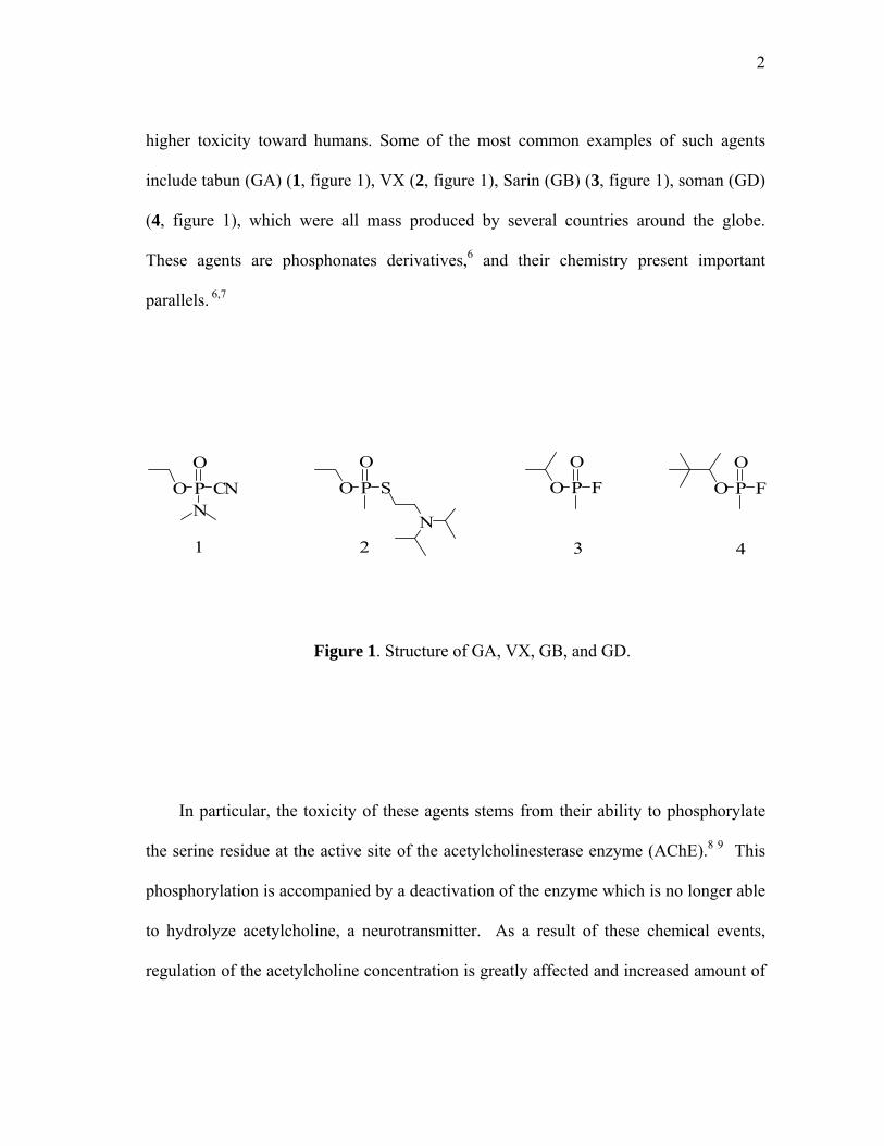
higher toxicity toward humans. Some of the most common examples of such agents
include tabun (GA) (1, figure 1), VX (2, figure 1), Sarin (GB) (3, figure 1), soman (GD)
(4, figure 1), which were all mass produced by several countries around the globe.
These agents are phosphonates derivatives,6 and their chemistry present important
parallels. 6,7
P
2 3 41
NO
OCN PO
OS
N
POO
F POO
F
Figure 1. Structure of GA, VX, GB, and GD.
In particular, the toxicity of these agents stems from their ability to phosphorylate
the serine residue at the active site of the acetylcholinesterase enzyme (AChE).8 9 This
phosphorylation is accompanied by a deactivation of the enzyme which is no longer able
to hydrolyze acetylcholine, a neurotransmitter. As a result of these chemical events,
regulation of the acetylcholine concentration is greatly affected and increased amount of
3
this neurotransmitter are present in the synapses. This triggers motor responses
including spasms which, depending on the nature of the organ affected, can have lethal
consequences. Following the decree of the chemical weapons convention treaty in 1997,
The United States has started to destroy stockpiles of these agents. Despite intense
research during the second half of the last century, the decontamination and
demilitarization of organophosphorus nerve agents is still, unfortunately a critical
problem.10 Moreover, these chemical agents have been used by terrorist groups and
continue to pose a threat to civilian populations. Hence efforts toward the development
of decontamination techniques are fully justified and have become the prime objective of
diverse research groups.11
1.2 General decontamination procedures
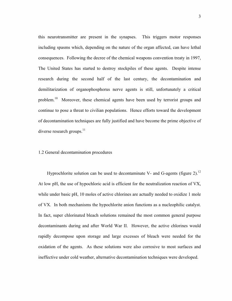
Hyprochlorite solution can be used to decontaminate V- and G-agents (figure 2).12
At low pH, the use of hypochloric acid is efficient for the neutralization reaction of VX,
while under basic pH, 10 moles of active chlorines are actually needed to oxidize 1 mole
of VX. In both mechanisms the hypochlorite anion functions as a nucleophilic catalyst.
In fact, super chlorinated bleach solutions remained the most common general purpose
decontaminants during and after World War II. However, the active chlorines would
rapidly decompose upon storage and large excesses of bleach were needed for the
oxidation of the agents. As these solutions were also corrosive to most surfaces and
ineffective under cold weather, alternative decontamination techniques were developed.

4
+ OH- + F-
+
OCl-
HClO
POO
FR POO
OHR
POO
S
N
POO
OHS
N
HOO O
Figure 2. Detoxification of G- agents and VX with Bleach.
These efforts led to the formulation of a new decontamination solution, namely
DS2, which was developed in 1960.13 DS2 is a polar non aqueous solution able to
dissolve any warfare agents. It is a noncorrosive solution, stable upon storage and active
from -26 oC to 52 oC. This solution in composed of 70% of diethylenetriamine, 28% of
ethylene glycol monomethyl ether and 2% of sodium hydroxide. The reactive
compound in this solution is in fact the conjugated base of ethylene glycol monomethyl
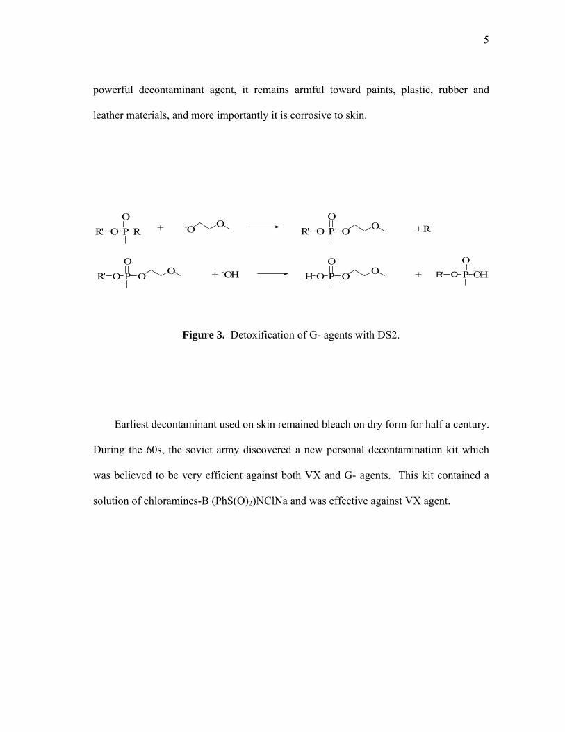
which vigorously reacts with organophosphonate chemical warfare agents (figure 3).
This reaction leads to the formation of a non-toxic phosphonate diester bearing a
monomethyl ethylene glycoxy group and which further reacts with hydroxide ions to
form the corresponding phosphonate monoesters.14 While DS2 has shown to be a
5
powerful decontaminant agent, it remains armful toward paints, plastic, rubber and
leather materials, and more importantly it is corrosive to skin.
P
+ R-+
+ -OH +
POO
RR' -O O
POO
R' O O
POO
R' O O
POO
H O OO
OHOR'
Figure 3. Detoxification of G- agents with DS2.
Earliest decontaminant used on skin remained bleach on dry form for half a century.
During the 60s, the soviet army discovered a new personal decontamination kit which
was believed to be very efficient against both VX and G- agents. This kit contained a
solution of chloramines-B (PhS(O)2)NClNa and was effective against VX agent.
6
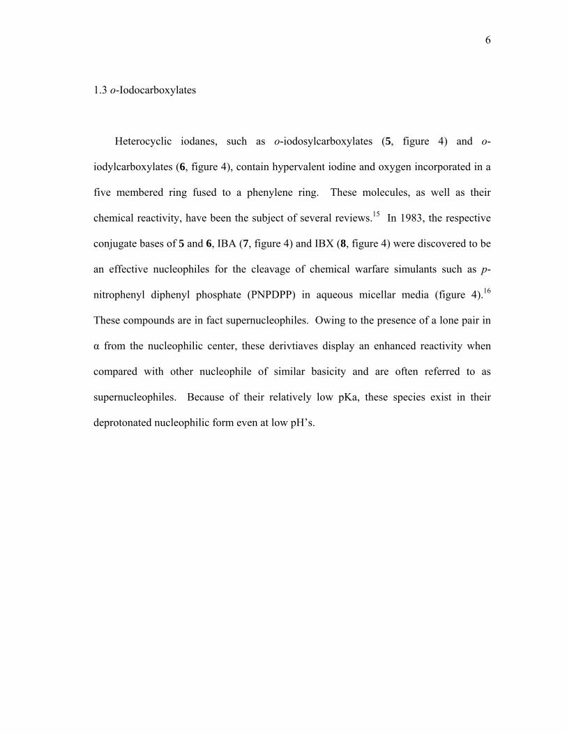
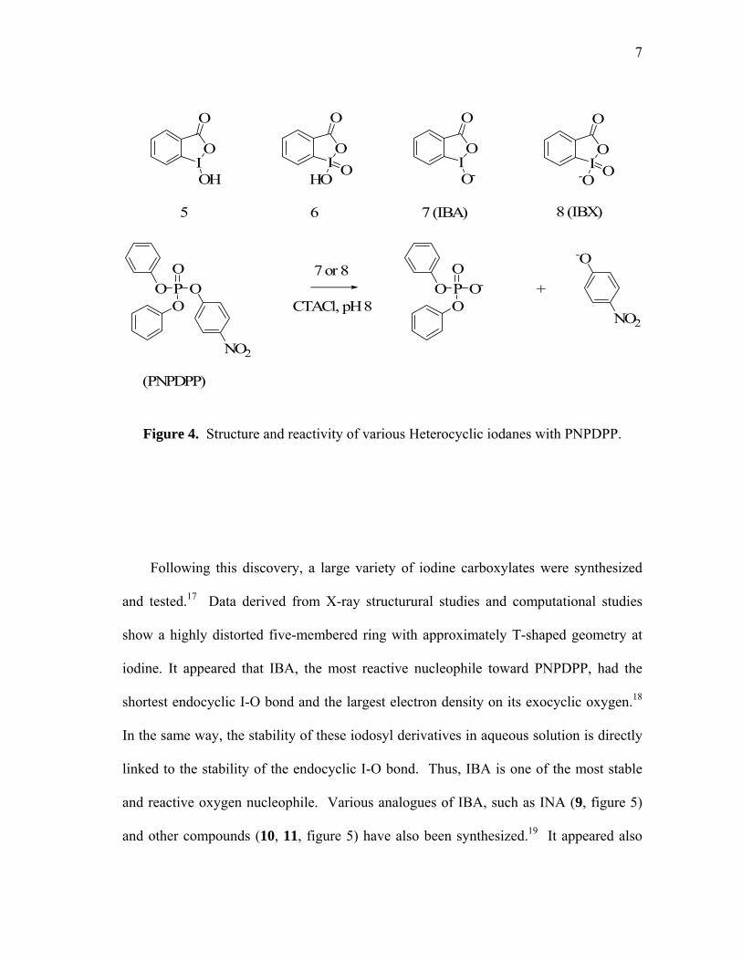
1.3 o-Iodocarboxylates
Heterocyclic iodanes, such as o-iodosylcarboxylates (5, figure 4) and o-
iodylcarboxylates (6, figure 4), contain hypervalent iodine and oxygen incorporated in a
five membered ring fused to a phenylene ring. These molecules, as well as their
chemical reactivity, have been the subject of several reviews.15 In 1983, the respective
conjugate bases of 5 and 6, IBA (7, figure 4) and IBX (8, figure 4) were discovered to be
an effective nucleophiles for the cleavage of chemical warfare simulants such as p-
nitrophenyl diphenyl phosphate (PNPDPP) in aqueous micellar media (figure 4).16
These compounds are in fact supernucleophiles. Owing to the presence of a lone pair in
α from the nucleophilic center, these derivtiaves display an enhanced reactivity when
compared with other nucleophile of similar basicity and are often referred to as
supernucleophiles. Because of their relatively low pKa, these species exist in their
deprotonated nucleophilic form even at low pH’s.
7
IO
O
OHIO
O
IO
O
O-IO
O
HO O -O O
5 6 7 (IBA) 8 (IBX)
7 or 8
CTACl, pH 8+
(PNPDPP)
PO
OOO
NO2
PO
O-OO
-O
NO2
Figure 4. Structure and reactivity of various Heterocyclic iodanes with PNPDPP.
Following this discovery, a large variety of iodine carboxylates were synthesized
and tested.17 Data derived from X-ray structurural studies and computational studies
show a highly distorted five-membered ring with approximately T-shaped geometry at
iodine. It appeared that IBA, the most reactive nucleophile toward PNPDPP, had the
shortest endocyclic I-O bond and the largest electron density on its exocyclic oxygen.18
In the same way, the stability of these iodosyl derivatives in aqueous solution is directly
linked to the stability of the endocyclic I-O bond. Thus, IBA is one of the most stable
and reactive oxygen nucleophile. Various analogues of IBA, such as INA (9, figure 5)
and other compounds (10, 11, figure 5) have also been synthesized.19 It appeared also

8
that 9 is 4 times more reactive than IBA. As the typical reaction is done in presence of
surfactants, the significant rate enhancement was explained by the additional aromatic
ring which increases the hydrophobicity and binding to micellar system where the
reaction occurs. 10 and 11 show an even greater nucleophilic reactivity. This was
suggested by the additional steric interaction between the 8-peri-H atoms and the
adjacent oxygen (10) and carbonyl (11) which where relieved by a shortened endocyclic
I-O bond leading to an enhanced negative charge and nucleophilicity at the O-.
9 10 11
IO
O
O-
H OIO
-OH
IO
O-
O
Figure 5. Structure of INA and various analogues.
Reactivity in association with surfactants has been thoroughly studied during the
past decades.20 It is now widely accepted that the rate enhancements observed in such
media are largely the result of increased concentrations of reactants in the small

9
interfacial volumes in which the reactions are believed to occur. This theory can be fully
revealed if the concentration of the reactants is kept constant, and the surfactant
concentration gradually increased (figure 6).21
Figure 6. Cleavage of 10-5 M parathion by 10-4 M IBA (■) or INA (○) as a function of
cetyltrimethylammonium chloride (CTACl) (M) at pH 8.0 and 25°C.

10
The rate constant versus cationic surfactant goes through a maximum which is the
balance point between the positive addition of reactive interfacial volumes and the
negative dilution of the nucleophile.
Finally, the incorporation of heterocyclic iodanes in cationic surfactants, such as the
duplex-IBA catalyst (12, figure 7), and solid supports, such as the silica-bound IBA (13,
figure 7),22 permitted another catalytic enhancement of the nucleophilic cleavage of
PNPDPP by a further increase of the local concentration of the nucleophilic iodane
moities at the micellar interface where the reaction occurs.
SiO
O
O
Si
Si
Si
12 13
IO
O
O-
N+R
2
IO
O
O-
ON+
Figure 7. Structure of duplex-IBA and silica-bound IBA.

11
1.4 Related nucleophiles
1.4.1 α effect nucleophiles
Other supernucleophiles have also been successfully used as nucleophilic catalysts
for the hydrolysis of organophosphorus esters. Just like iodine carboxylates, PNPDPP
hydrolysis is also largely enhanced by the addition of oximate anions such as
benzaldoximes (14, figure 8) and pyridinealdoxime (15, figure 8) in presence of the
cationic surfactants (CTACL).23 These oximes posses a rather high pKa (9-12) which
leads to a poor catalytic activity at neutral or slightly basic pH. However, pyridinium-
oxime derivatives, such as 2-PAM (16, figure 8) show an effective enhancement of their
catalytic activities at a lower pKa.24 The incorporation of amphiphilic oximes in cationic
surfactants (17, figure 8) also lead to the efficient phosphorylic cleavage of PNPDPP
with high turnover.25
Figure 8. Structure of various oximes.
14 15 16 17
N OH
O2N
NN OH
NN OH
+N+
n-C16H33 NOHBr-
12
Peroxyanions such as monoperoxyphthalate (18, figure 9) and hydroperoxide also
display an enhanced nucleophilic reactivity compared to other nucleophiles with the
same basicity due to α effect (increase nucleophilicity provoked by the proximity of an
unshared pair of electrons on an atom adjacent to the nucleophilic site). The
dephosphorylation of PNPDPP,26 and more importantly of VX,27 with HO2- in cationic
micelles displayed a very good reactivity. As the pKa of H2O2 is greater than 11, the use
of this nucleophile requires extreme conditions. Monoperoxyphthalate (pKa ≈ 8),
however, has shown a slower reactivity in cationic micelles for the hydrolysis of
PNPDPP as well as paraoxon (19, figure 9) and parathion (20, figure 9).28
1.4.2 Alkoxydes
Nucleophilic cleavage of PNPDPP by simple nucleophiles such as alkoxides is also
effective.29 The larger problem to overcome is the use of strongly basic non aqueous
solvents as the pKa of these alcohols are usually as high as 15. However, surfactants
with long alkyl chain terminated by hydroxyethylammonium (21, 22, figure 9) with
polyols as cosolvents were shown to efficiently cleave PNPDPP under mild conditions.30

13
18 19 (X=O Paraoxon)20 (X=S Parathion)
21 (R=H)22 (R=CH3)
O
OHO
OOH
PX
OO O
NO2
n-C14H29NR
OH
Figure 9. Structure of 18-22.
1.5 Enzyme catalyzed hydrolysis and mechanisms
Approximately 30 years ago, two types of enzymes were found to be capable of the
catalytic hydrolysis of organophosphotriesters. These bacterial phosphotriesterases
(PTE) isolated from, Pseudomonas diminuta and Flavobacterium have the ability to
hydrolyse a broad range of organophosphate pesticides and warfare agents with
extremely high turnover.31 In fact, organophosphorus warfare agent tolerance is directly
correlated to the concentration levels of PTE that can be found in various organisms.
For example, mammals, with high level of PTE, are more tolerant to organophosphorus
acetylcholine inhibitors than birds and insect lacking PTE.32

14
During the past decade the organophosphorus hydrolase (OPH), from Pseudomonas
diminuta, has received much attention.33 More specifically, it is able to hydrolyse most
warfare agents including 2, 3, 4, 19, and 20. OPH is also the only known enzyme able to
hydrolyse thiophosphorus esters at a significant rate.34 The native enzyme is known to
be composed of a zinc subunit. X-ray analyses of OPH were initiated in 1994 with the
first unintentional crystal structure of the Cd (II) substituted enzyme.35 Indeed, it was
immediately obvious, contrary to previous reports,36 that the active site was a dinuclear
Cd (II) moiety containing a bridging hydroxide. With the molecular model of the Cd (II)
substituted OPH, the next achievement was the growth of crystals of the active Zn
(II)/Zn (II) enzyme (figure 10).37 As shown by a crystal structure, this enzyme possesses
a deeply buried and unsymmetrical dinuclear active site in which the two metals are
bridged by a carbamylated lysine as well as a hydroxide. One of the zinc centers
coordinates to two histidines as well as an aspartic acid and features a more hindered
coordination sphere, while the other coordinates to the two other histidines and a
solvated hydroxide. The metal centers are separated by 3.3 Ǻ and the metal ligand bond
distances range from 1.9 to 2.3 Ǻ.
15
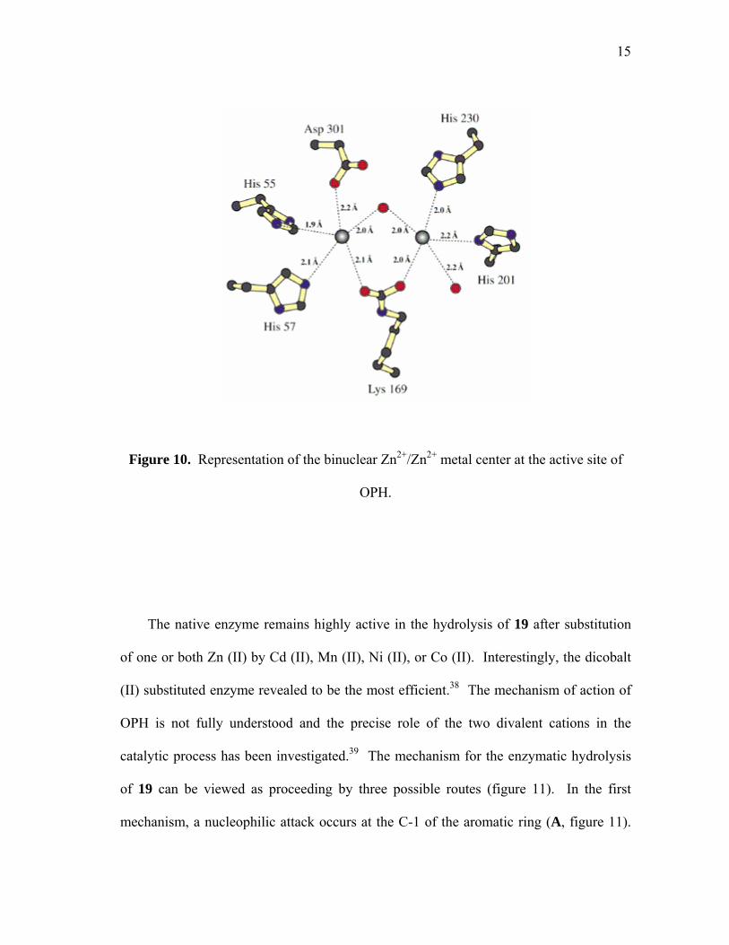
Figure 10. Representation of the binuclear Zn2+/Zn2+ metal center at the active site of
OPH.
The native enzyme remains highly active in the hydrolysis of 19 after substitution
of one or both Zn (II) by Cd (II), Mn (II), Ni (II), or Co (II). Interestingly, the dicobalt
(II) substituted enzyme revealed to be the most efficient.38 The mechanism of action of
OPH is not fully understood and the precise role of the two divalent cations in the
catalytic process has been investigated.39 The mechanism for the enzymatic hydrolysis
of 19 can be viewed as proceeding by three possible routes (figure 11). In the first
mechanism, a nucleophilic attack occurs at the C-1 of the aromatic ring (A, figure 11).

16
Eventhough this mechanism appears to be less likely to happen, the removal of the
intermediate stabilizing NO2 substituent diminished the overall reactivity by 5 orders of
magnitude.
OPO
OO
N+
-OO
-OHOPO
OO
N+
-OO-
OH
OPO
OO
N+
-OO
:X-Enzyme
OPO
XOEnzyme
-OH
OPO
OO
N+
-OO
-OH
OPO
OHO
-O
N+
-OO
+
A
B
C
Figure 11. Cartoon of the possible enzymatic hydrolysis mechanisms.

17
The other two mechanisms can be described by a Sn2 mechanism in which the water
molecule attacks the phosphoryl center and displaces the leaving group. The second
mechanism proposes the attack of a side chain of the protein, followed by another Sn2 by
water at the phosphorus regenerating the free enzyme (B, figure 11). This overall
mechanism results in a retention of configuration at the phosphorus.
The third mechanism involves a single displacement by a hydroxide molecule at the
phosphorus yielding the phosphoric acid product in a single step with an inversion of
configuration at the phosphorus center (C, figure 11). As OPH hydrolytically cleaves
the insecticides (S)-EPN (23) to produce (S)-O-ethyl phenylphosphonothioic acid (24)
with inversion of configuration, the later single Sn2 mechanism C was strongly
suggested.39
The replacement of any of the donating histidines leads to a dramatic 400-5000 fold
drop in organophosphorus triester hydrolysis, showing the importance of the use of both
metal center in the catalytic cycle.40 On the basis of these studies, two mechanisms have
been proposed involving either an intramolecular nucleophilic attack of the bridged
hydroxide onto the phosphorus center (D, figure 12), or the nucleophilic attack of a
solvated hydroxide to the organophosphorus bridging adduct (E, figure 12).

18
PX
YR
HisHis
LysHis
His
O
Zn
O
Zn
O
O H
Asp
HisHis
O
ZnOH
OAsp
LysHis
HisZn
P
O
XR
Y
ED
Figure 12. Proposed mechanisms for the hydrolysis of warfare agents at the active site
of OPH.
At this time, there is only few example of bidentate coordination of
organophosphorus triester to metal centers,41,42 while there are numerous example of η1-
coordinated phosphate triester. Diisopropylmethyl-phosphonate was found to coordinate
to a single Zn metal center of OPH.43
1.6 Organometallic catalysis and mechanisms
Hydrolysis of organophosphorus triesters has also been achieved using “green”
catalysts such as transition metal cations and their complexes.44 Much of the interest
was generated by the efficient use of Cu (II) cation for the degradation of the G- agent
stimulant diisopropyl phosphorofluoridate (23, figure 13) and the hydrolysis of

19
thiophosphoester such as 19.45 Other early available examples indicated that the divalent
Hg (II) cation constituted as well a rather potent candidate accelerating the hydrolysis
rate of 19 by a magnitude of 2 to 3.46 The use of lanthanide metal cation Ln (III) has
also been investigated. As the stability of highly charged metal cations in aqueous media
is always a concern, macrocyclic (25, figure 14)47 and cryptate (26, figure 14)48
lanthanides were prepared and tested with effective turnover for the hydrolytic
decomposition of 2,4-dinitrophenyl diethyl phosphate (24, figure 13) and PNPDPP
respectively.
PX
OO O
NO223 24
NO2PX
OO F
Figure 13. Structure of diisopropyl phosphorofluoridate and 2,4-dinitrophenyl diethyl
phosphate.
20
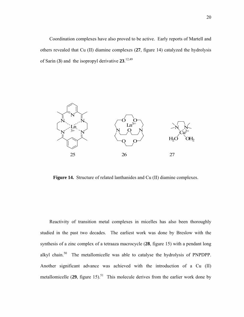
Coordination complexes have also proved to be active. Early reports of Martell and
others revealed that Cu (II) diamine complexes (27, figure 14) catalyzed the hydrolysis
of Sarin (3) and the isopropyl derivative 23.12,49
25 26 27
N
NN
N
NLn3+
OO
NON
OO
Ln3+N
Cu2+N
OH2H2O
Figure 14. Structure of related lanthanides and Cu (II) diamine complexes.
Reactivity of transition metal complexes in micelles has also been thoroughly
studied in the past two decades. The earliest work was done by Breslow with the
synthesis of a zinc complex of a tetraaza macrocycle (28, figure 15) with a pendant long
alkyl chain.50 The metallomicelle was able to catalyse the hydrolysis of PNPDPP.
Another significant advance was achieved with the introduction of a Cu (II)
metallomicelle (29, figure 15).51 This molecule derives from the earlier work done by
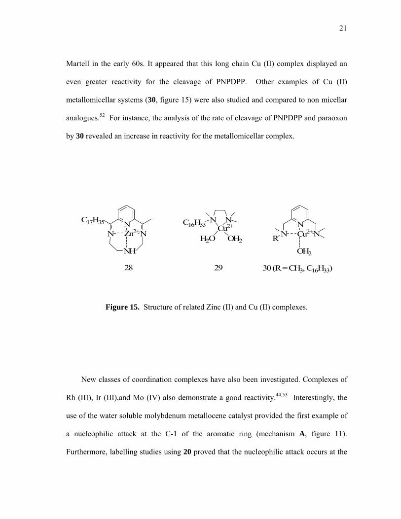
21
Martell in the early 60s. It appeared that this long chain Cu (II) complex displayed an
even greater reactivity for the cleavage of PNPDPP. Other examples of Cu (II)
metallomicellar systems (30, figure 15) were also studied and compared to non micellar
analogues.52 For instance, the analysis of the rate of cleavage of PNPDPP and paraoxon
by 30 revealed an increase in reactivity for the metallomicellar complex.
OH2
30 (R = CH3, C16H33)2928
Cu2+N
NR N
NCu2+
NC16H33
OH2H2ONH
Zn2+N
N N
C17H35
Figure 15. Structure of related Zinc (II) and Cu (II) complexes.
New classes of coordination complexes have also been investigated. Complexes of
Rh (III), Ir (III),and Mo (IV) also demonstrate a good reactivity.44,53 Interestingly, the
use of the water soluble molybdenum metallocene catalyst provided the first example of
a nucleophilic attack at the C-1 of the aromatic ring (mechanism A, figure 11).
Furthermore, labelling studies using 20 proved that the nucleophilic attack occurs at the

22
α-carbon of the organothiophosphate and not at the phosphorus center. It is also
important to note that several minerals including alumina, titania, goethite and clays can
surface catalyse the hydrolysis of thiophosphate triesters.
Another interesting nonmicellar approach to nucleophilic decomposition of
organophosphorus triesters employs the group 8 metal complexes (31, 32, figure 16).
Recently, Ryabov et all introduced Pd (II) and Pt (II) orthometalated aryl oxime
complexes ([MCl{C,N-(C6H4C(CH3) NOH)-2}(L)], M = Pd or Pt, L = DMSO or
pyridine) for the cleavage of thiophosphate triesters.54 It could be argued that palladium
and platinum are too soft Lewis acids, and that the square-planar configuration of the
metal centers which has never been reported in the enzymatic system is less likely to
demonstrate any decent catalytic activity. Nevertheless, Pd and Pt electrophilicity
coupled to the nucleophilicity of the coordinated oximate anion, provided an outstanding
reactivity toward thiophosphate triester hydrolysis such as 20 at pH 8.5. Although no
reaction intermediates have been isolated, it has been proposed that the activity of such
catalysts results from the coordination of parathion at the metal center followed by an
intramolecular nucleophilic attack by the neighboring oximate (figure 17).

23
31 32
N OHPd N OHPt
O
ClO
SClPy
Figure 16. Structure of related Pd (II) and Pt(II) orthometalated aryl oximes.
With a catalytic amount of the Pt (II) complex, first-order kinetic hydrolysis of 20
follows. The k2 were also measured for all of the above catalysts and compared to the
hydroxide catalyzed hydrolysis. It appeared that the catalytic activity of the Pd (II) and
Pt (II) complexes at a concentration as low as 10-4 M revealed an activity 106-107 times
higher than the hydroxide, representing by far the most efficient biomimetic system ever
encountered.
The proposed mechanism of the hydrolysis is depicted in F (figure 17). Initial
coordination to the metal activates the thioester toward intramolecular nucleophilic
attack of the oximate to the phosphorus center, providing rate enhancement of 109 fold
over the hydroxide catalyzed reaction at pH 8. Moreover, rapid decomposition of the
phosphorylated oxime intermediate must account for the observed turnover. In such
mechanism the role of Pd (II) and Pt (II) is double. Not only they increase the positive

24
charge on the phosphorus, but the added coordination to the oxime lowers its pKa
enhancing their nucleophilicity at neutral pH.
- HOPS(OR)2
- HOAr20F
N O-M
OH2
X
R
NO-MS
X
R
PRO OOR
NO2
NO
M
OH2
X
R
P SRO OR
Figure 17. Proposed mechanisms for the hydrolysis of parathion with Pd (II) and Pt (II)
complexes.
1.7 Objectives
As the lack of current “green” decontamination process still remains, recent
researches have been focused on the study of a large variety of methods for the
degradation of these compounds. Significant advances have been made with the use of

25
organic nucleophiles. Many iodosylcarboxylate derivatives have been prepared and
shown interesting catalytic properties for the hydrolysis of G- type agents. However,
these compounds are not active in the hydrolysis of V type agents as well as
thiophosphorus esters.
As of today, OPH remains the most active catalyst for the hydrolysis of
organophosphorus triesters remain the most efficient catalysts. Although, a large variety
of G- type warfare agents as well as V-types are efficiently decomposed by OPH, the
mass production of the enzyme remains a struggling problem. The active site has been
shown to consist of a unique binuclear metal center delivering a hydroxide for a Sn2
attack onto the phosphorus center.
Many research groups have also investigated a broad range of mononuclear
coordination and organometallics complexes as catalysts. These catalysts have been
shown to be highly efficient for the hydrolysis of the G- type agents. Furthermore, as
the molecular weight of these synthetic catalysts is very low compared to OPH, their
production remains very competitive even if used at higher loading. However, these
complexes have shown a lesser affinity for the cleavage of harder oxygen donor
pesticides compared to the thiono derivatives. Also, no synthetic complexes have been
proven suitable for the catalytic decontamination of the V- type nerve agents. Finally, it
is important to note that so far, very few dinuclear complexes that would mimic the OPH
active site have been investigated. 55

26
Based on the above, it has become the objective of this work to attempt the
preparation of dinuclear complexes that mimic the active site of PTE. In order to
progress toward this goal, the following tasks have been identified:
To synthesize chiral dinucleating ligands with pyridazine as a central core.
To synthesize dinucleating ligands with 1,4-(iminomethyl)benzene as central core.
To study the ligative behavior of the ligands toward various metal cations.
To study the interaction of the dinuclear metal complexes with phosphorus triesters.
To test the catalytic activity of the complexes in organophosphate ester hydrolysis.
27
CHAPTER II
EXPERIMENTAL PROCEDURES
2.1 General considerations
Methylparathion is highly toxic and should be handled in a well-ventilated fume
hood. All glassware exposed to methylparathion should be decontaminated with bleach.
All anhydrous reactions were carried out under nitrogen atmosphere in oven-dried (130
°C) glassware by using Schlenk and glovebox techniques unless noted otherwise. The
nitrogen source was passed over P2O5 immediately prior to entering any glassware. For
the handling and the use or synthesis of air-sensitive compounds, solvents and deuterated
solvents were freshly distilled prior to use. Tetrahydrofuran (THF) was freshly distilled
over NA/K amalgam. Acetonitrile (MeCN), chlorophorm (CHCl3) dichloromethane
(CH2Cl2), diethylether (Et2O), hexane, pentane, pyridine, and triethylamine (TEA) were
distilled from calcium hydride (CaH2) prior to use. Benzene (PhH), and toluene (PhMe)
over K. Methanol, Ethanol, and Ethyl acetate were used without purification. The
methylparathion solution was provided by A/S CHEMINOVA, LEMVIG, DK-7620,
Denmark as a gift. 2 were freshly prepared using literature protocols. 3,6-
distyrylpyridazine, O-TBDMS-(1R,2S)-(-)-norephedrine, O-TBDMS-(1R,2S)-(-)-
norephedrine, 3,6-pyridazinedicarboxaldehyde, O-benzyl-(1R,2S)-(-)-norephedrine, 2,3-
dimethoxy-benzene-1,4-dicarbaldehyse derivatives, (1R,2S) (-)-2-(N-norephedrinyl-
methyl)pyridine, (1S-2R)-1-[(pyridin-2-ylmethyl)-amino]-indan-2-ol, [2-(2-pyridyl)-

28
phenyl-C,N]palladium(II) acetate was prepared following the published synthesis.56
Other chemicals were commercially available and were purchased from Aldrich, or
VWR and used without purification.
2.2 Spectroscopy
Reactions were followed by thin layer chromatography (TLC) on EMD chemicals
silica gel 60 and aluminum oxide 60 F254 (250 µm thick). TLC's were visualized with a
UV lamp and stained using vanillin. Purifications of compounds were carried out by
flash chromatography using Natland silica gel (100-230 mesh). All NMR studies were
carried out on an Inova 300 and 500 MHz NMR spectrometer (300 and 500 MHz for 1H,
100.5 and 125.7 MHz for 13C, 121.4 MHz for 31P NMR). 85% H3PO4 was used as an
external standard for the solution 31P NMR spectra. The proton and carbon signals of
the deuterated solvent were used as internal standard for the 1H and 13C NMR spectra,
respectively. Solvents are indicated for each compound. 1H NMR coupling constants
(J) are reported on Hertz (Hz) and multiplicity is abbreviated as follows: app = apparent,
s = singlet, d = doublet, dd = doublet of doublets, t = triplet, q = quartet, m = multiplet,
br = broad signal. IR spectra were acquired using an ATI Mattson genesis series
spectrometer in the solvent indicated. Vibration frequencies are expressed in cm-1.
Elemental analyses were performed by Atlantic Microlab Inc. at Norcross, GA 30091.
Melting points were measured on a Laboratory Devices Mel-Temp apparatus and were
not corrected. All UV/Vis absorption spectra and spectrophotometric measurements

29
were recorded on a JASCO V530 UV/Vis spectrometer equipped with an automatic cell
changer. Single-crystal X-ray analysis for 2 was collected on a bruker SMART-CCD
diffractometer using graphite-monochromated Mo Kα radiation (=0.710 73 Ǻ).
Specimen of suitable size and quality was selected and mounted onto a glass fiber with
Apiezon grease. The structure was solved by direct methods, which successfully located
most of the non-hydrogen atoms. Subsequent refinement on F2 using the SHELXTL/PC
package (version 5.1) allowed location of the remaining non-hydrogen atoms.

30
CHAPTER III
LIGAND DESIGN
3.1 Introduction
Inspired by the role of dinuclear metalloenzymes in several biological processes,
recent developments in the area of Lewis acid catalysis have been concerned with the
study of bifunctional catalysts that mimic the functions of these enzymes.57 An
impressive collection of dinuclear complexes has been prepared and their catalytic
properties tested.58,59,60,61,62,63,64,65 While dinuclear metalloenzymes fulfill diverse
functions,66 a large proportion of the biomimetic studies focus on hydrolase catalysts
similar to those encountered in phosphoesterase.67 Structural analyses supported by
kinetic studies indicate that the catalytic properties of these complexes result from
synergistic effects that occur at the proximal metal centers of these derivatives.
The knowledge gathered from these studies has served to fuel a growing number of
investigations in the field of organic reaction catalysis. A large majority of the
complexes investigated so far utilize main group elements or early transition metals at
the active site of the catalyst. The group of Maruoka has been especially industrious in
this area and has described a series of achiral complexes that utilize 1,8-
biphenylenedioxy and 1,8-anthraquinonedioxy ligands in combination with hard metal
cations such as aluminum (III) and titanium (IV) (33, 34, figure 18).68 These complexes
are effective catalysts in a number of reactions that involve carbonyl substrates such as

31
the Mukayama aldol condensation. Supported by NMR studies, the activity of these
catalysts has been proposed to result from the double coordination of the carbonyl
oxygen atom of the substrate to the two juxtaposed metallic centers.69 This proposal,
which remains largely unsupported and lends itself to some debate, has been advanced to
rationalize the higher reaction rate observed when bifunctional rather than
monofunctional catalysts are used. Parallel studies carried out on thiocarbonyl
derivatives by Hawthorne suggest that analogous phenomena occur with mercury-based
polyfunctional Lewis acid catalysts (35, figure 18).61 While these studies are likely to
shine some light on the mechanisms that are at play in bifunctional Lewis acid catalysis,
the absence of chirality in the complexes will undoubtedly limit the scope of their use. It
is therefore logical that a number of chiral dinuclear complexes have now been
investigated for the asymmetric inductions of organic reactions.
C
C Me
Me
C
C
Me
Me
C C
Me Me
Hg Hg
HgO
O OOTi Ti (OiPr)3(iPrO)3
MeMeO O
O
R1 R2
Al Al(OiPr)2(OiPr)2
O
LALA
Drawing depicting the multipleactivation of a carbonyl.
33 34 35
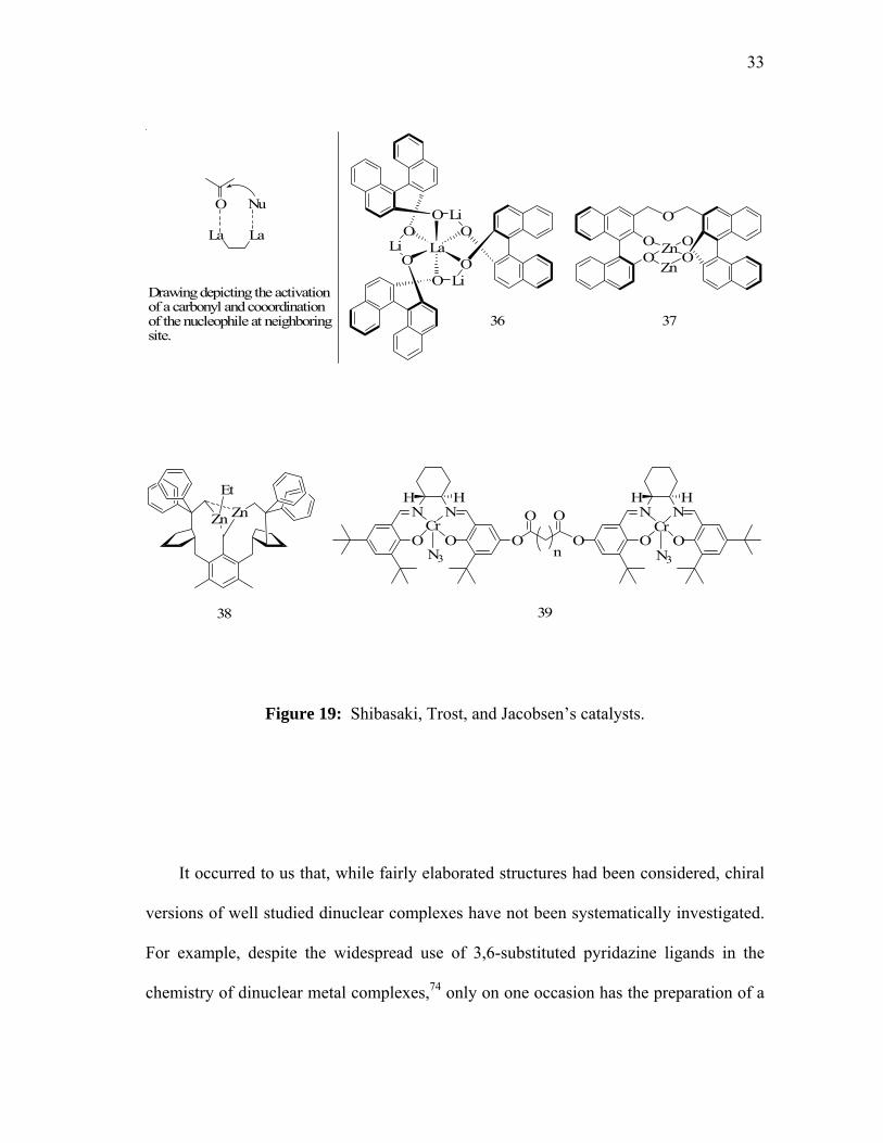
Figure 18: Maruoka, and Hawthorne’s catalysts.

32
A series of catalysts that utilize the 1,1’-(S)-binaphthol ligands to bridge two metal
centers (36, 37, figure 19) have been described by the group of Shibasaki who observed
unusually high enantiomeric excesses in various reactions including Michael addition
and nitro-aldol condensation.70 The results have been rationalized in terms of a
mechanism that involves the activation of the electrophilic substrate at one metal center
while the remaining metal center coordinates and transfers the nucleophile. Such
phenomena present direct analogies with mechanisms independently proposed by Corey
and Noyori earlier on.58,71 More recently, catalysts that function on the basis of the same
principles have been reported by Trost who demonstrated the use of chiral dinuclear zinc
catalysts of type 38 for enantioselective aldol reactions (figure 19).72 One of the clearest
examples for the occurrence of two-center catalysis has been obtained by the group of
Jacobsen who carry out a study on flexible dinculear Schiff base Cr(III) complexes 39 as
catalysts for the ring opening reactions of epoxides (figure 19).73 In particular,
compelling kinetic evidence has been obtained for the cooperation of the two metal
centers in the reaction of oxiranes with various nucleophiles including water and azide.
33
La
O
OO
O
O
OLi
Li
Li
O
OO Zn O
OZn
O
LaLa
Nu
Drawing depicting the activationof a carbonyl and cooordinationof the nucleophile at neighboringsite.
36 37
NH H
N
O O OO O
N NH H
On
Cr Cr
N3 N3
O OZn ZnEt
38 39
Figure 19: Shibasaki, Trost, and Jacobsen’s catalysts.
It occurred to us that, while fairly elaborated structures had been considered, chiral
versions of well studied dinuclear complexes have not been systematically investigated.
For example, despite the widespread use of 3,6-substituted pyridazine ligands in the
chemistry of dinuclear metal complexes,74 only on one occasion has the preparation of a

34
chiral example of such a ligand been reported (40, figure 20).75 Thus, it became the
objective of this work to prepare chiral dinucleating ligands derived from structurally
simple motifs and investigate the scope of their utility in phosphonate triester hydrolysis.
N N
O
N N
O
PhPh40
Figure 20: Pfaltz’s binuclear ligand design.
3.2 Dinuclear pyridazine based ligands
3.2.1 Ligands derived from 3,6-dihydroxymethylpyridazine
In order to insure the monomeric nature of the transition metal complexes, the
preparation of ligands able to present at least three basic sites to each metal center should
be prepared. For this reason, the introduction of aminoalcohol pendant arms at the
methyl positions of a 3,6-dimethylpyridazine was selected as a synthetic target. As part

35
of this work, we revisited the synthesis of 3,6-bis(hydroxymethyl)pyridazine (41, figure
21) which had been previously prepared in less than 25% yield starting from 2,5-
bis(hydroxymethyl)furan.76 Ozone was bubbled through a solution of 3,6-
distyrylpyridazine at -78ºC in methanol until the blue color of ozone appeared. Excess
of ozone was then removed by degassing, and an excess of sodium borohydride (3
equivalents) was added. The reaction mixture was warmed to ambient temperature
allowing the isolation of 41 as a white powder in 75% yield after chromatography.
N N N N
OOO O O
O
N N
OOO O O
O
N NHO OH
O3, MeOH
NaBH4, MeOH
-78oC to 25oC41
Figure 21. Synthesis of 3,6-di(hydroxymethyl)pyridazine.
36
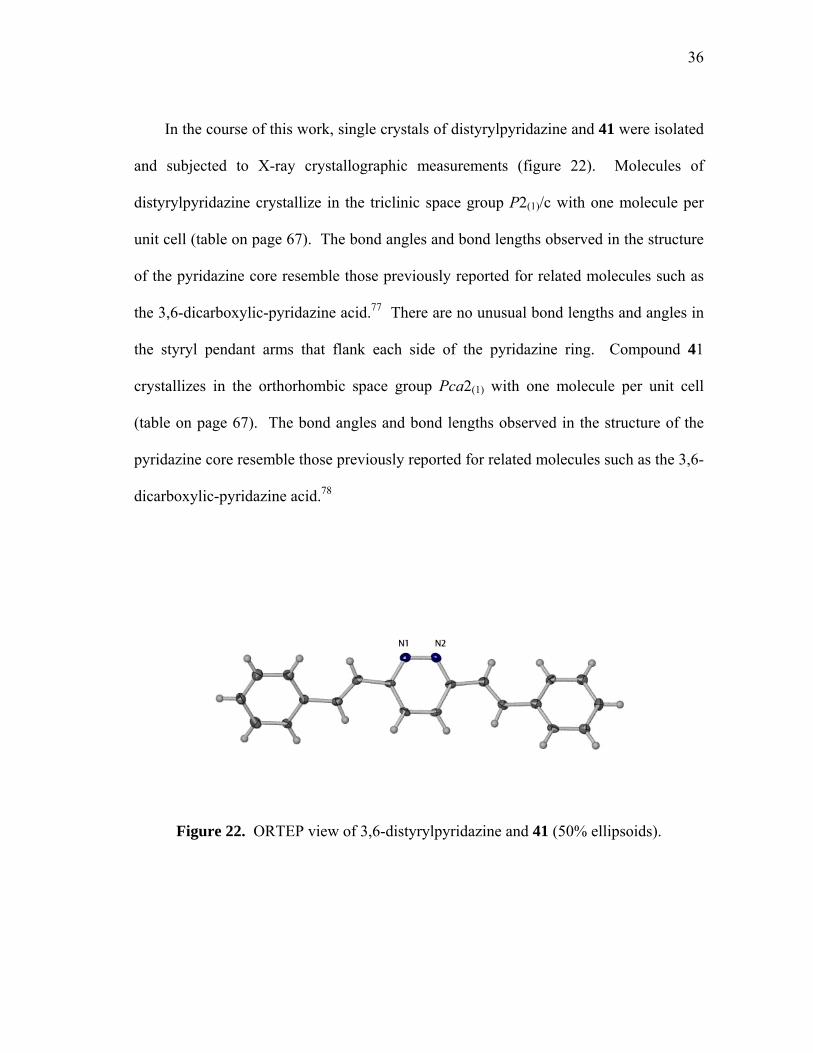
In the course of this work, single crystals of distyrylpyridazine and 41 were isolated
and subjected to X-ray crystallographic measurements (figure 22). Molecules of
distyrylpyridazine crystallize in the triclinic space group P2(1)/c with one molecule per
unit cell (table on page 67). The bond angles and bond lengths observed in the structure
of the pyridazine core resemble those previously reported for related molecules such as
the 3,6-dicarboxylic-pyridazine acid.77 There are no unusual bond lengths and angles in
the styryl pendant arms that flank each side of the pyridazine ring. Compound 41
crystallizes in the orthorhombic space group Pca2(1) with one molecule per unit cell
(table on page 67). The bond angles and bond lengths observed in the structure of the
pyridazine core resemble those previously reported for related molecules such as the 3,6-
dicarboxylic-pyridazine acid.78
Figure 22. ORTEP view of 3,6-distyrylpyridazine and 41 (50% ellipsoids).
37
Figure 22. Continued.
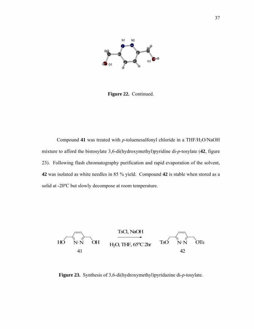
Compound 41 was treated with p-toluenesulfonyl chloride in a THF/H2O/NaOH
mixture to afford the bistosylate 3,6-di(hydroxymethyl)pyridine di-p-tosylate (42, figure
23). Following flash chromatography purification and rapid evaporation of the solvent,
42 was isolated as white needles in 85 % yield. Compound 42 is stable when stored as a
solid at -20ºC but slowly decompose at room temperature.
N NHO OH
TsCl, NaOH
H2O, THF, 65oC 2hr N NTsO OTs
4241
Figure 23. Synthesis of 3,6-di(hydroxymethyl)pyridazine di-p-tosylate.
38
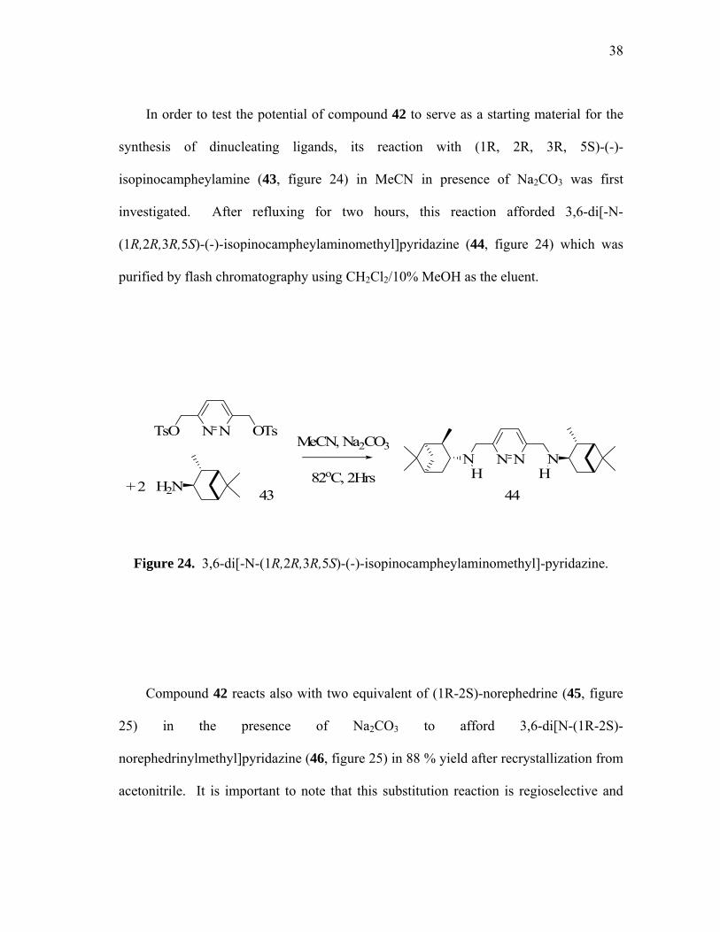
In order to test the potential of compound 42 to serve as a starting material for the
synthesis of dinucleating ligands, its reaction with (1R, 2R, 3R, 5S)-(-)-
isopinocampheylamine (43, figure 24) in MeCN in presence of Na2CO3 was first
investigated. After refluxing for two hours, this reaction afforded 3,6-di[-N-
(1R,2R,3R,5S)-(-)-isopinocampheylaminomethyl]pyridazine (44, figure 24) which was
purified by flash chromatography using CH2Cl2/10% MeOH as the eluent.
N NTsO OTsMeCN, Na2CO3
82oC, 2HrsN NN N
H HH2N+ 2 43 44
Figure 24. 3,6-di[-N-(1R,2R,3R,5S)-(-)-isopinocampheylaminomethyl]-pyridazine.
Compound 42 reacts also with two equivalent of (1R-2S)-norephedrine (45, figure
25) in the presence of Na2CO3 to afford 3,6-di[N-(1R-2S)-
norephedrinylmethyl]pyridazine (46, figure 25) in 88 % yield after recrystallization from
acetonitrile. It is important to note that this substitution reaction is regioselective and

39
that no O-substituted products or oligomers could be observed in the reaction mixture.
In a similar fashion, 42 reacts with (1S-2R)-1-amino-2-indanol (47, figure 25) to afford
3,6-di[-N-(1R-2S)-1-amino-2-indanolylmethyl]-pyridazine (48, figure 25) in 76 % yield.
As in the case of 44 and 46, the identity of 48 has been characterized by EA, 1H NMR,
13C NMR and FAB mass spectrometry.
N NTsO OTs
MeCN, Na2CO382oC, 2Hrs
N NNH HN N NNH HNMe
OHPh
Me
HOPh
OH HO46 48
NH2
Me
OHPh
H2N
HO
45 47
Figure 25. Synthesis of 3,6-di[N-(1R-2S)-norephedrinylmethyl]pyridazine and 3,6-di[-
N-(1R-2S)-1-amino-2-indanolylmethyl]pyridazine.
40
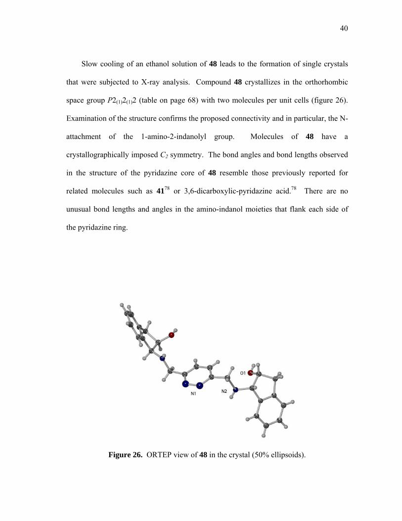
Slow cooling of an ethanol solution of 48 leads to the formation of single crystals
that were subjected to X-ray analysis. Compound 48 crystallizes in the orthorhombic
space group P2(1)2(1)2 (table on page 68) with two molecules per unit cells (figure 26).
Examination of the structure confirms the proposed connectivity and in particular, the N-
attachment of the 1-amino-2-indanolyl group. Molecules of 48 have a
crystallographically imposed C2 symmetry. The bond angles and bond lengths observed
in the structure of the pyridazine core of 48 resemble those previously reported for
related molecules such as 4178 or 3,6-dicarboxylic-pyridazine acid.78 There are no
unusual bond lengths and angles in the amino-indanol moieties that flank each side of
the pyridazine ring.
Figure 26. ORTEP view of 48 in the crystal (50% ellipsoids).
41
In order to generalize the synthesis of such dinucleating ligands, the reactions of 42
with various aminoalcohol reagents were investigated. These investigations lead to the
observation that 42 reacts smoothly with O-TBDMS-(1R,2S)-(-)-norephedrine (49,
figure 27), O-TBDMS-(1R,2S)-(-)-norephedrine (50, figure 27 ), (1R,2S)-(-)-ephedrine
(51, figure 27), and L-alanine methyl ester (52, figure 27), respectively, to afford a
variety of C2 symmetrical dinucleating ligands in high yield (53-56, figure 27). All of
these derivatives have been characterized by EA, 1H NMR, 13C NMR and FAB mass
spectrometry. Furthermore, molecules of 55 have a crystallographically imposed C2
symmetry (figure 28) (table on page 68). The bond angles and bond lengths observed in
the structure of the pyridazine core resemble the bis(hydroxymethyl)pyridazine and
those previously reported for related molecules such as 3,6-dicarboxylic-pyridazine
acid.78 Finally, there are no unusual bond lengths and angles in the ephedrine moieties
that flank each side of the pyridazine ring.
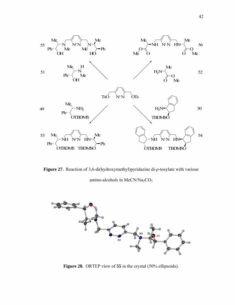
42
N NTsO OTs
N NNH HN N NNH HNMe
OTBDMSPh
Me
TBDMSOPh
OTBDMS TBDMSO
49 50
N NN NMe
OHPh
Me
HOPhMe Me
N NNH HNMe
OO
Me
OOMeMe
HN
Me
OHPh Me
NH2
Me
OTBDMSPh
H2N
TBDMSO
H2NMe
OOMe
51 52
53 54
55 56
Figure 27. Reaction of 3,6-di(hydroxymethyl)pyridazine di-p-tosylate with various
amino-alcohols in MeCN/Na2CO3
Figure 28. ORTEP view of 55 in the crystal (50% ellipsoids).

43
Compound 42 reacts also with bulky secondary amines such as 2-[N-
(1R,2R,3R,5S)-(-)-isopinocampheylaminomethyl]pyridine (57, figure 29) leading to the
formation of 58 (figure 29) also in high yield.
N NTsO OTs MeCN, Na2CO3
82oC, 2Hrs
N NN N
N N
+ 2 NH
N57 58
Figure 29. Reaction of 3,6-di(hydroxymethyl)pyridazine di-p-tosylate with 2-[N-
(1R,2R,3R,5S)-(-)-isopinocampheylaminomethyl]pyridine.
3.2.2 Dinuclear bis Schiff base ligands
In parallel to the investigations described in the above section, the preparation of
Schiff base ligands by condensation reactions of various aminoalcohols with 3,6-
pyridazinedicarboxaldehyde79 (59, figure 30) has also been investigated. The reaction of
59 with 45 proceeded at 65ºC in methanol to afford after work-up a low yield of the C2
symmetrical dinucleating ligand ((1R,2S)-(2-hydroxy-1-methyl-2-phenylethyl)-
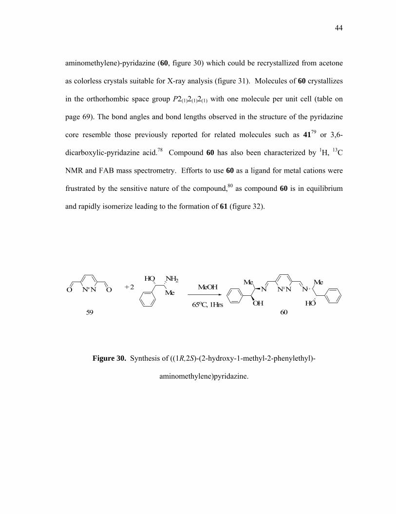
44
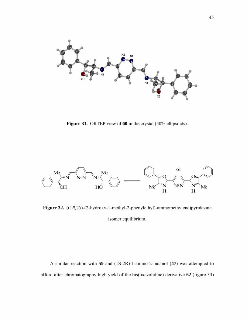
aminomethylene)-pyridazine (60, figure 30) which could be recrystallized from acetone
as colorless crystals suitable for X-ray analysis (figure 31). Molecules of 60 crystallizes
in the orthorhombic space group P2(1)2(1)2(1) with one molecule per unit cell (table on
page 69). The bond angles and bond lengths observed in the structure of the pyridazine
core resemble those previously reported for related molecules such as 4179 or 3,6-
dicarboxylic-pyridazine acid.78 Compound 60 has also been characterized by 1H, 13C
NMR and FAB mass spectrometry. Efforts to use 60 as a ligand for metal cations were
frustrated by the sensitive nature of the compound,80 as compound 60 is in equilibrium
and rapidly isomerize leading to the formation of 61 (figure 32).
N NO O MeOH
65oC, 1Hrs
N NN N+ 2
60
HO NH2
Me
59
Me
HO
Me
OH
Figure 30. Synthesis of ((1R,2S)-(2-hydroxy-1-methyl-2-phenylethyl)-
aminomethylene)pyridazine.
45
Figure 31. ORTEP view of 60 in the crystal (50% ellipsoids).
N NN NMe
HO
Me
OH N N
O
N N
O
Me MeH H
61
Figure 32. ((1R,2S)-(2-hydroxy-1-methyl-2-phenylethyl)-aminomethylene)pyridazine
isomer equilibrium.
A similar reaction with 59 and (1S-2R)-1-amino-2-indanol (47) was attempted to
afford after chromatography high yield of the bis(oxazolidine) derivative 62 (figure 33)
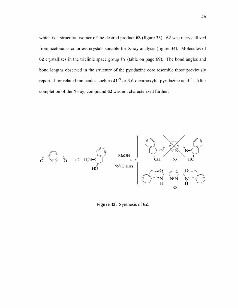
46
which is a structural isomer of the desired product 63 (figure 33). 62 was recrystallized
from acetone as colorless crystals suitable for X-ray analysis (figure 34). Molecules of
62 crystallizes in the triclinic space group P1 (table on page 69). The bond angles and
bond lengths observed in the structure of the pyridazine core resemble those previously
reported for related molecules such as 4179 or 3,6-dicarboxylic-pyridazine acid.78 After
completion of the X-ray, compound 62 was not characterized further.
N NO OMeOH
65oC, 1Hrs+ 2
N NN N
OH HOH2N
HO
N N
O
N N
O
H H62
63
Figure 33. Synthesis of 62.
47
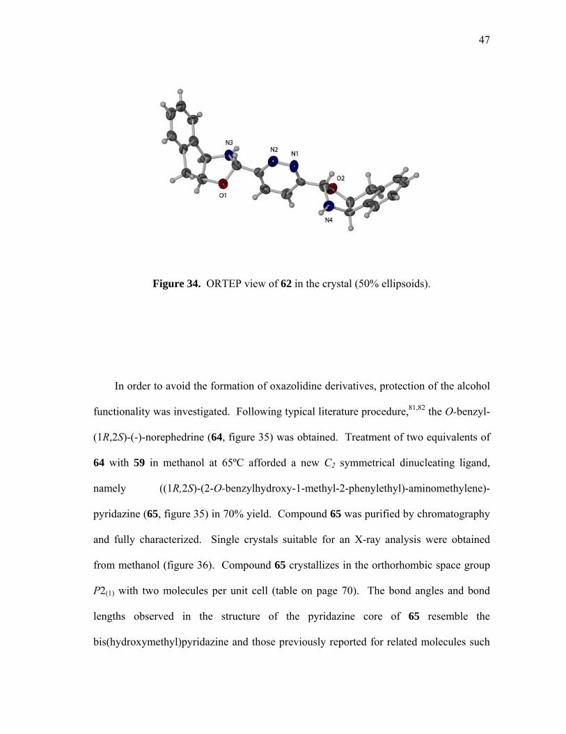
Figure 34. ORTEP view of 62 in the crystal (50% ellipsoids).
In order to avoid the formation of oxazolidine derivatives, protection of the alcohol
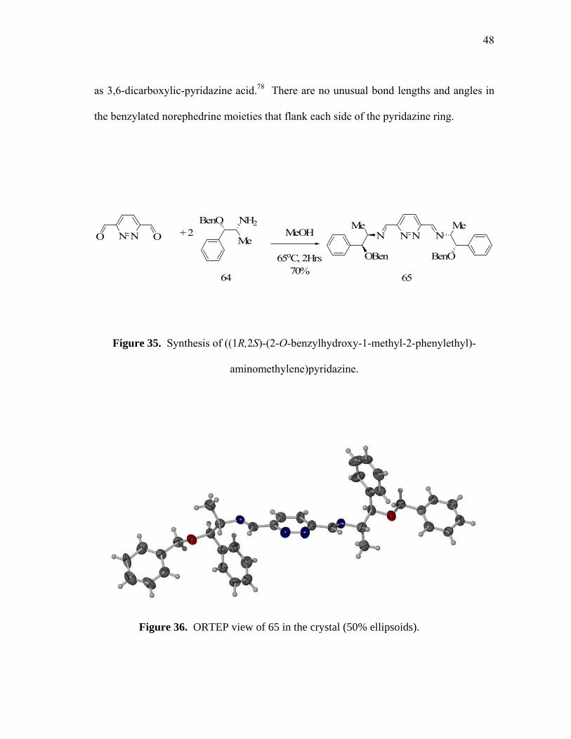
functionality was investigated. Following typical literature procedure,81,82 the O-benzyl-
(1R,2S)-(-)-norephedrine (64, figure 35) was obtained. Treatment of two equivalents of
64 with 59 in methanol at 65ºC afforded a new C2 symmetrical dinucleating ligand,
namely ((1R,2S)-(2-O-benzylhydroxy-1-methyl-2-phenylethyl)-aminomethylene)-
pyridazine (65, figure 35) in 70% yield. Compound 65 was purified by chromatography
and fully characterized. Single crystals suitable for an X-ray analysis were obtained
from methanol (figure 36). Compound 65 crystallizes in the orthorhombic space group
P2(1) with two molecules per unit cell (table on page 70). The bond angles and bond
lengths observed in the structure of the pyridazine core of 65 resemble the
bis(hydroxymethyl)pyridazine and those previously reported for related molecules such
48
as 3,6-dicarboxylic-pyridazine acid.78 There are no unusual bond lengths and angles in
the benzylated norephedrine moieties that flank each side of the pyridazine ring.
N NO O MeOH
65oC, 2Hrs70%
N NN N+ 2
65
BenO NH2
MeMe
BenO
Me
OBen
64
Figure 35. Synthesis of ((1R,2S)-(2-O-benzylhydroxy-1-methyl-2-phenylethyl)-
aminomethylene)pyridazine.
Figure 36. ORTEP view of 65 in the crystal (50% ellipsoids).
49
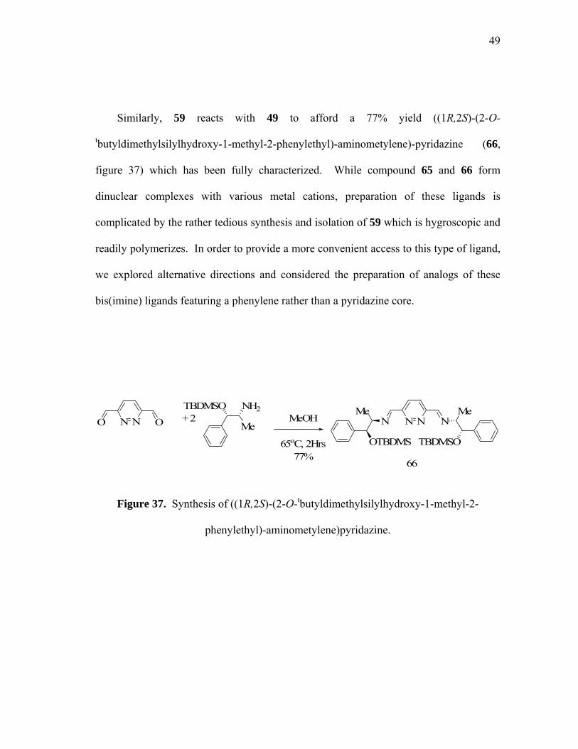
Similarly, 59 reacts with 49 to afford a 77% yield ((1R,2S)-(2-O-
tbutyldimethylsilylhydroxy-1-methyl-2-phenylethyl)-aminometylene)-pyridazine (66,
figure 37) which has been fully characterized. While compound 65 and 66 form
dinuclear complexes with various metal cations, preparation of these ligands is
complicated by the rather tedious synthesis and isolation of 59 which is hygroscopic and
readily polymerizes. In order to provide a more convenient access to this type of ligand,
we explored alternative directions and considered the preparation of analogs of these
bis(imine) ligands featuring a phenylene rather than a pyridazine core.
N NO O MeOH
65oC, 2Hrs77%
N NN N+ 2
66
TBDMSO NH2
MeMe
TBDMSO
Me
OTBDMS
Figure 37. Synthesis of ((1R,2S)-(2-O-tbutyldimethylsilylhydroxy-1-methyl-2-
phenylethyl)-aminometylene)pyridazine.

50
3.3 Dinuclear (iminomethyl)benzene ligands.
The ligands described in the above sections are well adapted to the formation of
dinuclear coordination complexes. In order to broaden the scope of our investigations,
we decided to investigate dinucleating ligands specifically designed for the preparation
of dinuclear organometallic complexes (Figure 38).
NN DD DD
D = donor group, M = metal moiety
NN DD DDM MMM
coordination complex organometallic complex
Figure 38: Dinuclear organometallic design.
2,3-dimethoxy-benzene-1,4-dicarbaldehyde derivative (67) was chosen as a starting
material. This choice was guided by the fact that this derivative should smoothly react
with secondary amines to afford a new type of dinucleating bis(Schiff base) ligands.
Secondly, the presence of two adjacent methoxy groups should only allow for the 1,2-
51
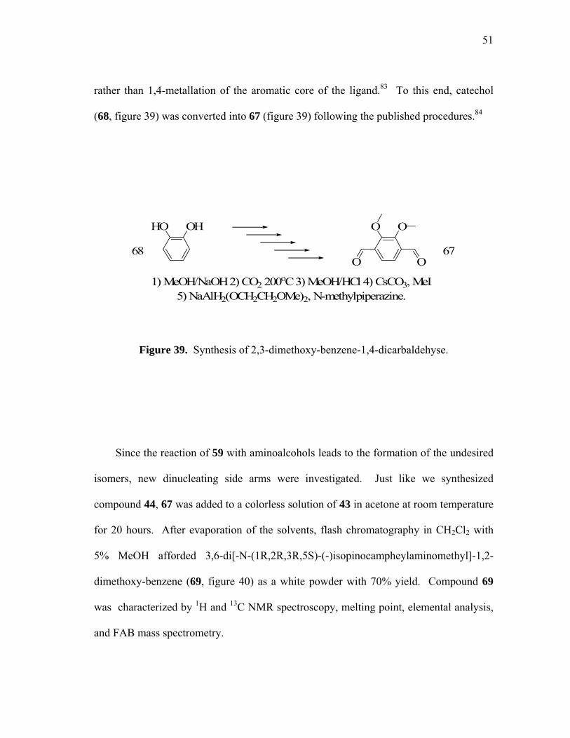
rather than 1,4-metallation of the aromatic core of the ligand.83 To this end, catechol
(68, figure 39) was converted into 67 (figure 39) following the published procedures.84
OHHO OO
O O
1) MeOH/NaOH 2) CO2 200oC 3) MeOH/HCl 4) CsCO3, MeI 5) NaAlH2(OCH2CH2OMe)2, N-methylpiperazine.
68 67
Figure 39. Synthesis of 2,3-dimethoxy-benzene-1,4-dicarbaldehyse.
Since the reaction of 59 with aminoalcohols leads to the formation of the undesired
isomers, new dinucleating side arms were investigated. Just like we synthesized
compound 44, 67 was added to a colorless solution of 43 in acetone at room temperature
for 20 hours. After evaporation of the solvents, flash chromatography in CH2Cl2 with
5% MeOH afforded 3,6-di[-N-(1R,2R,3R,5S)-(-)isopinocampheylaminomethyl]-1,2-
dimethoxy-benzene (69, figure 40) as a white powder with 70% yield. Compound 69
was characterized by 1H and 13C NMR spectroscopy, melting point, elemental analysis,
and FAB mass spectrometry.

52
OO
OO Acetone
RT, 20HrsN N
H2N+ 2 69
O O
Figure 40. Synthesis of 3,6-di[-N-(1R,2R,3R,5S)-(-)isopinocampheylaminomethyl]-
1,2-dimethoxy-benzene.
In a similar fashion, compound 67 can react easily with two equivalents of 2,4,6-
trimethylaniline (70, figure 41) in acetone to afford colorless crystals of 3,6-di[-N-(2,4,6-
trimethylphenyliminomethyl)]-1,2-dimethoxy-benzene (71, figure 41). This compound
was purified by recrystallization from pentane at -20oC in 85% yield. 71 has been
characterized by 1H and 13C NMR spectroscopy, melting point, elemental analysis, and
FAB mass spectrometry. In addition, its crystal structure has been determined.
Compound 71 crystallizes in the monoclinic space group P2(1)/n (table on page 70) with
one molecule per unit cell (figure 42). Examination of the structure confirms the
proposed connectivity as well as the presence of two imine functionalities. All bond
lengths and angles fall in the normal range and do not deserve further comments. We
can also notice that compared to the structure of the distyrylpyridazine, the mesityl
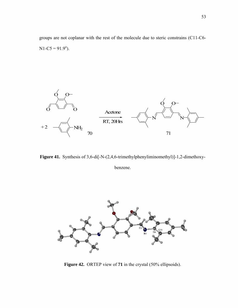
53
groups are not coplanar with the rest of the molecule due to steric constrains (C11-C6-
N1-C5 = 91.9o).
OO
OO Acetone
RT, 20HrsN N
+ 271
O O
NH270
Figure 41. Synthesis of 3,6-di[-N-(2,4,6-trimethylphenyliminomethyl)]-1,2-dimethoxy-
benzene.
Figure 42. ORTEP view of 71 in the crystal (50% ellipsoids).
54
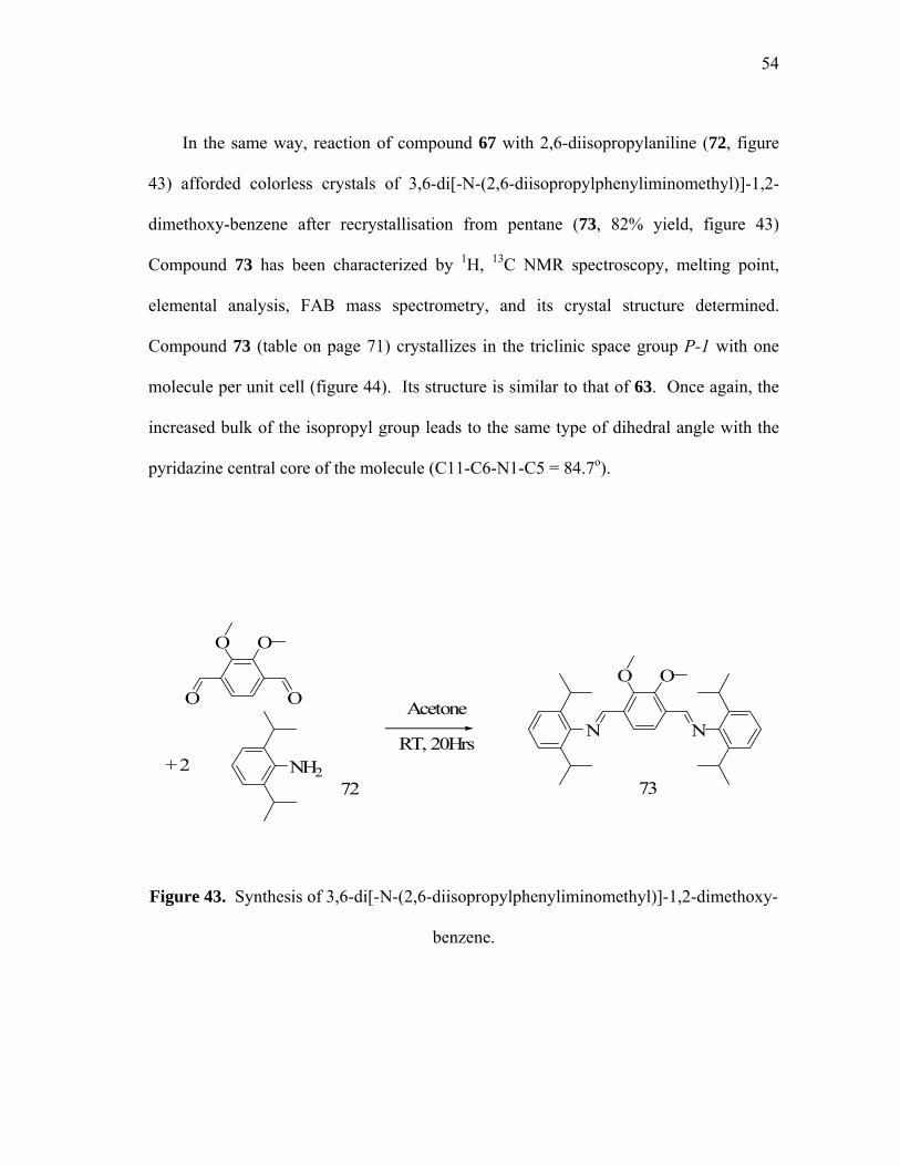
In the same way, reaction of compound 67 with 2,6-diisopropylaniline (72, figure
43) afforded colorless crystals of 3,6-di[-N-(2,6-diisopropylphenyliminomethyl)]-1,2-
dimethoxy-benzene after recrystallisation from pentane (73, 82% yield, figure 43)
Compound 73 has been characterized by 1H, 13C NMR spectroscopy, melting point,
elemental analysis, FAB mass spectrometry, and its crystal structure determined.
Compound 73 (table on page 71) crystallizes in the triclinic space group P-1 with one
molecule per unit cell (figure 44). Its structure is similar to that of 63. Once again, the
increased bulk of the isopropyl group leads to the same type of dihedral angle with the
pyridazine central core of the molecule (C11-C6-N1-C5 = 84.7o).
OO
OO Acetone
RT, 20HrsN N
+ 273
O O
NH272
Figure 43. Synthesis of 3,6-di[-N-(2,6-diisopropylphenyliminomethyl)]-1,2-dimethoxy-
benzene.

55
Figure 44. ORTEP view of 73 in the crystal (50% ellipsoids).
The t-butyl derivative 74 (figure 45) was prepared by reaction of 59 with t-
butylamine in methanol (75, figure 45). It was obtained in a 95% yield and did not
necessitate further purifications. Its identity has been confirmed by 1H and 13C NMR
spectroscopy, elemental analysis, and FAB mass spectrometry.

56
OO
OO MeOH, Na2CO3
RT, 20HrsN N
+ 274
O O
NH275
Figure 45. Synthesis of 3,6-di[-N-t-butyliminomethyl]-1,2-dimethoxy-benzene
3.4 Summary
New symmetric and asymmetric dinucleating ligands, revealed itself to be a success
as 15 new ligands have been successfully prepared by condensation reaction of primary,
secondary amines, as well as amino-alcohols to 3,6-Di(hydroxymethyl)Pyridazine di-p-
tosylate, 2,3-dimethoxy-benzene-1,4-dicarbaldehyde, and 3,6-
pyridazinedicarboxaldehyde. These methods allowed the rapid generation of a library of
enantiomerically pure dinucleating ligands.
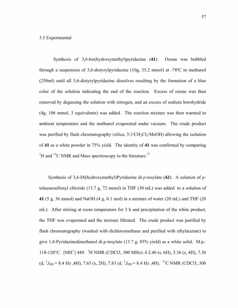
57
3.5 Experimental
Synthesis of 3,6-bis(hydroxymethyl)pyridazine (41): Ozone was bubbled
through a suspension of 3,6-distyrylpyridazine (10g, 35.2 mmol) at -78ºC in methanol
(250ml) until all 3,6-distyrylpyridazine dissolves resulting by the formation of a blue
color of the solution indicating the end of the reaction. Excess of ozone was then
removed by degassing the solution with nitrogen, and an excess of sodium borohydride
(4g, 106 mmol, 3 equivalents) was added. The reaction mixture was then warmed to
ambient temperature and the methanol evaporated under vacuum. The crude product
was purified by flash chromatography (silica, 3:1/CH2Cl2/MeOH) allowing the isolation
of 41 as a white powder in 75% yield. The identity of 41 was confirmed by comparing
1H and 13C NMR and Mass spectroscopy to the literature.77
Synthesis of 3,6-Di(hydroxymethyl)Pyridazine di-p-tosylate (42): A solution of p-
toluenesulfonyl chloride (13.7 g, 72 mmol) in THF (30 mL) was added to a solution of
41 (5 g, 36 mmol) and NaOH (4 g, 0.1 mol) in a mixture of water (20 mL) and THF (20
mL). After stirring at room temperature for 3 h and precipitation of the white product,
the THF was evaporated and the mixture filtrated. The crude product was purified by
flash chromatography (washed with dichloromethane and purified with ethylacetate) to
give 1,4-Pyridazinedimethanol di-p-tosylate (13.7 g, 85% yield) as a white solid. M.p.:
118-120°C. [MH+] 449. 1H NMR (CDCl3, 300 MHz): δ 2.46 (s, 6H), 5.36 (s, 4H), 7.36
(d, 3JHH = 8.4 Hz ,4H), 7.65 (s, 2H), 7.83 (d, 3JHH = 8.4 Hz ,4H). 13C NMR (CDCl3, 300
58
MHz): δ 21.8, 69.8, 126.2, 128.3, 130.3, 132.7, 145.7, 157.1. Elemental analysis calc.
(%) for C20H20N2O6S2: C 53.56, H 4.49, N 6.25; found: C 53.56, H 4.79, N 6.32.
Synthesis of 3,6-di[-N-(1R,2R,3R,5S)-(-)-isopinocampheylaminomethyl]-pyridazine
(44): A solution of 43 (1.1 g, 7.2 mmol) in acetonitrile was added to a solution of 42
(1.6 g, 3.6 mmol) and Na2CO3 (2.65g, 25 mmol) in acetonitrile. After reflux at 82oC for
2 h, the residue was filtrated and the solvent evaporated. The final product was purified
by flash chromatography (silica, 9:1/CH2Cl2/MeOH) as light brown oil (1.08 g, 73 %
yield). 1H NMR (CDCl3, 300 MHz): δ 0.94 (s, 6H), 1.11 (d, 3JHH = 7.5 Hz, 6H), 1.22 (s,
6H), 1.70 (m, 2H), 1.74 (m, 2H) 1.86 (m, 4H), 2.39 (m, 4H), 2.96 (m, 2H), 4.10 (d, 2JHH
= 15 Hz, 2H), 4.17 (d, 2JHH = 15 Hz, 2H), 7.57 (s, 2H) . 13C NMR (CDCl3, 300 MHz): δ
21.7, 28.0, 33.0, 36.6, 41.9, 45.3, 48.1, 51.5, 56.7, 126.8, 161.0. Elemental analysis calc.
(%) for C26H42N4: C 76.05, H 10.31, N 13.64; found: C 76.2, H 9.9, N 13.4.
Synthesis of 3,6-Di[-N-(1R-2S)-norephedrinylmethyl]pyridazine (46): A solution
of 45 (3.7 g, 25 mmol) in acetonitrile was added to a solution of 42 (5.6 g, 12.5mmol)
and Na2CO3 (5g, 50 mmol) in acetonitrile. After stirring at room temperature for 3 h, the
residue was filtrated and the solvent evaporated. The final product was purified by
recrystallization from CH3CN as a white solid (4.5 g, 88 % yield). M.p. 152-154°C.
[MH+] 407. 1H NMR (CDCl3, 300 MHz): δ 0.93 (d, 3JHH = 6.6 Hz, 6H), 3.03 (qd, 3JHH =
6.6 Hz 3JHH = 3.6 Hz, 2H), 4.17 (d, 2JHH = 14.7 Hz, 2H), 4.25 (d, 2JHH = 14.7 Hz, 2H),
4.81 (d, 3JHH = 3.6 Hz, 2H), 7.31 (m, 12H), 7.50 (s, 2H) . 13C NMR (CDCl3, 300 MHz):

59
δ 14.8, 51.0, 58.7, 73.8, 126.3, 126.7, 127.4, 128.3, 140.8, 160.7. Elemental analysis
calc. (%) for C24H30N4O2: C 70.91, H 7.44, N 13.78; found: C 70.5, H 7.7, N 13.4.
Synthesis of 3,6-Di[-N-(1R-2S)-1-amino-2-indanolylmethyl]pyridazine (48): The
same procedure as described for the preparation of ligand 46 was followed except that
47 (3.7 g, 25 mmol) was used. The final product was purified by recrystallization from
CH3CN as a white solid (3.8 g, 76 % yield). Colorless crystals suitable for X-ray
analysis were grown by recrystallization in ethanol at room temperature. M.p. 164-
166°C. [MH+] 403. 1H NMR (CDCl3, 300 MHz): δ 2.99 (dd, 3JHH = 3 Hz 2JHH = 13.5
Hz, 2H), 3.07 (dd, 3JHH = 4.8 Hz 2JHH = 13.5 Hz, 2H), 4.16 (d, 3JHH = 5.1 Hz, 2H), 4.30
(d, 2JHH = 15 Hz, 2H), 4.37 (d, 2JHH = 15 Hz, 2H), 4.45 (m, 2H), 7.27 (m, 6H), 7.38 (m,
2H), 7.58 (s, 2H). 13C NMR (pyridine-d6, 300 MHz): δ 40.5, 52.0, 66.6, 72.0, 125.4,
125.5, 126.6, 126.7, 127.8, 141.7, 144.6, 162.0. Elemental analysis calc. (%) for
C24H26N4O2: C 71.62, H 6.51, N 13.92; found: C 71.18, H 6.53, N 13.79.
Synthesis of 3,6-Di[-N-(1R-2S)-O-(tert-butyldimethylsilyl)-1-amino-2-
indanolylmethyl]pyridazine (53): A solution of 49 (2.23 g, 8.5 mmol) in acetonitrile was
added to a solution of 42 (1.88 g, 4.2 mmol) and Na2CO3 (2.65g, 25 mmol) in
acetonitrile. After reflux at 82oC for 2 h, the residue was filtrated and the solvent
evaporated. The final product was purified by flash chromatography (silica,
9:1/CH2Cl2/MeOH) as light brown oil (1.94 g, 74 % yield). 1H NMR (CDCl3, 300 MHz)
δ -0.17 (s, 6H), 0.05 (s, 6H), 0.90 (s, 18H), 1.05 (d, 3JHH = 3.9 Hz, 6H), 2.77 (m, 4H),

60
4.03 (d, 2JHH = 6.9 Hz, 2H), 4.01 (d, 2JHH = 6.9 Hz, 2H), 4.68 (d, 3JHH = 3 Hz, 2H), 7.28
(m, 10H), 7.35 (s, 2H) . 13C NMR (CDCl3, 300 MHz) δ -4.4, -4.2, 18.3, 27.1, 50.2, 57.8,
74.9, 126.1, 126.3, 127.2, 128.5, 160.3. Elemental analysis calc. (%) for C36H58N4O2Si2:
C 68.09, H 9.21, N 8.82; found: C 68.0, H 9.1, N 8.9.
Synthesis of 3,6-Di[-N-(1R-2S)-O-(tert-butyldimethylsilyl)norephedrinyl-methyl]-
pyridazine (54): A solution of 50 (1.81 g, 6.9 mmol) in acetonitrile was added to a
solution of 42 (1.52 g, 3.4 mmol) and Na2CO3 (2.65g, 25 mmol) in acetonitrile. After
reflux at 82oC for 2 h, the residue was filtrated and the solvent evaporated. The final
product was purified by flash chromatography (silica, 9:1/CH2Cl2/MeOH) as light brown
oil (1.75 g, 82 % yield). 1H NMR (CDCl3, 300 MHz): δ -0.18 (s, 6H), 0.05 (s, 6H), 0.90
(s, 18H), 1.05 (d, 3JHH = 6 Hz, 6H), 2.99 (m, 4H), 3.96 (d, 2JHH = 5.4 Hz, 2H), 4.22 (d,
2JHH = 14.7 Hz, 2H), 4.30 (d, 2JHH = 14.7 Hz, 2H), 4.65 (m, 2H), 7.22 (m, 8H), 7.68 (s,
2H). 13C NMR (CDCl3, 300 MHz): δ -4.5, -4.3, 18.3, 26.1, 40.0, 50.9, 65.1, 75.7, 125.1,
125.3, 126.5, 126.6, 127.9, 140.7, 143.0, 161.5. Elemental analysis calc. (%) for
C36H54N4O2Si2: C 68.52, H 8.63, N 8.88; found: C 68.7, H 8.5, N 8.7.
Synthesis of 3,6-Di[(2S)-ephedrinyl] pyridazine (55): A solution of 51
hydrochloride (1.3 g, 6.5 mmol) in acetonitrile was added to a solution of 42 (1.4 g, 3.2
mmol) and Na2CO3 (2.65g, 25 mmol) in acetonitrile. After reflux at 82oC for 3 h, the
residue was filtrated and the solvent evaporated. The final product was purified by flash
chromatography (silica, 9:1/CH2Cl2/MeOH) as a brown semi-solid (1.08 g, 78 % yield).

61
M.p. 72-75°C. 1H NMR (CDCl3, 300 MHz): δ 1.09 (d, 3JHH = 6.6 Hz, 6H), 2.24 (s, 6H),
2.94 (m, 2H), 3.88 (d, 2JHH = 8.7 Hz, 2H), 3.93 (d, 2JHH = 8.7 Hz, 2H), 4.82 (d, 3JHH = 3.3
Hz, 2H), 7.02 (s, 2H) 7.3 (m, 10H),. 13C NMR (CDCl3, 300 MHz): δ 10.0, 39.0, 58.5,
64.2, 74.8, 126.5, 126.9, 127.4, 128.3, 143.2, 161.1. Elemental analysis calc. (%) for
C26H34N4O2: C 71.86, H 7.89, N 12.89; found: C 71.8, H 7.3, N 12.4.
Synthesis of 3,6-Di[l-Alaninemethylester]pyridazine (56): A solution of 52
hydrochloride (0.91 g, 6.5 mmol) in acetonitrile was added to a solution of 42 (1.55 g,
3.2 mmol) and Na2CO3 (2.65g, 25 mmol) in acetonitrile. After reflux at 82oC for 3 h,
the residue was filtrated and the solvent evaporated. The final product was purified by
flash chromatography (silica, 9:1/CH2Cl2/MeOH) as dark brown oil (0.81g, 82 % yield).
1H NMR (CDCl3, 300 MHz): δ 1.36 (d, 3JHH = 7.2 Hz, 6H), 3.45 (q, 3JHH = 7.2 Hz, 2H),
3.74 (s, 6H), 4.02 (d, 2JHH = 14.4 Hz, 2H), 4.17 (d, 2JHH = 14.4 Hz, 2H), 7.56 (s, 2H) .
13C NMR (CDCl3, 300 MHz): δ 19.2, 51.4, 52.2, 56.3, 126.7, 160.5, 175.8. Elemental
analysis calc. (%) for C14H22N4O4: C 54.18, H 7.15, N 18.05; found: C 53.9, H 7.9, N
17.6.
Synthesis of 3,6-Di[l-Alanine]pyridazine (56’): To a solution of 56 (0.6 g, 2.0
mmol) in methanol was added a 1N solution of NaOH (4ml, 4.0 mmol). After stirring at
25oC for 4h the solvent was evaporated. The final product was purified by flash
chromatography (silica, MeOH) as light brown oil (0.52 g, 92 % yield). 1H NMR (D2O,
300 MHz): δ 1.26 (d, 3JHH = 3.9 Hz, 6H), 3.23 (q, 3JHH = 3.9 Hz, 2H), 3.93 (d, 2JHH = 8.4
62
Hz, 2H), 4.08 (d, 2JHH = 8.4 Hz, 2H), 7.80 (s, 2H) . 13C NMR (D2O, 500 MHz): δ 18.5,
50.4, 58.7, 129.0 (quaternary carbons not detected). Elemental analysis calc. (%) for
C12H18N4O2: C 51.06, H 6.43, N 19.85; found: C 50.7, H 6.5, N 19.5.
Synthesis of 2-[(1R,2R,3R,5S)-(-)-isopinocampheylaminomethyl]-Pyridine (57): To
a solution of 43 (0.8 g, 5.2 mmol) in methanol was added the 2-picolyl chloride (8.5 g,
5.2 mmol). After maintained at Ph 9 by addition of concentrated NaOH solution at 65oC
for 20h, the solvents were evaporated. The final product was purified by flash
chromatography (silica, 19:1/CH2Cl2/MeOH) as light brown oil (1.0 g, 78 % yield). 1H
NMR (CDCl3, 300 MHz): δ 0.95 (s, 3H), 1.07 (d, 3JHH = 8.9 Hz, 3H), 1.18 (s, 3H), 1.89
(m, 1H), 2.05 (m, 4H), 2.36 (m, 2H), 3.26 (m, 1H), 4.09 (s, 2H), 7.11 (m, 1H), 7.47 (m,
1H), 7.60 (s, 1H), 8.46 (m, 1H). 13C NMR (CDCl3, 300 MHz): δ 21.7, 23.6, 28.0, 33.0,
36.8, 38.8, 41.9, 45.3, 48.1, 51.5, 56.7, 126.8, 160.9. Elemental analysis calc. (%) for
C16H24N2: C 78.64, H 9.90, N 11.46; found: C 78.4, H 9.4, N 11.6.
Synthesis of 3,6-Di(hydroxymethyl)Pyridazine di-p-tosylate with 2-[N-
(1R,2R,3R,5S)-(-)-isopinocampheylaminomethyl]pyridine (58): A solution of 57 (1.46
g, 6.0 mmol) in acetonitrile was added to a solution of 42 (1.34 g, 3.0 mmol) and
Na2CO3 (2.65g, 25 mmol) in acetonitrile. After reflux at 82oC for 2 h, the residue was
filtrated and the solvent evaporated. The final product was purified by flash
chromatography (silica, 19:1/CH2Cl2/MeOH) as light brown oil (1.75 g, 65 % yield). 1H
NMR (CDCl3, 300 MHz): δ 0.80 (s, 6H), 0.93 (d, 3JHH = 9.6 Hz, 6H), 1.16 (s, 6H), 1.78
63
(m, 2H), 1.97 (m, 8H), 2.26 (m, 4H), 3.12 (m, 2H), 3.81 (d, 2JHH = 14 Hz, 2H), 3.96 (d,
2JHH = 14 Hz, 2H), 4.05 (s, 2H), 7.11 (m, 2H), 7.47 (m, 2H), 7.60 (s, 2H), 7.61 (m, 2H),
8.46 (m, 2H). 13C NMR (CDCl3, 300 MHz): δ 21.1, 23.4, 27.5, 28.3, 33.8, 39.4, 41.2,
41.8, 48.2, 55.1, 57.2, 59.2, 122.1, 123.1, 127.0, 136.5, 149.1, 160.8, 161.5. Elemental
analysis calc. (%) for C38H52N6: C 76.98 H 8.84, N 14.18; found: C 77.1, H 8.9, N 14.1.
Synthesis of ((1R,2S)-(2-hydroxy-1-methyl-2-phenylethyl)-aminomethylene)-
pyridazine (60): A solution of 45 (0.76 g, 5 mmol) in MeOH was added to a light
yellow solution of 59 (0.35 g, 2.5 mmol) also in MeOH. After stirring for 6 hours at
65oC, the solution was evaporated. The remaining yellow crude product was washed
with ET2O and the final product recrystallized in Acetone leading to the formation of
colorless crystal suitable for X-ray analysis. [MH]+ 403. 1H NMR (CDCl3, 300 MHz): δ
1.14 (d, 3JHH = 6Hz, 6H), 3.56 (dq, 3JHH = 6Hz, 3JHH = 3.3Hz, 2H), 5.04 (d, 3JHH = 3.3Hz,
2H), 7.23-7.32 (m, 6H) 8.04 (s, 2H). 13C NMR (CDCl3, 300 MHz): δ 17.1, 72.7, 78.9,
126.0, 126.8, 127.8, 128.4, 158,5 (Cq non detected). Elemental analysis calc. (%) for
C24H26N4O2: C 71.62 H 6.51, N 13.92; found: C 77.76, H 6.89, N 13.71.
Synthesis of ((1R,2S)-(2-O-benzylhydroxy-1-methyl-2-phenylethyl)-amino-
methylene)pyridazine (65): A solution of 64 (0.76 g, 5 mmol) in MeOH was added to a
light yellow solution of 59 (0.35 g, 2.5 mmol) in H2O. After stirring for 2 hours at 65oC,
the solution was stored in the fridge at -20oC for 10 Hours until white crystal of 55
precipitated. The remaining yellow solution was filtered off, and the final product
64
washed with ethanol. Few colorless crystals were suitable for X-ray analysis. [MH]+
583. 1H NMR (CDCl3, 300 MHz) δ 1.35 (d, 3JHH = 6.6Hz, 6H), 3.60 (dq, 3JHH = 6.6Hz,
3JHH = 6.6Hz, 2H), 4.17 (d, 2JHH = 12Hz, 2H) , 4.36 (d, 3JHH = 6.6Hz, 2H), 4.44 (d, 2JHH =
12Hz, 2H) 6.97-7.25 (m, 20H), 7.7 (s, 2H), 8.32 (s, 2H). Elemental analysis calc. (%)
for C38H38N4O2: C 78.32, H 6.57, N 9.61; found: C 77.96, H 6.46, N 9.44.
Synthesis of ((1R,2S)-(2-O-tbutyldimethylsilylhydroxy-1-methyl-2-
phenylethyl)aminometylene)-pyridazine (66): A solution of 49 (1.09 g, 4.1 mmol) in
methanol (75ml) was added to a solution of 59 (0.28 g, 2 mmol) in water (15ml). After
reflux at 65oC for 2 h, the methanol was evaporated and the oily residue extracted with
CH2Cl2 (3x50 ml). The organic layer was then drying with MgSO4 et evaporated. The
final product was purified by flash chromatography (silica, 9:1/hexane/ethylacetate) as
light brown oil (0.97 g, 77 % yield). 1H NMR (CDCl3, 300 MHz): δ -0.15 (s, 6H), -0.11
(s, 6H), 0.91 (s, 18H), 1.31 (d, 3JHH = 6.3 Hz, 6H), 3.78 (m, 2H), 4.86 (d, 3JHH = 5.2 Hz,
2H), 7.28 (m, 10H), 8.18 (s, 2H), 8.44 (m, 2H). 13C NMR (CDCl3, 300 MHz): δ -4.9, -
4.7, 17.7, 18.2, 25.8, 73.0, 78.5, 124.4, 126.9, 127.2, 127.7, 142.5, 157.5, 158.5.
Elemental analysis calc. (%) for C36H54N4O2Si2: C 68.52, H 8.63, N 8.88; found: C 68.3,
H 8.7, N 8.6.
Synthesis of 3,6-di[-N-(1R,2R,3R,5S)-(-)isopinocampheylaminomethyl]-1,2-
dimethoxy-benzene (69): A solution of 67 (0.97 g, 5 mmol) in acetone was added to a
solution of 43 (1.5 g, 10 mmol) in acetone. After stirring at room temperature for 20

65
hours, the solvent was evaporated. The final product was purified by flash
chromatography (silica, 19:1/CH2Cl2/MeOH) as a white powder (1.62 g, 70 % yield).
M.p. 121-123°C. [MH]+ 465. 1H NMR (CDCl3, 300 MHz): δ 1.05 (d, 3JHH = 7.5Hz,
6H), 1.13 (s, 6H), 1.29 (s, 6H), 1.91 (m, 4H), 2.03 (m, 4H), 2.14 (m, 2H), 2.37 (m, 2H),
2.42 (m, 2H), 3.64 (m, 2H), 3.94 (s, 6H), 7.91 (s, 2H), 8.57 (s, 2H). 13C NMR (CDCl3,
300 MHz): δ 19.8, 23.6, 28.0, 33.9, 35.8, 38.9, 41.7, 43.3, 47.5, 61.6, 70.8, 122.5, 132.2,
152.8, 153.1. Elemental analysis calc. (%) for C30H44N2O2: C 77.54, H 9.54, N 6.03;
found: C 77.21, H 9.09, N 5.66.
Synthesis of 3,6-di[-N-(2,4,6-trimethylphenyliminomethyl)]-1,2-dimethoxy-
benzene (71): A solution of 67 (1.36 g, 7 mmol) in acetone was added to a solution of
70 (1.90 g, 14 mmol) in acetone. After stirring at room temperature for 20 hours, the
solvent was evaporated. The final product was purified by recrystallization in pentane at
-20oC (2-3 times) as colorless crystals (2.55 g, 85 % yield). M.p. 161-164°C. [MH]+
429. 1H NMR (CDCl3, 300 MHz): δ 2.15 (s, 12H), 2.31 (s, 6H), 3.93 (s, 6H), 6.92 (s,
4H), 8.08 (s, 2H), 8.60 (s, 2H). 13C NMR (CDCl3, 300 MHz): δ 18.3, 20.7, 62.0, 122.2,
127.0, 128.7 132.7, 133.2, 149.1, 154.0, 158.4. Elemental analysis calc. (%) for
C28H32N2O2: C 78.47, H 7.53, N 6.54; found: C 78.38, H 7.66, N 6.15.
Synthesis of 3,6-di[-N-(2,6-diisopropylphenyliminomethyl)]-1,2-dimethoxy-
benzene (73): A solution of 67 (1 g, 10 mmol) in acetone was added to a solution of 72
(1.8 g, 10 mmol) in acetone. After stirring at room temperature for 20 hours, the
66
solvents were evaporated. After dissolving the remaining yellow precipitate in CH2Cl2,
the starting material 72 was separated by washing thoroughly with 1m HCl solution (4
times). The remaining organic layer was then dried over anhydrous MgSO4, filtrated,
and evaporated. The final product was purified by recrystallization in pentane at room
temperature (2.1 g, 82 % yield). M.p. 175-178°C. [MH]+ 513. 1H NMR (CDCl3, 300
MHz): δ 1.18 (d, 3JHH = 6.9Hz, 24H), 2.99 (septuplet, 3JHH = 6.9Hz, 4H), 4.01 (s, 6H),
7.18 (m, 6H), 8.08 (s, 2H), 8.61 (s, 2H). 13C NMR (CDCl3, 300 MHz): δ 23.5, 28.0,
61.8, 122.4, 123.1, 124.3, 132.6, 137.6, 149.4, 154.2, 157.8. Elemental analysis calc.
(%) for C34H44N2O2: C 79.65, H 8.65, N 5.46; found: C 79.14, H 8.74, N 5.44.
Synthesis of 3,6-di[-N-t-Butyliminomethyl]-1,2-dimethoxy-benzene (74): A
solution of 67 (1.95 g, 10 mmol) in MeOH was added to a solution of 75 (1.5 g, 20
mmol) and Na2CO3 (3.2 g, 30 mmol) in MeOH. After stirring at room temperature for
20 hours, the residue was filtrated and the solvent evaporated. The final product was
isolated as a white powder with no further purifications (2.89 g, 95 % yield). M.p. 133-
135°C. [MH]+ 305. 1H NMR (CDCl3, 300 MHz) δ 1.28 (s, 18H), 3.88 (s, 6H), 7.66 (s,
2H), 8.59 (s, 2H). 13C NMR (CDCl3, 300 MHz) δ 29.7, 57.8, 61.6, 122.0, 132.7, 150.9,
152.9. Elemental analysis calc. (%) for C18H28N2O2: C 71.02, H 9.27, N 9.20; found: C
70.7, H 8.6, N 8.9.

67
Table 1: Crystal data for 3,6-distyrylpyridazine and 41.
Crystal data 3,6-distyrylpyridazine 41 Formula C20 H16 N2 C6 H8 N2 O2 Mr 284.35 140.14 Crystal size (mm3) 0.25x0.20x0.12 0.3x0.15*0.15 Crystal system Triclinic Orthorhombic Space group P2(1)/c Pca2(1) a (Å) 5.726(2) 13.0247(15) b (Å) 18.449(7) 4.2030(5) c (Å) 13.645(5) 11.9215(13) α (°) 90 90 β (°) 96.637(7) 90 γ (°) 90 90 V (Å3) 1431.7(9) 652.62(13) Z 4 4 ρcalc (Mg/m3) 1.319 1.426 µ(Mo Kα)(mm-1) 0.078 0.109 F(000) (e) 600 296 Data Collection T/K 273 383(2)Scan mode ω ωhkl range -6<=h<=6
-19<=k<=21 -14<=l<=16
-17<=h<=12 -4<=k<=5 -15<=l<=15
Measured refl. 7250 3720 Unique refl., [Rint] 2509 [R(int) = 0.1226] 1459 [R(int) = 0.0295] Refl. used for refinement 2509 1459 Absorption correction none none Tmin/Tmax none none Refinement Refined parameters 199 99R1, wR2 [I>2σ(I)] R1 = 0.1191, wR2 = 0.2741 R1 = 0.0507, wR2 = 0.1283 ρfin (max/min) (eÅ-3) 0.767 and -0.456 0.460 and -0.185
a R1 = (Fo - Fc)/Fo.b wR2 = {[w(Fo2 - Fc
2)2]/[w(Fo2)2]}1/2; w = 1/[�2(Fo
2) + (ap)2 + bp]; p
= (Fo2 + 2Fc
2)/3 with: a = 0.19 and b = 0 for 3,6-distyryl.; a = 0.0916 and b = 0 for 41.

68
Table 2: Crystal data for complexes 48 and 55.
Crystal data 48 55 Formula C12 H13 N2 O C26 H34 N4 O2 Mr 201.24 434.57 Crystal size (mm3) 0.37x0.19x0.11 0.42x0.20x0.18 Crystal system Orthorhombic Monoclinic Space group P2(1)2(1)2 C2 a (Å) 37.790(9) 13.829(4) b (Å) 4.6788(11) 5.8709(19) c (Å) 5.6130(13) 14.814(5) α (°) 90 90 β (°) 90 106.715(5) γ (°) 90 90 V (Å3) 992.4(4) 1151.9(7) Z 4 2 ρcalc (Mg/m3) 1.347 1.253 µ(Mo Kα)(mm-1) 0.088 0.080 F(000) (e) 428 468 Data Collection T/K 383(2) 273(2)Scan mode ω ωhkl range -50<=h<=47
-6<=k<=5 -6<=l<=7
-15<=h<=4 -5<=k<=6 -13<=l<=16
Measured refl. 5906 1621 Unique refl., [Rint] 2258 [R(int) = 0.0579] 1240 [R(int) = 0.0319] Refl. used for refinement 2258 1240 Absorption correction none none Tmin/Tmax none none Refinement Refined parameters 61 149R1, wR2 [I>2σ(I)] R1 = 0.1046, wR2 = 0.2706 R1 = 0.0499, wR2 = 0.1380 ρfin (max/min) (eÅ-3) 0.620 and -0.493 0.199 and -0.203
a R1 = (Fo - Fc)/Fo.b wR2 = {[w(Fo2 - Fc
2)2]/[w(Fo2)2]}1/2; w = 1/[�2(Fo
2) + (ap)2 + bp]; p
= (Fo2 + 2Fc
2)/3 with: a = 0.1305 and b = 2.2654 for 48; a = 0.055 and b = 2.0994 for 55.

69
Table 3: Crystal data for complexes 60 and 62.
Crystal data 60 62 Formula C24 H28 N4 O2.5 C24 H22 N4 O2 Mr 412.50 398.46 Crystal size (mm3) 0.27x0.30x0.30 0.06x0.07x0.04 Crystal system Orthorhombic Triclinic Space group P2(1)2(1)2(1) P1 a (Å) 9.2103(7) 5.5582(17) b (Å) 11.0209(8) 9.698(3) c (Å) 21.4829(17) 11.646(4) α (°) 90 75.915(5) β (°) 90 90.000(6) γ (°) 90 73.348(5) V (Å3) 2180.6(3) 581.7(3) Z 4 1 ρcalc (Mg/m3) 1.256 1.137 µ(Mo Kα)(mm-1) 0.083 0.074 F(000) (e) 880 210 Data Collection T/K 173(2) 173(2)Scan mode ω ωhkl range -10<=h<=9
-12<=k<=12 -18<=l<=23
-6<=h<=7 -9<=k<=13 -15<=l<=15
Measured refl. 9795 3925 Unique refl., [Rint] 3132 [R(int) = 0.0392] 3331 [R(int) = 0.0685] Refl. used for refinement 3132 3331 Absorption correction none none Tmin/Tmax none none Refinement Refined parameters 275 307R1, wR2 [I>2σ(I)] R1 = 0.0554, wR2 = 0.1344 R1 = 0.0750, wR2 = 0.1751 ρfin (max/min) (eÅ-3) 0.345 and -0.233 0.362 and -0.248
a R1 = (Fo - Fc)/Fo.b wR2 = {[w(Fo2 - Fc
2)2]/[w(Fo2)2]}1/2; w = 1/[�2(Fo
2) + (ap)2 + bp]; p
= (Fo2 + 2Fc
2)/3 with: a = 0.0983 and b = 0 for 60; a = 0.0921 and b = 0 for 62.
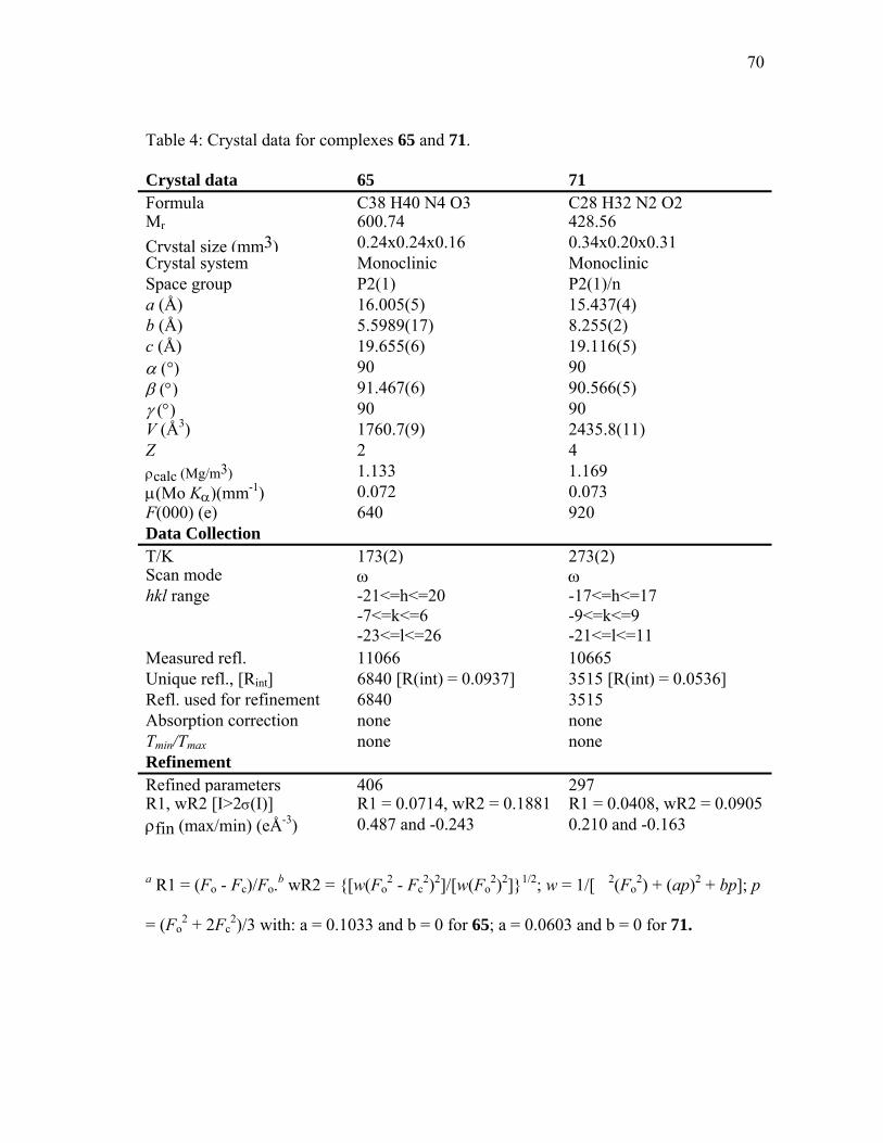
70
Table 4: Crystal data for complexes 65 and 71.
Crystal data 65 71 Formula C38 H40 N4 O3 C28 H32 N2 O2 Mr 600.74 428.56 Crystal size (mm3) 0.24x0.24x0.16 0.34x0.20x0.31 Crystal system Monoclinic Monoclinic Space group P2(1) P2(1)/n a (Å) 16.005(5) 15.437(4) b (Å) 5.5989(17) 8.255(2) c (Å) 19.655(6) 19.116(5) α (°) 90 90 β (°) 91.467(6) 90.566(5) γ (°) 90 90 V (Å3) 1760.7(9) 2435.8(11) Z 2 4 ρcalc (Mg/m3) 1.133 1.169 µ(Mo Kα)(mm-1) 0.072 0.073 F(000) (e) 640 920 Data Collection T/K 173(2) 273(2)Scan mode ω ωhkl range -21<=h<=20
-7<=k<=6 -23<=l<=26
-17<=h<=17 -9<=k<=9 -21<=l<=11
Measured refl. 11066 10665 Unique refl., [Rint] 6840 [R(int) = 0.0937] 3515 [R(int) = 0.0536] Refl. used for refinement 6840 3515 Absorption correction none none Tmin/Tmax none none Refinement Refined parameters 406 297R1, wR2 [I>2σ(I)] R1 = 0.0714, wR2 = 0.1881 R1 = 0.0408, wR2 = 0.0905 ρfin (max/min) (eÅ-3) 0.487 and -0.243 0.210 and -0.163
a R1 = (Fo - Fc)/Fo.b wR2 = {[w(Fo2 - Fc
2)2]/[w(Fo2)2]}1/2; w = 1/[�2(Fo
2) + (ap)2 + bp]; p
= (Fo2 + 2Fc
2)/3 with: a = 0.1033 and b = 0 for 65; a = 0.0603 and b = 0 for 71.
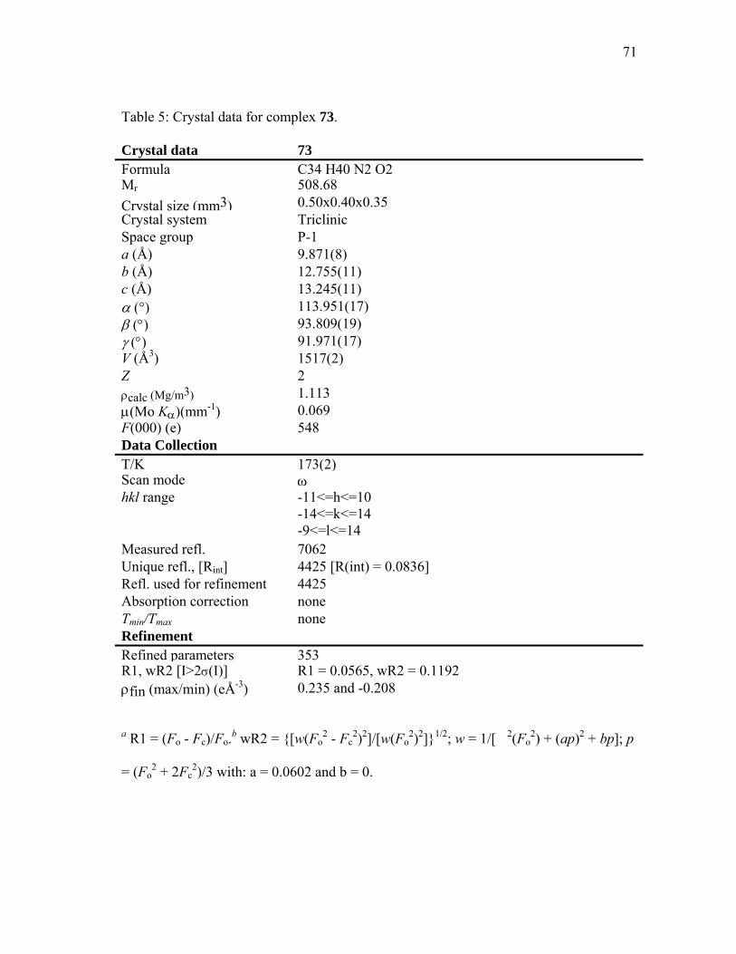
71
Table 5: Crystal data for complex 73.
Crystal data 73 Formula C34 H40 N2 O2Mr 508.68 Crystal size (mm3) 0.50x0.40x0.35 Crystal system Triclinic Space group P-1 a (Å) 9.871(8) b (Å) 12.755(11) c (Å) 13.245(11) α (°) 113.951(17) β (°) 93.809(19) γ (°) 91.971(17) V (Å3) 1517(2) Z 2 ρcalc (Mg/m3) 1.113 µ(Mo Kα)(mm-1) 0.069 F(000) (e) 548 Data Collection T/K 173(2)Scan mode ω hkl range -11<=h<=10
-14<=k<=14 -9<=l<=14
Measured refl. 7062 Unique refl., [Rint] 4425 [R(int) = 0.0836] Refl. used for refinement 4425 Absorption correction none Tmin/Tmax none Refinement Refined parameters 353R1, wR2 [I>2σ(I)] R1 = 0.0565, wR2 = 0.1192 ρfin (max/min) (eÅ-3) 0.235 and -0.208
a R1 = (Fo - Fc)/Fo.b wR2 = {[w(Fo2 - Fc
2)2]/[w(Fo2)2]}1/2; w = 1/[�2(Fo
2) + (ap)2 + bp]; p
= (Fo2 + 2Fc
2)/3 with: a = 0.0602 and b = 0.
72
CHAPTER IV
COORDINATION CHEMISTRY
4.1 Introduction
Organophosphorus pesticides are for the most part thiophosphate triesters and
undergo oxidation to yield the more toxic oxon derivatives. As a result, their destruction
is typically achieved by hydrolysis which leads to the formation of the corresponding
thiophosphate diesters. The hydrolysis reaction takes up to several months in water
under neutral pH condition85 but is greatly accelerated in the presence of the dinuclear
metalloenzyme phosphotriesterase (PTE).86 So far, all of the coordination complexes
that have been investigated as hydrolysis catalysts are monofunctional.87 As these
complexes typically display only marginal activity, our main goal has become to
synthesize a series of dinuclear complexes in order to test their catalytic properties in the
hydrolysis reaction of phosphotriester. In this work, we report the coordination
chemistry of the dinucleating ligands defined in chapter III.
4.2 Investigation of the coordination chemistry of the bis-Schiff base ligands
With the preparation of dinuclear complexes as a goal, it became important to
investigate the ligative behavior of the different ligands toward metal cation moieties. In
a titration experiment monitored by 1H NMR, portions of 0.5 mol-equivalent of bismuth

73
trichloride were added to a CD3CN solution of the dinucleating ligand 66 (Figure 46).
At 0.5 equivalents of cations, the appearance of a new set of sharp 1H NMR signals
indicates the formation of a new species which most likely correspond to a bismuth
complex of 66. Further addition of BiCl3 to the solution leads to an increase in the
intensity of the signal corresponding to the new complex. At two equivalents of added
BiCl3, the signals corresponding to the free ligand have completely disappeared and
further addition of BiCl3 does not lead to further changes of the spectrum.
Figure 46. NMR spectra of 66 in the presence of increasing amount of BiCl3.
74
These observations are in agreement with the formation of a dinuclear bismuth
complex of 66. The detection of a single signal for the aromatic protons suggests that
the new complex (76, figure 47) retains C2 symmetry. The proposed structure 76 is in
agreement with studies by Keppler who synthesized and characterized this new type of
binuclear Bi (III) compounds (77, figure 48).88
CD3CN, BiCl3
RT, 10min
N NN N
76
Me
O
Me
O
N NN NMe
TBDMSO
Me
OTBDMS
t-But-Bu
BiCl3
BiCl3
Figure 47. Coordination reaction of 66 with BiCl3.
75
NN N
MeNR2
SBi Cl
Cl
NNN
MeR2N
SBiCl
Cl
77
Figure 48. Coordination chemistry of BiCl3.
The formation of a dinuclear complex is also observed by NMR when uranyl nitrate
is added to the dinucleating ligand 66 (figure 49). From 0 equivalent to 2 equivalents of
added uranyl nitrate, a new set of lines appears while the original peaks corresponding to
the free ligand decrease. After addition of 2 equivalents, a well-resolved spectrum of the
pure complex is observed (78, figure 50) which does not changes after further additions
of uranyl nitrate. The proposed structure 78 is in agreement with the studies by
Indrasenan and his work on uranyl coordination chemistry (79, figure 51). 89
76
CD3CN, UO2(NO3)2
RT, 10min
N NN NMe
O
Me
O
N NN NMe
TBDMSO
Me
OTBDMS
t-But-Bu
U UO
O O
O
78 + 4NO3-
Figure 49. Coordination reaction of 66 with uranyl nitrate.
Figure 50. NMR spectra of 66 in the presence of increasing amount of UO22+.

77
NN
U
OH
O
O
NO3NO3
79
Figure 51. Coordination chemistry of UO2 (II).
While ambiguous results were obtained with Zn2+, titration of ligand 66 in CD3CN
with scandium (III) and yttrium (III) triflates monitored by 1H-NMR suggested the
formation of 2:2 complexes (80, 81, figure 52, and 54 respectively). Indeed, when
portions of 0.5 mol equivalents of the triflate salts were added to the solution containing
the ligand, extensive broadenings and consequent shifts of the resonance are observed.
At 1 mol equivalent of cations, well-resolved spectra, which do not change upon further
additions, are observed suggesting the formation the symmetrical complexes (figures 53
and 55. Also, the signal of the aromatic protons indicated that the C2 symmetry of the
ligands was retained which reinforce the proposed structures of 2/2 complexes.
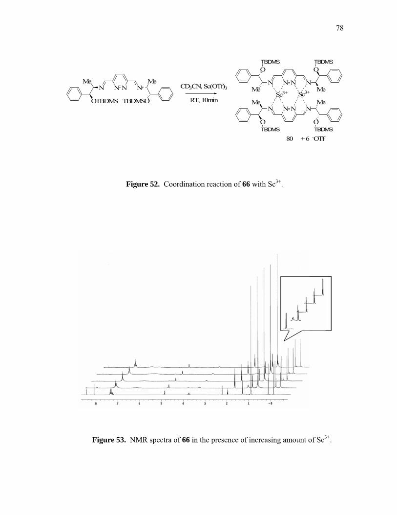
78
CD3CN, Sc(OTf)3
RT, 10min
N NN NN NN N
Me
TBDMSO
Me
OTBDMSSc3+ Sc3+
O
Me
O
Me
NN NN
O
Me
O
Me
80 + 6 -OTf
TBDMS TBDMS
TBDMSTBDMS
Figure 52. Coordination reaction of 66 with Sc3+.
Figure 53. NMR spectra of 66 in the presence of increasing amount of Sc3+.
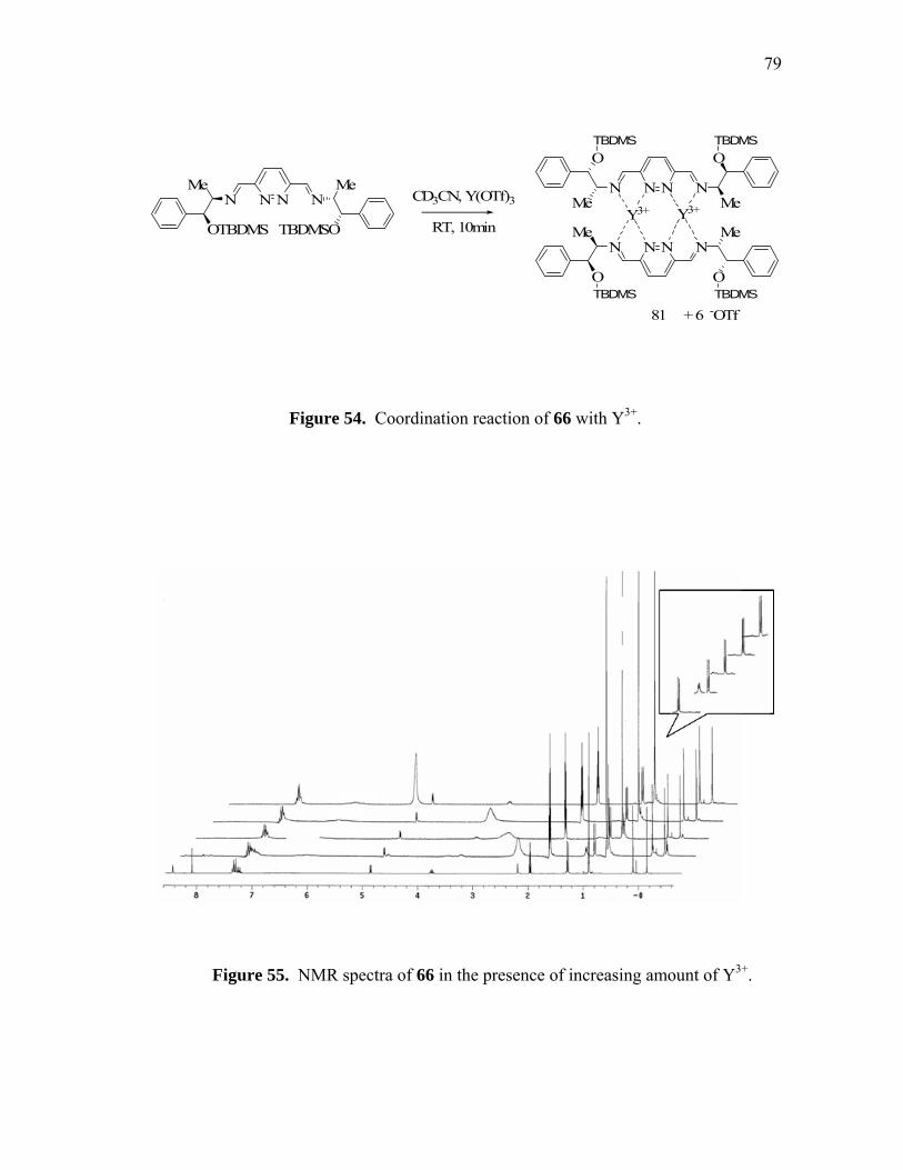
79
CD3CN, Y(OTf)3
RT, 10min
N NN NN NN N
Me
TBDMSO
Me
OTBDMSY3+ Y3+
O
Me
O
Me
NN NN
O
Me
O
Me
81 + 6 -OTf
TBDMS TBDMS
TBDMSTBDMS
Figure 54. Coordination reaction of 66 with Y3+.
Figure 55. NMR spectra of 66 in the presence of increasing amount of Y3+.

80
4.3 Coordination chemistry of the ligands derived from 3,6-bis(hydroxymethyl)-
pyridazine
4.3.1 NMR studies
Synthesis of a dinuclear aluminum complex was achieved by slow addition of a solution
of 44 in pentane to an excess of trimethylaluminum in pentane followed by cooling to -
40ºC resulted in the formation of a white precipitate. Analysis of this precipitate by 1H
NMR in pyridine-d5 confirmed the formation of a dinuclear aluminum complex (82,
figure 56, figure 57).
AlMe3, pentane
-40oC, 1HrsN NN N
82
N NN NH H Al Al
Me Me Me Me
Figure 56. Coordination reaction of 44 with AlMe3.
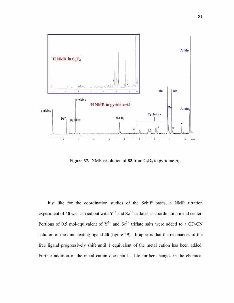
81
Figure 57. NMR resolution of 82 from C6D6 to pyridine-d5.
Just like for the coordination studies of the Schiff bases, a NMR titration
experiment of 46 was carried out with Y3+ and Sc3+ triflates as coordination metal center.
Portions of 0.5 mol-equivalent of Y3+ and Sc3+ triflate salts were added to a CD3CN
solution of the dinucleating ligand 46 (figure 59). It appears that the resonances of the
free ligand progressively shift until 1 equivalent of the metal cation has been added.
Further addition of the metal cation does not lead to further changes in the chemical

82
shifts of the proton of the ligand. These observations suggest the formation of 2:2
complexes 83 (figure 58) whose structure is fluxional in the presence of excess ligand.
Just like in the case of complexes 80 and 81, the signal of the aromatic protons also
indicates that the C2 symmetry of the ligand is retained in the 2/2 complexes.
CD3CN, M(OTf)3
RT, 10min
N NN NN NNH HN
Me
HO
Me
OH3+M M3+
HO
Me
OH
Me
NN NN
OH
Me
HO
Me
83 + 6 -OTf
Figure 58. Coordination reaction of 46 with Y and Sc triflate.

83
Figure 59. NMR spectra of 46 in the presence of increasing amount of Sc3+.
4.3.2 Synthesis of dinuclear copper complexes
Because of the ubiquity of copper in metalloenzymes and artificial catalyst, it was
decided to use this metal in order to probe the ligating properties of the 46 and 48. Thus,
in order to evaluate the stoichiometry of the metal complexes, spectrometric titration
experiment were undertaken. Incremental addition of Cu2+ in ethanol to a solution of 46
or 48 resulted in the appearance of a band at 365 nm (Figure 60, 61).

84
Figure 60. Spectrometric titration of 46 (10-4 M) with CuCl2 in EtOH. Absorbance
variation at 365 nm as a function of the number of equivalents of CuCl2 added.
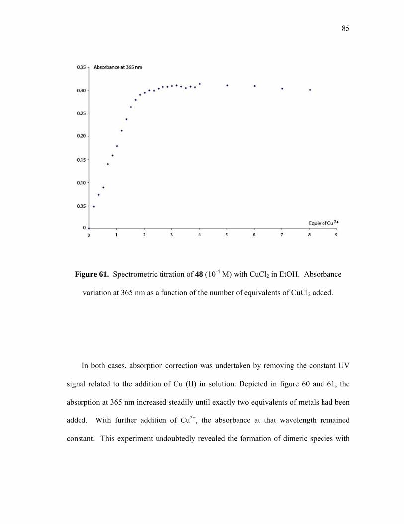
85
Figure 61. Spectrometric titration of 48 (10-4 M) with CuCl2 in EtOH. Absorbance
variation at 365 nm as a function of the number of equivalents of CuCl2 added.
In both cases, absorption correction was undertaken by removing the constant UV
signal related to the addition of Cu (II) in solution. Depicted in figure 60 and 61, the
absorption at 365 nm increased steadily until exactly two equivalents of metals had been
added. With further addition of Cu2+, the absorbance at that wavelength remained
constant. This experiment undoubtedly revealed the formation of dimeric species with
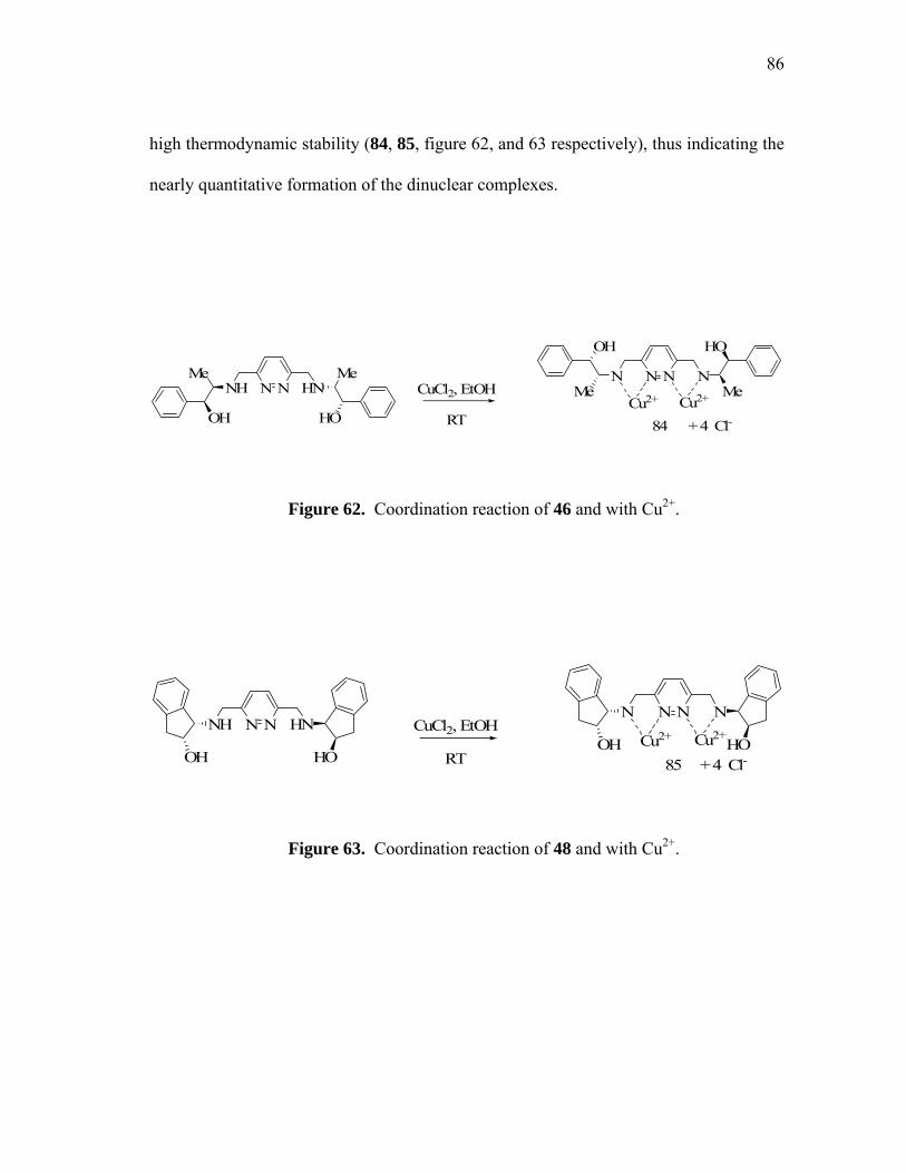
86
high thermodynamic stability (84, 85, figure 62, and 63 respectively), thus indicating the
nearly quantitative formation of the dinuclear complexes.
CuCl2, EtOH
RT
N NN NN NNH HN
Me
HO
Me
OHCu2+ Cu2+
HO
Me
OH
Me
84 + 4 Cl-
Figure 62. Coordination reaction of 46 and with Cu2+.
CuCl2, EtOH
RT
N NN NN NNH HN
Cu2+ Cu2+
85 + 4 Cl-OH HOOH HO
Figure 63. Coordination reaction of 48 and with Cu2+.

87
In the case of 48, the existence of dinuclear complexes could also be confirmed by
ESI mass spectrometry of methanol solutions containing 48 and two equivalents of
Cu(OTf)2. The base peak, observed at 675 amu, results from the loss of two molecule of
HOTf and one OTf - anion from the parent complex 48·(CuOTf2)2 (figure 64). Loss of
an additional HOTf molecule results in a peak observed at 525 amu.
Figure 64. ESI mass spectrometry of 48 and with Cu2+.

88
In order to compare the activity of the dinuclear catalysts to that of mononuclear
analogs, mononucleating ligands such as 86 and 87 were also prepared. These ligands,
which are designed to simulate a half of ligands 84 and 85, were prepared following the
published procedures. 90,91 Just like we did for compound 84 and 85, the spectrometric
titration using CuCl2 was carried out. Incremental addition of Cu2+ in ethanol to a
solution of 86 or 87 resulted in the appearance of a band at 700 nm (Figure 65, 67)
whose intensity increased until almost exactly one equivalent of copper cation had been
added.
Figure 65. Spectrometric titration of 86 (10-2 M) with CuCl2 in EtOH. Absorbance
variation at 700 nm as a function of the number of equivalents of CuCl2 added.

89
N NH
HO
N N
OCu2+
CuCl2, EtOH
RT
86 88
Figure 66. Coordination reaction of 86 and with Cu2+.
Figure 67. Spectrometric titration of 87 (8x10⁻³ M) with CuCl2 in EtOH. Absorbance
variation at 700 nm as a function of the number of equivalents of CuCl2 added.

90
87 89
N NH
HO
CuCl2, EtOH
RTN N
OCu2+
Figure 68. Coordination reaction of 87 and with Cu2+.
In both cases, absorption at that wavelength increased steadily until almost one
equivalent of metal had been added thus indicating the formation of 1:1 complexes (88,
89, figure 66, and 68 respectively).
4.4 Summary
In conclusion, with the use of simple techniques such as 1H NMR and UV titration
we report the coordination chemistry of our chiral dinucleating ligands with C2
symmetry in which two metal centers can be embedded.
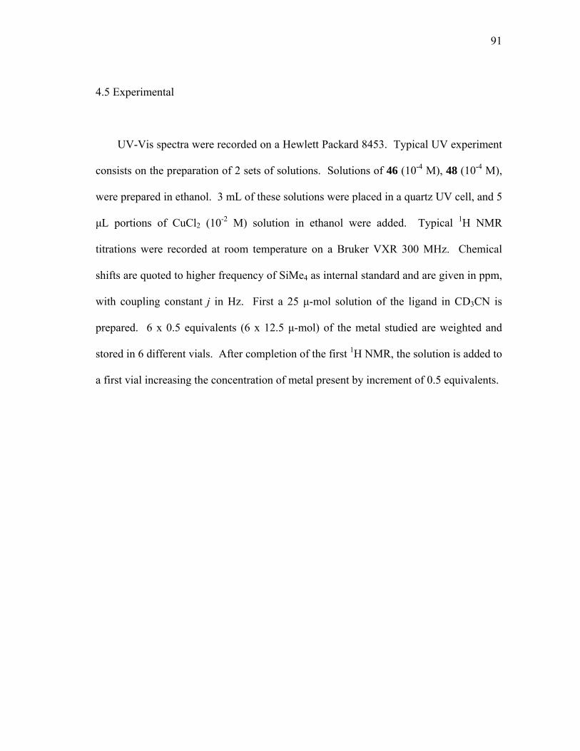
91
4.5 Experimental
UV-Vis spectra were recorded on a Hewlett Packard 8453. Typical UV experiment
consists on the preparation of 2 sets of solutions. Solutions of 46 (10-4 M), 48 (10-4 M),
were prepared in ethanol. 3 mL of these solutions were placed in a quartz UV cell, and 5
µL portions of CuCl2 (10-2 M) solution in ethanol were added. Typical 1H NMR
titrations were recorded at room temperature on a Bruker VXR 300 MHz. Chemical
shifts are quoted to higher frequency of SiMe4 as internal standard and are given in ppm,
with coupling constant j in Hz. First a 25 µ-mol solution of the ligand in CD3CN is
prepared. 6 x 0.5 equivalents (6 x 12.5 µ-mol) of the metal studied are weighted and
stored in 6 different vials. After completion of the first 1H NMR, the solution is added to
a first vial increasing the concentration of metal present by increment of 0.5 equivalents.
92
CHAPTER V
ORTHOMETALATED PD (II) COMPLEXES
5.1 Introduction
As stated in the introductory chapter of this thesis, organophosphorus esters that
inhibit the acetylcholine esterase enzyme are used as nerve agents.1 Because of the toxic
natures of these chemicals,92,93 much attention has been devoted to the development of
methods that allow for their neutralization.94 In this regard, hydrolysis reactions that
produce non-toxic phosphate or phosphonate anions have been investigated. In the case
of parathion (O,O-diethyl O-p-nitrophenyl thiophosphate), a pesticide whose
environmental presence is generating growing concerns,95 the hydrolysis reaction
produces p-nitrophenol and the corresponding thiophosphate diester which is non-toxic.
This hydrolysis is slow and takes up to several months at neutral pH.86 In the presence
of Lewis acidic transition metal catalysts, the rate of this reaction is increased. While
coordination complexes typically display a marginal activity,44,46,88 recent reports
presented in the first chapter of this thesis suggest that organometallic catalysts can be
remarkably active.96 In particular, the group of Ryabov has investigated several
cyclometallated palladium and platinum aryl oxime complexes ([MCl{C,N-
(C6H4C(CH3)=NOH)-2}(L)], M = Pd or Pt, L = DMSO or pyridine) that greatly
accelerate the hydrolysis of parathion.55 Although no reaction intermediates have been
isolated, it has been proposed that the activity of such catalysts results from the
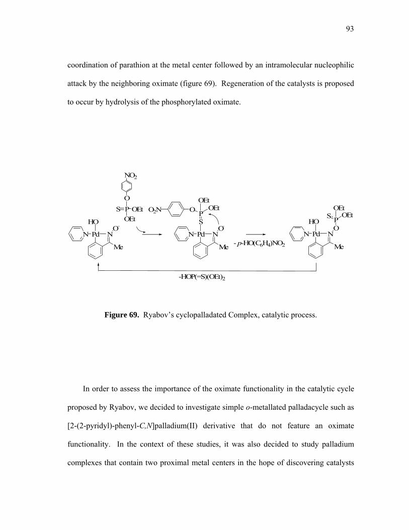
93
coordination of parathion at the metal center followed by an intramolecular nucleophilic
attack by the neighboring oximate (figure 69). Regeneration of the catalysts is proposed
to occur by hydrolysis of the phosphorylated oximate.
N
SP
PdO
N
O OEtOEt
O2N
N
HO
PdO
N
PS OEtOEt
N
HO
PdO
N
-HOP(=S)(OEt)2
S PO
OEtOEt
NO2
- p-HO(C6H4)NO2Me Me Me
Figure 69. Ryabov’s cyclopalladated Complex, catalytic process.
In order to assess the importance of the oximate functionality in the catalytic cycle
proposed by Ryabov, we decided to investigate simple o-metallated palladacycle such as
[2-(2-pyridyl)-phenyl-C,N]palladium(II) derivative that do not feature an oximate
functionality. In the context of these studies, it was also decided to study palladium
complexes that contain two proximal metal centers in the hope of discovering catalysts

94
whose efficiency is increased by cooperative effects occuring at neighboring metal
centers. Such cooperative effects have been proposed for the hydrolysis of
organophosphorus esters at the dinuclear active site of the phosphotriesterase
metalloenzyme (PTE).
5.2 Mononuclear palladium (II) complexes
The reaction of methylparathion (20, Figure 70) with of [2-(2-pyridyl)-phenyl-
C,N]palladium(II) acetate (90, figure 70) leads to the isolation of the corresponding o-
palladated complex di-µ-(dimethylthiophosphate-S,S)-bis[2-(2-pyridyl)phenyl-
C,N]dipalladium(II) (91, figure 70).
THF/H2O, RT, 12hrs N
POO
O
2
SP
O
OO
NO2
N2
PdAcO
90
20
91
PdS
Figure 70. Coordination reaction of [2-(2-pyridyl)-phenyl-C,N]palladium(II) with
methylparathion.
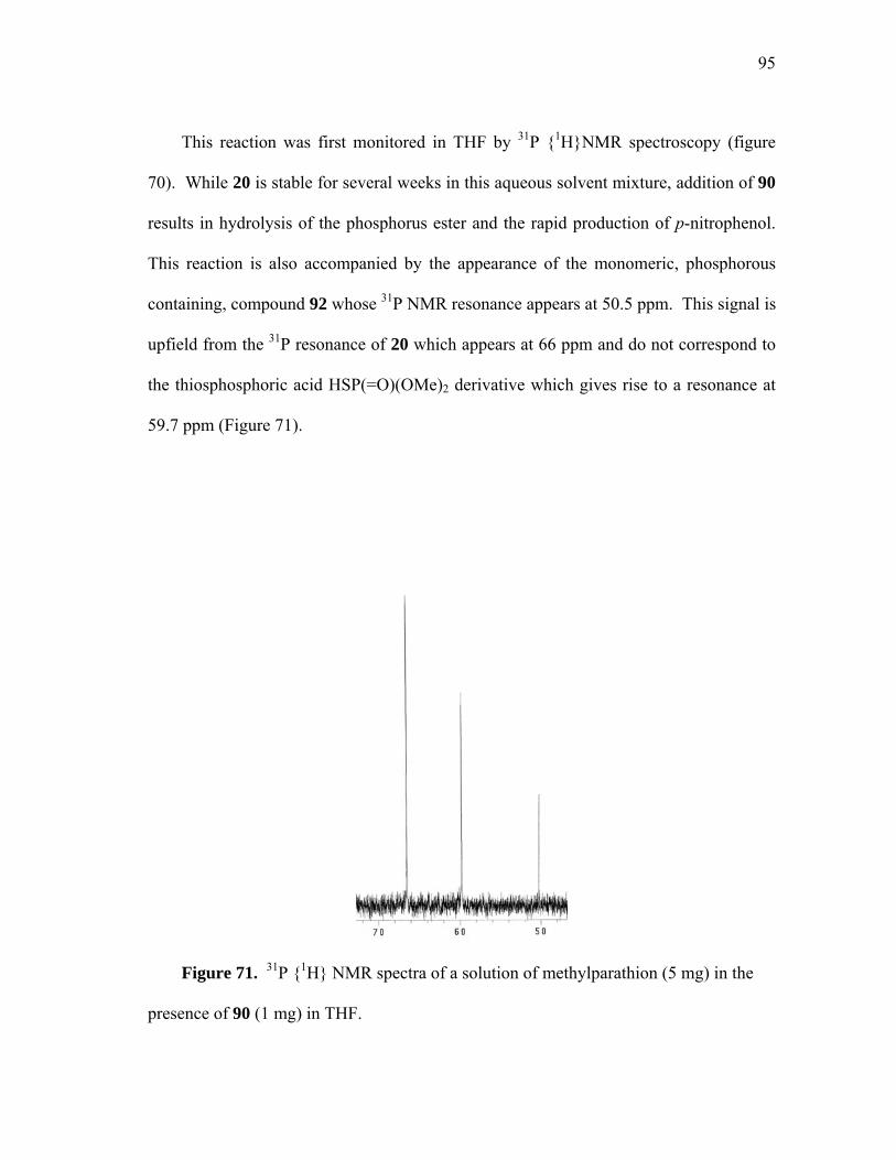
95
This reaction was first monitored in THF by 31P {1H}NMR spectroscopy (figure
70). While 20 is stable for several weeks in this aqueous solvent mixture, addition of 90
results in hydrolysis of the phosphorus ester and the rapid production of p-nitrophenol.
This reaction is also accompanied by the appearance of the monomeric, phosphorous
containing, compound 92 whose 31P NMR resonance appears at 50.5 ppm. This signal is
upfield from the 31P resonance of 20 which appears at 66 ppm and do not correspond to
the thiosphosphoric acid HSP(=O)(OMe)2 derivative which gives rise to a resonance at
59.7 ppm (Figure 71).
Figure 71. 31P {1H} NMR spectra of a solution of methylparathion (5 mg) in the
presence of 90 (1 mg) in THF.
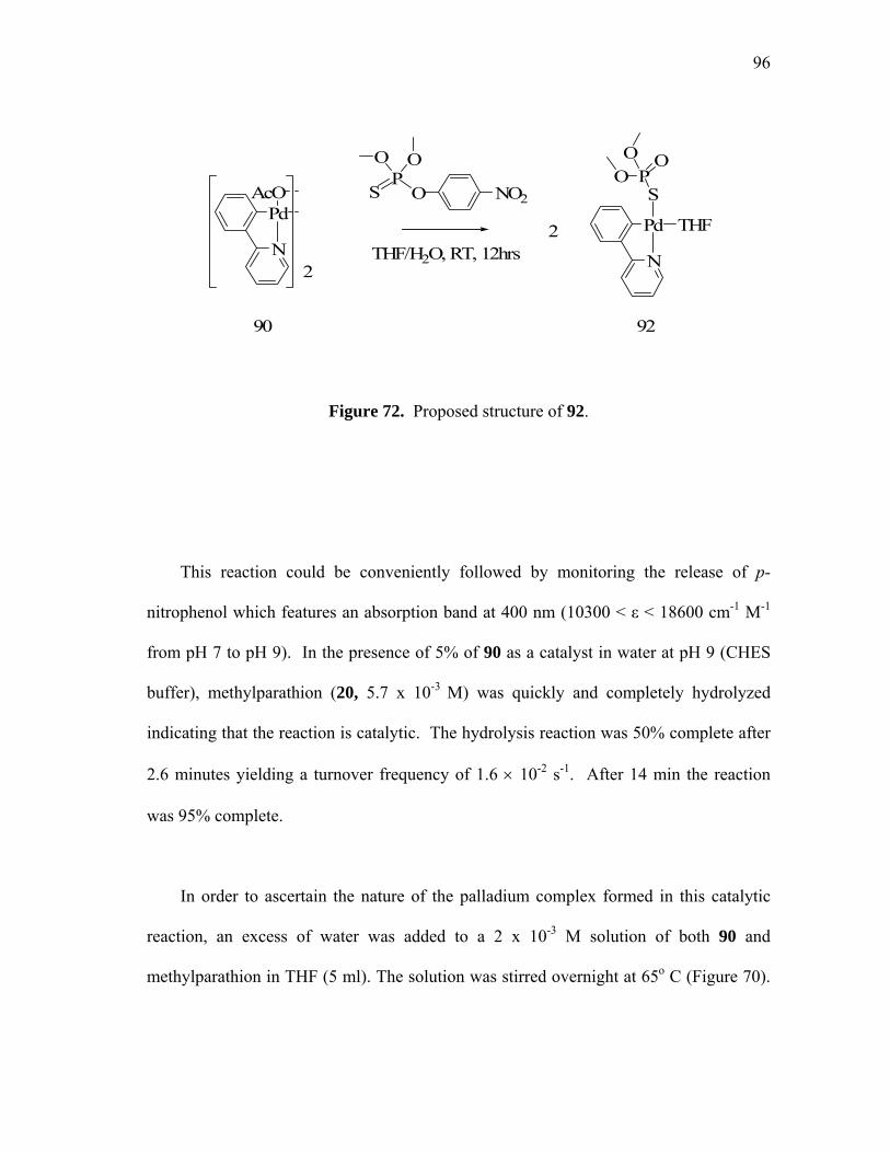
96
THF/H2O, RT, 12hrs N
Pd
SP
OOO
2
SP
O
OO
NO2
N2
PdAcO
90 92
THF
Figure 72. Proposed structure of 92.
This reaction could be conveniently followed by monitoring the release of p-
nitrophenol which features an absorption band at 400 nm (10300 < ε < 18600 cm-1 M-1
from pH 7 to pH 9). In the presence of 5% of 90 as a catalyst in water at pH 9 (CHES
buffer), methylparathion (20, 5.7 x 10⁻³ M) was quickly and completely hydrolyzed
indicating that the reaction is catalytic. The hydrolysis reaction was 50% complete after
2.6 minutes yielding a turnover frequency of 1.6 × 10⁻² s-1. After 14 min the reaction
was 95% complete.
In order to ascertain the nature of the palladium complex formed in this catalytic
reaction, an excess of water was added to a 2 x 10⁻³ M solution of both 90 and
methylparathion in THF (5 ml). The solution was stirred overnight at 65o C (Figure 70).

97
After evaporation of the solvents, colorless needles of 91 were isolated by slow
evaporation in DMF at room temperature (Figure 73).
Figure 73. Structure of complex 91 in the crystal (50% ellipsoid). Selected bond
lengths [Å] and angles [deg]. Pd-C(1) 2.049(11), Pd-N 2.060(11), Pd-S 2.418(3), Pd-
S#1 2.467(3), S-P 2.089(5), S-Pd#1 2.467(3), P-O(1) 1.478(10), P-O(3) 1.594(10), P-
O(2) 1.621(9). C(1)-Pd-N 80.9(5), C(1)-Pd-S 96.7(3), N-Pd-S 175.5(3), C(1)-Pd-S#1
177.3(3), N-Pd-S#1 98.6(3), S-Pd-S#1 83.98(11), P-S-Pd 101.75(17), P-S-Pd#1
97.72(16), Pd-S-Pd#1 96.02(11), C(7)-N-Pd 115.7(8), C(11)-N-Pd 125.2(10), C(2)-C(1)-
Pd 127.1(10), C(6)-C(1)-Pd 114.9(8).
98
Complex 91 crystallizes in the triclinic space group P-1 (Table on page 114). In
this complex, the two square planar palladium moieties (sum of angle of Pd = 360.2(2)˚)
are linked by the sulfur atoms of the bridging dimethylthiophosphate ligands. The Pd-S
distances of 2.418(3) and Pd-S 2.467(3) Å are comparable to those observed in related
complexes such as (R,S)-di-µ-(dimethylthiophosphate-S,S)-bis{2-[2-(4-
carbomethoxy)oxazolinyl]-phenyl-C,N}dipalladium(II) (av. Pd-S = 2.40 Å)12 which also
features bridging thiophosphate groups. The remaining coordination sites of the
palladium center are occupied by the phenylpyridine carbon and nitrogen donor atoms.
The resulting Pd-C(1) (2.049(11) Å) and Pd-N (2.060(11) Å) bonds are comparable to
those observed for [2-(2-pyridyl)phenyl]palladium(II) chloride [LPd(µ−Cl)2PdL]. 97
Interestingly, the solid state 31P{1H} NMR of 91 revealed two different sets of data
(figure 74). The first set corresponds to the “eclipsed” regio-isomer 93 (figure 75),
where the two phosphorus nucleus have a different environment (29.3 ppm and 35.9
ppm). The second set correspond to the staggered regio-isomer 94 (figure 75), where a
unique peak is observed (32.9 ppm). Indeed in such conformation, the two phosphorus
nuclei have an identical environment.
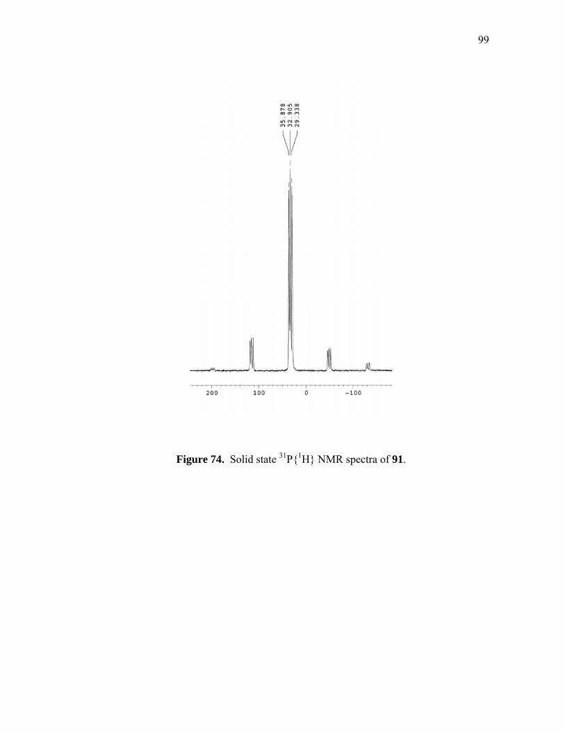
99
Figure 74. Solid state 31P{1H} NMR spectra of 91.
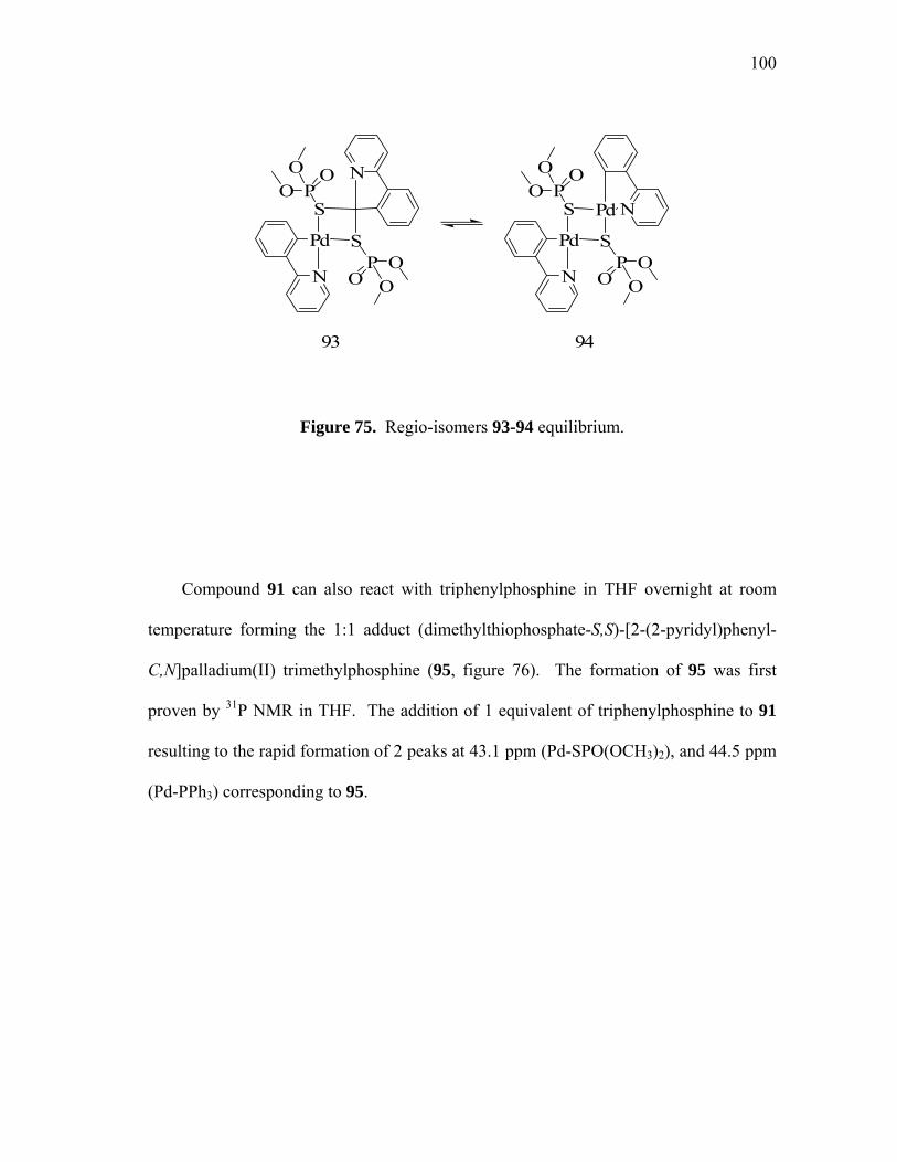
100
93
N
Pd
SP
OOO
N
SP
O OO
N
Pd
SP
OOO
NPd
SP
O OO
94
Figure 75. Regio-isomers 93-94 equilibrium.
Compound 91 can also react with triphenylphosphine in THF overnight at room
temperature forming the 1:1 adduct (dimethylthiophosphate-S,S)-[2-(2-pyridyl)phenyl-
C,N]palladium(II) trimethylphosphine (95, figure 76). The formation of 95 was first
proven by 31P NMR in THF. The addition of 1 equivalent of triphenylphosphine to 91
resulting to the rapid formation of 2 peaks at 43.1 ppm (Pd-SPO(OCH3)2), and 44.5 ppm
(Pd-PPh3) corresponding to 95.

101
N
Pd
SP
OOO
2
THF/H2O, RT, 12hrs
Triphenylphosphine
N
Pd
SP
OOO
PPh
PhPh
95
Figure 76. Coordination reaction of di-µ-(dimethylthiophosphate-S,S)-bis[2-(2-
pyridyl)phenyl-C,N]dipalladium(II) with triphenylphosphine.
Crystals of 95 were isolated and studied by X-ray analysis (Figure 77). Complex 95
crystallizes in the triclinic space group P-1 (Table on page 114). In this complex, we
can observe the typical square planar palladium environment (sum of angle of Pd =
360.36(52)˚), with the sulfur bound thiophosphate and triphenylphosphine ligands in a
forced cis-arrangement. The resulting Pd-P (2.261(15) Å) and Pd-S (2.414(14) Å)
distances fall within the expected range. 98 The remaining coordination sites of the
palladium center are occupied by the phenylpyridine carbon and nitrogen donor atoms.
The Pd-N distances of 2.089(4) Å and Pd-C 2.024(5) Å are comparable to those
observed in 91.
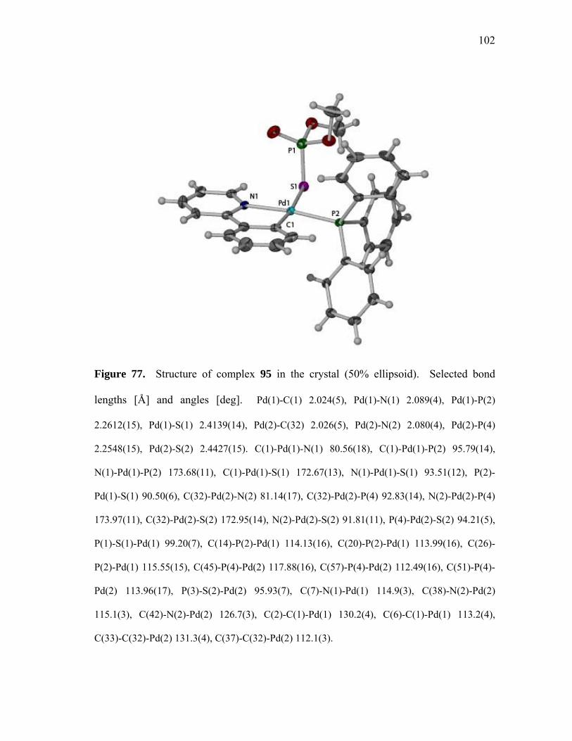
102
Figure 77. Structure of complex 95 in the crystal (50% ellipsoid). Selected bond
lengths [Å] and angles [deg]. Pd(1)-C(1) 2.024(5), Pd(1)-N(1) 2.089(4), Pd(1)-P(2)
2.2612(15), Pd(1)-S(1) 2.4139(14), Pd(2)-C(32) 2.026(5), Pd(2)-N(2) 2.080(4), Pd(2)-P(4)
2.2548(15), Pd(2)-S(2) 2.4427(15). C(1)-Pd(1)-N(1) 80.56(18), C(1)-Pd(1)-P(2) 95.79(14),
N(1)-Pd(1)-P(2) 173.68(11), C(1)-Pd(1)-S(1) 172.67(13), N(1)-Pd(1)-S(1) 93.51(12), P(2)-
Pd(1)-S(1) 90.50(6), C(32)-Pd(2)-N(2) 81.14(17), C(32)-Pd(2)-P(4) 92.83(14), N(2)-Pd(2)-P(4)
173.97(11), C(32)-Pd(2)-S(2) 172.95(14), N(2)-Pd(2)-S(2) 91.81(11), P(4)-Pd(2)-S(2) 94.21(5),
P(1)-S(1)-Pd(1) 99.20(7), C(14)-P(2)-Pd(1) 114.13(16), C(20)-P(2)-Pd(1) 113.99(16), C(26)-
P(2)-Pd(1) 115.55(15), C(45)-P(4)-Pd(2) 117.88(16), C(57)-P(4)-Pd(2) 112.49(16), C(51)-P(4)-
Pd(2) 113.96(17), P(3)-S(2)-Pd(2) 95.93(7), C(7)-N(1)-Pd(1) 114.9(3), C(38)-N(2)-Pd(2)
115.1(3), C(42)-N(2)-Pd(2) 126.7(3), C(2)-C(1)-Pd(1) 130.2(4), C(6)-C(1)-Pd(1) 113.2(4),
C(33)-C(32)-Pd(2) 131.3(4), C(37)-C(32)-Pd(2) 112.1(3).
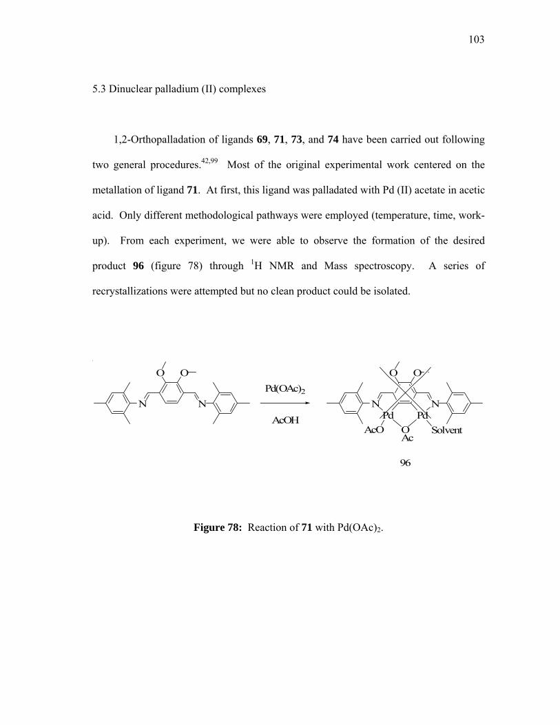
103
5.3 Dinuclear palladium (II) complexes
1,2-Orthopalladation of ligands 69, 71, 73, and 74 have been carried out following
two general procedures.42,99 Most of the original experimental work centered on the
metallation of ligand 71. At first, this ligand was palladated with Pd (II) acetate in acetic
acid. Only different methodological pathways were employed (temperature, time, work-
up). From each experiment, we were able to observe the formation of the desired
product 96 (figure 78) through 1H NMR and Mass spectroscopy. A series of
recrystallizations were attempted but no clean product could be isolated.
N N
O OPd(OAc)2
AcOHN N
O O
PdOAc
PdAcO Solvent
96
Figure 78: Reaction of 71 with Pd(OAc)2.
104
In the same way, the reaction of 71 with Na2PdCl4 in CH2Cl2 leads to formation of
the palladium chloride complex which presumably adopts a polymeric structure.
Although we have not been able to thoroughly characterize this product. The 1H NMR
in DMSO shows poorly resolved peaks corresponding to the mesityl (CH3, 25ppm; CHar
6.9 ppm), the methoxy (CH3, 3.81 ppm), as well as the pyridazine (CH=N, 8.39 ppm;
CHar, 6.9 ppm) H atoms. However, elemental analysis data confirms the proposed
formulation and indicate the absence of any additional ligands. In order to obtain a more
tractable dinuclear palladium complex, the chlorinated intermediate was allowed to react
with TlACAC. This reaction was carried out on the crude chloride complex produced by
the reaction of 63 with Na2PdCl4. The crude product was treated with 3 equivalents of
TlACAC which resulted in the precipitation of TlCl. The product was extracted in
pentane and recrystallized from hexane at room temperature which afforded red crystals
of 97 (figure 79) in a 26% yield. This derivative has been fully characterized by EA, 1H
NMR, 13C NMR and ES mass spectrometry.
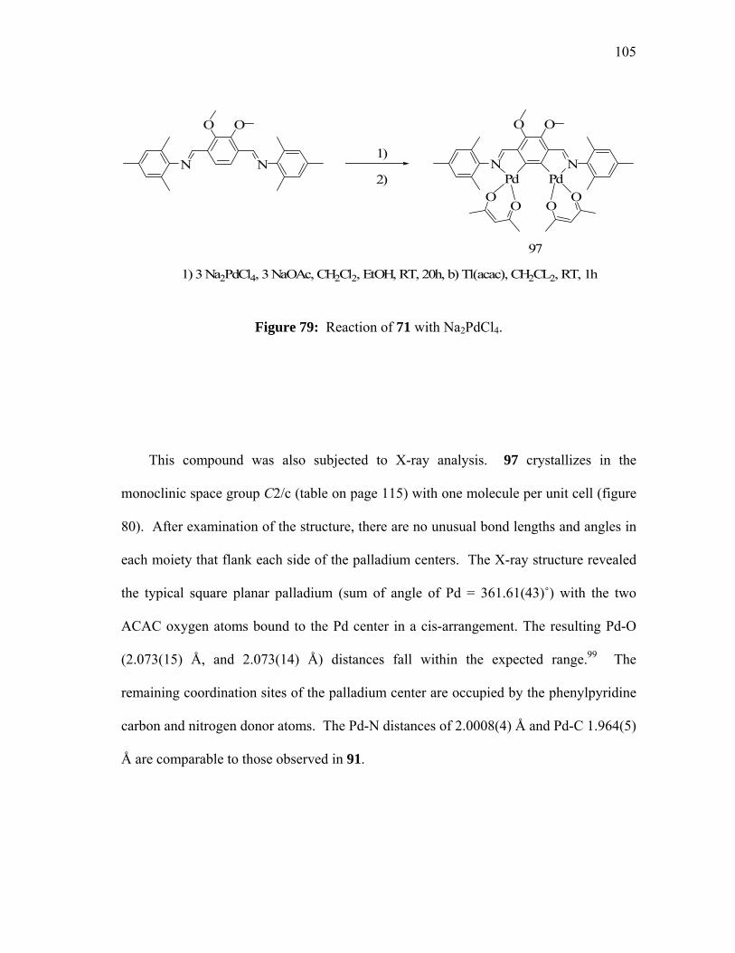
105
N N
O O
N N
O O
Pd PdO
OOO
1) 3 Na2PdCl4, 3 NaOAc, CH2Cl2, EtOH, RT, 20h, b) Tl(acac), CH2CL2, RT, 1h
97
1)
2)
Figure 79: Reaction of 71 with Na2PdCl4.
This compound was also subjected to X-ray analysis. 97 crystallizes in the
monoclinic space group C2/c (table on page 115) with one molecule per unit cell (figure
80). After examination of the structure, there are no unusual bond lengths and angles in
each moiety that flank each side of the palladium centers. The X-ray structure revealed
the typical square planar palladium (sum of angle of Pd = 361.61(43)˚) with the two
ACAC oxygen atoms bound to the Pd center in a cis-arrangement. The resulting Pd-O
(2.073(15) Å, and 2.073(14) Å) distances fall within the expected range.99 The
remaining coordination sites of the palladium center are occupied by the phenylpyridine
carbon and nitrogen donor atoms. The Pd-N distances of 2.0008(4) Å and Pd-C 1.964(5)
Å are comparable to those observed in 91.

106
Figure 80. ORTEP view of 97 in the crystal (50% ellipsoids). Selected bond lengths
[Å] and angles [deg]. N-Pd 2.008(10), Pd-C(1) 1.964(11), Pd-O(1) 2.073(10), Pd-O(2)
2.073(11). C(5)-N-Pd 115.5(8), C(6)-N-Pd 124.5(8), C(1)-Pd-N 80.6(4), C(1)-Pd-O(1)
173.8(5), N-Pd-O(1) 95.9(4), C(1)-Pd-O(2) 93.8(5), N-Pd-O(2) 158.0(4), O(1)-Pd-O(2)
91.3(4), C(16)-O(1)-Pd 128.2(14), C(18)-O(2)-Pd 124.9(14), C(2)-C(1)-Pd 114.4(8),
C(1)#1-C(1)-Pd 128.6(4).
Following the same experimental procedure, ligands 69 and 73 were doubly
palladated with Na2PdCl4 in EtOH. These reactions also produced the polymeric
chloride complexes which upon treatment with TlACAC produced complex (98, figure
81) and (99, figure 82) in a 46% and 55 % yield respectively. Both of these compounds
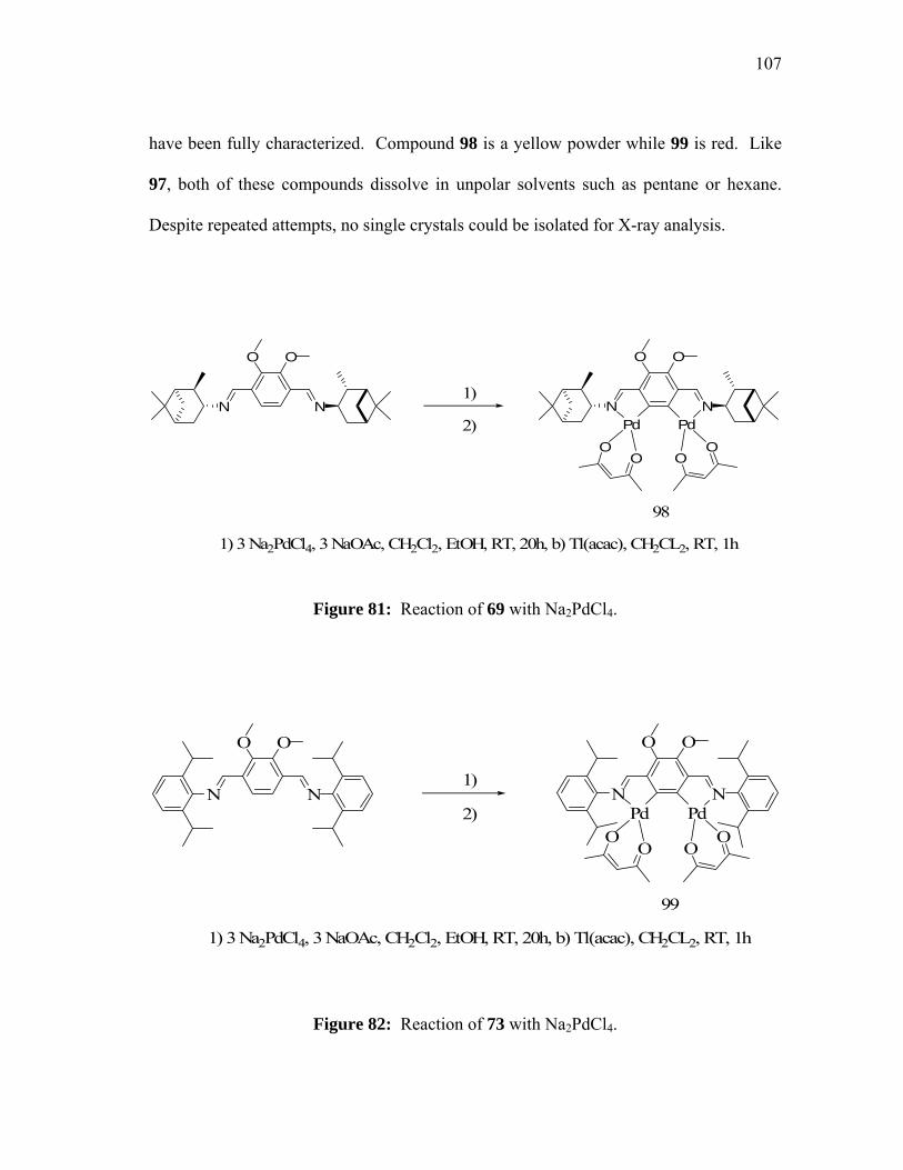
107
have been fully characterized. Compound 98 is a yellow powder while 99 is red. Like
97, both of these compounds dissolve in unpolar solvents such as pentane or hexane.
Despite repeated attempts, no single crystals could be isolated for X-ray analysis.
N N
O O
Pd Pd
OOO
O
1) 3 Na2PdCl4, 3 NaOAc, CH2Cl2, EtOH, RT, 20h, b) Tl(acac), CH2CL2, RT, 1h
98
1)
2)N N
O O
Figure 81: Reaction of 69 with Na2PdCl4.
N N
O O
N N
O O
Pd PdO
OOO
1) 3 Na2PdCl4, 3 NaOAc, CH2Cl2, EtOH, RT, 20h, b) Tl(acac), CH2CL2, RT, 1h
99
1)
2)
Figure 82: Reaction of 73 with Na2PdCl4.

108
5.4 Summary
Reaction of methylparathion with [2-(2-pyridyl)-phenyl-C,N]palladium(II) acetate
leads to the formation of the corresponding dimethylthiophosphate complex 83 which
has been isolated in a 74% yield. Complex 83 has been characterized by NMR, and
elemental analysis. As indicated by X-ray analysis, this complex assumes a dinuclear
structure with the two palladium phenylpyridine moieties linked by two S-bridging
dimethylthiophosphate. Compound 83 also reacts with triphenylphosphine leading to the
formation of the 1:1 adduct 86 which can be isolated and crystallized with high yield
(85%). Orthopalladation of ligands 61, 63, 65 can be achieved by reacting a 3
equivalents excess of Na2PdCl4, in a 1:1 CH2Cl2/ethanol mixture. The isolation of the
desired product was successfully achieved by displacing the chlorines with thallium
ACAC.
5.5 Experimental
General considerations:
Methylparathion is highly toxic and should be handled in a well-ventilated fume
hood. All glassware exposed to methylparathion should be decontaminated with bleach.
Solvents were dried by standard method. All 31P NMR studies were carried out on an
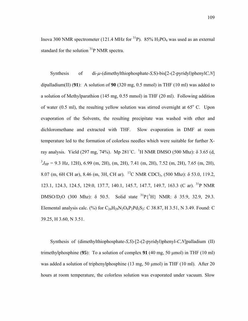
109
Inova 300 NMR spectrometer (121.4 MHz for 31P). 85% H3PO4 was used as an external
standard for the solution 31P NMR spectra.
Synthesis of di-µ-(dimethylthiophosphate-S,S)-bis[2-(2-pyridyl)phenylC,N]
dipalladium(II) (91): A solution of 90 (320 mg, 0.5 mmol) in THF (10 ml) was added to
a solution of Methylparathion (145 mg, 0.55 mmol) in THF (20 ml). Following addition
of water (0.5 ml), the resulting yellow solution was stirred overnight at 65o C. Upon
evaporation of the Solvents, the resulting precipitate was washed with ether and
dichloromethane and extracted with THF. Slow evaporation in DMF at room
temperature led to the formation of colorless needles which were suitable for further X-
ray analysis. Yield (297 mg, 74%). Mp 281˚C. 1H NMR DMSO (500 Mhz): δ 3.65 (d,
3JHP = 9.3 Hz, 12H), 6.99 (m, 2H), (m, 2H), 7.41 (m, 2H), 7.52 (m, 2H), 7.65 (m, 2H),
8.07 (m, 6H CH ar), 8.46 (m, 3H, CH ar). 13C NMR CDCl3, (500 Mhz): δ 53.0, 119.2,
123.1, 124.3, 124.5, 129.0, 137.7, 140.1, 145.7, 147.7, 149.7, 163.3 (C ar). 31P NMR
DMSO/D2O (300 Mhz): δ 50.5. Solid state 31P{1H} NMR: δ 35.9, 32.9, 29.3.
Elemental analysis calc. (%) for C26H28N2O6P2Pd2S2: C 38.87, H 3.51, N 3.49. Found: C
39.25, H 3.60, N 3.51.
Synthesis of (dimethylthiophosphate-S,S)-[2-(2-pyridyl)phenyl-C,N]palladium (II)
trimethylphosphine (95): To a solution of complex 91 (40 mg, 50 µmol) in THF (10 ml)
was added a solution of triphenylphosphine (13 mg, 50 µmol) in THF (10 ml). After 20
hours at room temperature, the colorless solution was evaporated under vacuum. Slow

110
recrystallization of the white residue in acetone led to the formation of colorless needles
which were suitable for further X-ray analysis. Yield (45 mg, 85%). Mp. 281˚C. 1H
NMR CDCl3 (300 Mhz): δ 3.32 (d, 3JHP = 7.2 Hz, 6H), 6.47 (m, 1H), 6.57 (m, 1H), 6.94
(m, 1H), 7.28 (m, 2H), 7.37 (m, 6H), 7.43 (m, 3H), 7.55 (m, 1H), 7.78 (m, 1H), 7.83 (m,
7H). 13C NMR CDCl3 (300 Mhz): δ 53.0 119.2, 123.1, 124.3, 124.5, 129.0, 137.6,
140.1, 145.7, 147.7, 149.6, 163,2 (Cq non detected). 31P NMR THF (300 Mhz): δ 43.1,
44.5. Elemental analysis calc. (%) for C31H29NO3P2SPd: C 56.07, H 4.40, N 2.11.
Found C 55.89, H 4.33, N 2.25.
Synthesis of 3,6-(2,4,6-trimethylphenyliminomethyl)-1,2-dimethoxy-1,2-
phenylenedipalladium-(II)-bis(2,4-pentanedionate) (97): A solution of Na2PdCl4 (1.54
g, 5.25 mmol) and NaOAc (0.43 g, 5.25 mmol) was prepared in 10 ml ethanol and added
to a solution of 71 (0.75g, 1.75 mmol). The reaction mixture was stirred at room
temperature for 20 hours before being filtered under vacuum and washed 3 times with
pentane. The resulting red crude material was thoroughly dried and use for the next step
without further purifications. The red powder was then dissolved in 10 ml of CH2Cl2 and
3 equivalent of TlACAC (1.59 g, 5.25 mmol) was directly added. The rapid precipitation
of TlCl2 was observed and the reaction vessel was stirred for 1 hour at room
temperature. Typical workup was followed with filtration of the precipitate, and
evaporation of solvent under vacuum. The red crude product was then dissolved in
pentane, filtered, and dried. After recrystallization in hexane at room temperature, red
crystals of 97 (0.38g, 26%) were isolated. Mp 241-243˚C. [M-Cl]+ 673. 1H NMR C6D6
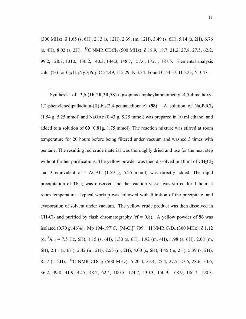
111
(300 MHz): δ 1.65 (s, 6H), 2.13 (s, 12H), 2.39, (m, 12H), 3.49 (s, 6H), 5.14 (s, 2H), 6.76
(s, 4H), 8.02 (s, 2H). 13C NMR CDCl3 (500 MHz): δ 18.9, 18.7, 21.2, 27.8, 27.5, 62.2,
99.2, 128.7, 131.0, 136.2, 140.3, 144.3, 148.7, 157.6, 172.1, 187.5. Elemental analysis
calc. (%) for C38H44N2O6Pd2: C 54.49, H 5.29, N 3.34. Found C 54.37, H 5.23, N 3.47.
Synthesis of 3,6-(1R,2R,3R,5S)-(-)isopinocampheylaminomethyl-4,5-dimethoxy-
1,2-phenylenedipalladium-(II)-bis(2,4-pentanedionate) (98): A solution of Na2PdCl4
(1.54 g, 5.25 mmol) and NaOAc (0.43 g, 5.25 mmol) was prepared in 10 ml ethanol and
added to a solution of 69 (0.81g, 1.75 mmol). The reaction mixture was stirred at room
temperature for 20 hours before being filtered under vacuum and washed 3 times with
pentane. The resulting red crude material was thoroughly dried and use for the next step
without further purifications. The yellow powder was then dissolved in 10 ml of CH2Cl2
and 3 equivalent of TlACAC (1.59 g, 5.25 mmol) was directly added. The rapid
precipitation of TlCl2 was observed and the reaction vessel was stirred for 1 hour at
room temperature. Typical workup was followed with filtration of the precipitate, and
evaporation of solvent under vacuum. The yellow crude product was then dissolved in
CH2Cl2 and purified by flash chromatography (rf = 0.8). A yellow powder of 98 was
isolated (0.70 g, 46%). Mp 194-197˚C. [M-Cl]+ 709. 1H NMR C6D6 (300 MHz): δ 1.12
(d, 3JHH = 7.5 Hz, 6H), 1.15 (s, 6H), 1.30 (s, 6H), 1.92 (m, 4H), 1.98 (s, 6H), 2.08 (m,
6H), 2.11 (s, 6H), 2.42 (m, 2H), 2.55 (m, 2H), 4.00 (s, 6H), 4.45 (m, 2H), 5.39 (s, 2H),
8.57 (s, 2H). 13C NMR CDCl3 (500 MHz): δ 20.4, 23.4, 25.4, 27.5, 27.6, 28.6, 34.6,
36.2, 39.8, 41.9, 42.7, 48.2, 62.4, 100.5, 124.7, 130.3, 150.9, 168.9, 186.7, 190.3.

112
Elemental analysis calc. (%) for C40H56N2O6Pd2: C 54.99, H 6.46, N 3.21. Found C
55.11, H 6.23, N 2.84.
Synthesis of 3,6-(2,6-diisopropylphenyliminomethyl)-4,5-dimethoxy-1,2-
phenylene-dipalladium-(II)-bis(2,4-pentanedionate) (99): A solution of Na2PdCl4 (1.54
g, 5.25 mmol) and NaOAc (0.43 g, 5.25 mmol) was prepared in 10 ml ethanol and added
to a solution of 73 (0.90 g, 1.75 mmol). The reaction mixture was stirred at room
temperature for 20 hours before being filtered under vacuum and washed 3 times with
pentane. The resulting red crude material was thoroughly dried and use for the next step
without further purifications. The red powder was then dissolved in 10 ml of CH2Cl2 and
3 equivalent of TlACAC (1.59 g, 5.25 mmol) was directly added. The rapid precipitation
of TlCl2 was observed and the reaction vessel was stirred for 1 hour at room
temperature. Typical workup was followed with filtration of the precipitate, and
evaporation of solvent under vacuum. The red crude product was then dissolved in
pentane, filtered, and dried. After several attempts to recrystallize 99 (0.89 g, 55%) no
crystals suitable for X-ray analysis were isolated. [M-Cl]+ 757. 1H NMR C6D6 (300
MHz): δ 1.14 (d, 3JHH = 2.1 Hz, 6H), 1.16 (d, 3JHH = 3.6 Hz, 6H) 1.23 (d, 3JHH = 6.9 Hz,
6H), 1.32 (d, 3JHH = 6.6 Hz, 6H), 1.54 (s, 6H), 1.92 (s, 6H), 3.43 (m, 2H), 3.62 (m, 2H),
3.84 (s, 6H), 5.09 (s, 2H), 7.17 (m, 4H), 7.28 (m, 2H), 8.18 (s, 2H). 13C NMR CDCl3
(500 MHz): δ 23.5, 24.1, 25.0, 26.8, 27.5, 28.1, 62.4, 99.2, 123.2, 123.3, 127.4, 140.5,
142.0, 142.4, 144.0, 148.9, 157.9, 171.7, 185.4, 186.7. Elemental analysis calc. (%) for
C44H56N2O6Pd2: C 57.33, H 6.12, N 3.04. Found C 57.40, H 6.04, N 2.82.
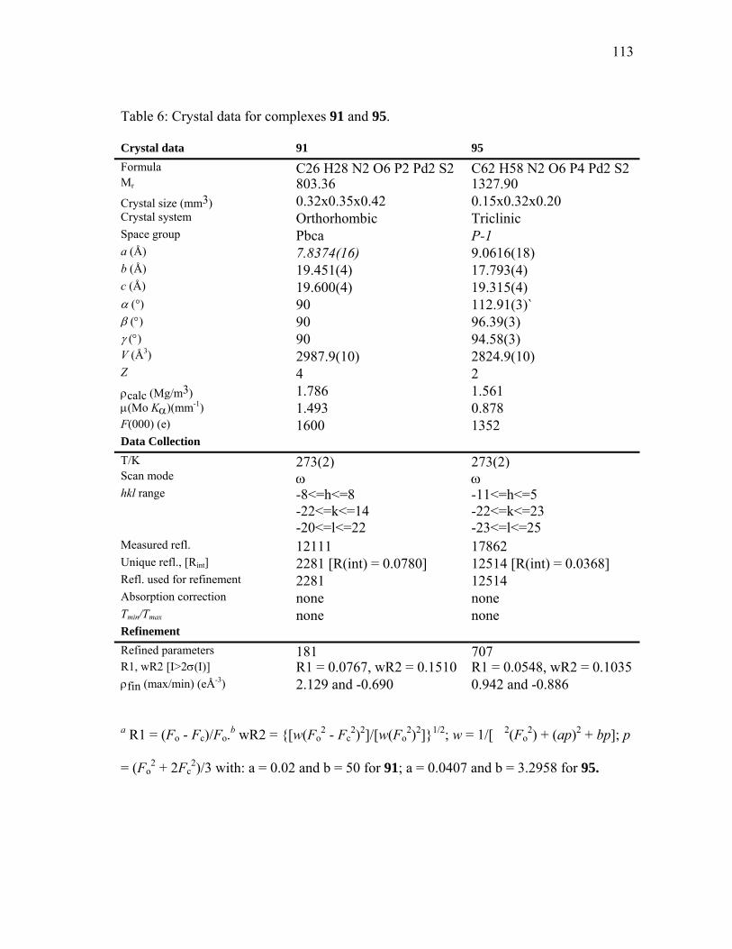
113
Table 6: Crystal data for complexes 91 and 95.
Crystal data 91 95 Formula C26 H28 N2 O6 P2 Pd2 S2 C62 H58 N2 O6 P4 Pd2 S2Mr 803.36 1327.90 Crystal size (mm3) 0.32x0.35x0.42 0.15x0.32x0.20 Crystal system Orthorhombic Triclinic Space group Pbca P-1 a (Å) 7.8374(16) 9.0616(18) b (Å) 19.451(4) 17.793(4) c (Å) 19.600(4) 19.315(4) α (°) 90 112.91(3)` β (°) 90 96.39(3) γ (°) 90 94.58(3) V (Å3) 2987.9(10) 2824.9(10) Z 4 2 ρcalc (Mg/m3) 1.786 1.561 µ(Mo Kα)(mm-1) 1.493 0.878 F(000) (e) 1600 1352 Data Collection T/K 273(2) 273(2)Scan mode ω ωhkl range -8<=h<=8
-22<=k<=14 -20<=l<=22
-11<=h<=5 -22<=k<=23 -23<=l<=25
Measured refl. 12111 17862 Unique refl., [Rint] 2281 [R(int) = 0.0780] 12514 [R(int) = 0.0368] Refl. used for refinement 2281 12514 Absorption correction none none Tmin/Tmax none none Refinement Refined parameters 181 707R1, wR2 [I>2σ(I)] R1 = 0.0767, wR2 = 0.1510 R1 = 0.0548, wR2 = 0.1035 ρfin (max/min) (eÅ-3) 2.129 and -0.690 0.942 and -0.886
a R1 = (Fo - Fc)/Fo.b wR2 = {[w(Fo2 - Fc
2)2]/[w(Fo2)2]}1/2; w = 1/[�2(Fo
2) + (ap)2 + bp]; p
= (Fo2 + 2Fc
2)/3 with: a = 0.02 and b = 50 for 91; a = 0.0407 and b = 3.2958 for 95.

114
Table 7: Crystal data for complex 97.
Crystal data 97 Formula C38 H44 N2 O6 Pd2 Mr 837.55
Crystal size (mm3) 0.24x0.20x0.18 Crystal system Monoclinic Space group C2/c a (Å) 22.043(10) b (Å) 16.271(7) c (Å) 10.094(5) α (°) 90 β (°) 98.834(9) γ (°) 90 V (Å3) 3578(3) Z 4
ρcalc (Mg/m3) 1.555 µ(Mo Kα)(mm-1) 1.053 F(000) (e) 1704 Data Collection T/K 273(2) Scan mode ω hkl range -23<=h<=25
-14<=k<=18 -11<=l<=11
Measured refl. 8296 Unique refl., [Rint] 2812 [R(int) = 0.0777] Refl. used for refinement 2812 Absorption correction Empirical Tmin/Tmax 0.4749 and 0.4066 Refinement Refined parameters 217 R1, wR2 [I>2σ(I)] R1 = 0.0878, wR2 = 0.1946 ρfin (max/min) (eÅ-3) 1.292 and -0.904
a R1 = (Fo - Fc)/Fo.b wR2 = {[w(Fo2 - Fc
2)2]/[w(Fo2)2]}1/2; w = 1/[�2(Fo
2) + (ap)2 + bp]; p
= (Fo2 + 2Fc
2)/3 with: a = 0.1308 and b = 0.
115
CHAPTER VI
METAL CATALYSED ORGANOPHOSPHATE ESTER HYDROLYSIS
6.1. Introduction
As mentioned in the introductory paragraph of this dissertation, the
organophosphorus hydrolase (OPH), from Pseudomonas diminuta, is very active in the
hydrolysis of various organophosphorus esters including organothiphosphates. The X-
ray structure of this enzyme indicates the presence of a dinuclear active site containing
two Zn2+ cations bridged by a hydroxide.100 The proposed mechanism suggests that a
water molecule is activated and oriented at the more buried metal center while the
organophosphate coordinates at the more accessible metal site. It has been suggested
that hydrolysis occurs by a SN2 mechanism in which a water molecule attacks the
phosphoryl center and displaces the leaving group. A number of studies have recently
focused on optimizing the rate of hydrolysis observed with the native enzyme but it is
important to note that so far, no dinuclear complexes that mimic the function of OPH
have been reported or investigated. 101
In the context of this thesis, it was decided to determine whether or not some of the
dinuclear complexes presented in the preceding chapters could serve to mimic the
activity of OPH in the hydrolysis of methylparathion. As part of these studies, a number
of metal ions and complexes have been tested.
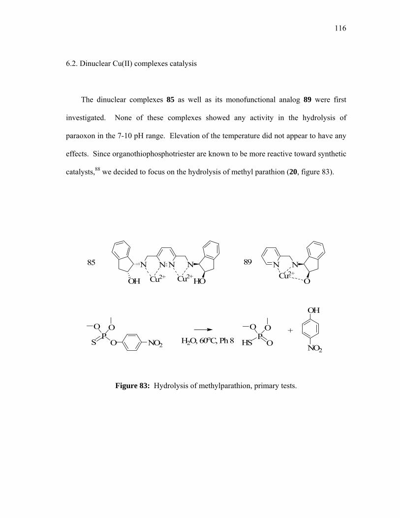
116
6.2. Dinuclear Cu(II) complexes catalysis
The dinuclear complexes 85 as well as its monofunctional analog 89 were first
investigated. None of these complexes showed any activity in the hydrolysis of
paraoxon in the 7-10 pH range. Elevation of the temperature did not appear to have any
effects. Since organothiophosphotriester are known to be more reactive toward synthetic
catalysts,88 we decided to focus on the hydrolysis of methyl parathion (20, figure 83).
H2O, 60oC, Ph 8+
SP
O
OO
NO2 HSP
O
OO
OH
NO2
N NN N
Cu2+ Cu2+OH HO
85 89 N N
OCu2+
Figure 83: Hydrolysis of methylparathion, primary tests.
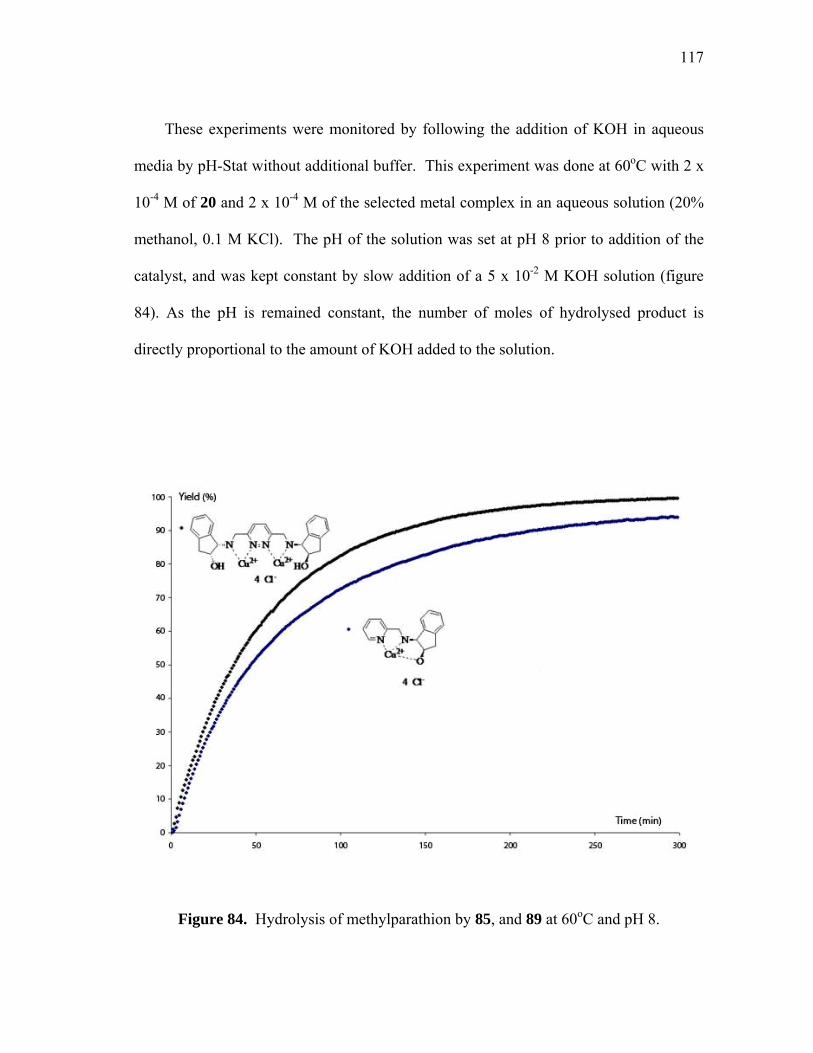
117
These experiments were monitored by following the addition of KOH in aqueous
media by pH-Stat without additional buffer. This experiment was done at 60oC with 2 x
10-4 M of 20 and 2 x 10⁻⁴ M of the selected metal complex in an aqueous solution (20%
methanol, 0.1 M KCl). The pH of the solution was set at pH 8 prior to addition of the
catalyst, and was kept constant by slow addition of a 5 x 10⁻² M KOH solution (figure
84). As the pH is remained constant, the number of moles of hydrolysed product is
directly proportional to the amount of KOH added to the solution.
Figure 84. Hydrolysis of methylparathion by 85, and 89 at 60oC and pH 8.
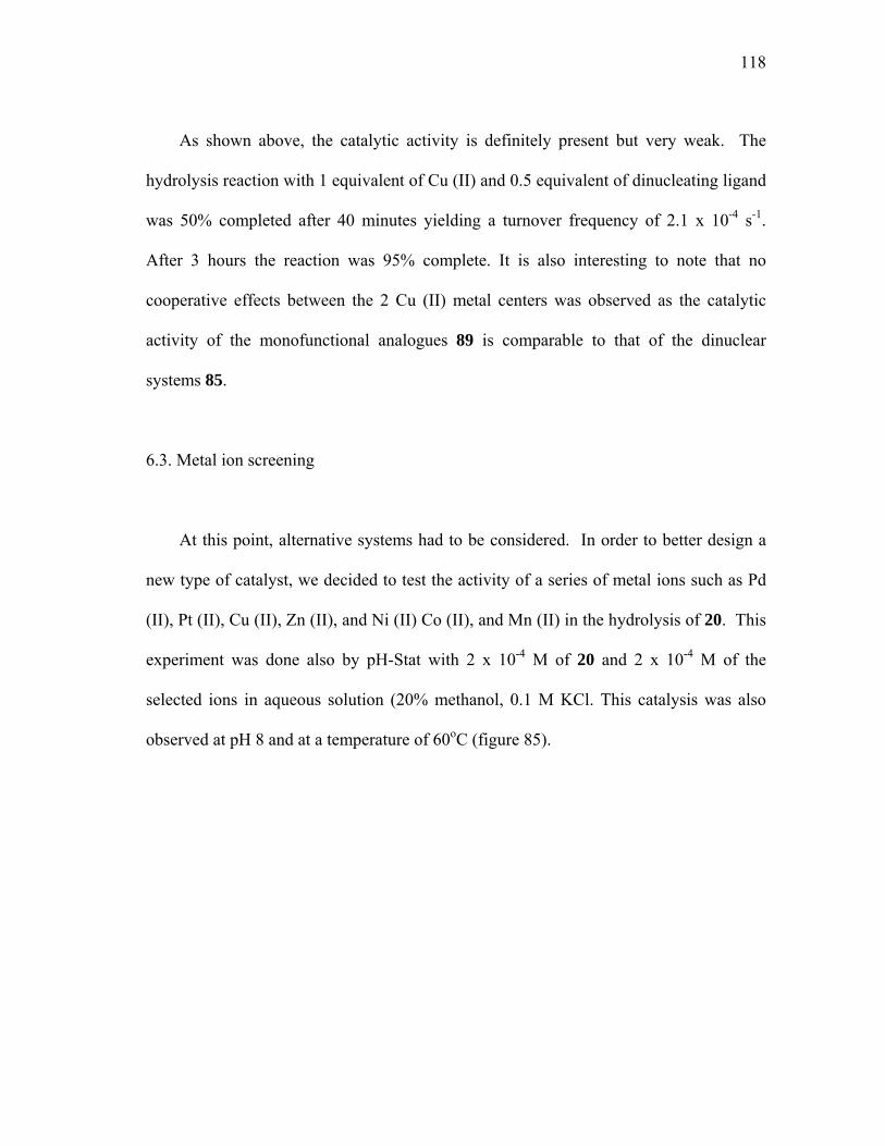
118
As shown above, the catalytic activity is definitely present but very weak. The
hydrolysis reaction with 1 equivalent of Cu (II) and 0.5 equivalent of dinucleating ligand
was 50% completed after 40 minutes yielding a turnover frequency of 2.1 x 10⁻⁴ s-1.
After 3 hours the reaction was 95% complete. It is also interesting to note that no
cooperative effects between the 2 Cu (II) metal centers was observed as the catalytic
activity of the monofunctional analogues 89 is comparable to that of the dinuclear
systems 85.
6.3. Metal ion screening
At this point, alternative systems had to be considered. In order to better design a
new type of catalyst, we decided to test the activity of a series of metal ions such as Pd
(II), Pt (II), Cu (II), Zn (II), and Ni (II) Co (II), and Mn (II) in the hydrolysis of 20. This
experiment was done also by pH-Stat with 2 x 10⁻⁴ M of 20 and 2 x 10⁻⁴ M of the
selected ions in aqueous solution (20% methanol, 0.1 M KCl. This catalysis was also
observed at pH 8 and at a temperature of 60oC (figure 85).
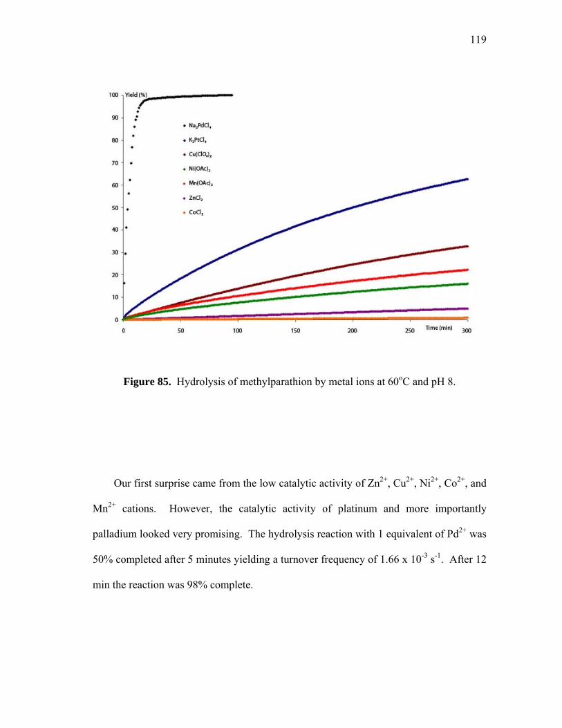
119
Figure 85. Hydrolysis of methylparathion by metal ions at 60oC and pH 8.
Our first surprise came from the low catalytic activity of Zn2+, Cu2+, Ni2+, Co2+, and
Mn2+ cations. However, the catalytic activity of platinum and more importantly
palladium looked very promising. The hydrolysis reaction with 1 equivalent of Pd2+ was
50% completed after 5 minutes yielding a turnover frequency of 1.66 x 10⁻³ s-1. After 12
min the reaction was 98% complete.

120
6.4. Dinuclear Pd(II) complexes catalysis
Based on the above observations, we decided to investigate the use of the dinuclear
palladium complexes 97, 98, and 99 as catalysts for the hydrolysis of paraoxon (19), and
parathion (20). For our first experiments, catalyst 97 was chosen and tested. The
reactions were followed by monitoring the absorbance of the p-nitrophenol released after
addition of the palladium catalyst to a buffered solution of the organophosphotriester.
Just like for the Cu (II) complexes 85 and 89, no catalytic activity was observed from pH
7 to pH 10 for the hydrolysis reaction of paraoxon. However, when the same experiment
was carried out with the more reactive substrate parathion, catalysis was readily
observed. These experiments were monitored by following the release of p-nitrophenol
in aqueous media by UV at 400 nm. This experiment was done at room temperature
with 3.5 × 10⁻⁵ M of 20 and 1.75 × 10⁻⁶ M of catalyst (10% Pd) in a 5.7 x 10⁻³ CHESS
solution (pH 9). We rapidly noticed that even with the use of only 0.1 equivalents on
catalyst, the hydrolysis of 20 was observed at a high rate (figure 79). The hydrolysis
reaction by 97 was 50% completed after 29 minutes yielding a turnover frequency of
2.87 x 10⁻³ s-1. After 2.6 hours the reaction was 90% complete. The exact same
experimental procedure was followed for catalysts 98 and 99. It appears that catalyst 98
yielded the most promising results yet (figure 86). The hydrolysis reaction by 98 was
50% completed after 10 minutes yielding a turnover frequency of 8.33 x 10⁻³ s-1. After
34 min the reaction was 95% complete. As for the last catalyst 99, only a turnover
frequency of 2.66 x 10⁻⁴ s-1 was observed (figure 86).

121
H2O, RT, Ph 9+
SP
O
OO
NO2 HSP
O
OO
OH
NO2
N N
O O
Pd PdO
OOO
N N
O O
Pd PdO
OOO
N N
O O
Pd PdO
OOO
Catalyst (5%)
97
98
99
Figure 86. Hydrolysis of methylparathion by 97, 98, and 99 at RT at pH 9.
122
Figure 86. Hydrolysis of methylparathion by 97, 98, and 99 at RT at pH 9.
In order to obtain a more detailed insight in the kinetics involved in this reaction,
catalytic experiments were carried out at various substrate and catalyst (97)
concentrations at pH 9 (CHES, at 5.7 mM), and were analyzed using the initial slope
methods. As indicated by this kinetic analysis, the reaction rate follows a second order
kinetics with a first order dependence on substrate (figure 87) and palladium (figure 88)
(v = k2[Pd][parathion]). The k2 value of 190 M-1s-1 obtained from these measurements is
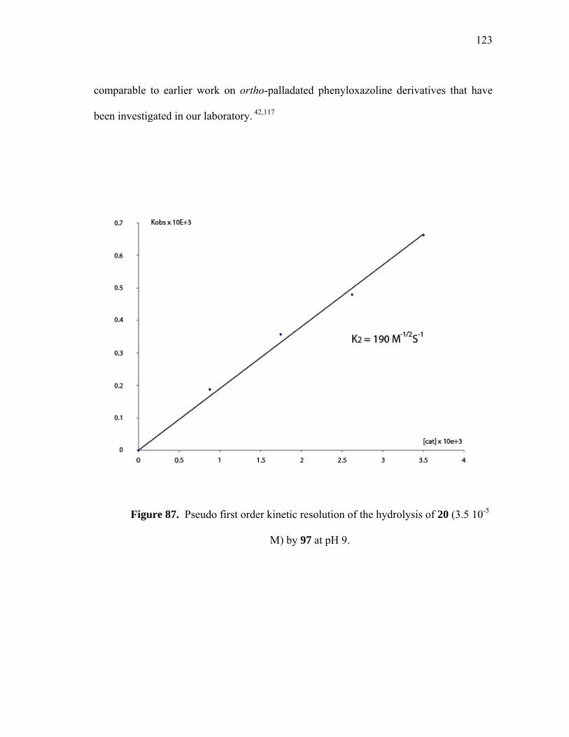
123
comparable to earlier work on ortho-palladated phenyloxazoline derivatives that have
been investigated in our laboratory. 42,117
Figure 87. Pseudo first order kinetic resolution of the hydrolysis of 20 (3.5 10-5
M) by 97 at pH 9.

124
Figure 88. Pseudo first order kinetic resolution of the hydrolysis of 20 by 97 (1.75 10-6
M) at pH 9.
6.5. Mononuclear Pd(II) complexes catalysis
In order to compare the activity of the dinculear catalysts 97, 98, and 99 to that of
a mononuclear derivative, the monofunctional palladium complex 91 was investigated.
(figure 89). The catalysis experiments were carried out also at various methylparathion
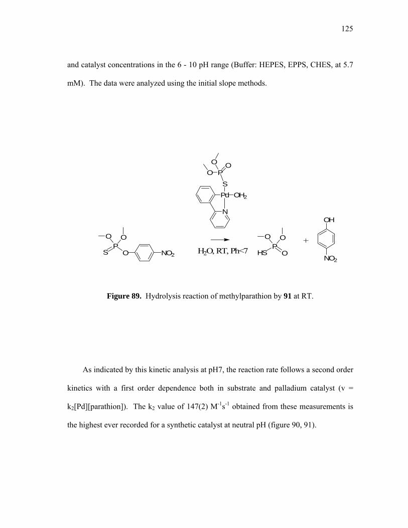
125
and catalyst concentrations in the 6 - 10 pH range (Buffer: HEPES, EPPS, CHES, at 5.7
mM). The data were analyzed using the initial slope methods.
H2O, RT, Ph<7+
SP
O
OO
NO2 HSP
O
OO
OH
NO2
N
POO
O
PdS
OH2
Figure 89. Hydrolysis reaction of methylparathion by 91 at RT.
As indicated by this kinetic analysis at pH7, the reaction rate follows a second order
kinetics with a first order dependence both in substrate and palladium catalyst (v =
k2[Pd][parathion]). The k2 value of 147(2) M-1s-1 obtained from these measurements is
the highest ever recorded for a synthetic catalyst at neutral pH (figure 90, 91).

126
Figure 90. Pseudo first order kinetic resolution of the hydrolysis of methylparathion
(3.5 10-5 M) by 91 at pH 7.

127
Figure 91. Pseudo first order kinetic resolution of the hydrolysis of methylparathion by
91 (1.75 10-6 M) at pH 7.
In fact, the k2 value reported herein at neutral pH is comparable to those reported by
Ryabov for organoplatinum and palladium complexes in the 8.5 – 9.5 pH range. Since
91 is the only species observed in solution, it is logical to assume that the catalyst is a
mononuclear palladium species. In an effort to understand the structure of 91 in
solution, a number of physical measurements have been carried out. Pulse field gradient
spin-echo (PGSE) has emerged as a valid method for the determination of diffusion

128
coefficients and hydrodynamic radii of organometallic compounds.102 When the PGSE
experiment of complex 91 in DMSO-d6 was carried out (figure 92), the
dimethylthiophosphate and phenylpyridine moity were found to diffuse at the same rate
indicating that dimethylthiophosphate anion is still ligated to the palladium center. This
result suggests that, in solution, dimethylthiophosphate is still bound to the palladium
center during the catalytic process (figure 93).
Figure 92. 1H PGSE analysis of 91 (1 mg) in DMSO-d6.

129
OHO2N
SP
O
OO
NO2
H2O
H2O
H2O
N
Pd
SP
OOO
2
N
Pd
SP
OOO
N
Pd
SP
OOO
S PO
O
OH2
N
Pd
SP
OOO
S PO
OOH O NO2
H2O
-SP
O
OO
Figure 93. Proposed mechanism for the hydrolysis of 20 by 91 at neutral pH.

130
At pH9, the reaction rate follows another kinetic pathway with the same first order
dependence in substrate (figure 94) but a half order dependence in palladium catalyst
(figure 95). The catalytic activity of compound 91 has a turnover frequency of 1.6 × 10⁻²
s-1. The data can be fit to an expression for the hydrolysis of methylparathion by
compound 91 as in the following equation: k2Keq1/2 = 3.02(1) M-1/2s-1 (v =
k2Keq1/2[Pd]1/2[parathion]; Keq = [monomer]2 /[Dimer]). These observations can be
reconciled on the basis of a mechanism in which the active form of the catalyst is a
monomer whose concentration is controlled by an equilibrium with a dimer (figure 96).
In order to further investigate such drastic increase in the catalytic activity of the
system from neutral to alkaline media, the pH of the catalytic hydrolysis of 20 by
compound 91 was studied between pH 7 and 9. This profile reveals the existence of 2
pH independent plateaus connected by an intermediate regime where the rate of
hydrolysis depends on the pH (figure 97). Furthermore, the UV spectrum of 91
depending on the pH was investigated (figure 98). The corresponding plot showed the
decrease of a UV band at 258 nm establishing that a new species was forming in
solution: Complex 91 at neutral pH, and the hydroxo derivative 100 at higher pH (figure
99).
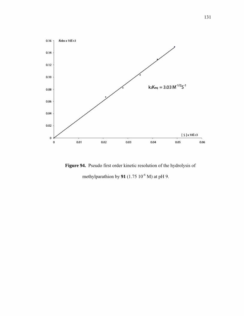
131
Figure 94. Pseudo first order kinetic resolution of the hydrolysis of
methylparathion by 91 (1.75 10-6 M) at pH 9.
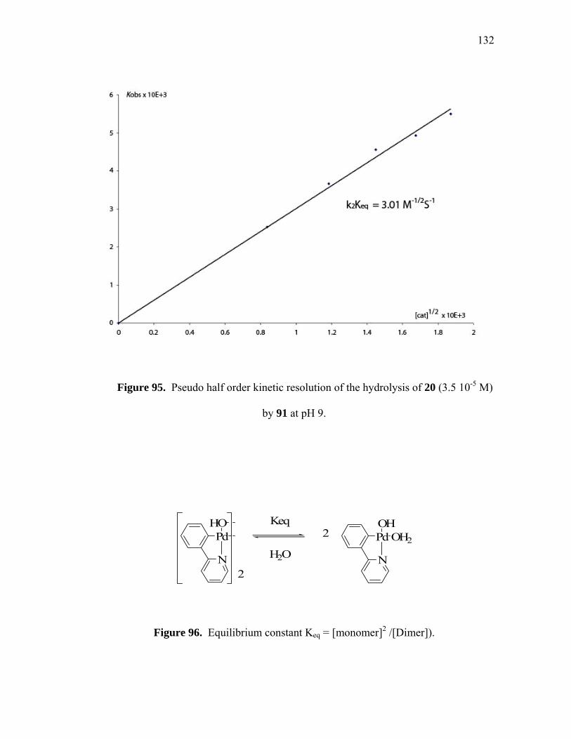
132
Figure 95. Pseudo half order kinetic resolution of the hydrolysis of 20 (3.5 10-5 M)
by 91 at pH 9.
N
Keq
N2
H2O
2PdHO
PdOH
OH2
Figure 96. Equilibrium constant Keq = [monomer]2 /[Dimer]).

133
Figure 97. Expression of the Kobs vs pH for the hydrolysis of methylparathion by 91
([methylparathion] = 3.5 10-5 M; [91] = 1.75 10-6 M).

134
Figure 98. UV recording of 91 (35 µmol) in aqueous buffer with 16% dioxane from pH
7 to pH 9.5.
N
HO- PdOH
OH2
N
POO
O
PdS
OH2
100
Figure 99: Complex 91 equilibrium between neutral and basic pH.
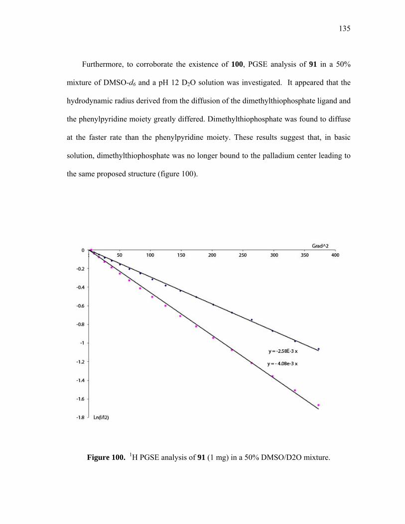
135
Furthermore, to corroborate the existence of 100, PGSE analysis of 91 in a 50%
mixture of DMSO-d6 and a pH 12 D2O solution was investigated. It appeared that the
hydrodynamic radius derived from the diffusion of the dimethylthiophosphate ligand and
the phenylpyridine moiety greatly differed. Dimethylthiophosphate was found to diffuse
at the faster rate than the phenylpyridine moiety. These results suggest that, in basic
solution, dimethylthiophosphate was no longer bound to the palladium center leading to
the same proposed structure (figure 100).
Figure 100. 1H PGSE analysis of 91 (1 mg) in a 50% DMSO/D2O mixture.
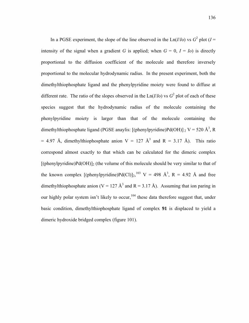
136
In a PGSE experiment, the slope of the line observed in the Ln(I/Io) vs G2 plot (I =
intensity of the signal when a gradient G is applied; when G = 0, I = Io) is directly
proportional to the diffusion coefficient of the molecule and therefore inversely
proportional to the molecular hydrodynamic radius. In the present experiment, both the
dimethylthiophosphate ligand and the phenylpyridine moiety were found to diffuse at
different rate. The ratio of the slopes observed in the Ln(I/Io) vs G2 plot of each of these
species suggest that the hydrodynamic radius of the molecule containing the
phenylpyridine moiety is larger than that of the molecule containing the
dimethylthiophosphate ligand (PGSE anaylis: [(phenylpyridine)Pd(OH)] 2 V = 520 Å3, R
= 4.97 Å, dimethylthiophosphate anion V = 127 Å3 and R = 3.17 Å). This ratio
correspond almost exactly to that which can be calculated for the dimeric complex
[(phenylpyridine)Pd(OH)]2 (the volume of this molecule should be very similar to that of
the known complex [(phenylpyridine)Pd(Cl)]2,103 V = 498 Å3, R = 4.92 Å and free
dimethylthiophosphate anion (V = 127 Å3 and R = 3.17 Å). Assuming that ion paring in
our highly polar system isn’t likely to occur,104 these data therefore suggest that, under
basic condition, dimethylthiophosphate ligand of complex 91 is displaced to yield a
dimeric hydroxide bridged complex (figure 101).
137
N
HO-Pd
O
O
N
PdH
HN
POO
O
2
PdS
Figure 101. Formation of the dimeric hydroxide bridged complex
This proposal is in agreement with the 31P {1H}NMR spectrum of 91 in a 50%
mixture of DMSO-d6 and a pH 12 D2O/DO- solution. Indeed, this spectrum reveals the
presence of a resonance at 59.7 ppm which correspond to the chemical shift of the free -
SP(=O)(OMe)2 derivative.
From the latest analytical and kinetic studies the following mechanism for the
hydrolysis of methylparathion has been proposed (figure 102). In a first step, compound
91 monomerize to the active species hydroxy-bis[2-(2-pyridyl)phenyl-
C,N]dipalladium(II), hydrate is complexed by the methylparathion followed by rapid
hydrolysis and release of the nontoxic thiophosphate.

138
N
Pd OHOH2
H2O Keq
N
PdHO
2
N
Pd OH
SP
O
OO
NO2
O-
NO2N
Pd OH2
SP
OH
OO
N
Pd OH2
SP
O
OO
-SP
O
OO
SP
O
OO
NO2
H2O
HO-
H2O
HO-
H2O
Figure 102. Proposed mechanism for the hydrolysis of 20 by 91 at basic pH.
139
6.6. Summary
The results presented in this chapter indicate that both mononuclear and dinuclear
organopalladium catalysts are competent in the hydrolysis of parathion and by far
outperform copper (II) coordination complexes. Another important aspect of the work
described in this chapter concerns the fact that dinuclear palladium complexes do not
appear more active than mononuclear palladium complexes. In fact, the mononuclear
palladium catalyst 91 is very potent and features the highest activity ever recorded for an
artificial parathion hydrolysis catalyst. Remarkably, this catalyst is active at slightly
acidic and neutral pH where the reaction follows a second order rate law with a first
order dependence in palladium catalyst and parathion (v(pH=9) = k2(pH=9)[parathion][cat]
with k2(pH=9) = 147 M-1s-1). It becomes even more active at pH 9 where the rate law
displays a half order dependence in catalyst and a first order dependence in parathion
(v(pH=9) = k2(pH=9)Keq1/2[cat]1/2[parathion] with k2(pH=9)Keq
1/2 = 3.02 M-1/2s-1). While the
change in the kinetic rate law does not allow for a direct comparison of the activity, the
initial rate of the catalytic reaction at pH 9 is faster than that of pH 7 by an acceleration
factor A given by:
A = v(pH=9)/ v(pH=7) = k2(pH=9)Keq1/2 / (k2(pH=7) × [cat]1/2)
With a palladium catalyst concentration of 1.75 10-5 M, the acceleration factor A is
equal to 4.78. In fact, initial rate of the catalytic reaction at pH 9 is comparable to that

140
observed by Ryabov with more expensive platinum catalysts (k2 = 200-900 M-1s-1).
Because of the half order dependence in palladium catalysts observed in the case of 91,
the acceleration factor A observed at lower palladium concentration would be even
greater so that 91 would outperform the platinum catalysts reported by Ryabov at
micromolar catalyst concentrations. For example, at pH 9 and with a catalyst
concentration of 3.5 10-6 M, the initial rate of the reaction with 91 as a catalyst is faster
than that employing the platinum catalyst 32 (figure 13, k2 = 914 M-1s-1) by an
acceleration factor A given by:
A = v(cat=91)/ v(cat=32) = k2(cat=91)Keq1/2 / (k2(cat=32) × [cat]1/2) = 1.76
6.7. Experimental
Catalytic hydrolysis of methylparathion by complex 82: In a typical experiment,
the cell was filled with 2.0 mL of a buffer solution (HEPES, 6 < pH < 7; EPPS, 7.5 < pH
<8.5; CHES, 9 < pH < 10 ; 10 mM) and 0.278 mL of a stock solution of catalyst in
dioxane ([Pd] = 2 × [82] = 4.4 × 10⁻⁵ M). Following addition of 0.278 mL of a stock
solution of methylparathion ([methylparathion] = 4.4 × 10⁻⁴ M in dioxane), the total
volume of the solution was adjusted to 3.5 mL by addition of water and the reaction was
followed by monitoring the release of p-nitrophenol which features an absorption band
at 400 nm (ε = 18600 M-1 cm-1 at pH 9). The dependence of the reaction rate on
methylparathion was established by varying the methylparathion concentration at
141
constant catalyst concentration (([Pd] = 3.5 x 10⁻⁶ M). The dependence of the reaction
rate on catalyst was established by varying the catalyst concentration at constant
methylparathion concentration (3.5 x 10⁻⁵ M).
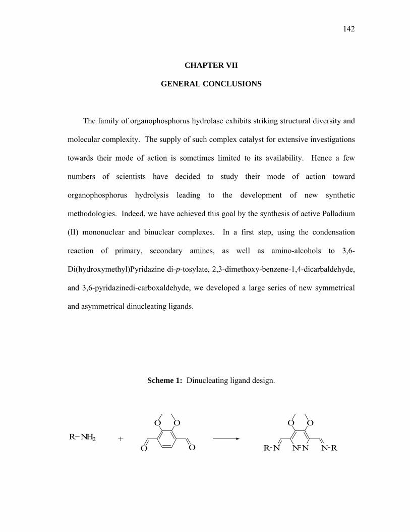
142
CHAPTER VII
GENERAL CONCLUSIONS
The family of organophosphorus hydrolase exhibits striking structural diversity and
molecular complexity. The supply of such complex catalyst for extensive investigations
towards their mode of action is sometimes limited to its availability. Hence a few
numbers of scientists have decided to study their mode of action toward
organophosphorus hydrolysis leading to the development of new synthetic
methodologies. Indeed, we have achieved this goal by the synthesis of active Palladium
(II) mononuclear and binuclear complexes. In a first step, using the condensation
reaction of primary, secondary amines, as well as amino-alcohols to 3,6-
Di(hydroxymethyl)Pyridazine di-p-tosylate, 2,3-dimethoxy-benzene-1,4-dicarbaldehyde,
and 3,6-pyridazinedi-carboxaldehyde, we developed a large series of new symmetrical
and asymmetrical dinucleating ligands.
Scheme 1: Dinucleating ligand design.
O O
O O
N NN NR R
O O
NH2R +

143
Scheme 1: Continued.
N NO O
N NTsO OTs
R2
HO NH2
R1
NHR1
R2
NH2R
R2
HO NH
R1
R3
O
HO NH2
R1
O
O NH2
R1
R2
+
N NN NR R
N NNH HNR R
N NN NR RR' R'
With the use of simple techniques such as 1H NMR and UV titration, these
dinucleating ligands revealed interesting coordination chemistry properties chelating 2
metal centers such as Cu (II) while preserving the C2 symmetry of the complexes. The
orthopalladation reaction of our latest ligands was also a success. By reacting ligands
61, 63, 65 with 3 equivalent of Na2PdCl4 in a 1:1 CH2Cl2/ethanol mixture followed by
the displacement of the chlorines with Thallium Acetonylacetonate, the isolations of the
desired products were successfully achieved. Reaction of methylparathion and with [2-
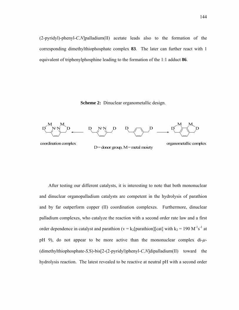
144
(2-pyridyl)-phenyl-C,N]palladium(II) acetate leads also to the formation of the
corresponding dimethylthiophosphate complex 83. The later can further react with 1
equivalent of triphenylphosphine leading to the formation of the 1:1 adduct 86.
Scheme 2: Dinuclear organometallic design.
NN DD DD
D = donor group, M = metal moiety
NN DD DDM MMM
coordination complex organometallic complex
After testing our different catalysts, it is interesting to note that both mononuclear
and dinuclear organopalladium catalysts are competent in the hydrolysis of parathion
and by far outperform copper (II) coordination complexes. Furthermore, dinuclear
palladium complexes, who catalyze the reaction with a second order rate law and a first
order dependence in catalyst and parathion (v = k2[parathion][cat] with k2 = 190 M-1s-1 at
pH 9), do not appear to be more active than the mononuclear complex di-µ-
(dimethylthiophosphate-S,S)-bis[2-(2-pyridyl)phenyl-C,N]dipalladium(II) toward the
hydrolysis reaction. The latest revealed to be reactive at neutral pH with a second order
145
rate law and a first order dependence in catalyst and parathion (v = k2[parathion][cat]
with k2 = 150 M-1s-1). It becomes even more active at pH 9 where the rate law displays a
half order dependence in catalyst and a first order dependence in parathion (v =
k2Keq1/2[cat]1/2[parathion] with k2Keq
1/2 = 3.02 M-1/2s-1). Remarkably, this mononuclear
catalyst is very potent and features the highest activity ever recorded for a synthetic
parathion hydrolysis catalyst.
146
REFERENCES
1 (a) A Guide to Organophosphorus Chemistry Quin, L. D. Wiley, New York, 2000. (b)
Pesticides Remediation in Soils and Water Roberts, T.; Keaney, P. C. Wiley, New York,
1998.
2 Algeria, H. A.; Shaw, T, J, Environ. Sci. Technol. 1999, 33, 850.
3 Gilliom, R. J.; Barbash, J. E.; Kolpin, D. W. Larson, S. J. Environ. Sci. Technol. 1999,
33, 164 A.
4 Squillace, P. J.; Scott, J. C.; Moran, M. J.; Nolan, B. T.; Kolpin, D. W. Environ. Sci.
Technol. 2002, 36, 1923.
5 Water Resources Assessment Matzner, R.; Waldman, E. U.S. Environmental Protection
Agency, Washington DC, 1999.
6 Military Chemical and Biological Agents, Chemical and Toxicological Properties
Compton, J. A. The Telford Press, Caldwell, NJ, 1987.
7 Chemical Warfare Agents: Toxicology and Treatments Marrs, T. C.; Maynard, R. L.
Sidell, F. R. Wiley, New York, 1996, p1-26.
8 Benschop, H. P.; de Jong, L. P. A. Acc. Chem. Res. 1988, 21, 368.
9 Chemical Warfare Agents Somani, S. M. Academic Press, New York, 1992, Chapter
IV.
10 Sultatos, L. G. J. Toxicol. Environ. Health 1994, 43, 271.
147
11 (a) Ember, L. S. Chem. Eng. News 1997, 75(11), 22. (b) Yang, Y. C. Acc. Chem. Res.
1999, 32, 109.
12 (a) Gustafson, R. L.; Martell, A. E. J. Am. Chem. Soc. 1962, 84, 2309. (b) Ward, J. R.;
Yang, Y. C.; Wilson, R. B. Jr.; Burrows, W. D.; Ackman, L. L. Bioorg. Chem. 1988, 16,
12. (c) Epstein, J.; Bauer, V. E.; Saxe, M.; Demek, M. M. J. Am. Chem. Soc. 1956, 78,
4068.
13 Development of Decontamination Solution DS2 Jackson, J. B. CWLR, 2368, April,
1960.
14 Reactions of Chemical Warfare Agents with DS2 Beaudry, W. T.; Szafraniec, L. L.;
Leslie, D. R. CRDEC-TR-364, June, 1992.
15 (a) Verma, K. K.; Gupta, A. K. Talanta 1982, 102, 18. (b) De Munari, S.; Frigerio,
M.; Santagostino, M. J. Org. Chem. 1996, 61, 9272. (c) Nicolaou, K. C.; Baran, P. S.;
Zhong, Y. L. J. Am. Chem. Soc. 2001, 123, 3183. (d) Wirth, T. Angew. Chem., Int. Ed.
2001, 40, 2812.
16 (a) Moss, R. A.; Alwis, K. W.; Bizzigotti, G. O. J. Am. Chem. Soc. 1983, 105, 681.
(b) Moss, R. A.; Alwis, K. W.; Shin, J. S. J. Am. Chem. Soc. 1984, 106, 2651.
17 (a) Segues, B.; Rico-Lattes, I Bull. Soc. Chim. Fr. 1996, 133,.925. (b) Stang, P. J.;
Zhdankin, V. V. Chem. Rev. 1996, 96, 1123.
18 (a) The Chemistry of Halides, Pseudo Halides and Azides Koser, G. F. Wiley, New
York, 1995. (b) Katritzky, A. R.; Savage, G. P; Palenik, G. J.; Qian, K.; Zhang, Z. J.

148
Chem. Soc. Perkin Trans. 2 1990, 1657. (c) Moss, R. A.; Bracken, K.; Emge, T. J. J.
Org. Chem. 1995, 60, 7739.
19 Moss, R. A.; Zhang, H.; Chatterjee, S.; Krogh-Jespersen, K. Tetrahedron. Lett. 1993,
34, 1729.
20 Surfactants and Polymers in Aqueous Solutions Jönsson, B.; Lindman, B.; Holmberg,
K.; Krongberg, B. Wiley, New York, 1998.
21 Moss, R. A.; Morales-Rojas, H. Langmuir 2000, 16, 6485.
22 (a) Panetta, C. A.; Garlick, S. M.; Durst, H. D.; Longo, F. R.; Ward, J. R. J. Org.
Chem. 1990, 55, 5202. (b) Moss, R. A.; Fujiyama, R.; Zhang, H.; Chung, Y. C.;
McSorley, K. Langmuir, 1993, 9, 2902.
23 (a) Bunton, C. A.; Ihara, Y. J. Org. Chem. 1977, 42, 2865. (b) Bunton, C. A.; Nelson,
S. E.; Quan, C. J. Org. Chem. 1982, 47, 1157.
24 Terrier, F.; MacCormack, P.; Kizilian, E.; Halle, J. C.; Demersemen, P.; Guir, F.;
Lion, C. J. J. Chem. Soc. Perkin trans. 2 1991, 153.
25 Biresaw, G.; Bunton, C. A. J. Phys. Chem. 1986, 90, 5849.
26 Bunton, C. A.; Mhala, M. M.; Moffatt, J. R.; J. Phys. Org. Chem. 1990, 9, 2832.
27 Yang, Y. C.; Berg, F. J.; Szafraniec, L. L.; Beaudry, W. T.; Bunton, C. A.; Kumar, A.
J. Chem. Soc. Perkin Trans. 2 1997, 607.
28 (a) Lion, C.; Hedayatullah, M.; Bauer, J. P.; Charvy, C.; Delmas, G.; Despangne, B.;
Sentenac-Roumanou, H. Bull. Soc. Chem. Belg. 1991, 100, 555. (b) Bhattacharya, S.;
Snehalatha, K. J. Org. Chem. 1997, 62, 2198.
149
29 DeBruin, K. E.; Tang, C. I. W.; Johnson, D. M.; Wilde, R. L. J. Am. Chem. Soc. 4989,
111, 5871.
30 Oumar-Mahamat, H.; Slebocka-Tilk, H.; Brown, R. S. Can. J. Chem. 1999, 77, 1577.
31 Review of Pesticide Disposal Research Munnecke, D. M.; Day, R.; Trask, H. W. U.S.
Environmental Protection Agency, Washington DC, 1976.
32 Vilanova, E.; Sogorb, M. A. Crit. Rev. Toxicol. 1999, 29, 21.
33 Raushel, F. M.; Holden, H. M Advances in Enzymology and Related Areas of
Molecular Biology 2000, 74, 51.
34 (a) Dumas, D. P.; Caldwell, S. R.; Wild, J. R.; Raushel, F. M. J. Biol. Chem. 1989,
264, 19659. (b) Dumas, D. P.; Durst, H. D.; Landis, W. G.; Raushel, F. M.; Wild, J. R.
Arch. Biochem. Biophys. 1990, 277, 155.
35 Benning, M. M.; Kuo, J. M.; Raushel, F. M.;. Holden, H. M. Biochemistry 1994, 33,
15001.
36 Omburo, G. A.; Kuo, J. M.; Mullins, L. S.; Raushel, F. M. J. Biol. Chem. 1992, 267,
13278.
37 Vanhooke, J. L.; Benning, M. M.; Raushel, F. M.; Holden, H. M. Biochemistry 1996,
35, 6020.
38 Chae, M. Y.; Postula, J. F.; Raushel, F. M. Bioorg. Med. Chem. Lett. 1994, 4, 1473.
39 Lewis, V. E.; Donarksi, W. J.; Wild, J. R.; Raushel, F. M. Biochemistry 1988, 27,
1591.
40 Lai, K.; Dave, K. I.; Wild, J. R. J. Biol. Chem. 1994, 269, 16579.

150
41 Kursheva, L. I.; Il’in, A. M.; Batyeva, E. S.; Kataeva, O. N.; Gubaidullin, A. T. Russ.
J. Gen. Chem. 2001, 71, 484.
42 Kim, M.; Gabbai, F.P J. Chem. Soc., Dalton Trans. 2004, articles ASAP.
43 Shi, Q.; Cao, R.; Hong, M. C.; Wang, Y. Y.; Shi, Q. Z. Trans. Met. Chem. 2001, 26,
657.
44 (a) Wadsworth, W. S. J. Org. Chem. 1981, 46, 4080. (b) Hendry, P.; Sargeson, A. M.
Aust. J. Chem. 1986, 39, 1177. (c) Morrow, J. R.; Trogler, W. C. Inorg, Chem. 1989, 28,
2330. (d) Chin, J. Acc. Chem. Res. 1991, 24, 145.
45 Wagner-jauregg, T.; Hackley, B. E.; Lies, T. A.; Owen, O. O.; Proper, R. J. J. Am.
Chem. Soc. 1955, 77, 922.
46 (a) Zeinali, M.; Torrents, A. Environ. Sci. Technol. 1998, 32, 2338. (b) Smolen, J. M.;
Stone, A. L. Environ. Sci. Technol. 1997, 31, 1664.
47 Hay, R. W.; Govan, N. Chem. Comm. 1990, 714.
48 Oh, S. J.; Yoon, C. W.; Park, J. W. J. Chem. Soc. Perkin Trans. 2 1996, 329.
49 Courtney, R. C.; Gustafson, R. L.; Westerback, S. J.; Hyytiainen, H.; Chabarek, S. C.;
Martell, A. E. J. Am. Chem. Soc. 1957, 79, 3030.
50 Gellman, S. H.; Petter, R.; Breslow, R. J. Am. Chem. Soc. 1986, 108, 2388.
51 (a) Bunton, C. A.; Scrimin, P.; Tecilla, P. J. Chem. Soc. Perkin Trans. 2 1996, 419.
(b) Scrimin, P.; Tecilla, P.; Tonellato, U. J. Org. Chem. 1991, 56, 161.
52 Scrimin, P.; Ghirlanda, G.; Tecilla, P.; Moss, R. A. Langmuir 1996, 12, 6235.
53 Kuo, L. Y.; Perera, N. M. Inorg. Chem. 2000, 39, 3117.

151
54 Kazankov, G. M; Sergeeva, V. S.; Efremenko, E. N.; Alexandrova, L.; Varfolomeev,
S. D.; Ryabov, A. D. Angew. Chem., Int. Ed. 2000, 39, 3117.
55 Carlsson, H.; Haukka, M.; Nordlander, E. Inorg. Chem. 2004, 20(5), 5681-5687.
56 Gutierrez, M. A.; Newkome, G. R.; Selbin J. J. Organometallic Chem. 1980, 202,
341-350.
57 (a) Sträter, N.; Lipscomb, W. N.; Klabunde, T.; Krebs, B. Angew. Chem., Int. Ed.
Engl. 1996, 35, 2024. (b) Wilcox, D. E. Chem. Rev. 1996, 96, 2435.
58 (a) Williams, N. H.; Takasaki, B.; Wall, M.; Chin, J. Acc. Chem. Res. 1999, 32(6),
485-493. (b) Van Den Beuken, E. K.; Feringa, B. L. Tetrahedron 1998, 54(43), 12985-
13011. (c) Corey, E. J.; Bakshi, R. K.; Shibata, S.; Chen, C. P.; Singh, V. K. J. Am.
Chem. Soc. 1987, 109(25), 7925-6. (d) Corey, E. J.; Bakshi, R. K.; Shibata, S. J. Am.
Chem. Soc. 1987, 109(18), 5551-3. (e) Noyori, R.; Kitamura, M. Angew. Chem., Int. Ed.
Engl. 1991, 30(1), 34-48.
59 (a) Ooi, T.; Takahashi M.; Maruoka, K. J. Am. Chem. Soc. 1996, 118, 11307-11308.
(b) Ooi, T.; Kii, S.; Hanawa, H.; Maruoka, K. Tetrahedron Lett. 1998, 39, 3729-3732.
(c) Ooi, T.; Saito, A.; Maruoka, K. Tetrahedron Lett. 1998, 39, 3745-3748. (d) Ooi, T.;
Asao, N.; Maruoka, K. J. Syn. Org. Chem. Jpn. 1998, 56, 377-385. (e) Maruoka, K..
Catal. Today 2001, 66, 33-45.
60 (a) Wuest, J. D. Acc. Chem. Res. 1999, 32, 81-89. (b) Wuest, J. D.; Zacharie, B. J.
Am. Chem. Soc. 1985, 107, 6121-6123
61 Lee, H.; Diaz, M.; Hawthorne, M. F Tetrahedron Lett. 1999, 40, 7651-7655.

152
62 (a) Jacobsen, E. N. Acc. Chem. Res. 2000, 33, 421-431. (b) Annis, D. A.; Jacobsen, E.
N. J. Am. Chem. Soc. 1999, 121, 4147-4154. (c) Konsler, R. G.; Karl, J.; Jacobsen, E. N.
J. Am. Chem. Soc. 1998, 120, 10780-10781. (d) Fields, L. B.; Jacobsen, E. N.
Tetrahedron: Asymmetry 1993, 4, 2229-40.
63 (a) Matsunaga, S.; Das, J.; Roels, J.; Vogl, E. M.; Yamamoto, N.; Iida, T.; Yamaguchi,
K.; Shibasaki, M. J. Am. Chem. Soc. 2000, 122, 2252-2260. (b) Shibasaki, M.; Groger,
H. Top. Organomet. Chem. 1999, 2, 199-232. (c) Shibasaki, M. Enantiomer 1999, 4,
513-527. (d) Yamada, K.-I.; Harwood, S. J.; Groger, H.; Shibasaki, M. Angew. Chem.,
Int. Ed. 1999, 38, 3504-3506. (e) Yamasaki, S.; Iida, T.; Shibasaki, M. Tetrahedron
1999, 55, 8857-8867. (f) Shibasaki, M.; Sasai, H. Adv. Asymmetric Synth. 1998, 3, 191-
233. (g) Shibasaki, M.; Sasai, H.; Arai, T.; Iida, T. Pure Appl. Chem. 1998, 70, 1027-
1034. (h) Shibasaki, M.; Sasai, H.; Arai, T. Angew. Chem., Int. Ed. Engl. 1997, 36,
1237-1256.
64 Kii, S.; Maruoka, K. Tetrahedron Lett. 2001, 42, 1935-1939.
65 For a recent example of a chiral dinucleating ligand see: Trost, B. M.; Ito, H.; Silcoff,
E. R. J. Am. Chem. Soc. 2001, 123, 3367-3368.
66 Oxidation, MMO, Oxygen transport, transesterification.
67 (a) Koike, T.; Inoue, M.; Kimura, E.; Shiro, M. J. Am. Chem. Soc. 1996, 118, 3091.
(b) Young M. J.; Wahnon D.; Hynes R. C.; Chin J. J. Am. Chem. Soc. 1995, 117, 9441.
(c)Takasi, B. T.; Chin, J. J. Am. Chem. Soc. 1995, 117, 8582. (d) Oh, S. J.; Song, K. H.;
Whang, D.; Kim, K.; Yoon, T. H.; Moon, H.; Park, J. W. Inorg. Chem. 1996, 35, 3780.

153
(e) Hettich, R.; Schneider, H.-J. J. Am. Chem. Soc. 1997, 119, 5638. (f) Ott, R.; Krämer,
R. Angew. Chem., Int. Ed. 1998, 37, 1957. (g) Bracken, K.; Moss, R. A.; Ragunathan,
K. G. J. Am. Chem. Soc. 1997, 119, 9323. (h) Kim, A. D.; Burstyn, J. N. Inorg. Chem.
1996, 35, 2792. (i) Molenveld, P.; Engbersen, J. F. J.; Kooijman, H.; Spek, A. L.;
Reinhoudt, D. N. J. Am. Chem. Soc. 1998, 120, 6726. (j) Gajda, T.; Duepre, Y.;
Toeroek, I.; Harmer, J.; Schweiger, A.; Sander, J.; Kuppert, D.; Hegetschweiler, K.
Inorg. Chem. 2001, 40(19), 4918-4927. (k) Kövári, E.; Krämer, R. J. Am. Chem. Soc.
1996, 118, 12704. (l) Liu, S.; Hamilton, A. D. Chem. Commun. 1999, 587.
68 (a) Maruoka, K. Catal. Today 2001, 66(1), 33-45. (b) Hanawa, H.; Kii, S.; Asao, N.;
Maruoka, K. Tetrahedron Lett. 2000, 41(29), 5543-5546. (c) Asao, N.; Kii, S.; Hanawa,
H.; Maruoka, K. Tetrahedron Lett. 1998, 39(22), 3729-3732.
69 Ooi, T.; Takahashi, M.; Maruoka, K. J. Am. Chem. Soc. 1996, 118(45), 11307-11308.
70 (a) Shibasaki, M.; Sasai, H.; Arai, T. Angew. Chem., Int. Ed. Engl. 1997, 36(12),
1237-1256. (b) Yoshikawa, N.; Kumagai, N.; Matsunaga, S.; Moll, G.; Ohshima, T.;
Suzuki, T.; Shibasaki, M. J. Am. Chem. Soc. 2001, 123(16), 3856. (c) Kumagai, N.;
Matsunaga, S.; Yoshikawa, N.; Ohshima, T.; Shibasaki, M. Org. Lett. 2001, 3(10), 1539-
1542.
71 (a) Noyori, R.; Kitamura, M.; Nakamura, A. Angew. Chem. Int. Ed. Engl., 1991, 30,
49. (b) Kitamura, M.; Suga, S.; Niwa, M.; Noyori, R. J. Am. Chem. Soc., 1995, 117,
4832-4842.
72 Trost, B. M.; Silcoff, E. R.; Ito, H. Org. Lett. 2001, 3(16), 2497-2500.

154
73 Jacobsen, E. N. Acc. Chem. Res. 2000, 33(6), 421-431.
74 (a) Barrios, A. M.; Lippard, S. J. Inorg. Chem. 2001, 40, 1060-1064. (b) Manzur, J.;
Garcia, A. M.; Gomez, B.; Spodine, E. Polyhedron 2000, 19, 2367-2372. (c)
Robichaud, P.; Thompson, L. K. Inorg. Chim. Acta 1984, 85, 137-142. (d) Thompson,
L. K.; Mandal, S. K.; Rosenberg, L.; Lee, F. L.; Gabe, E. J. Inorg. Chim. Acta 1987, 133,
81-91. (e) Sullivan, D. A.; Palenik, G. J. Inorg. Chem. 1977, 16, 1127-1133. (f)
Thompson, L. K.; Chako, V. T.; Elvidge, J. A.; Lever, A. B. P.; Parish, R. V. Can. J.
Chem. 1969, 47, 4141-4152.
75 (a) Fahrni, C. J.; Pfaltz, A. Helv. Chim. Acta 1998, 81(3), 491-506. (b) Fahrni, C. J.;
Pfaltz, A.; Neuburger, M.; Zehnder, Ma. Helv. Chim. Acta 1998, 81(3), 507-524.
76.Brooker, S.; Kelly, R. J. J. Chem. Soc., Dalton Trans. 1996, 10, 2117-2122.
77 Sueur, S.; Lagrenee, M.; Abraham, F.; J. Heterocyclic Chem., 1987, 24, 1285-1289.
78 Abraham, F.; Mernari, B.; Lagrenee, M.; Sueur, S. Acta Crystallogr.,Sect.C 1988, 44,
1267.
79 Wiley, R. H. J. Macromol. Sci., Chem. 1987, A24(10), 1183-90.
80 Fulop, F.; Dahlqvist, M.; Pihlaja, K. Acta Chem. Scand. 1991, 45(3), 273-5.
81 Hu, X. E.; Cassady, J. M. Synth. Commun. 1995, 25(6), 907-13.
82 Cabezon, B.; Cao, J.; Raymo, F. M.; Stoddart, J. F.; White, A. J. P.; Williams, D. J.
Chem. Eur. J. 2000, 6(12), 2262-2273.
83 Metz, B.; Moras, D.; Weiss, R. J. Am. Chem. Soc. 1971, 93, 1808-1809.

155
84 (a) Fukuda, Y.; Seto, S.; Furuta, H.; Ebisu, H.; Oomori, Y.; Terashima, S.
J.Med.Chem. 2001, 44, 1396-1406. (b) Weitl, F. L.; Raymond, K. N.; Durbin, P. W. J.
Med. Chem. 1981, 24, 203.
85 Lartiges, S. B.; Garrigues, P. P. Environ. Sci. Technol. 1995, 29, 1246-1254.
86 (a) Donarski, W. J.; Dumas, D. P.; Heitmeyer, D. P.; Lewis, V. E.; Raushel, F. M.
Biochemistry 1989, 28, 4650-4655. (b) Caldwell, S. R.; Newcomb, J. R.; Schlecht, K.
A; Raushel, F. M. Biochemistry 1991, 30, 7438-7444.
87 (a) Morales-Rojas, H.; Moss, R. A. Chem. Rev. 2002, 102, 2497-2522. (b) Ketellaar,
J. A. A.; Gersmann, H. R.; Beck, M. Nature 1956, 177, 392-393. (c) Wan, H. B.; Wong,
M. K.; Mok, C. Y. Pestic. Sci. 1994, 42, 93-99. (d) Hartshorn, C. M.; Singh, A.; Chang,
E. L. J. Mater. Chem. 2002, 12, 602-605. (e) Menger, F. M.; Gan, L. H.; Johnson, E.;
Durst, D. H. J. Am. Chem. Soc. 1987, 109, 2800-2803. (f) Moss, R. A.; Gong, P. K.;
Morales-Rojas, H. Org. Lett. 2002, 4, 1835-1838. (g) Hay, R. W.; Clifford, T.; Richens,
D. T.; Lightfoot, P. Polyhedron 2000, 19, 1485-1492.
88 Diemer, R.; Dittes, U.; Nuber, B.; Seifried, V.; Opferkuch, W.; Keppler, B. K. Metal-
Based Drugs 1995, 2(5), 271-92.
89 Kuncheria, B.; Devi, G. S.; Indrasenan, P. Inorganica Chimica Acta 1989, 155(2),
255-8.
90 Hong, Y.; Gao, Y.; Nie, X.; Zepp, C. M. Tet. Lett. 1994, 35, 6631-6634.
91 Klosa, J. J.Prakt.Chem. 1967, 36, 1-4.
92 Alegria H. A.; Shaw, T. J. Environ. Sci. Technol. 1999, 33, 850-856.
156
93 Gilliom, R. J.; Barbash, J. E.; Kolpin, D. W.; Larson, S. J. Environ. Sci. Technol. A.
1999, 33, 164 A-169 A.
94 (a) Morales-Rojas, H.; Moss, R. A. Chem. Rev. 2002, 102, 2497-2521. (b) Yang, Y.
C.; Baker, J. A.; Ward, J. R. Chem. Rev, 1992, 92, 1729-1743.
95 Reichman, R.; Mahrer, Y.; Wallach, R. Environ. Sci. Technol. 2000, 34, 1321-1330.
96 Kuo, L. Y.; Perera, N. M. Inorg. Chem. 2000, 39, 2103-2106.
97 Constable, E. C.; Thompson, A. M. W. C.; Leese, T. A.; Reese, D. G. F.; Tocher, D.
A. Inorganica Chimica Acta 1991, 182(1), 93-100.
98 Qin, Y.; Selvaratnam, S.; Vittal, J. J.; Leung, P. Organometallics 2002, 21(24), 5301-
5306.
99 Kim, M.; Liu, Q.; Gabbai, F. P. Organometallics 2004, articles ASAP.
100 (a) Hong, H.-B.; Raushel, F. M. Biochemistry 1996, 35, 10904. (b) Shim, H.; Hong,
S.-B.; Raushel, F. M. J. Biol. Chem. 1998, 273, 17445.
101 Carlsson, H.; Haukka, M.; Nordlander, E. Inorg. Chem. 2004, 43(18), 5681-5687.
102 (a) Valentini, M.; Pregosin, P. S.; Ruegger, H. Organometallics 2000, 19, 2551-2555.
(b) Valentini, M.; Pregosin, P. S.; Ruegger, H. Dalton Trans. 2000, 24, 4507-4510. (c)
Sandstr m, J. Dynamic NMR Spectroscopy; Academic Press: New York, 1982, 77-92.
(d) Kaplan, J. I. Fraenkel, G. NMR of Chemically Exchanging Systems; Academic Press:
New York, 1980, 71-128.
103 Klement, U. Zeitschrift fuer Kristallographie 1993, 208(2), 299-301.

157
104 Martínez-Viviente, E.; Rüegger, E.; Pregosin, P. S.; López-Serrano, J.
Organometallics, 2002, 21, 5841-5846.

158
APPENDIX
Pulsed Field Gradent Spin-Echo NMR.
All measurements were performed on a Varian Inova 500 MHz spectrometer using
Varian's diffusion software package. Samples were recorded in d6-DMSO/D2O at 293 K
without spinning.
In a typical PGSE experiment, the following spin echo sequence is applied:
As depicted above, a π/2 pulse magnetization is first created and followed by an
intermediate π pulse at t = τ. The latest generates a simple echo at t = 2τ which would
result to a standard NMR spectrum after acquisition if no further modifications were
159
applied. In a PGSE experiment, a first pulsed-linear-field gradient G (length: δ = 6.5
ms) is generated at τ/2 which provokes a linear one-dimensional dephasing of the spins,
the magnitude of which varies along the direction of the applied field gradient. After a
time ∆ = τ and after application of the π pulse, the same pulsed-linear-field gradient is
applied which linearly rephase the spins before the acquisition. Simply put, if spins
move out of their starting position between the 2 pulsed gradients, their acquisitions are
lost as their rephasing does not occur so that they do not contribute to the intensity of the
spins ultimately detected.
During a typical experiment, all factors are kept constant while the gradient strength
(G) is arrayed between 0 and 14 G/cm and calibrated using d5-DMSO. Following data
collection, the spectra are carefully integrated and the resulting data points (Ln(I/Io) vs
G2) are subjected to a least-squares fit which produces a regression line whose slope is
proportional to the diffusion coefficient and inversely proportional to the molecular
hydrodynamic radius rH (D = (kT)/(6πηrH)
160
VITA PERSONAL: Alexandre Picot c/o Dr. F. P. Gabbaï Born: July 21, 1975, Toulouse, France Department of Chemistry Texas A&M University College Station, TX 77843-3255 EDUCATION: 1999-2004 Texas A&M University, Texas, Ph.D., Chemistry 1998-1999 Université Paul Sabatier at Toulouse, France, M.S. Chemistry 1994-1998 Université Paul Sabatier at Toulouse, France, B.S. Chemistry PUBLICATIONS: “Bismuth(III) bis(trifluoromethanesulfonyl)amide” Picot, Alexandre; Repichet, Sigrid; Le Roux, Christophe; Dubac, Jacques; Roques, Nicolas. Journal of Fluorine Chemistry 2002, 116(2), 129-134. “Composition and compound based on salts of metals and acids having a sulfonyl group borne by a perhalogenated carbon and their use as Lewis acid catalysts” Roques, Nicolas; Dubac, Jacques; Le Roux, Christophe; Repichet, Sigrid; Bernard, Jean-Marie; Maestro, Jean-Pierre; Vidal, Thierry; Peyronneau, Magali; Picot, Alexandre; Mazieres, Stephane. PCT Int. Appl. 2002, WO 0205954. “C2-chiral dinucleating ligands with a 3,6-disubstituted pyridazine core” Picot, Alexandre; Gabbaï, Francois P. Tetrahedron Letters 2001, 43(1), 11-13. “Interaction of the Bifunctional Lewis Acid 1,2-Bis(chloromercurio)-tetrafluorobenzene with Aldehydes, Nitriles, and Epoxides” Beckwith, Julie D.; Tschinkl, Martin; Picot, Alexandre; Tsunoda, Mitsukimi; Bachman, Robert; Gabbaï, Francois P. Organometallics 2001, 20(14), 3169-3174. “Dinucleating ligands derived from 3,6-bis(hydroxymethyl)pyridazine” Picot, Alexandre; Gabbaï, Francois P. To be submitted. “Synthesis of cyclometalated binuclear palladium complexes” Picot, Alexandre; Gabbaï, Francois P. To be submitted. “di-µ-(dimethylthiophosphate-S,S)-bis[2-(2-pyridyl)phenyl-C,N]dipalladium(II), a remarkably active precatalyst for the hydrolysis of methylparathion” Picot, Alexandre; Gabbaï, Francois P. To be submitted.