Embed Size (px)
Citation preview

RESEARCH ARTICLE Open Access
Casitas B-lineage lymphoma linker helixmutations found in myeloproliferativeneoplasms affect conformationLori Buetow1, Giancarlo Tria2,4, Syed Feroj Ahmed1, Andreas Hock1, Hao Dou1,3, Gary J. Sibbet1,Dmitri I. Svergun2 and Danny T. Huang1*
Abstract
Background: Casitas B-lineage lymphoma (Cbl or c-Cbl) is a RING ubiquitin ligase that negatively regulates proteintyrosine kinase (PTK) signalling. Phosphorylation of a conserved residue (Tyr371) on the linker helix region (LHR)between the substrate-binding and RING domains is required to ubiquitinate PTKs, thereby flagging them fordegradation. This conserved Tyr is a mutational hotspot in myeloproliferative neoplasms. Previous studies haverevealed that select point mutations in Tyr371 can potentiate transformation in cells and mice but not all possiblemutations do so. To trigger oncogenic potential, Cbl Tyr371 mutants must perturb the LHR-substrate-bindingdomain interaction and eliminate PTK ubiquitination. Although structures of native and pTyr371-Cbl are available,they do not reveal how Tyr371 mutations affect Cbl’s conformation. Here, we investigate how Tyr371 mutationsaffect Cbl’s conformation in solution and how this relates to Cbl’s ability to potentiate transformation in cells.
Results: To explore how Tyr371 mutations affect Cbl’s properties, we used surface plasmon resonance to measureCbl mutant binding affinities for E2 conjugated with ubiquitin (E2–Ub), small angle X-ray scattering studies toinvestigate Cbl mutant conformation in solution and focus formation assays to assay Cbl mutant transformationpotential in cells. Cbl Tyr371 mutants enhance E2–Ub binding and cause Cbl to adopt extended conformationsin solution. LHR flexibility, RING domain accessibility and transformation potential are associated with the extentof LHR-substrate-binding domain perturbation affected by the chemical nature of the mutation. More disruptivemutants like Cbl Y371D or Y371S are more extended and the RING domain is more accessible, whereas Cbl Y371Fmimics native Cbl in solution. Correspondingly, the only Tyr371 mutants that potentiate transformation in cells arethose that perturb the LHR-substrate-binding domain interaction.
Conclusions: c-Cbl’s LHR mutations are only oncogenic when they disrupt the native state and fail to ubiquitinatePTKs. These findings provide new insights into how LHR mutations deregulate c-Cbl.
Keywords: Ubiquitin, Cbl, Myeloproliferative neoplasms, SAXS, Transformation potential
Abbreviations: Cbl, Casitas B-lymphoma lineage; Dmax, Maximum particle distance; E2 ~ Ub, E2 conjugated toubiquitin via a thioester; EOM, Ensemble optimization method; GA, Genetic algorithm; GST, Glutathione S-transferase;LH, Linker helix; LHR, Linker helix region; LL1, Linker loop 1; LL2, Linker loop 2; MDS/MPN, Myelodysplasticsyndromes-myeloproliferative neoplasms; MM, Molecular mass; N-Cbl, N-terminal fragment of Cbl comprisingresidues 47–435; PTK, Protein tyrosine kinase; Rflex, Quantification of protein flexibility estimated by EOM;Rg, Radius of gyration; SAXS, Small angle X-ray scattering; SPR, Surface plasmon resonance; TK, Tyrosine kinase;TKBD, Tyrosine kinase binding domain; Ub, Ubiquitin; UbcH5B–Ub, Ubiquitin stably conjugated to UbcH5B viaan isopeptide bond; WT, Wild-type
* Correspondence: [email protected] Research UK Beatson Institute, Garscube Estate, Switchback Road,Glasgow G61 1BD, UKFull list of author information is available at the end of the article
© 2016 Buetow et al. Open Access This article is distributed under the terms of the Creative Commons Attribution 4.0International License (http://creativecommons.org/licenses/by/4.0/), which permits unrestricted use, distribution, andreproduction in any medium, provided you give appropriate credit to the original author(s) and the source, provide a link tothe Creative Commons license, and indicate if changes were made. The Creative Commons Public Domain Dedication waiver(http://creativecommons.org/publicdomain/zero/1.0/) applies to the data made available in this article, unless otherwise stated.
Buetow et al. BMC Biology (2016) 14:76 DOI 10.1186/s12915-016-0298-6

BackgroundDysregulated signalling is a prominent feature in cellulartransformation and tumorigenesis. The proto-oncogeneCasitas B-lineage lymphoma (Cbl or c-Cbl), encodes anE3 ubiquitin ligase that downregulates PTK-directed cellsignaling through ubiquitination, thereby targeting thesekinases for lysosomal or proteasomal degradation [1, 2].Cbl is a member of the Cbl family of proteins, so charac-terized based on a highly conserved N-terminal regionthat contains the structural components required forubiquitin ligase activity. In simpler eukaryotic organisms,
such as Caenorhabditis elegans and Dictyostelium discoi-deum, only one Cbl protein is present, but in mammalsthere are three, including Cbl, Cbl-b and Cbl-c.The conserved N-terminus of Cbl family proteins con-
tains a substrate tyrosine kinase-binding domain (TKBD),a linker helix region (LHR) and a RING domain (Fig. 1a).The TKBD confers specificity to Cbl’s ligase activity basedon the selective recruitment of phosphorylated sub-strates containing an (N/D)XpY(S/T)XXP, DpYR orRA(V/I)XNQpY(S/T) motif [3]. The RING domain mediatesthe transfer of ubiquitin (Ub) from an E2 Ub-conjugating
TKBD RING
LL1 LL2
LHR
N C
a
Tyr
LH
b
LL1
LHTKBD
RING
Native unphosphorylatedclosed & autoinhibited
LL2RING
Native unphosphorylatedopen & competent
LL2
Phosphorylatedactivated
TKBD
RING
LL1
LL2
pTyr
c
Tyr
LL1
LHTKBD TyrLH
Tyrp
d
LL1
LHTKBD
RINGLL2
X
RINGLL2
LHTKBD X
LL1
TKBD
LL1
LL2
pTyr
RING
LH
X
LH Tyr371 mutantclosed & autoinhibited
LH Tyr371 mutantopen & competent
LH Tyr371 mutantunlocked & intermediate activity
Peptide substrate mimetic
Fig. 1 Model depicting linker helix (LH)-mediated regulation of Cbl ligase. a Domains of Cbl’s N-terminal ubiquitin ligase region. A blue rectangleand navy wavy line correspondingly depict the TKBD and its substrate-binding site. The LHR and its components are indicated with LL1 colouredpurple, LL2 green and LH yellow. A yellow hexagon depicts the LH Tyr. The RING is coloured orange with the two loops representing the E2-binding site. b Model of unphosphorylated Cbl coloured as in (a). When Tyr371 is unphosphorylated, the LH is clamped to the TKBD and move-ment of the RING restricted to the TKBD face opposing the substrate-binding site. Rotation about LL2 allows Cbl to fluctuate between an autoin-hibited state where the E2-binding site is occluded (left) and a catalytically competent state where E2 is able to bind (right). c Model ofphosphorylated Cbl coloured as in (a). A brown hexagon and circle represent the phosphorylated LH Tyr. When Tyr371 is phosphorylated, theLH clamps into the RING domain and LL1 rotates 180°, juxtaposing the RING domain and substrate-binding face of the TKBD. The Tyr371 bindingsite on the TKBD cannot accommodate a phosphate moiety, thereby eliminating the LH-TKBD interaction. Whether the RING domain is flexiblein solution is unknown. d Model depicting possible states of Cbl Tyr 371 mutants coloured as in (a). The LH Tyr371 mutant is shown as an X ina red square. Cbl Y371X mutants are predicted to favour different states depending on the chemical nature of the substitution. Less disruptivemutants are expected to maintain the unphosphorylated equilibrium (left and middle), whereas more disruptive mutants are expected to perturbthe LH-TKBD interaction, leaving the RING in an open, catalytically competent conformation with greater access to space surrounding the TKBD (right)
Buetow et al. BMC Biology (2016) 14:76 Page 2 of 14

enzyme to the substrate [4, 5]. Within the LHR is aconserved tyrosine (Tyr371 in Cbl) that is crucial forregulating ligase activity. Phosphorylation of this tyro-sine enhances ligase activity and is essential for ubiqui-tination of receptor PTKs [6–11]. In addition to thehighly conserved N-terminus, Cbl and Cbl-b also haveextensive C-termini that confer adaptor-like functionsto these proteins based on the ability to mediate mul-tiple protein-protein interactions. These include a pro-line rich region that mediates interactions with SH3domain-containing proteins and a tyrosine rich regionthat, upon phosphorylation, becomes a binding motiffor other SH2 domain-containing proteins [12]. Cbland Cbl-b terminate with an ubiquitin-associated do-main, which is crucial for homo- and heterodimeriza-tion of these two Cbl proteins [13, 14].Cbl abnormalities are associated with a number of
human cancers. High expression of Cbl is commonlyobserved in several human breast cancer cell lines aswell as primary breast and prostate cancer tissues [15, 16].In addition, Cbl mutations are observed in myelodysplas-tic syndromes-myeloproliferative neoplasms (MDS/MPN)and non-small cell lung cancers [17, 18]. Cbl is suggestedto have different roles in pathological processes dependingon its function as a ligase or adaptor. Breast and prostatecancer cell line oncogenic characteristics are reducedwhen Cbl is knocked down, and, in breast cancer cells,independent of its ligase activity, Cbl mediates suppres-sion of transcriptional activity induced by the tumoursuppressor transforming growth factor-β1 [15, 16]. Incontrast, more in-depth studies of select MDS/MPN mu-tants suggest that these mutants potentiate transformationbecause they fail to ubiquitinate and thereby downregulatePTK signalling [19, 20].The highest frequency of missense mutations in MDS/
MPN clinical samples occurs at Tyr371; identified muta-tions include Cbl Y371S, Y371H, Y371C, and Y371D. CblY371S and Y371H have been investigated extensively inindependent studies [19, 20]. Cbl Y371S promotes colonyformation in soft-agar assays and induces tumour forma-tion when injected into nude mice. Likewise, Cbl Y371Hpromotes cytokine-independent growth in cells but onlywhen endogenous Cbl is knocked down. Both displaydefects in their ability to ubiquitinate the PTK EGFR.For Tyr371 mutations in the LHR, although defectivePTK ubiquitination contributes to aberrant cell signal-ling, it is not sufficient to potentiate transformation.Cbl Y371F shows reduced EGFR ubiquitination in vitroand in cells [6, 21], but it is significantly less efficient inpromoting colony formation in soft agar assays anddoes not induce tumour formation when injected intomice [21, 22].A number of factors contribute to how phosphoryl-
ation of Tyr371 regulates Cbl’s ligase activity. In the
unphosphorylated state, Cbl’s RING domain rotates be-tween an open, catalytically competent conformationand a closed conformation (Fig. 1b) [5, 6]. In the closedconformation, the E2-binding site on the RING domainis occluded and Cbl is autoinhibited. Although the RINGcan bind E2 thioesterified with Ub (E2 ~Ub; ~ indicatesthioester bond) in the unphosphorylated state, theRING domain is restricted to the TKBD face oppositethe substrate-binding site by interactions between theLHR and TKBD. The LHR comprises a long loop(called linker loop 1 or LL1, residues 353–363) followedby an α-helix (LH, residues 364–374) and then a sec-ond shorter loop (LL2, residues 375–380). Tyr371 is onthe LH, and, in the unphosphorylated state, Tyr371 andseveral other residues from the LH, including Tyr368, bindto the TKBD, limiting movement between the RING andTKBD to rotation about LL2. Phosphorylation of Tyr371eliminates this LHR-TKBD interaction – modellingsuggests the Tyr371 binding pocket on the TKBD is toosmall to accommodate a phosphate group. Instead,pTyr371-LHR forms a new set of interactions with theRING domain that stabilizes E2 ~ Ub during Ub trans-fer [6, 7]. Movement within LL2 is more restricted bythis new set of interactions but a conformationalchange in LL1 juxtaposes the RING domain with thesubstrate-binding face of the TKBD (Fig. 1c). In so-lution, whether LL1 is flexible and adopts multipleconformations or prefers the crystallographically ob-served conformation where the RING is juxtaposedwith the substrate binding face of the TKBD isunclear.We postulate that Y371 mutations might have differ-
ent oncogenic potentials due to flexibility differences inCbl’s linker region mediated by the LHR-TKBD inter-action (Fig. 1d). We predict that, while a large aromaticresidue like Phe cannot duplicate the Tyr371-TKBD inter-actions, neither will it perturb those interactions; hence,Cbl Y371F can adopt and readily maintain conformationsobserved in the unphosphorylated state. On the otherhand, mutations, such as Y371S, introduce a polar residueinto a hydrophobic pocket and are likely to perturb orocclude the LH-TKBD interaction, thereby unlocking theLHR from the TKBD and leaving Cbl in an intermediateactivity state where the E2 ~Ub binding site on the RINGdomain is exposed but lacking the pTyr371-LHR E2 ~Ubstabilizing component (Fig. 1c,d). To investigate the in-fluence of mutations and phosphorylation on Cbl’s LHRflexibility and RING accessibility in solution, we per-formed small-angle X-ray scattering (SAXS) analysis andsurface plasmon resonance (SPR) assays on pTyr371-Cbl,unphosphorylated Cbl and a selection of Cbl variants.We then performed focus formation assays to explorethe relationship between this flexibility and oncogenicpotential.
Buetow et al. BMC Biology (2016) 14:76 Page 3 of 14
ResultsCbl’s LHR-TKBD interactions affect RING binding affinityfor E2 ~ UbPrevious studies suggest Cbl Y371 mutants have thepotential to perturb or abrogate LHR-E2 interactionsessential for ligase activity [5, 21]. To investigate thispossibility, we used SPR to measure binding of the sub-strate E2 UbcH5B conjugated to Ub (UbcH5B–Ub;hyphen indicates a non-hydrolyzable amide linkage) tovariants of the N-terminal fragment of Cbl comprisingthe TKBD, linker region and RING domain (N-Cbl,residues 47–435), as performed previously [6, 23]. Wetested the unphosphorylated N-Cbl variants containingthe Tyr371 mutations identified in MDS/MPN clinicalsamples as well as N-Cbl Y371A, which behaves similarlyto the MDS/MPN Tyr371 mutants [21], and N-Cbl Y371Fbased on its reduced transformation potential in cells andmice. In addition, we tested Cbl Y371E, which has previ-ously been shown to stimulate Cbl activity in vitro [8].N-Cbl Y368F was also included in our assays becausethis mutation is required to generate homogeneouslyphosphorylated pTyr371-N-Cbl; previous studies dem-onstrate this mutation does not affect the structure orligase activity in vitro or in cells [6, 21, 22]. We alsoincluded N-Cbl M222E, which disrupts the closed con-formation without perturbing the TKDB-LHR interaction[6], so that we could investigate changes induced by rota-tion about LL2. We predicted that, if the RING domainwere unfolded, no binding of UbcH5B–Ub to N-Cbl Y371variants is expected, whereas, if these variants simplyweaken the LHR-TKBD interaction, Cbl Y371 mutantswill adopt the closed conformation less frequently andthus bind UbcH5B–Ub more tightly than wild-type N-Cbl. None of the N-Cbl Y371 mutants is expected tobind UbcH5B–Ub as tightly as pTyr371-N-Cbl becausepTyr371 eliminates the closed, autoinhibited conformationand forms additional interactions with UbcH5B ~Ub. Allof the N-Cbl variants in our SPR assay bindUbcH5B–Ub with similar or higher affinity than wild-type N-Cbl (84 μM) but more weakly than pTyr371-N-Cbl (2.6 μM, Table 1), suggesting mutations in theLHR do not affect the competency of the RING do-main to bind UbcH5B–Ub. Other mutations in theLHR also affect LHR-TKBD interactions as evidencedby the higher binding affinity of N-Cbl Y368F(49 μM) for UbcH5B–Ub than wild-type N-Cbl.The SPR data demonstrate that the chemical charac-
teristics of the side chain of the mutated amino acid inthe N-Cbl Y371 variants influence binding affinity forUbcH5B–Ub: the greater potential the mutation has toperturb the Tyr371 binding pocket on the TKBD, thehigher the binding affinity for UbcH5B–Ub. The highestbinding affinities are observed when Tyr is substitutedwith an amino acid that has a shorter, non-aromatic and
polar or charged side chain like N-Cbl Y371S (16 μM),Y371E (9 μM), and Y371D (10 μM); the binding affin-ities of these mutants are comparable to N-Cbl M222E(14 μM), which disrupts the RING-TKBD interaction inthe closed, autoinhibited conformation. In contrast, whenTyr371 is substituted with Phe, the UbcH5B–Ub bindingaffinity is comparable to wild-type (N-Cbl Y371F, 84 μM)and the LHR-TKBD interaction is unperturbed, as demon-strated by the crystallographic structure of N-Cbl Y371F(Additional file 1: Figure S1, Table S1). For the remainingMDS/MPN mutants, polar and aromatic or short andhydrophobic amino acids are substituted for Tyr371 (N-Cbl Y371H, Y371A and Y371C). These are expected toweaken the interactions within the Tyr371 binding pocketbut not as effectively as the group of mutants with shortand polar or charged side chains. Thus, the UbcH5B–Ubbinding affinity of these mutants is expected to fall be-tween wild-type and the more disruptive mutants and thisis indeed what we observe in our SPR assays: N-CblY371H (39 μM), Y371A (30 μM) and Y371C (36 μM).
Characterization of wild-type and pTyr371 Cbl by SAXSInitially, we sought to investigate how Tyr371 phosphoryl-ation affects the conformation of Cbl in solution by per-forming SAXS analysis on pTyr371-N-Cbl and wild-typeN-Cbl. The monomeric state of these N-Cbl variants wasconfirmed by the SAXS-derived overall parameters (Fig. 2,Table 2, Additional file 1: Table S2) and gel filtration chro-matography (data not shown). The deduced molecularmasses (MMs) ranged from 41 to 48 kDa and are compar-able with a predicted monomer mass of ~45 kDa forN-Cbl.The SAXS-derived radius of gyration (Rg) of wild-type
N-Cbl variant is ~2.4 nm whereas pTyr371-N-Cbl is~2.8 nm, and the calculated maximum particle distances(Dmax) are ~7.0 and ~9.2 nm, respectively. These differences
Table 1 Dissociation constants (Kd) for interactions between Cblvariants and UbcH5B–Ub
Immobilized GST-Cbl variant Analyte Kd (μM)
WT UbcH5B–Ub 84 ± 2
Y371F UbcH5B–Ub 84 ± 2
Y371H UbcH5B–Ub 39 ± 1
Y371C UbcH5B–Ub 36 ± 1
Y371A UbcH5B–Ub 30 ± 1
Y371S UbcH5B–Ub 16 ± 0.7
Y371D UbcH5B–Ub 10 ± 0.5
Y371E UbcH5B–Ub 9 ± 0.4
pY371 Y368F UbcH5B–Ub 2.6 ± 0.6
Y368F UbcH5B–Ub 49 ± 2
M222E UbcH5B–Ub 14 ± 0.6
Standard errors of the mean are indicated
Buetow et al. BMC Biology (2016) 14:76 Page 4 of 14

indicate significant conformational changes in N-Cbl uponphosphorylation of Tyr371. The presence of an extended tailin the P(r) function of pTyr371-N-Cbl (Fig. 2b, Additionalfile 1: Figure S2) suggests rearrangements of the RING do-main that make the particle more elongated as compared tothe native one. In addition, the normalized Kratky plots(Fig. 2c) for wild-type and pTyr371-N-Cbl demonstrate sig-nificant differences. The wild-type reveals the peak at s Rg ≈√3, as expected for globular particles [24], whereas forpTyr371-N-Cbl the peak position is significantly shifted sug-gesting the presence of structural disorder.
Fig. 2 SAXS analysis of wild-type (WT) and pTyr371-N-Cbl. a Top: WT experimental scattering data (dark red) versus theoretical scatteringcurve from crystal structure (PDB:2Y1M) before (dashed green line; χ = 2.05) and after normal modes refinement (solid red line; χ = 0.94).Bottom: pTyr371-N-Cbl experimental scattering data (orange) versus theoretical scattering from the EOM 2.0 ensemble (orange line; χ = 0.77). b P(r)distribution of WT (solid red) and pTyr371-N-Cbl (dashed orange). c Normalized Kratky plot of WT and pTyr371-N-Cbl coloured as in (a). d EOM 2.0distributions of the random pool (black) and the ensembles of WT and pTyr371-N-Cbl coloured as in (b)
Table 2 SAXS parameters for wild-type N-Cbl and pTyr371-N-Cbl
Parameter Wild-type pTyr371
Rg [P(r)] 2.4 nm 2.8 nm
Rg [Guinier] 2.4 nm 2.8 nm
Dmax 7.0 nm 9.2 nm
VPorod 66.5 nm3 71.5 nm3
VDammif 86.5 nm3 95.5 nm3
MMPorod 43 kDa 46 kDa
MMDammif 44 kDa 47 kDa
Rg radius of gyration, Dmax maximum particle distances, V volume, MMmolecular mass
Buetow et al. BMC Biology (2016) 14:76 Page 5 of 14

The theoretical scattering computed from the native,unphosphorylated crystal structure (PDB:2Y1M) givesan overall reasonable fit to the experimental data butalso displays some systematic deviations (Fig. 2a). Thesedeviations point to possible differences between solu-tion and crystalline states of the native protein. Thecrystal structure was therefore refined using an iterativenormal mode analysis [25]. This procedure yielded a
model which was similar to the original structure (rootmean square deviation 1.015 Å) and provided a significantimprovement of the fit from χ = 2.05 to 0.95. Ab initioshape reconstruction further confirms the globular stateof unphosphorylated Cbl (Fig. 3, Additional file 1:Figure S3).Given the expected flexibility of pTyr371-N-Cbl,
structural modelling was performed using an EnsembleOptimization Method (EOM) [45]. Here, ensembles ofmodels with variable conformations are selected from apool of randomly generated models such that the scat-tering from the ensemble fits the experimental data,and the distributions of the overall parameters (e.g. Rg)in the selected pool are compared to the original pool.As a negative control, EOM was first used on the datafrom N-Cbl and the selected ensembles showed predom-inantly compact conformations (Fig. 2d). In contrast, theselected ensembles for pTyr371-N-Cbl displayed broaderdistributions of predominantly extended conformations,which were on average more extended than the randompool (Fig. 2d). These data indicate that pTyr371-N-Cbl isextended and flexible in solution, suggesting that phos-phorylation of the LH allows the RING domain to adoptmultiple conformations in solution and access multiplesurfaces of the TKBD (Fig. 4).
Characterization of N-Cbl mutants by SAXSTo study how LHR-TKBD interactions influence the flexi-bility of LL1 in solution, we performed SAXS on a con-centration series of our N-Cbl Tyr371 mutants, N-CblY368F and N-Cbl M222E. The monomeric state of allN-Cbl variants was confirmed by the SAXS-derivedoverall parameters (Additional file 1: Table S2) and gelfiltration chromatography (data not shown) with the ex-ception of N-Cbl Y371C, where strong concentration-dependent effects were observed; for this reason, N-CblY371C was omitted from further SAXS analysis. For theremaining N-Cbl variants, the deduced MMs ranged from41 to 48 kDa pointing to a monomeric state in solution.Comparison of the SAXS-derived parameters as well
as quantification of the flexibility (Rflex) estimated byusing EOM revealed a link between the conformation ofN-Cbl in solution and the extent of perturbation of theTyr371-TKBD interaction similar to the trend observedin our SPR data (Tables 1 and 3 and Fig. 5). Our N-CblY371 variants were classified into four categories, depend-ing on the extent of LHR-TKBD perturbation observedbased on our SPR findings: complete (pTyr371-N-Cbl),strongly-perturbing (Y371S, Y371D, Y371E), moderatelyperturbing (Y371A, Y371H, Y371C), and non-perturbing(Y371F). pTyr371-N-Cbl, which abolishes the LHR-TKBD interaction, appears the most elongated, whereasunphosphorylated, wild-type N-Cbl appears more com-pact. Compared to wild-type N-Cbl, the strongly
Fig. 3 Comparison of crystal structure of N-Cbl (PDB 2Y1M) andthe model derived from iterative normal mode analysis. Theunphosphorylated, catalytically competent N-Cbl from the crystalstructure (PDB 2Y1M) is coloured cyan and is superposed with thenormal mode analysis-refined model. The TKBD is shown in blue, theRING domain in orange and the LHR in yellow as a surface model
Buetow et al. BMC Biology (2016) 14:76 Page 6 of 14
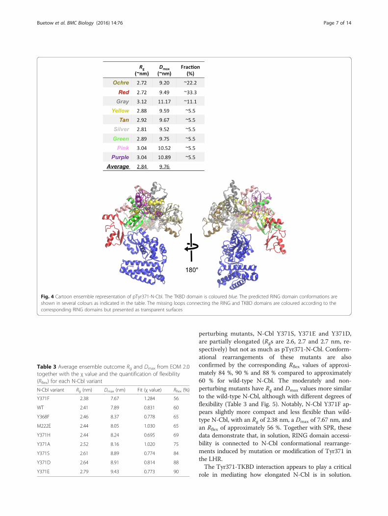
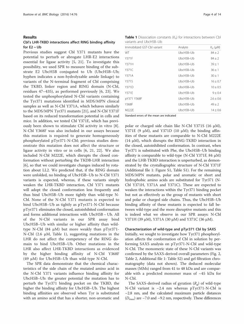
perturbing mutants, N-Cbl Y371S, Y371E and Y371D,are partially elongated (Rgs are 2.6, 2.7 and 2.7 nm, re-spectively) but not as much as pTyr371-N-Cbl. Conform-ational rearrangements of these mutants are alsoconfirmed by the corresponding Rflex values of approxi-mately 84 %, 90 % and 88 % compared to approximately60 % for wild-type N-Cbl. The moderately and non-perturbing mutants have Rg and Dmax values more similarto the wild-type N-Cbl, although with different degrees offlexibility (Table 3 and Fig. 5). Notably, N-Cbl Y371F ap-pears slightly more compact and less flexible than wild-type N-Cbl, with an Rg of 2.38 nm, a Dmax of 7.67 nm, andan Rflex of approximately 56 %. Together with SPR, thesedata demonstrate that, in solution, RING domain accessi-bility is connected to N-Cbl conformational rearrange-ments induced by mutation or modification of Tyr371 inthe LHR.The Tyr371-TKBD interaction appears to play a critical
role in mediating how elongated N-Cbl is in solution.
Fig. 4 Cartoon ensemble representation of pTyr371-N-Cbl. The TKBD domain is coloured blue. The predicted RING domain conformations areshown in several colours as indicated in the table. The missing loops connecting the RING and TKBD domains are coloured according to thecorresponding RING domains but presented as transparent surfaces
Table 3 Average ensemble outcome Rg and Dmax from EOM 2.0together with the χ value and the quantification of flexibility(Rflex) for each N-Cbl variant
N-Cbl variant Rg (nm) Dmax (nm) Fit (χ value) Rflex (%)
Y371F 2.38 7.67 1.284 56
WT 2.41 7.89 0.831 60
Y368F 2.46 8.37 0.778 65
M222E 2.44 8.05 1.030 65
Y371H 2.44 8.24 0.695 69
Y371A 2.52 8.16 1.020 75
Y371S 2.61 8.89 0.774 84
Y371D 2.64 8.91 0.814 88
Y371E 2.79 9.43 0.773 90
Buetow et al. BMC Biology (2016) 14:76 Page 7 of 14
Although N-Cbl Y368F and M222E have higher bindingaffinities for UbcH5B–Ub than wild-type, at SAXS reso-lution both have characteristics comparable to wild-type,suggesting they are similarly compact in solution. Al-though N-Cbl M222E has a comparable binding affinity tothe strongly-perturbing N-Cbl Y371 mutants, in solution,it appears more compact, as both Rg and Dmax are similarto wild-type N-Cbl and flexibility is relatively limited(Rflex ~60 %). N-Cbl Y368F has a higher binding affinityfor UbcH5B–Ub than wild-type, but at the level of
resolution provided by SAXS its characteristics andcompactness are similar to those of the wild-type pro-tein, suggesting similar behaviour in solution.
Transformation potential of Cbl Y371 mutants in cellsTo test the transforming potential of Cbl Y371 mutantsin cells, we performed focus formation assays using 3T3cells stably transfected with N-terminally FLAG-taggedCbl variants. Relative protein expression levels of eachvariant were assessed by immunoblotting (Fig. 6a). Foci
Fig. 5 EOM 2.0 analysis of N-Cbl Tyr371 mutants, N-Cbl Y368F and N-Cbl M222E. Mutants were clustered according to their ability to perturb theLHR-TKBD interaction based on SAXS parameters shown in Table 3. a Strongly perturbing mutants. b Moderately perturbing mutants. c Weakly ornon-perturbing mutants
Buetow et al. BMC Biology (2016) 14:76 Page 8 of 14

were visualized with sulforhodamine B staining andcounted manually with a DNA Safe Imager (Invitrogen).Afterwards, the sulforhodamine B stain was extractedand the A564 measured to compare relative cell densities[26, 27]. Foci counts and relative cell densities were ana-lyzed by one-way ANOVA followed by Dunnett’s testwith wild-type Cbl as the control. Cbl70Z was includedamong the variants tested as a positive transformingcontrol [22]. Other variants tested included the same setused in our biochemical assays except in a full-lengthcontext.Foci were observed in cells transfected with Cbl Y371
mutants and Cbl70Z, but not in wild-type, Cbl Y368F-or Cbl M222E-transfected cells (Fig. 6). When analyzedby one-way ANOVA followed by Dunnett’s test, signifi-cant differences (P < 0.05) in number of foci and celldensities were observed between cells infected with wild-type Cbl and all the Y371 mutants except Cbl Y371F(Fig. 6c,d, Additional file 1: Figure S4). Significantly morefoci were present in Cbl70Z, Cbl Y371E and the MDS/MPN Cbl Y371 mutant set and the relative cell densitywas significantly greater compared to wild-type Cbl; how-ever, there is no correlation between mutations that pro-mote moderate or strong LHR-TKBD perturbations andtransformation potential. Based on the ability to form foci,all the Cbl variants except Cbl Y368F and M222E have the
ability to potentiate transformation in cells. Based onstatistical analyses of the number of foci and relative celldensity, Cbl70Z, Cbl Y371E and the MDS/MPN Cbl Y371mutant set have significantly higher numbers of foci thanwild-type Cbl. For Cbl Y371F, neither the number of focinor the relative cell density are significantly different thanin cells infected with wild-type Cbl; these results suggestthis mutant may potentiate transformation but not to thesame extent as the other Cbl Y371 mutants or Cbl70Z.
DiscussionIn this work, we employed SAXS and SPR to characterizehow modifications and mutations in Cbl affect the con-formation and accessibility of the RING domain in solu-tion and subsequently investigate the relationship betweenthese characteristics and the ability of Cbl variants topotentiate transformation in cells. Our results show that amutation (M222E) within the RING-TKBD interfaceregulates accessibility of the RING domain only, whereasmutation or modification of Tyr371 within the LHR medi-ates the LHR-TKBD interaction, thereby regulating RINGdomain accessibility as well as the space sampled by theRING domain relative to the TKBD. The chemical natureof the substitution or modification controls the LHR-TKBD interaction and shifts the equilibrium between thenative conformation where the RING is restricted to the
Fig. 6 Transformation potential of Cbl variants in focus formation assays. a Immunoblot of lysates from 3T3 fibroblasts infected with FLAG-taggedCbl variants using α-FLAG antibody (top) and α-actin (bottom) antibody as a loading control. b Sulforhodamine B-stained 3T3 fibroblasts infectedwith indicated Cbl variants. c Mean number of foci formed by Cbl variant-infected 3T3 fibroblasts shown in a bar graph (n = 3). No foci werepresent in 3T3 cells infected with wild-type Cbl, Cbl M222E or Cbl Y368F. Double asterisks (**) denote significant differences (P < 0.05) betweenindicated Cbl variant and wild-type using ANOVA followed by Dunnett’s multiple comparisons test. Error bars indicate standard deviation. d As in(c) but for A564 of extracted sulforhodamine B from Cbl-infected 3T3 fibroblasts
Buetow et al. BMC Biology (2016) 14:76 Page 9 of 14

face of the TKBD opposing the substrate-binding site andopen conformations where the RING domain can accessother surfaces of the TKBD and potentially other regionsof Cbl. None of the Tyr371 mutants can adopt thepTyr371 conformation that is critical for activation ofE2 ~ Ub and RTK ubiquitination, but all of the Tyr371mutants perturb the LHR-TKBD conformation observedin the native state except Cbl Y371F. Likewise, all of theTyr371 mutants except Cbl Y371F clearly potentiatetransformation in our focus formation assays, whereas CblY371F forms foci but statistical analyses of the number offoci formed and relative cell densities suggest that thismutant has less transformation potential than the others.Previous studies have shown that Cbl Y371F is not trans-forming in soft agar colony formation assays nor does itpromote tumour formation in nude mice [21, 22]. Thesedata indicate that the flexibility of the LHR features in theCbl Tyr371 mutant transformation mechanism.Met222 is a key residue in maintaining the RING-
TKBD interaction in native Cbl. A Met to Glu mutationwas used to perturb this interaction and investigate theimportance of the RING-TKBD interaction on Cbl’sconformation in solution. Our previous work and SPRstudies show that mutation of Met222 in the TKBD toGlu increases Cbl’s E2–Ub binding affinity by approxi-mately 8-fold and enhances Cbl’s in vitro autoubiquitina-tion rate [6]. Though Cbl M222E has a higher bindingaffinity for UbcH5B–Ub and is more active than wild-type Cbl, SAXS analysis demonstrates that Cbl M222Ehas parameters (Rg, Dmax and Rflex) comparable to wild-type Cbl, suggesting both adopt a compact conformation.In cells, Cbl M222E does not enhance EGFR ubiquitina-tion (data not shown) nor does it potentiate transform-ation in our focus formation assays. The lack of effect ofthis mutant in these assays suggests structural regulationof Cbl’s ligase activity requires more than a simple rotationthat exposes the E2-binding site on the RING domain.Previous work has shown that phosphorylation of the
conserved LHR Tyr in the Cbl family enhances activityin vitro and is essential for receptor PTK ubiquitinationin cells, where Cbl’s E3 ligase activity features promin-ently in EGFR downregulation via lysosomal degradation[8, 10, 11]. The rate and pattern of Cbl-mediated EGFRubiquitination determines whether EGFR is directed tothe lysosome or recycled to the membrane. X-ray crys-tallography and NMR studies of Cbl and Cbl-b haveshown that this phosphorylation event eliminates RING-mediated autoinhibition, optimally positions Ub for trans-fer and juxtaposes the RING domain with the TKBDsubstrate-binding site [6, 7, 9]. Notably, in the crystalstructures of Cbl and Cbl-b bound to Zap70 peptide andE2 or E2 ~Ub, the conformations of RING domain rela-tive to the TKBD are identical. Here, we show that, insolution, when Tyr371 is phosphorylated, the Rg and Dmax
increase and EOM analysis demonstrates the linker regionbecomes flexible, allowing the RING domain to samplethe space surrounding the TKBD rather than maintain thecrystallographically observed conformation. This is con-sistent with other E3 ligases, where translational androtational movement of the RING domain are requiredfor Ub transfer to select substrate lysine sites (monoubi-quitination and multiubiquitination) as well as polyUbchain formation [28]. Cbl binds EGFR directly through itsTKBD and indirectly through growth factor receptorbinding 2-mediated interactions in the region C-terminalto the RING domain [12]. Cbl-mediated ubiquitination ofEGFR is observed for both binding sites, highlighting apotential essential role for RING flexibility in Cbl sub-strate ubiquitination.Our SAXS and SPR data show that mutations within
the LHR perturb the native LHR-TKBD conformationand that the extent of perturbation is dependent on thechemical nature of the amino acid substitution. In thenative state, Trp258, Ala262, Val263, and Met274 form ahydrophobic pocket around the aromatic ring of Tyr371and the side chain of Ser227 forms a hydrogen bondwith the hydroxyl group of Tyr371. The more disruptivethe mutation is to the hydrophobic environment, thegreater the perturbation in the LHR-TKBD interaction.When Tyr371 is mutated to an amino acid with acharged side chain like Asp or Glu or a small, polar sidechain like Ser, the UbcH5B–Ub binding affinity is approxi-mately 8-fold enhanced compared to native Cbl and theRg and Dmax increase to values comparable to pTyr371-Cbl. Additionally, SAXS analysis reveals these Tyr371mutants are the most flexible compared to native Cbl.Mutation of Tyr371 to smaller hydrophobic amino acidslike Cys or Ala only moderately perturbs the LHR-TKBDinteraction as evidenced by an approximately 2-fold en-hancement in the UbcH5B–Ub binding affinity and themoderate increase in Rg, Dmax and Rflex compared to na-tive Cbl. Likewise, when Tyr371 is mutated to His, onlymoderate to weak perturbation of the LHR-TKBD inter-action occurs. There is an approximately 2-fold enhance-ment in the UbcH5B–Ub binding affinity and SAXSanalysis shows evidence of more flexibility (slightly highervalues of Rg, Dmax and Rflex) compared to native Cbl.When Tyr371 is mutated to Phe, the hydrophobic interac-tions are not disturbed and the native state is maintained:the UbcH5B–Ub binding affinity, Rg, Dmax and Rflex
values are comparable to native Cbl. In addition, previ-ous in vitro autoubiquitination assays show native Cbland Cbl Y371F have comparable catalytic efficiencies[6]. Thus, in solution, it appears as though Cbl Y371Fmimics the behaviour of native Cbl.Mutation of Tyr371 is not the only site at which the
LHR-TKBD interaction can be perturbed. Our previouswork and current data show that mutation of Tyr368 to
Buetow et al. BMC Biology (2016) 14:76 Page 10 of 14
Phe also disrupts the LHR-TKDB interaction [6]. CblY368F binds UbcH5B–Ub approximately 2-fold moretightly than native Cbl and has an approximately 2-foldenhancement in catalytic efficiency for in vitro autoubi-quitination. In addition, this mutant displays a slightincrease in all SAXS-based parameters (including Rflex)compared to native Cbl. However, in contrast to the CblTyr371 mutants, Cbl Y368F does not promote focusformation in our assays nor does it compromise EGFRubiquitination in cells [6] and previous work has alsoshown this mutant does not form colonies in soft agarassays nor promote tumour growth in nude mice [21, 22].Though it is clear that Tyr368-TKBD interactions alsoplay a role in the conformations Cbl adopts in solution,the significance of this interaction in Cbl’s ligase activity incells is unclear. Other studies have shown that Cbl ΔY368is defective in EGFR ubiquitination and has the ability topotentiate transformation in cells and in mice [21, 22]. Inaddition, Cbl Y368C has also been identified in MDS/MPN clinical samples, and, like Cbl Y371C and Y371S,has been shown to potentiate transformation in cells andin mice; however, whether PTK ubiquitination is compro-mised remains unknown [19]. As observed for Tyr371, thechemical nature of the mutation at Tyr368 might factorinto regulation of Cbl’s ligase activity as well as transform-ation potential.While the ability to ubiquitinate PTKs is essential for
Cbl-mediated downregulation of PTK signalling, previouswork has clearly shown that it is not the only prerequisitefor Cbl-dysfunction driven transformation. Thien et al.[21] propose that the LHR-TKBD stability of Cbl mutantscontributes to their ability to potentiate transformationbased on the analysis of a number of linker region andRING mutants: Cbl ΔY368, ΔY371 and Y371A promotetransformation but not Cbl Y368F or Y371F. Cbl Y368F isslightly more flexible than native Cbl but retains the abilityto ubiquitinate receptor PTKs, whereas Cbl Y371F cannotubiquitinate receptor PTKs but is able to maintain thenative conformation in solution. Thus, it seems that bothcharacteristics contribute to the transformation potentialof Cbl mutants. Cbl functions as both an ubiquitin ligaseand adaptor in receptor PTK-mediated cell signalling.Although enzymatic activity is slower in the absence ofTyr371 phosphorylation, Cbl is still functional [6]. It maybe that when the LHR-TKBD interaction is perturbed, Cblubiquitinates substrates that bind to regions other thanthe canonical TKBD-binding site or the more flexible mu-tants might ubiquitinate “dead end” lysines on substratessuch that biologically viable ubiquitination chains are notformed. Alternatively, perturbed LHR-TKBD interactionsmight disrupt oligomerization or interactions with otherproteins or lead to the non-sequential recruitment ofsubstrates to exposed surfaces that interfere with con-trolled signalling.
ConclusionsWe have shown that LHR-TKBD interactions regulateCbl’s ligase activity and conformations in solution. Mu-tations or modifications within the LHR prevent Cbl frommaintaining a closed conformation where the RING do-main is restricted to a surface opposite the TKBD-substratebinding site; instead, depending on the nature of thealteration, Cbl becomes flexible and the RING domaincan access multiple surfaces of the TKBD. Transformationonly occurs when mutations within the LHR (1) perturbthe native state and (2) fail to ubiquitinate PTKs.
MethodsProtein preparationFor crystallization, N-Cbl Y371F (residue 47–435) wasexpressed, purified and stored as described previously[6]. For SPR and SAXS analysis, N-Cbl variants werecloned into a modified form of pGEX4T1 (GE Health-care) containing an N-terminal histidine glutathione S-transferase (His-GST) tag followed by a thrombin orTEV-protease cleavage site and expressed in E. coli BL21(DE3) Gold. pTyr371-N-Cbl was generated as describedpreviously [6]. For SPR analysis, all N-Cbl variants werepurified by Ni-NTA followed by glutathione-affinity chro-matography. Anion exchange chromatography was subse-quently used to separate His-GST-pTyr371-N-Cbl fromunphosphorylated His-GST-N-Cbl. For SAXS studies, N-Cbl variants were then treated with TEV or thrombinprotease to cleave the His-GST tag and further purified byNi-NTA pass-back followed by anion exchange and sizeexclusion chromatography. Proteins for SAXS were storedin 25 mM Tris-HCl (pH 7.6), 500 mM NaCl and 1 mMDTT at –80 °C. UbcH5B S22R C85K–Ub (referred to asUbcH5B–Ub) was expressed, generated and purified asdescribed previously [7]. His-GST-tagged protein concen-trations were determined by Bradford assay using BSA asa standard and all other concentrations were determinedusing a NanoVue Spectrophotometer (GE Healthcare).
Crystallization and structural determinationCrystals were obtained by mixing N-Cbl Y371F (10 mg/mL)with an equal volume of reservoir solution containing0.1 M Tris-HCl, pH 8.5, 3.0–3.1 M sodium formate,and 5 mM DTT using hanging drop vapour diffusion at8 °C. The crystals were flash-frozen in 0.1 M Tris-HCl,pH 8.5, 3.0–3.1 M sodium formate, 5 mM DTT, 8 %(v/v) glycerol, 8 % (v/v) ethylene glycol, and 8 % (v/v)sucrose. Data were collected with beamline I04 atDiamond Light Source, integrated with automated XDS[29] and scaled using the CCP4 program suite [30]. Initialphases were obtained by molecular replacement withPHASER using native N-Cbl (PDB:2Y1M) [6]. N-CblY371F crystals belong to space group C2221 with six
Buetow et al. BMC Biology (2016) 14:76 Page 11 of 14
molecules in the asymmetric unit. The model was built inCOOT [31] and refined using PHENIX [32].
Biacore analysisCbl–UbcH5B–Ub binding experiments were conductedas described previously [6, 23]. GST-Cbl variants werecoupled to CM-5 chips and binding was measured at aconcentration range of 0–120 μM of UbcH5B–Ub. Thedata were analyzed using Biacore T200 Evaluation soft-ware package (Biacore Life Sciences) and Scubber2.0c(BioLogic Software).
Plasmids and cell cultureN-terminally FLAG-tagged Cbl variants were generatedby PCR amplification and ligation into a pBABE vector[33] by In-Fusion (Clontech) according to the manufac-turer’s instructions. Subsequently, these Cbl variantswere transiently transfected into Phoenix Eco cells usingLipofectamine 2000 (Thermo Fisher Scientific) accordingto the manufacturer’s instructions. Infection was per-formed in 3T3 (mouse fibroblast) cells followed by puro-mycin (2 μg/mL) selection. Cells were cultured in DMEMcontaining 20 mM glutamine and 10 % donor bovineserum in a 37 °C incubator at 5 % CO2.
Focus formation assays and sulforhodamine B extractionTo perform the focus formation assays, 3T3 cells wereseeded onto 10 cm2 dishes and infected with the Cblvariants on the following day. This was followed by asecond round of infection after 24 hours and the mediachanged on the following day. After 3 days of selectionwith puromycin, the 3T3 cells were split and 0.5 × 10⁵
cells seeded onto 3.5 cm2 dishes and cultured for 18 daysbefore being fixed in methanol and stained with sulfor-hodamine B. Foci were then counted manually with aDNA Safe Imager (Invitrogen). Afterwards, each dishwas incubated with 1.1 mL of 10 mM Tris, pH 10.5 for5 minutes with rocking at room temperature to extractsulforhodamine B and the absorbance of the extracteddye was measured at 564 nm. Prism (Graph Pad, MacV5.0C) was used for statistical analyses (one-way ANOVAfollowed by Dunnett multiple comparisons testing withP < 0.05 treated as the cutoff for significant differences).
ImmunoblottingAfter 1 week, total protein was isolated from one 3.5 cm2
dish of each Cbl variant in whole cell lysis buffer contain-ing 50 mM Tris, pH 7.4, 150 mM NaCl, 1 mM EDTA,1 % Igepal CA-630 (Sigma), and 10 % glycerol. Proteinswere separated under reducing conditions using SDSpolyacrylamide gel electrophoresis and transferred ontoa nitrocellulose membrane (GE Healthcare Life Sciences).Blots were probed with rabbit anti-FLAG (Sigma Aldrich,
F7425) and goat anti-actin (SantaCruz Biotechnology,sc-1616) antibodies, incubated with donkey anti-goatIRDye 800CW and goat anti-rabbit IRDye 680LT sec-ondary antibodies (LI-COR Biosciences, 925_32214 and925_68021), and visualized using an Odyssey CLx ImagingSystem (LI-COR Biosciences).
Small angle X-ray scatteringSynchrotron X-ray scattering data of Cbl mutants werecollected at EMBL P12 beamline (DESY, Hamburg) usinga robotic sample changer [34]. All the mutants were mea-sured in the same buffer (500 mM NaCl, 25 mM Tris-HCl(pH 7.6), 1 mM DTT) in a concentration series rangingfrom either 0.5–10 mg/mL (0.5, 1, 2, 5, and 10 mg/mL) or0.5–6.6 mg/mL (0.5, 1, 1.6, 3.3, and 6.6 mg/mL). SAXSdata were recorded at 10 °C using a PILATUS 2 M pixeldetector (DECTRIS, Baden, Switzerland) at a sample-detector distance of 3.1 m and a wavelength of 0.15 nm.This setup covers a range of momentum transfer of0.1 < s < 5 nm–1 (s = 4π sin(θ)/λ, where 2θ is the scatteringangle). Initially, the data were pre-processed using anautomatic pipeline [35] and further analyzed using PRI-MUS [36, 37]. The forward scattering I(0) as well as the Rg
were calculated using the Guinier approximation assum-ing that, at very small angles (s < 1.3/Rg), the intensity isrepresented as I(s) = I(0) · exp(-(sRg)
2/3) [38]. Linearity inthe Guinier region was used to exclude sample aggrega-tion. The pair-distance distribution function P(r), fromwhich the Dmax and Rg were estimated, was computedusing GNOM [39]. Qualitative assessment of compactnessversus structural disorder was made by transforming thescattering profiles in the so-called Kratky representation[I(s)s2 vs. s] [40] and its normalized version [(sRg)
2*I(s)/I(0)vs. sRg] [41]. The MM was derived from (1) the excludedvolume of the hydrated particle using the Porod invariant[36] and (2) the excluded volumes of the ab initio models.Ab initio models were created with DAMMIF [42] usinglow resolution data (s < 2 nm–1). The algorithm constructsbead models yielding a scattering profile with the lowestpossible discrepancy (χ) to the experimental data whilekeeping beads interconnected and the model compact.Twenty independent ab initio reconstructions were per-formed for each Cbl mutant and then averaged usingDAMAVER [43]. Superimpositions between ab initio re-constructions and available atomic models were madeusing the software SUPCOMB [44]. The flexibility wasanalyzed using EOM 2.0 [45] – an enhanced version ofthe EOM [46], which assumes coexistence of a range ofconformations in solution for which an average scatteringintensity fits the experimental data. In EOM 2.0, a pool of10,000 independent models is created as first step withthe aim to approximate the (otherwise infinite) conform-ational space for a protein exhibiting disorder. For eachmodel in the pool the theoretical scattering curve is
Buetow et al. BMC Biology (2016) 14:76 Page 12 of 14
automatically computed with CRYSOL [47]. After-wards, genetic algorithm (GA) is used to select ensembleswith varying numbers of conformers (usually from 5 to40) by calculating the average theoretical profile andfitting it to the experimental SAXS data. For each Cblmutant, the GA was repeated 100 independent times andthe ensemble with the lowest discrepancy reported as thebest solution out of 100 final ensembles. Furthermore, 100repetitions of GA allowed the computation of Rg and Dmax
distributions so that structural information about theflexibility could be extracted. Distributions with Rg averagevalues above the Rg average values calculated for the poolare classified as extended whereas models with valuesbelow the average as compact. The width of the distribu-tion is also used to derive the flexibility of the particle,whereby a narrow distribution indicates a rather rigid par-ticle and broader distributions are associated with higherflexibility [46]. Using EOM 2.0, systematic quantificationof the flexibility was made using the metric Rflex – whichcomputes the Shannon information entropy of thedistributions [45]. All the software used for the SAXSdata analysis belongs to the ATSAS 2.5 package [36].
Additional file
Additional file 1: Figure S1. Comparison of wild-type and N-Cbl Y371F.Figure S2. Comparison of SAXS scattering data for wild-type (WT, blue)and pTyr371-N-Cbl (yellow). Figure S3. Comparison of ab initio and relaxedcrystal models. Figure S4. Confidence intervals (95 %) for the differencebetween group means. Table S1. Data collection and refinement statistics.(DOCX 15797 kb)
AcknowledgementsWe would like to thank W. Clark and A. Keith for in-house DNA sequencing,DLS for access to beamline I04 (mx8659), EMBL for the access to P12 beam-line, and A. Schuettelkopf, M. Gabrielsen and O. Byron for discussions.
FundingThis work was supported by Cancer Research UK. DTH was supported byEuropean Research Council (grant number 647849).
Availability of data and materialsThe coordinates and structure factors for N-Cbl Y371F have been depositedin Protein Data Bank (PDB: 5J3X in http://www.rcsb.org).
Authors’ contributionsLB, DTH and HD performed protein purification. HD carried out proteincrystallization and X-ray data collection. LB performed structure determin-ation. SFA, AH and LB conducted focus formation assays. LB, DTH and GTperformed SAXS experiments. GT and DIS conducted SAXS analyses. GJS per-formed and analyzed SPR experiments. LB and DTH wrote the manuscript.All authors read and approved the final manuscript.
Competing interestsThe authors declare that they have no competing interests.
Author details1Cancer Research UK Beatson Institute, Garscube Estate, Switchback Road,Glasgow G61 1BD, UK. 2EMBL c/o DESY, Notkestrasse 85, Geb, 25a, 22603Hamburg, Germany. 3Present address: Institute of Medical Genetics, School ofMedicine, Shandong University, No. 44 Wenhuaxi Road, Jinan, Shandong250012, People’s Republic of China. 4Present address: Multimodal Molecular
Imaging Institute, Nanoscopy Division, Maastricht University,Universiteitssingel 50, 6229 ER Maastricht, The Netherlands.
Received: 31 March 2016 Accepted: 12 August 2016
References1. Mohapatra B, Ahmad G, Nadeau S, Zutshi N, An W, Scheffe S, Dong L,
Feng D, Goetz B, Arya P, et al. Protein tyrosine kinase regulation byubiquitination: critical roles of Cbl-family ubiquitin ligases. Biochim BiophysActa. 2013;1833(1):122–39.
2. Swaminathan G, Tsygankov AY. The Cbl family proteins: ring leaders inregulation of cell signaling. J Cell Physiol. 2006;209(1):21–43.
3. Ng C, Jackson RA, Buschdorf JP, Sun Q, Guy GR, Sivaraman J. Structural basisfor a novel intrapeptidyl H-bond and reverse binding of c-Cbl-TKB domainsubstrates. EMBO J. 2008;27(5):804–16.
4. Joazeiro CA, Wing SS, Huang H, Leverson JD, Hunter T, Liu YC. The tyrosinekinase negative regulator c-Cbl as a RING-type, E2-dependent ubiquitin-protein ligase. Science. 1999;286(5438):309–12.
5. Zheng N, Wang P, Jeffrey PD, Pavletich NP. Structure of a c-Cbl-UbcH7 complex:RING domain function in ubiquitin-protein ligases. Cell. 2000;102(4):533–9.
6. Dou H, Buetow L, Hock A, Sibbet GJ, Vousden KH, Huang DT. Structuralbasis for autoinhibition and phosphorylation-dependent activation of c-Cbl.Nat Struct Mol Biol. 2012;19(2):184–92.
7. Dou H, Buetow L, Sibbet GJ, Cameron K, Huang DT. Essentiality of a non-RING element in priming donor ubiquitin for catalysis by a monomeric E3.Nat Struct Mol Biol. 2013;20(8):982–6.
8. Kassenbrock CK, Anderson SM. Regulation of ubiquitin protein ligase activityin c-Cbl by phosphorylation-induced conformational change andconstitutive activation by tyrosine to glutamate point mutations. J BiolChem. 2004;279(27):28017–27.
9. Kobashigawa Y, Tomitaka A, Kumeta H, Noda NN, Yamaguchi M, Inagaki F.Autoinhibition and phosphorylation-induced activation mechanisms ofhuman cancer and autoimmune disease-related E3 protein Cbl-b. Proc NatlAcad Sci U S A. 2011;108(51):20579–84.
10. Levkowitz G, Waterman H, Ettenberg SA, Katz M, Tsygankov AY, Alroy I,Lavi S, Iwai K, Reiss Y, Ciechanover A, et al. Ubiquitin ligase activity andtyrosine phosphorylation underlie suppression of growth factor signalingby c-Cbl/Sli-1. Mol Cell. 1999;4(6):1029–40.
11. Ryan PE, Sivadasan-Nair N, Nau MM, Nicholas S, Lipkowitz S. The N terminusof Cbl-c regulates ubiquitin ligase activity by modulating affinity for theubiquitin-conjugating enzyme. J Biol Chem. 2010;285(31):23687–98.
12. Schmidt MH, Dikic I. The Cbl interactome and its functions. Nat Rev Mol CellBiol. 2005;6(12):907–18.
13. Peschard P, Kozlov G, Lin T, Mirza IA, Berghuis AM, Lipkowitz S, Park M,Gehring K. Structural basis for ubiquitin-mediated dimerization andactivation of the ubiquitin protein ligase Cbl-b. Mol Cell. 2007;27(3):474–85.
14. Kozlov G, Peschard P, Zimmerman B, Lin T, Moldoveanu T, Mansur-Azzam N,Gehring K, Park M. Structural basis for UBA-mediated dimerization of c-Cblubiquitin ligase. J Biol Chem. 2007;282(37):27547–55.
15. Kang JM, Park S, Kim SJ, Hong HY, Jeong J, Kim HS, Kim SJ. CBL enhancesbreast tumor formation by inhibiting tumor suppressive activity of TGF-betasignaling. Oncogene. 2012;31(50):5123–31.
16. Knight JF, Shepherd CJ, Rizzo S, Brewer D, Jhavar S, Dodson AR, Cooper CS,Eeles R, Falconer A, Kovacs G, et al. TEAD1 and c-Cbl are novel prostatebasal cell markers that correlate with poor clinical outcome in prostatecancer. Br J Cancer. 2008;99(11):1849–58.
17. Kales SC, Ryan PE, Nau MM, Lipkowitz S. Cbl and human myeloid neoplasms:the Cbl oncogene comes of age. Cancer Res. 2010;70(12):4789–94.
18. Tan YH, Krishnaswamy S, Nandi S, Kanteti R, Vora S, Onel K, Hasina R, Lo FY,El-Hashani E, Cervantes G, et al. CBL is frequently altered in lung cancers: itsrelationship to mutations in MET and EGFR tyrosine kinases. PLoS One.2010;5(1):e8972.
19. Sanada M, Suzuki T, Shih LY, Otsu M, Kato M, Yamazaki S, Tamura A, Honda H,Sakata-Yanagimoto M, Kumano K, et al. Gain-of-function of mutated C-CBLtumour suppressor in myeloid neoplasms. Nature. 2009;460(7257):904–8.
20. Niemeyer CM, Kang MW, Shin DH, Furlan I, Erlacher M, Bunin NJ, Bunda S,Finklestein JZ, Sakamoto KM, Gorr TA, et al. Germline CBL mutations causedevelopmental abnormalities and predispose to juvenile myelomonocyticleukemia. Nat Genet. 2009;42:794–800.
Buetow et al. BMC Biology (2016) 14:76 Page 13 of 14
21. Thien CB, Walker F, Langdon WY. RING finger mutations that abolish c-Cbl-directed polyubiquitination and downregulation of the EGF receptor areinsufficient for cell transformation. Mol Cell. 2001;7(2):355–65.
22. Andoniou CE, Thien CB, Langdon WY. Tumour induction by activated ablinvolves tyrosine phosphorylation of the product of the cbl oncogene.EMBO J. 1994;13(19):4515–23.
23. Dou H, Buetow L, Sibbet GJ, Cameron K, Huang DT. BIRC7-E2 ubiquitinconjugate structure reveals the mechanism of ubiquitin transfer by a RINGdimer. Nat Struct Mol Biol. 2012;19(9):876–83.
24. Receveur-Brechot V, Durand D. How random are intrinsically disorderedproteins? A small angle scattering perspective. Curr Protein Pept Sci.2012;13(1):55–75.
25. Panjkovich A, Svergun DI. Deciphering conformational transitions ofproteins by small angle X-ray scattering and normal mode analysis. PhysChem Chem Phys. 2016;18(8):5707–19.
26. Skehan P, Storeng R, Scudiero D, Monks A, McMahon J, Vistica D, Warren JT,Bokesch H, Kenney S, Boyd MR. New colorimetric cytotoxicity assay foranticancer-drug screening. J Natl Cancer Inst. 1990;82(13):1107–12.
27. Vichai V, Kirtikara K. Sulforhodamine B colorimetric assay for cytotoxicityscreening. Nat Protoc. 2006;1(3):1112–6.
28. Duda DM, Borg LA, Scott DC, Hunt HW, Hammel M, Schulman BA. Structuralinsights into NEDD8 activation of cullin-RING ligases: conformational controlof conjugation. Cell. 2008;134(6):995–1006.
29. Kabsch W. Xds. Acta Crystallogr D Biol Crystallogr. 2010;66(Pt 2):125–32.30. Winn MD, Ballard CC, Cowtan KD, Dodson EJ, Emsley P, Evans PR,
Keegan RM, Krissinel EB, Leslie AG, McCoy A, et al. Overview of theCCP4 suite and current developments. Acta Crystallogr D BiolCrystallogr. 2011;67(Pt 4):235–42.
31. Emsley P, Lohkamp B, Scott WG, Cowtan K. Features and development ofCoot. Acta Crystallogr D Biol Crystallogr. 2010;66(Pt 4):486–501.
32. Adams PD, Afonine PV, Bunkoczi G, Chen VB, Davis IW, Echols N, Headd JJ,Hung LW, Kapral GJ, Grosse-Kunstleve RW, et al. PHENIX: a comprehensivePython-based system for macromolecular structure solution. ActaCrystallogr D Biol Crystallogr. 2010;66(Pt 2):213–21.
33. Morgenstern JP, Land H. Advanced mammalian gene transfer: hightitre retroviral vectors with multiple drug selection markers and acomplementary helper-free packaging cell line. Nucleic Acids Res.1990;18(12):3587–96.
34. Round AR, Franke D, Moritz S, Huchler R, Fritsche M, Malthan D, Klaering R,Svergun DI, Roessle M. Automated sample-changing robot for solutionscattering experiments at the EMBL Hamburg SAXS station X33. J ApplCrystallogr. 2008;41:913–7.
35. Franke D, Kikhney AG, Svergun DI. Automated acquisition and analysis ofsmall angle X-ray scattering data. Nucl Instrum Methods A. 2012;689:52–9.
36. Petoukhov MV, Franke D, Shkumatov AV, Tria G, Kikhney AG, Gajda M, GorbaC, Mertens HDT, Konarev PV, Svergun DI. New developments in the ATSASprogram package for small-angle scattering data analysis. J Appl Crystallogr.2012;45:342–50.
37. Konarev PV, Volkov VV, Sokolova AV, Koch MHJ, Svergun DI. PRIMUS: aWindows PC-based system for small-angle scattering data analysis. J ApplCrystallogr. 2003;36:1277–82.
38. Guinier A. La diffraction des rayons X aux tres petits angles; application a l'etudede phenomenes ultramicroscopiques. Ann Phys (Paris). 1939;12:161–237.
39. Svergun DI. Determination of the regularization parameter in indirect-transform methods using perceptual criteria. J Appl Crystallogr.1992;25:495–503.
40. Glatter O, Kratky O, editors. Small angle x-ray scattering. London: Academic;1982.
41. Durand D, Vives C, Cannella D, Perez J, Pebay-Peyroula E, Vachette P,Fieschi F. NADPH oxidase activator p67(phox) behaves in solution as amultidomain protein with semi-flexible linkers. J Struct Biol. 2010;169(1):45–53.
42. Franke D, Svergun DI. DAMMIF, a program for rapid ab-initio shapedetermination in small-angle scattering. J Appl Crystallogr. 2009;42:342–6.
43. Volkov VV, Svergun DI. Uniqueness of ab initio shape determination insmall-angle scattering. J Appl Crystallogr. 2003;36:860–4.
44. Kozin MB, Svergun DI. Automated matching of high- and low-resolutionstructural models. J Appl Crystallogr. 2001;34:33–41.
45. Tria G, Mertens HD, Kachala M, Svergun DI. Advanced ensemblemodelling of flexible macromolecules using X-ray solution scattering.IUCrJ. 2015;2(Pt 2):207–17.
46. Bernado P, Mylonas E, Petoukhov MV, Blackledge M, Svergun DI. Structuralcharacterization of flexible proteins using small-angle X-ray scattering. J AmChem Soc. 2007;129(17):5656–64.
47. Svergun D, Barberato C, Koch MHJ. CRYSOL - A program to evaluate x-raysolution scattering of biological macromolecules from atomic coordinates.J Appl Crystallogr. 1995;28:768–73.
• We accept pre-submission inquiries
• Our selector tool helps you to find the most relevant journal
• We provide round the clock customer support
• Convenient online submission
• Thorough peer review
• Inclusion in PubMed and all major indexing services
• Maximum visibility for your research
Submit your manuscript atwww.biomedcentral.com/submit
Submit your next manuscript to BioMed Central and we will help you at every step:
Buetow et al. BMC Biology (2016) 14:76 Page 14 of 14